bronchiectasis severity and prediction of airway impulse
TRANSCRIPT

Page 1/31
Impulse Oscillometry May Be Useful in Evaluation ofBronchiectasis Severity and Prediction of AirwayReversibility.Cuiyan Tan
Fifth A�liated Hospital of Sun Yat-sen UniversityDonghai Ma
Fifth A�liated Hospital of Sun Yat-sen UniversityKongqiu Wang
Fifth A�liated Hospital of Sun Yat-sen UniversityChangli Tu
Fifth A�liated Hospital of Sun Yat-sen UniversityMeizhu Chen
Fifth A�liated Hospital of Sun Yat-sen UniversityXiaobin Zheng
Fifth A�liated Hospital of Sun Yat-sen UniversityYingjian Liang
Fifth A�liated Hospital of Sun Yat-sen UniversityYiying Huang
Fifth A�liated Hospital of Sun Yat-sen UniversityZhenguo Wang
Fifth A�liated Hospital of Sun Yat-sen UniversityJian Wu
Fifth A�liated Hospital of Sun Yat-sen UniversityJin Huang
Fifth A�liated Hospital of Sun Yat-sen UniversityJing Liu ( [email protected] )
Fifth A�liated Hospital of Sun Yat-sen University https://orcid.org/0000-0003-4638-409X
Research
Keywords: bronchiectasis, impulse oscillometry, spirometry, plethysmography, airway reversibility
Posted Date: May 10th, 2021
DOI: https://doi.org/10.21203/rs.3.rs-445238/v1

Page 2/31
License: This work is licensed under a Creative Commons Attribution 4.0 International License. Read Full License

Page 3/31
AbstractBackground
Impulse oscillometry (IOS) can be used to evaluateairway impedance in patients with obstructive airwaydiseases. Previous studies have demonstrated that IOS parameters differ betweenbronchiectasis patientsand healthy controls. This study aims to explore the usefulness of IOS in assessing disease severity andairway reversibility in bronchiectasis.
Method
Seventy-four patients with non-cystic �brosis bronchiectasis who visited our Respiratory Medicineoutpatient clinic were consecutively recruited. Spirometry, plethysmography and IOS tests wereperformed. Patients were strati�ed into mild, moderate and severe disease according to Reiff, Bhalla, BSI,FACED, and BRICS scores. Airway reversibility was measured by bronchodilation test (BDT) and the resultwas classi�ed as positive or negative.. ROC curves of IOS parameters was used to assess the usefulnessof IOS parameters in predicting airway reversibility. Correlations between the IOS, spirometric lungfunction and bronchiectasis severity parameters were analysed.
Results
Many IOS parameters, such as airway resistance at 5Hz (R5), small airways resistance (R5–R20), totalairway reactance (X5), resonance frequency (Fres), total airway impedance at 5Hz (Z5), and peripheralresistance (Rp) increased with increased bronchiectasis severity according to the FACED, BSI and Reiffscores. Large airway resistance (R20) and central resistance (Rc) were not signi�cantly different amonggroups with differentbronchiectasis severity. The difference between R5 and R20 (R5-R20) showed 81.0%sensitivity, and 69.8%speci�city in predicting the airway reversibility in bronchiectasis with AUC of 0.794(95%CI, 0.672-0.915).
Conclusion
IOS measurements are useful indicators of bronchiectasis severity and may be useful for predicting theairway reversibility.
IntroductionImpulse oscillometry (IOS) is a non-invasive and relatively simple method that can be used to detectairway abnormalities. IOS measures airway resistance and reactance using a variant of the forcedoscillation technique, and re�ect the mechanical properties of both the small and large airways, thusproviding information about sites where the obstruction occurs (peripheral or central), severity andrespiratory dynamics characteristics. The performance of IOS consists of exerting external pressuresignals at different frequencies on the normal tidal breathing and requires minimal patient cooperation,[1]

Page 4/31
Bronchiectasis is a chronic airway disease where there is abnormal enlargement of some airways in thelungs. This is normally diagnozed by identi�cation of typical radiographic features in high resolutioncomputed tomography (HRCT) scans, such as bronchial enlargement and wall thickening. Thesechanges are consequences of a complex interaction between various factors, including geneticspredisposition, airway dysfunction, in�ammatory response, structural diseases and chronic infections.[2]Multiple scoring systems have been developed to evaluate the severity of bronchiectasis. The most oftenused are the modi�ed Reiff[3], FACED[4] and BSI[5] scores. The modi�ed Reiff score is completely basedon radiological changes such as bronchial enlargement and number of lobes with pathology, while theFACED and BSI scores integrate radiological and clinical variables, and have been validated to predict therisk of exacerbation, hospital admission and mortality.
The applications of IOS in evaluation of lung functions, especially in patients with obstructive lungdiseases, are emerging due to its ease to use. IOS measurements suggest that asthma patients presentwith increased airway resistance and reactance. Preforming IOS in asthma patients is useful for thedetection of small airway dysfunction and monitoring asthma control [7].[6]. However, the usefulness ofIOS in bronchiectasis remain largely unexplored. To our knowledge, there are limited studies that haveanalyzedthe correlation between IOS parameters and bronchiectasis severity. An early studydemonstrated that IOS can be used to differentiate bronchiectasis patients from healthy controls.[8]Recently, Yamamoto et al. [9], showed that certain IOS parameters correlated with disease severity ofbronchiectasis.
This study aims to assess the usefulness of IOS parameters in evaluating the severity of bronchiectasisby analysing the correlation between the lung function parameters, IOS parameters, and bronchiectasisseverity score. Finally, we also aimed to estimate the role of IOS in predicting airway reversibility in non-cystic �brosis bronchiectasis patients.
Materials And MethodsPatient recruitment
Patients with non-cystic �brosis bronchiectasis who visited the outpatient clinic of Respiratory Medicineat a university-a�liated tertiary hospital during two calendar years were consecutively and prospectivelyincluded in this study. The healthy control group included staff members, interns, graduate students, andpeople without respiratory symtoms who underwent health check-ups and had a normal chest CT scan..
The diagnosis of bronchiectasis was based on the presentation of typical clinical symptoms (chroniccough, purulent sputum and repeated haemoptysis), signs of localised or persistent moist rales onpulmonary auscultation, and one of the following �ndings on high-resolution computed tomography(HRCT): (1) bronchial wall thickening and an enlarged lumen, where the ratio of the bronchus diameter tothat of the accompanying artery is 1:1; (2) irregular cystic dilation with a gas-�uid level.

Page 5/31
The exclusion criteria were as follows: (1) acute exacerbation within the past four weeks, de�ned asincreased amount of sputum, aggravated cough or shortness of breath, haemoptysis, fever (bodytemperature >37.3°C), new onset of moist rales, or new infectious lesion on chest CT; (2) presence ofallergic diseases (including but not limited to asthma, allergic rhinitis, allergic purpura, and urticaria),diffuse pan-bronchiolitis, allergic bronchopulmonary aspergillosis (ABPA), Kartagener syndrome,pneumoconiosis, or a history of lung resection and chest wall surgery; (3) presence of malignant tumours,severe cardiac dysfunction, chronic renal failure, or neuromuscular system diseases.
The study was approved by our hospital’s Ethics Committee. All patients provided written informedconsent to participate in the present study.
Methods
Plethysmography, spirometry, and IOS were performed according to the European Respiratory Society(ERS) and the American Thoracic Society (ATS) lung function test guidelines.[10] All subjects wereoffered an opportunity to familiarise themselves with the testing equipment by watching a video anddemonstration by technicians and also undergoing a pilot test within 5 days prior to the actual test. Thepilot test results were not documented. Patients who used inhalation medication were instructed todiscontinue the medication for at least 24 hours before the test. On the day of the actual pulmonaryfunction test, reversibility was tested using 400 µg of salbutamol, and the parameters were measuredbefore and after salbutamol administration. The non-force-dependent parameters obtained by IOS weremeasured �rst, then the slow lung capacity (SVC) and �nally, the force-dependent parameters.
Plethysmography and spirometry
The MasterScreen (CareFusion Co, California, US) instrument was used for plethysmography andspirometry. Plethysmography was performed to determine the following lung function parameters:residual volume (RV), total lung capacity (TLC) and RV/TLC ratio. Spirometry was used to determine thefollowing parameters: the vital capacity (VC), forced vital capacity (FVC), forced vital capacity in onesecond (FEV1), FEV1/FVC ratio, peak expiratory �ow (PEF), maximal expiratory �ow (MEF) at 75% of theVC (MEF75), MEF at 50% of the VC (MEF50), MEF at 25% of the VC (MEF25), and maximal mid-expiratory�ow (MMEF75/25). The baseline lung function level was expressed as the percentage of the predictedvalue (% pred). Pulmonary function tests were performed in strict accordance with the latest ATS/ERSguidelines.[10]
Impulse oscillometry (IOS)
The SentrySuite® IOS system (CareFusion Co, California, US) with SentrySuite® software was used forIOS measurement. The system was calibrated each day before the measurement. The followingparameters were collected: central resistance (Rc), peripheral resistance (Rp), total respiratory impedance(Z5), resistance at 5 Hz (R5), resistance at 10 Hz (R10), resistance at 15 Hz (R15), resistance at 20 Hz(R20), resistance at 25 Hz (R25), resistance at 35 Hz (R35), reactance at 5 Hz (X5), and resonant

Page 6/31
frequency (Fres). The difference between R5–R20, which was an indicator of small airway obstruction,was also calculated.,
De�nition of bronchodilatation (BDT) positive
The patients were divided into two subgroups based on their BDT results; a positive and a negative BDTcohort, and subgroup analyses were performed. According to the ATS/ ERS[11] de�nition, a signi�cantbronchodilator response, i.e. positive BDT was de�ned as an improvement of ≥ 12% and ≥ 200mL ineither FEV1 or FVC after inhalation of bronchodilator compared to before..
Bronchiectasis severity strati�cation
A number of scoring systems were adopted to stratify the severity of bronchiectasis, including theBhalla[12], BRICS[13], modi�ed Reiff[3], Bronchiectasis Severity Index (BSI)[5], and FACED[4] scores.Bhalla score involves nine CT features, and is calculated by subtracting the CT scores from 25, yielding ascore that ranges from 3 to 25 (mild 16 to 25; moderate 9 to 15, severe 3 to 8 points). BRICS is asimpli�ed radiological score based on two components: bronchial enlargement and number ofbronchopulmonary segments with emphysema. The BRICS scoreranges from 0 to 5 (mild 1pt; moderate 2to 3 pts, severe >3 pts). The modi�ed Reiff scores involves the evaluation of the severity of theenlargement of airways by comparing the diameter of the airway with the diameter of the adjacentpulmonary artery, and points are awarded for each lobe (1 = < 2 times, 2 =2-3 times, 3 = >3 times thediameter of the adjacent pulmonary artery). Since the lingula is treated as a separate lobe, the maximumscore is 18 points (mild 0 to 6; moderate 7 to 12, severe 13 to 18 points).
The Bhalla, BRICS and modi�ed Reiff scores are mainly based on the radiological changes ofbronchiectasis. The BSI and FACED scores on the other hand incorporate clinical variables withradiological changes to predict clinical outcomes of bronchiectasis. The BSI score incorporates HRCTscores, age, FEV1, dyspnoea, body mass index, exacerbations, hospital admissions and colonisation witheither Pseudomonas aeruginosa or other pathogenic organisms, to produce a score that ranges from 0 to26 (mild 0 to 4; moderate 5 to 8; severe ≥9 points). Finally, the FACED score incorperates �ve variables:the FEV1, age, colonisation of Pseudomonas, lobe engagement and dyspnoea scores, to produce a scorethat ranges from 0 to 7 (mild 0 to 2; moderate 3 to 4, severe 5 to 8 points).
We also listed individual parameter such as lobe engagement, type of enlargement, the predominantfeature of bronchial wall thickening and enlargement, mucus plugs, atelectasis or lung consolidation, thenumber of segments with emphysema, as well as the number of bullae.
Statistical analysis
Categorical variables were presented as frequencies or percentages, and compared using the Chi-squaretest (or Fisher’s exact test when appropriate). Depending on the distribution, continuous variables werepresented as means and standard deviations (SD) when data was normally distributed or as medianswith interquartile ranges (IQR) then the distribution was not normal. Comparison between two groups was

Page 7/31
performed by the Student’s t-test if the data was normally distributed or Mann–Whitney U-test when thedistribution as not normal. The normality of the distribution was tested using the Shapiro-Wilk test. TheKruskal–Wallis test or one-way ANOVA test were used to compare IOS parameters and lung functionamong patients presenting with different severities of bronchiectasis strati�ed by the FACED, BSI,modi�ed Reiff, Bhalla and BRICS scores. Spearman’s rank correlation coe�cient was used for bivariatecorrelation analysis between IOS parameters, spirometric parameters and broncheistasis severity scores.IOS parameters that signi�cantly differed between the BDT positive and negative cohorts were then usedto predict airway reversibilityReceiver operating characteristic (ROC) curves were performed, and the areaunder the curve (AUC) of the different IOS parameters was comparedusing DeLong test.[14] Wilcoxonmatched-pairs signed-rank test was used for comparison the IOS parameter before and afterbronchodilatation. For all analyses, p-values <0.05 were considered statistically signi�cant. All statisticalanalyses were performed using Stata version 15.1(StataCorp; Texas; USA).
ResultsPatient demographics and basic clinical characteristics
A total of 134 patients with bronchiectasis were screened for inclusion, and 91 patients met the inclusioncriteria. Seventeen patients were further excluded due to the following concerns: ten patients did notcooperate during plethysmography, four patients did not undergo the IOS test, and three patients hadinaccurate or poor reproducibility of lung function results. Finally, 74 patients completed both the IOS andplethysmography tests. (Flow chart in supplementary Fig. 1)
The age of the included bronchiectasis patients ranged between 26 and 81 years old, with a mean age of60.4 ± 11.2 years and 41(55.4%) were males. The mean duration of the disease since diagnosis was 6.7 ± 9.1years. The control group comprised of 121 subjects ranged between 20 and 88 years old, with amean age of 57.5 ± 13.5 years and68 (56.2%) were males. No statistically signi�cantdifferences wereobserved between the bronchiectasis cohort and healthy controls regarding age, gender, and smokinghabits. (Table 1) Sixteen (21.6%) bronchiectasis had a history of tuberculosis, which is signi�nicantlymore common than the control group (p = 0.01). Thirty-�ve (47.3%) patients with bronchiectasis had atleast one acute exacerbation and 8 (10.8%) patients had more than three exacerbations during theprevious year. The sputum culture demonstrated the chronic colonisation of Pseudomonas aeruginosa in11 (14.9%) patients. (Table 1)

Page 8/31
Table 1Patients’ baseline characteristics versus healthy control.
Parameters Bronchiectasis patients
n = 74
Healthy controls
n = 121
P value
Age, yrs 60.4 (11.2) 57.5 (13.5) 0.118
Gender, male 41 (55.4%) 68 (56.2%) 0.914
Smoking 0.293
Non-smoker 44 (59.5%) 81 (66.9%)
Active smoker 30 (40.5%) 40 (33.1%)
Disease years 6.7 (9.1) NA NA
History of tuberculosis 16 (21.6%) 7 (5.8%) 0.01
Comorbidity
Hypertension 20 (27.0%) 45 (37.2) 0.146
Diabetes Mellitus 5 (6.8%) 13 (10.7%) 0.353
Chronic Heart Disease 9 (12.2%) 19 (15.7%) 0.496
No. of exacerbation 0.9 (1.2) NA NA
≥1 35 (47.3%) - -
≥2 19 (25.7%) - -
≥3 8 (10.8%) - -
Hospital admission 0.1 (0.3) NA NA
mMRC dyspnea score NA NA
0 2 (2.7%) - -
1 33 (44.6%) - -
2 25 (33.8%) - -
3 11 (14.9%) - -
4 3 (4.1%) - -
Sputum culture NA NA
None 57 (77.0%) - -
Pseudomonas aeruginosa 11 (14.9%) - -
Data appeared either with n (%) or mean (SD). mMRC, modi�ed Medical Research Council.

Page 9/31
Parameters Bronchiectasis patients
n = 74
Healthy controls
n = 121
P value
Klebsiella pneumoniae 2 (2.7%) - -
Candida albicans 4 (5.4%) - -
Data appeared either with n (%) or mean (SD). mMRC, modi�ed Medical Research Council.
Most of the included patients had mild to moderate bronchiectasis based on radiological features, withmedian Bhalla, BRICS and modi�ed Reiff scores of 17, 2 and 5 points, respectively. The BSI and FACEDscores which can predict the clinical outcome were 5 and 1 point, respectively, which correspond to mildbronchiectasis. Cylindrical bronchiectasis accounted for 56 (75.7%) of all cases. Some complications ofCT features were also evaluated: 26 (35.1%) patients presented with atelectasis or lung consolidation, 21(28.4%) with mucus plugging, 34 (46.0%) patients presented with emphysema in at least one segmentand the presence of bullae were observed in 7 (9.5%) cases. (Table 2)

Page 10/31
Table 2Summary of bronchiectasis severity according to clinical and radiological criteria (n = 74).
Comprehensive
Parameters
Bronchiectasis
patients
Individual
Parameter
Bronchiectasis
patients
Bhalla score, median (IQR) 17.0 (14.0,19.0)
No. of affected lobes 4 (2, 5)
Bhalla strati�cation Type
Mild 49 (66.2%) cylindrical 56 (75.7%)
Moderate 23 (31.1%) cystic 18 (24.3%)
Severe 2 (2.7%) Bronchial wall thickening
BRICS score, median (IQR) 2.0 (1.0, 3.0) slight 17 (23.0%)
BRICS strati�cation moderate 44 (59.5%)
Mild 25 (33.8%) severe 13 (17.6%)
Moderate 39 (52.7%) Bronchial dilatation grade
Severe 10 (13.5%) slight 42 (56.8%)
Reiff score, median (IQR) 5.0 (3.0, 8.0) moderate 16 (21.6%)
Reiff strati�cation severe 16 (21.6%)
Mild 47 (63.5%) Atelectasis or consolidation
Moderate 18 (24.3%) Yes 26 (35.1%)
Severe 9 (12.2%) No 48 (64.9%)
BSI score, median (IQR) 5.0 (3.0, 8.0) With mucus plugs 21 (28.4%)
BSI strati�cation No. of segments withemphysema
Mild 36 (48.6%) 0 40 (54.1%)
Moderate 27 (36.5%) 1 27 (36.5%)
Severe 11 (14.9%) 2 7 (9.5%)
FACED score, median(IQR)
1.0 (1.0, 3.0) No. of Bullae
FACED strati�cation 0 67 (90.5%)
Mild 48 (64.9%) 1 3 (4.1%)
Categorical data appeared as n (%), continuous variable expressed with median (IQR). Left part,comprehensive parameter, right part, individual parameters.

Page 11/31
Comprehensive
Parameters
Bronchiectasis
patients
Individual
Parameter
Bronchiectasis
patients
Moderate 18 (24.3%) 2 1 (1.4%)
Severe 8 (10.8%) 3 3 (4.1%)
Categorical data appeared as n (%), continuous variable expressed with median (IQR). Left part,comprehensive parameter, right part, individual parameters.
IOS parameters differ in differentbronchiectasis severity groups strati�ed by multidimensional scoresbased on both radiology and clinical characteristics.
Correlation between IOS parameters andthe modi�ed Reiff, BSI and FACED scores are shown in Fig. 1.The results demonstrated that as the severity of bronchiectasis increased, IOS parameters R5, R5-R20,Fres, Z5 and Rp also tended to increase, while the X5 decreased in value as bronchiectasis severityadvanced. All these IOS parameters signi�cantly differed among the three different bronchiectasisseverity cohorts except for R20 Rc. Similar results were observed regarding the BSI and FACED scores.(Fig. 1)
With regard to the lung function measured by spirometry and plethysmography, patients with a mildgrade of bronchiectasis had best FVC, FEV1 and FEV1/FVC ratio, compared with those who hadmoderate and severe bronchiectasis (p < 0.001 for all variables) according toFACED, modi�ed Reiff andBSI scores. Air-trapping markers RV and RV/TLC ratio signi�cantly increased in patients with moderateand severe bronchiectasis than the mild ones, regardless of the severity scale used (FACED, modi�edReiff or BSI scores). While the MEF75, MEF50, MEF25MMEF75/25, as well as PEF all decreased as thebronchiectasis severity increased according to the FACED score. (Supplementary Table 1 to 5)
Mutual comparison between the mild, moderate, and severe cohorts of bronchiectasis in terms ofdifferent evaluation scales and detailed IOS parameters, spirometry and plethysmography data wereshown in supplementary Table 1 to 5.
IOS parameters predict airway reversibility.
As shown in Table 3, bronchiectasis patients had higher airway resistance (Rc, Rp, Z5, R5, R20 and R5-R20) and airway reactance (X5 and Fres) on IOS, compared to health controls. The differences werestatistically signi�cant (p < 0.001). Spirometry parameters FVC, FEV1, and FEV1/FVC ratio were alsolower in broncheistasis patients compared to health controls (p < 0.001). RV and RV/TLC ratio weresigni�cantly increased in the bronchiectasis group (p < 0.001). MEF75, MEF50, MEF25, and MMEF75/25were signi�cantly decreased in bronchiectasis (p < 0.001 for all variables).

Page 12/31
Table 3Baseline respiratory impedance and lung function between the bronchodilator positive and negative
cohort.Parameters Bronchiectasis vs Control Bronchiectasis (BE) patients (n
= 74)
Non-BEcontrol
(n = 121)
BE patients
(n = 74)
p-
value
BDT negative
(n = 53)
BDT positive
(n = 21)
p-value
Rc, kpa/l/s 0.2 (0.1–0.3) 0.3 (0.2–0.3) < 0.001
0.3 (0.2–0.3) 0.3 (0.2–0.3) 0.91
Rp, kpa/l/s 0.3 (0.1–0.3) 0.4 (0.3–0.7) < 0.001
0.3 (0.3–0.5) 0.7 (0.4–0.9) < 0.001
Z5, kpa/l/s 0.3 (0.3–0.4) 0.5 (0.4–0.7) < 0.001
0.5 (0.4–0.6) 0.7 (0.6–0.9) < 0.001
R5, kpa/l/s 0.3 (0.3–0.4) 0.5 (0.4–0.6) < 0.001
0.5 (0.4–0.5) 0.6 (0.5–0.8) < 0.001
R20,kpa/l/s
0.2 (0.3–0.4) 0.3 (0.3–0.4) < 0.001
0.3 (0.3–0.4) 0.3 (0.3–0.4) 0.34
R5-R20,kpa/l/s
0.0 (0.0-0.1) 0.1 (0.1–0.3) < 0.001
0.1 (0.1–0.2) 0.3 (0.2–0.4) < 0.001
X5, kpa/l/s -0.1 (-0.1–0.1)
-0.2 (-0.4–0.1) < 0.001
-0.1 (-0.3–0.1)
-0.4 (-0.5–0.3) < 0.001
Fres, Hz 11.2 (9.5–14.4)
20.0 (15.4–25.6)
< 0.001
17.5 (15.2–23.4)
25.7 (19.9–30.6)
< 0.001
FEV1,%pred
96.7 (13.4) 70.4 (27.1) < 0.001
78.5 (25.7) 49.9 (18.5) < 0.001
FVC, %pred 100.1 (15.6) 82.9 (20.9) < 0.001
86.4 (21.6) 74.1 (16.0) 0.021
FEV1/FVC 81.2 (8.4) 66.8 (15.8) < 0.001
72.3 (13.7) 53.3 (12.2) < 0.001
RV, %pred 100.7 (83.9-115.3)
126.7 (101.0-165.1)
< 0.001
117.4 (96.5-137.8)
171.9 (138.3-196.1)
< 0.001
TLC, %pred 97.3 (88.5-108.5)
99.2 (88.9-112.3)
0.27 97.6 (85.5-106.2)
106.7 (101.5-118.4)
0.002
Data presented as mean (SD) or median (IQR) as appropriate; Rc, central resistance; Rp, peripheralresistance; Z5, respiratory impedance at 5 Hz; R5 and R20, respiratory system resistance at 5 and20Hz, respectively; X5, respiratory system reactance at 5Hz; Fres, resonant frequency; FEV1, forcedexpiratory volume in one second; FVC, forced vital capacity; RV, residual volume; TLC, total lungcapacity; MEF, maximal expiratory �ow; MMEF, maximal mid-expiratory �ow; PEF, peak expiratory�ow; VC IN, inspiratory vital capacity.
Page 13/31
Parameters Bronchiectasis vs Control Bronchiectasis (BE) patients (n = 74)
Non-BEcontrol
(n = 121)
BE patients
(n = 74)
p-
value
BDT negative
(n = 53)
BDT positive
(n = 21)
p-value
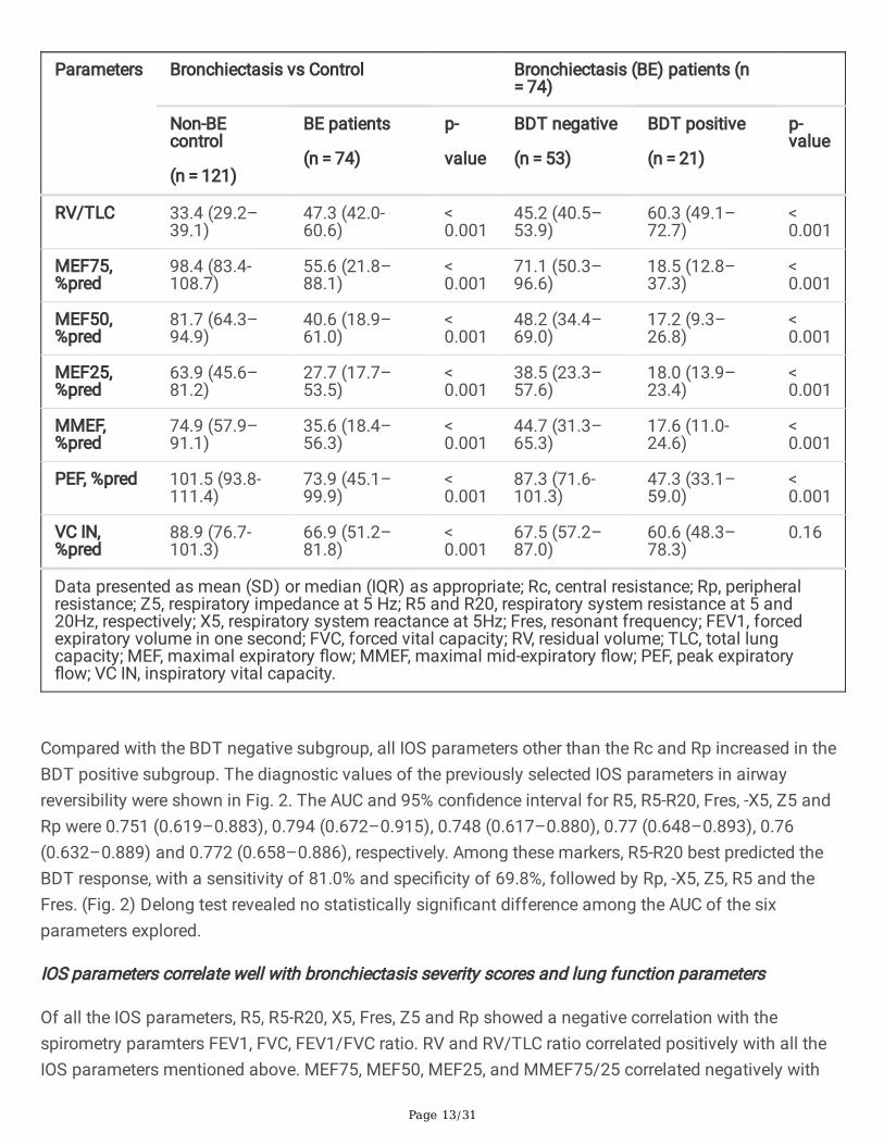
RV/TLC 33.4 (29.2–39.1)
47.3 (42.0-60.6)
< 0.001
45.2 (40.5–53.9)
60.3 (49.1–72.7)
< 0.001
MEF75,%pred
98.4 (83.4-108.7)
55.6 (21.8–88.1)
< 0.001
71.1 (50.3–96.6)
18.5 (12.8–37.3)
< 0.001
MEF50,%pred
81.7 (64.3–94.9)
40.6 (18.9–61.0)
< 0.001
48.2 (34.4–69.0)
17.2 (9.3–26.8)
< 0.001
MEF25,%pred
63.9 (45.6–81.2)
27.7 (17.7–53.5)
< 0.001
38.5 (23.3–57.6)
18.0 (13.9–23.4)
< 0.001
MMEF,%pred
74.9 (57.9–91.1)
35.6 (18.4–56.3)
< 0.001
44.7 (31.3–65.3)
17.6 (11.0-24.6)
< 0.001
PEF, %pred 101.5 (93.8-111.4)
73.9 (45.1–99.9)
< 0.001
87.3 (71.6-101.3)
47.3 (33.1–59.0)
< 0.001
VC IN,%pred
88.9 (76.7-101.3)
66.9 (51.2–81.8)
< 0.001
67.5 (57.2–87.0)
60.6 (48.3–78.3)
0.16
Data presented as mean (SD) or median (IQR) as appropriate; Rc, central resistance; Rp, peripheralresistance; Z5, respiratory impedance at 5 Hz; R5 and R20, respiratory system resistance at 5 and20Hz, respectively; X5, respiratory system reactance at 5Hz; Fres, resonant frequency; FEV1, forcedexpiratory volume in one second; FVC, forced vital capacity; RV, residual volume; TLC, total lungcapacity; MEF, maximal expiratory �ow; MMEF, maximal mid-expiratory �ow; PEF, peak expiratory�ow; VC IN, inspiratory vital capacity.
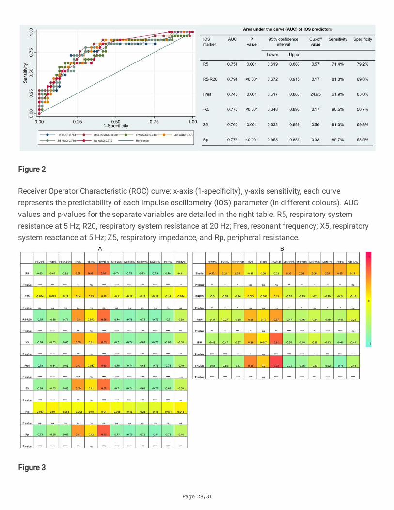
Compared with the BDT negative subgroup, all IOS parameters other than the Rc and Rp increased in theBDT positive subgroup. The diagnostic values of the previously selected IOS parameters in airwayreversibility were shown in Fig. 2. The AUC and 95% con�dence interval for R5, R5-R20, Fres, -X5, Z5 andRp were 0.751 (0.619–0.883), 0.794 (0.672–0.915), 0.748 (0.617–0.880), 0.77 (0.648–0.893), 0.76(0.632–0.889) and 0.772 (0.658–0.886), respectively. Among these markers, R5-R20 best predicted theBDT response, with a sensitivity of 81.0% and speci�city of 69.8%, followed by Rp, -X5, Z5, R5 and theFres. (Fig. 2) Delong test revealed no statistically signi�cant difference among the AUC of the sixparameters explored.
IOS parameters correlate well with bronchiectasis severity scores and lung function parameters
Of all the IOS parameters, R5, R5-R20, X5, Fres, Z5 and Rp showed a negative correlation with thespirometry paramters FEV1, FVC, FEV1/FVC ratio. RV and RV/TLC ratio correlated positively with all theIOS parameters mentioned above. MEF75, MEF50, MEF25, and MMEF75/25 correlated negatively with

Page 14/31
respiratory reactance and resistance, but not R20 and Rc. A strong correlation was observed between thetwo indicators of small airway dysfunction, the frequency dependence of resistance (FDR, R5-20) andMMEF75/25 (rs=-0.79, p < 0.0001). Fres demonstrated the strongest correlation with FEV1 (rs=-0.78, p < 0.0001), followed by FDR (rs=-0.75, p < 0.0001). (Fig. 3)
Regarding the correlation between bronchiectasis severity scores and lung function parameters, resultsshowed that FEV1, FVC, FEV1/FVC ratio correlated negatively with the BRICS, modi�ed Reiff, BSI andFACED scores. As low Bhalla scores re�ected high severity, a positive correlation was demonstratedbetweenBhalla score and lung function parameters. Similarly, MEF75, MEF50, MEF25, MMEF75/25 werealso negatively associated with bronchiectasis severity scores. The RV/TLC ratio showed a moderatecorrelation with BSI scores (rs = 0.61, p < 0.0001) and a strong correlation with FACED scores (rs = 0.72, p < 0.0001). (Fig. 3)
Regarding the correlation between IOS parameters and bronchiectasis severity scores, the modi�ed Reiffscores correlated positively with R5, R5-R20, Fres, Z5, and Rp,and negatively with X5. A similar correlationwas observed between BSI scores and IOS parameters. Compared with both the modi�ed Reiff and BSIscores, the FACED scores showed moderate correlation with IOS parameters. However, only a few IOSparameters (X5, Fres and Rp) correlated with Bhalla scores. No correlation between IOS parameters andBRICS scores was detected (data not shown). Overall, we noticed that the airway reactance (X5 and Fres)showed a stronger correlation with BSI scores than airway resistence. The modi�ed Reiff and FACEDscores, on the other hand, showed stronger correlation with Rp than airway reactance measurements (X5and Fres). (Fig. 4) In addition, the number of bronchiectasis lobes showed a weak correlation with IOSparameters R5, R5-R20, Fres, X5, Z5, and Rp. (Fig. 5)
IOS parameters do not correlate with Pseudomonas infection or hospital admission.
As shown in Table 4, patients chronically colonized with Pseudomonas had longer disease duration(median: 12.5 vs 3.0 years, p < 0.001). Acute exacerbation (p = 0.005) and hospital admission (p = 0.017)occurred more frequently in the Pseudomonas positive subgroup than the negative. Bronchiectasispatients with chronic Pseudomonas infection also presented lower FVC, VC, and PEF than thePseudomonas negative subgroup. However, there were no signi�cant differences in IOS parametersbetween Pseudomonas positive and negative subgroups Patients with previous hospital admission hadlonger disease duration and had a higher number of acute exacerbations than the hospital admissionfree group. Nevertheless, there were no signi�cant differences in IOS parameters between patients withprevious hospital admission and those without,, although the traditional lung parameters FVC, FEV1signi�cantly decreased in patients with previous hospital admission. (Table 5)

Page 15/31
Table 4Comparison between bronchiectasis patients in terms of the chronic colonisation of Pseudomonas
aeruginosa.Parameters Pseudomonas negative
(n = 63)
Pseudomonas positive
(n = 11)
p-value
Age, yrs 60.5 (10.5) 59.7 (15.5) 0.83
Gender 0.47
Male 36 (57.1%) 5 (45.5%)
Female 27 (42.9%) 6 (54.5%)
BMI, kg·m− 2 23.0 (4.6) 20.5 (4.1) 0.10
Disease years 3.0 (1.0, 5.0) 12.5 (5.0, 18.0) < 0.001
Exacerbation 0.0 (0.0, 1.0) 2.0 (1.0, 3.0) 0.005
Hospital admission 0.0 (0.0, 0.0) 0.0 (0.0, 1.0) 0.017
mMRC score 0.051
0 2 (3.2%) 0 (0.0%)
1 31 (49.2%) 2 (18.2%)
2 21 (33.3%) 4 (36.4%)
3 8 (12.7%) 3 (27.3%)
4 1 (1.6%) 2 (18.2%)
IOS and lung function parameters
Rc, kpa/l/s 0.3 (0.2, 0.3) 0.3 (0.3, 0.3) 0.15
Rp, kpa/l/s 0.3 (0.3, 0.6) 0.7 (0.3, 0.9) 0.060
Z5, kpa/l/s 0.5 (0.4, 0.7) 0.6 (0.5, 0.8) 0.11
R5, kpa/l/s 0.5 (0.4, 0.6) 0.6 (0.4, 0.6) 0.22
R20, kpa/l/s 0.3 (0.3, 0.4) 0.4 (0.3, 0.4) 0.64
R5-R20, kpa/l/s 0.1 (0.1, 0.3) 0.2 (0.1, 0.3) 0.22
X5, kpa/l/s -0.2 (-0.3, -0.1) -0.3 (-0.5, -0.2) 0.056
Rc, central resistance; Rp, peripheral resistance; Z5, respiratory impedance at 5 Hz; R5 and R20,respiratory system resistance at 5 and 20Hz, respectively; X5, respiratory system reactance at 5Hz;Fres, resonant frequency; FEV1, forced expiratory volume in one second; FVC, forced vital capacity; RV,residual volume; TLC, total lung capacity; MEF, maximal expiratory �ow; MMEF, maximal mid-expiratory �ow; PEF, peak expiratory �ow; VC IN, inspiratory vital capacity.

Page 16/31
Parameters Pseudomonas negative
(n = 63)
Pseudomonas positive
(n = 11)
p-value
Fres, Hz 18.3 (15.3, 25.2) 23.2 (20.2, 26.1) 0.14
FEV1, %pred 72.5 (27.0) 57.9 (25.5) 0.098
FVC, %pred 85.0 (19.8) 71.3 (23.8) 0.044
FEV1/FVC 67.0 (15.7) 65.9 (17.0) 0.83
RV, %pred 130.0 (98.5, 165.3) 121.2 (107.9, 157.9) 0.68
TLC, %pred 100.1 (87.4, 112.4) 93.4 (88.9, 105.0) 0.27
RV/TLC 46.8 (41.3, 60.5) 53.9 (46.7, 64.1) 0.085
MEF75, %pred 58.7 (22.6, 94.9) 37.7 (17.7, 57.8) 0.11
MEF50, %pred 44.4 (18.9, 64.8) 31.1 (12.0, 46.0) 0.21
MEF25, %pred 28.0 (18.4, 53.2) 25.5 (14.2, 55.1) 0.67
MMEF, %pred 36.5 (19.4, 61.4) 29.1 (12.2, 42.5) 0.30
PEF, %pred 77.9 (47.3, 100.4) 56.2 (32.0, 74.4) 0.034
VC IN, %pred 68.5 (57.2–87.0) 58.8 (29.7–75.1) 0.039
Rc, central resistance; Rp, peripheral resistance; Z5, respiratory impedance at 5 Hz; R5 and R20,respiratory system resistance at 5 and 20Hz, respectively; X5, respiratory system reactance at 5Hz;Fres, resonant frequency; FEV1, forced expiratory volume in one second; FVC, forced vital capacity; RV,residual volume; TLC, total lung capacity; MEF, maximal expiratory �ow; MMEF, maximal mid-expiratory �ow; PEF, peak expiratory �ow; VC IN, inspiratory vital capacity.

Page 17/31
Table 5Comparison between bronchiectasis patients with and without hospital admission.
Parameters No hospital admission
(n = 64)
Hospital admission
(n = 10)
p-value
Age, yrs 59.6 (11.5) 65.3 (8.7) 0.14
Gender 0.32
Male 34 (53%) 7 (70%)
Female 30 (47%) 3 (30%)
BMI, kg·m− 2 23.1 (4.5) 19.5 (3.9) 0.017
Disease years 3.0 (1.0-5.5) 13.5 (3.0–20.0) 0.020
Exacerbation 0.0 (0.0–1.0) 2.0 (1.0–3.0) 0.002
Hospital admission 0.0 (0.0–0.0) 1.0 (1.0–1.0) < 0.001
mMRC < 0.001
0 2 (3%) 0 (0%)
1 33 (52%) 0 (0%)
2 21 (33%) 4 (40%)
3 7 (11%) 4 (40%)
4 1 (2%) 2 (20%)
IOS and lung function parameters
Rc, kpa/l/s 0.3 (0.2–0.3) 0.2 (0.2–0.3) 0.18
Rp, kpa/l/s 0.3 (0.3–0.6) 0.8 (0.3-1.0) 0.094
Z5, kpa/l/s 0.5 (0.4–0.7) 0.7 (0.4–0.9) 0.21
R5, kpa/l/s 0.5 (0.4–0.6) 0.6 (0.4–0.8) 0.43
R20, kpa/l/s 0.3 (0.3–0.4) 0.3 (0.3–0.4) 0.21
R5-R20, kpa/l/s 0.1 (0.1–0.2) 0.3 (0.1–0.4) 0.074
X5, kpa/l/s -0.2 (-0.3–0.1) -0.4 (-0.6–0.1) 0.067
Fres, Hz 19.5 (15.3–25.1) 24.4 (18.3–29.2) 0.13
Rc, central resistance; Rp, peripheral resistance; Z5, respiratory impedance at 5 Hz; R5 and R20,respiratory system resistance at 5 and 20Hz, respectively; X5, respiratory system reactance at 5Hz;Fres, resonant frequency; FEV1, forced expiratory volume in one second; FVC, forced vital capacity; RV,residual volume; TLC, total lung capacity; MEF, maximal expiratory �ow; MMEF, maximal mid-expiratory �ow; PEF, peak expiratory �ow; VC IN, inspiratory vital capacity.

Page 18/31
Parameters No hospital admission
(n = 64)
Hospital admission
(n = 10)
p-value
FEV1, %pred 73.1 (25.8) 52.8 (30.1) 0.026
FVC, %pred 86.0 (18.8) 63.4 (23.6) 0.001
FEV1/FVC 67.2 (14.8) 64.2 (21.6) 0.58
RV, %pred 128.2 (100.8-164.3) 124.1 (101.0-180.0) 0.89
TLC, %pred 99.7 (90.6-112.5) 92.5 (83.2-104.5) 0.12
RV/TLC 46.8 (41.7–59.0) 63.5 (46.8–72.1) 0.060
MEF75, %pred 57.8 (27.2–89.6) 25.6 (10.1–88.0) 0.082
MEF50, %pred 43.0 (20.3–62.3) 20.1 (8.3–46.7) 0.12
MEF25, %pred 27.6 (18.5–53.3) 43.8 (13.0-53.5) 0.72
MMEF, %pred 36.5 (20.8–58.9) 21.6 (8.3–48.9) 0.18
PEF, %pred 77.3 (49.2–100.0) 36.6 (27.4–88.8) 0.060
VC IN, %pred 67.5 (56.3–85.3) 59.7 (29.7–75.1) 0.072
Rc, central resistance; Rp, peripheral resistance; Z5, respiratory impedance at 5 Hz; R5 and R20,respiratory system resistance at 5 and 20Hz, respectively; X5, respiratory system reactance at 5Hz;Fres, resonant frequency; FEV1, forced expiratory volume in one second; FVC, forced vital capacity; RV,residual volume; TLC, total lung capacity; MEF, maximal expiratory �ow; MMEF, maximal mid-expiratory �ow; PEF, peak expiratory �ow; VC IN, inspiratory vital capacity.
DiscussionResults of the present study supported previous �ndings that IOS parameters correlated with the severityof bronchiectasis assessed by scoring tools that incoorperate clinical characteristics, such as the FACEDand BSI scores. Both the airway resistance and reactance increased as bronchiectasis severity advanced.Furthermore, the present study explored the correlation between IOS parameters and airway reversibility. Alarge proportion of patients (28%) in the cohort showed positive responses to the β2 agonist salbutamol.IOS parameter R5-R20 was found to best predict BDT response. To the best of our knowledge, this wasthe �rst study that demonstrated the usefulness of IOS in the assessment of airway reversibility in non-cystic �brosis bronchiectasis.
Bronchiectasis is a heterogenous disease. Many factors contribute to the development of bronchiectasis,including the early infection by Mycobacteria tuberculosis and chronic colonisation by microorganismwith potential pathogenicity (MPP).[2] In the present study, 21.6% of the recruited bronchiectasis patientspresented with a history of tuberculosis infection. Airway pathogenswere isolated in 23% of patients, andPseudomonas aeruginosa was found to be the major MMP in our study. Infections are a common cause

Page 19/31
of bronchiesctassi. In a Greek study, previous infection (25.2%) and previous tuberculosis (TB) (22.3%)were the most commonly identi�ed underlying conditions in bronchiectasis.[15] Data from a recent largecohort study suggests that previous infection was the ethiology of bronchiectasis in 40.4% of the casesin the Caucasian population. The presence of TB was observed in 13.5% of the patients, while 25.6% ofthese patients had chronic infection related to Pseudomonas aeruginosa.[16]. Another study in aTaiwanese population indicated that 12% of bronchiectasis cases could be attributed to an early infectionof Mycobacteria tuberculosis.[17] The apparent differences in percentage could be explained bygeographical differencesand relatively small sample sizes. Interestingly, in the European study, thepresence of Hemophilus in�uenzae and Pseudomonas aeruginosa were identi�ed as the most commonpathogens, while in the US, non-tuberculous mycobacteria (NTM) and Pseudomonas aeruginosa werefound to be relatively common, while Hemophilus in�uenzae were less often detectedin bronchiectasis.[5,18]
Bronchial enlargement, bronchial wall thickening, and other individual features of bronchiectasis could beeasily recognised on a HRCT scan of the thorax. However, systematic quanti�cation of theseabnormalities could be more di�cult. In this study, we use multidimensional scoring tools to stratify theseverity of bronchiectasis into mild, moderate, and severe. Accurate strati�cation of bronchiectasis couldallow clinicians to take relevant preventive measures and prescribe adequate treatment and medication.[19, 20] In the present study, 85.1% of patients were categorised into mild (48.6%) and moderate (36.5%)broncheistasis according to the BSI[5] scores, which mean that the mortality and hospitalisation rate in 4years were predicted to rise to 11.3% and 19.4%, respectively.[19] Similar results are obtained using theFACED[4] scores, where 89.2% were categorised into mild (64.9%) and moderate (24.3%) bronchiestasis.Patients with mild and moderate disease severity according to FACED were predicted to have a 5-yearmortality of 4% and 25%, respectively.[19] The modi�ed Reiff[3] score, which could better re�ectairwaydamages instead categorized 87.8% of the included patients as mild or moderate disease. These resultswere in line with the study by Radovanovic et al.[21], and consistent with the results usingFACED and BSIscores. Complication such as atelectasis and lung consolidation are not uncommonin bronchiectasis. Inthe present study, more than one-third of the patients showed atelectasis or lung consolidation on CTscan. In cystic �brosis patients, such abnormalities might be considered a biomarker that predicts laterdevelopment of bronchiectasis if detected in childhood.[22]
Airway reversibility tests showed that that 21 out of 74 (28%) bronchiesctasis patients in the presentstudy were BDT positive. The British Thoracic Society guideline[20] for non-cystic �brosis bronchiectasisrecommends reversibility testing to identify co-existing obstructive conditions. An Australian studysuggested that 17% of hospitalised bronchiectasis patients had bronchodilator reversibility in their largeairways (FEV1). Furthermore, 41% of the subjects also showed reversibility in their small airways.However, the study population was relatively small and many patients presented with asthma (44%) orchronic obstructive lung disease (COPD) (59%) comorbidities[23] However, after excluding all patientswith comorbid asthma and COPD, another study on patients with stable bronchiectasis still demonstrateda large proportion with positive BDT.[24] Previous studies had shown that BDT could be positive in up to25% of patients with bronchiectasis.[18, 24, 25]

Page 20/31
Spirometry, which is currently ultilized in the clinic for measurement of lung function primarily re�ectair�ow characteristics, and the results depend on the central airway diameter, lung volume, lung elasticretraction force, respiratory muscle strength, and forced expiratory pattern[26]. Spirometry parameters likeFEV1 and FVC, which are commonly used to assess the degree of airway obstruction in obstructivepulmonary disease such as asthma and COPD has inherient limitations when used to evaluate theseverity of bronchiectasis because the pathophysiological mechanisms of bronchiectasis are differentfrom those of obstructive lung disease. In contrast, IOS mainly evaluates the airway diameter andmeasures small airway function. Preformance of IOS does not require forced exhalation and is notrelated to respiratory muscle strength. The results of the present study show that IOS parameters thatre�ect airway resistance and reactance increased with increased severity of bronchiectasis, suggestingthat IOS can be useful in the assessment bronchiectasis severity. Our data are consistent with the resultsby Yamamoto et al.[9]. Besides FACED and BSI scores, we also validated IOS’s usefulness in assessingthe severity of bronchiectasis strati�ed by predominately radiological scoring system such as themodi�ed Reiff score. Our results con�rmed the value of IOS parameters in differentiating the degree ofradiological airway changes. However, in our study, theRc and R20 were not statistically signi�cantamong the different bronchiectasis severity subgroups. Notably, these two parameters mainly measurecentral airway resistance. These results suggest that the degree of central airway obstruction is similar inbronchiestasis regardless of disease severity. In summary, IOS parameters correlate well with bothradiographic and clinical severity of bronchiectasis.
In the present study, bronchiectasis patients with positive BDT had decreased FEV1 and FEV1/FVC ratioon spirometry, demonstrating an obstructive ventilatory dysfunction. When we look at the IOS parameters,the small airway resistance indicator R5-R20 increased, but the large airway resistance indicators Rc andR20 remain the same. This suggested that airway obstruction in bronchiestasis mainly occurs inperipheral airways and not central ones. This could be further con�rmed by the decrease of the MEF25and MMEF75/25, parameters re�ecting the changes in the small airways’ �ow rate. In addition, the airwaytrapping markers RV and RV/TLC ratio increased in bronchiectasis. The pathophysiological features ofbronchiectasis are heterogeneous, both obstructive and restrictive ventilation dysfunction may present.However, a multicenter, prospective and observational study suggests that airway trapping and lungdiffusing capacity impairment are the most common lung function abnormalities in bronchiectasis.[21]
ROC curves were drawn to �nd out which IOS parameter best predicted airway reversibility inbronchiesctasis. The AUC of IOS parameters indicated that the small airway resistance indicator R5-R20was superior to other IOS paremeters in differenting between patients with positive BDT and negativeBDT. In bronchiectasis, air�ow obstruction was found to be predominantly due to pathologies in the smalland medium airways.[27] An infection process in the small airways could lead to the release ofin�ammatory mediators such as proteases and elastases.[28] As consequences, the airway epitheliumwould be in�ltrated by in�ammatory cytokines and free radicals, promoting bronchial wall thickening andobstruction of small airways, as well as persistent bronchial dilatation.[29] The above condition wouldlead to reduction in the effective cross-sectional area of the peripheral airways, thus increasing airwayresistance, and impairing the lung elastic capacity accordingly. Guan et al.[24] showed that airway

Page 21/31
resersibility in bronchiectasis patients was associated with poorer lung function. Furthermore, a study onCOPD patients showed that the reactance indices X5, Fres and the resistance Z5 and R5-R20 could helpidentify patients with FEV1% predicted less than 50%.[30]
In the present study, all IOS parameters except for R20 and Rp all correlated negatively with lungspirometry variables and positively with airway trapping markers. Previous studies have demonstrated agood correlation between IOS parameters and spirometric parameters in diseases like asthma, COPD andcystic �brosis.[30–32] In bronchiectasis, lower FEV1 is associated with increased bronchiectasis severity.[33] Also, the extent of bronchiectasis and bronchial wall thickening is inversely associated with lungfunction.[34] Our results revealed that IOS parameters correlated well with both lung function and diseaseseverity. This suggests that IOS may serve as an useful alternative to assess disease severity andmonitor disease progression n bronchiectasis. However, unlike traditional spirometeric parameters, IOSwas unable to distinguish patients with Pseudomonas aeruginosa infection from those withoutinfections. IOS parameters also seem to be less in�uenced by the previous condition like hospitaladmission.
The present study has some limitations. First, it was a single-centre investigation, and the �ndings couldhave been affected by selection bias. Second, although IOS is an emerging tool in the analysis of airwayresistance and reactance in bronchiectasis, there are relatively few studies in this area, and no referencestandard for IOS measurements has been established. Further study based on a large population isrequired to corroborate our �ndings and to establish standarised guidelines.
ConclusionIn conclusion, this study supports the use of IOS to assess the severity of bronchiectasis. IOS parameterscorrelate well with disease severity strati�ed using multidimensional assessment tools that takes intoaccount both clinical and radiological features. Furthermore, IOS parameters are shown to be effective inthe prediction of airway reversibility in patients with bronchiectasis. In the future, IOS may function as analternative to spirometry, to evaluate lung function in adults with bronchiectasis.
AbbreviationsABPA: Allergic bronchopulmonary aspergillosis
ATS: American Thoracic Society
AUC: Area under the curve
BDR: Bronchodilator response
BDT: Bronchial dilation test
BSI: Bronchiectasis Severity Index

Page 22/31
COPD: Chronic obstructive pulmonary disease
ERS: European Respiratory Society
FDR: Frequency dependence of resistance
FEV1: Forced expiratory volume in one second
Fres: Resonant frequency
FVC: Forced vital capacity
GINA: Global Initiative for Asthma
HRCT: High-resolution computed tomography
IOS: Impulse oscillometry
IQR: Interquartile range
MEF75: Maximal expiratory �ow at 75% of the FVC
MEF50: Maximal expiratory �ow at 50% of the FVC
MEF25: Maximal expiratory �ow at 25% of the FVC
MMEF: Maximal mid-expiratory �ow
PCCM: Pulmonary and Critical Care Medicine
PEF: Peak expiratory �ow
Rc: Central resistance
Rp: Peripheral resistance
RV: Residual volume
R5: Resistance at 5 Hz
R20: Resistance at 20 Hz
TLC: Total lung capacity
X5: Reactance at 5 Hz
Z5: Impedance value at 5 Hz.

Page 23/31
DeclarationsAvailability of data and materials
The datasets used and analysed during the current study are available from the corresponding author onreasonable request.
Ethics approval and consent to participate
The study was approved by the Ethics Committee of the Fifth A�liated Hospital of Sun Yat-senUniversity. All patients provided written informed consent to participate in the study. (approve number[2015] K02-1).
Consent for publication
Not applicable
Funding
JL was supported by the Zhuhai science and technology innovation Bureau, Novel coronavirus epidemicprevention and control emergency research (grant numbers ZH22036302200021PWC) and theGuangdong Basic and Applied Basic Research Foundation (2020A1515011147). XZ received fundingfrom the Fundamental Research Funds for the Central Universities (19ykpy51); the Medical Scienti�cResearch Foundation of Guangdong Province of China (A2020176) and the Science and TechnologyPlanning Project for medicine of Zhuhai City (ZH2202200012HJL). The authors state that neither thestudy design, results, interpretation of the �ndings, nor any other subject discussed in the submittedmanuscript was dependent on �nancial support.
Competing interests
The authors declare that they have no competing interests.
Authors' contributions
CT participated in drafting the work, acquisition, analysis, and interpretation of data. DM participated indrafting the work, acquisition, analysis, and interpretation of data. CT, MC, KW, XZ, YH, ZW, JWparticipated in the data acquisition and interpretation. JH participated in the conception and design of thework. JL conceived of the study, and participated in its design and coordination and helped to draft themanuscript. All authors read and approved the �nal manuscript.
Acknowledgements
We would like to extend our gratitude to Ms Chaolin Chen, Ms Shaofen Gao, Ms Xiaoling Huang and MsShican Yuan (the lung function laboratory of The Fifth A�liated Hospital of Sun Yat-sen University,Zhuhai, China) for their PFT data collection.

Page 24/31
References1. Bickel S, Popler J, Lesnick B, Eid N. Impulse oscillometry: Interpretation and practical applications.
Chest American College of Chest Physicians; 2014; 146: 841–847.
2. Flume PA, Chalmers JD, Olivier KN. Advances in bronchiectasis: endotyping, genetics, microbiome,and disease heterogeneity. Lancet Elsevier Ltd; 2018; 392: 880–890.
3. Reiff DB, Wells AU, Carr DH, Cole PJ, Hansell DM. CT �ndings in bronchiectasis: limited value indistinguishing between idiopathic and speci�c types. Am. J. Roentgenol. 1995; 165: 261–267.
4. Martinez-Garcia MA, de Gracia J, Vendrell Relat M, Giron R-M, Maiz Carro L, de la Rosa Carrillo D,Olveira C. Multidimensional approach to non-cystic �brosis bronchiectasis: the FACED score. Eur.Respir. J. 2014; 43: 1357–1367.
5. Chalmers JD, Goeminne P, Aliberti S, McDonnell MJ, Lonni S, Davidson J, Poppelwell L, Salih W, PesciA, Dupont LJ, Fardon TC, De Soyza A, Hill AT. The bronchiectasis severity index. An internationalderivation and validation study. Am. J. Respir. Crit. Care Med. 2014; 189: 576–585.
�. Postma DS, Brightling C, Baldi S, Van den Berge M, Fabbri LM, Gagnatelli A, Papi A, Van der Molen T,Rabe KF, Siddiqui S, Singh D, Nicolini G, Kraft M, Pizzichini E, Cukier A, Stelmach R, Olivenstein R,Zhang Q, Badorrek P, Gessner C, Scichilone N, Chetta A, Paggiaro P, Milleri S, D’Amato M, SpanevelloA, Foschino MP, Boersma WG, Broeders M, Vroegop JS, et al. Exploring the relevance and extent ofsmall airways dysfunction in asthma (ATLANTIS): baseline data from a prospective cohort study.Lancet Respir. Med. Lancet Publishing Group; 2019; 7: 402–416.
7. Cottini M, Licini A, Lombardi C, Berti A. Clinical Characterization and Predictors of IOS-De�ned Small-Airway Dysfunction in Asthma. J. Allergy Clin. Immunol. Pract. American Academy of Allergy,Asthma and Immunology; 2020; 8: 997-1004.e2.
�. Guan W -j., Yuan J -j., Gao Y -h., Li H -m., Zheng J -p., Chen R -c., Zhong N -s. Impulse Oscillometryand Spirometry Small-Airway Parameters in Mild to Moderate Bronchiectasis. Respir. Care AmericanAssociation for Respiratory Care; 2016; 61: 1513–1522.
9. Yamamoto Y, Miki K, Tsujino K, Kuge T, Matsuki T, Fukushima K, Oshitani Y, Kagawa H, Yoshimura K,Miki M, Kida H. Evaluation of disease severity in bronchiectasis using impulse oscillometry. ERJOpen Res. European Respiratory Society (ERS); 2020; 6: 00053–02020.
10. Miller MR, Hankinson J, Brusasco V, Burgos F, Casaburi R, Coates A, Crapo R, Enright P, van derGrinten CPM, Gustafsson P, Jensen R, Johnson DC, MacIntrye N, McKay R, Navajas D, Pedersen OF,Pellegrino R, Viegi G, Wagner J. Standardisation of spirometry. Eur. Respir. J. European RespiratorySociety; 2005. p. 319–338.
11. Pellegrino R, Viegi G, Brusasco V, Crapo RO, Burgos F, Casaburi R, Coates A, van der Grinten CPMM,Gustafsson P, Hankinson J, Jensen R, Johnson DC, MacIntyre N, McKay R, Miller MR, Navajas D,Pedersen OF, Wanger J. Interpretative strategies for lung function tests. Eur. Respir. J. EuropeanRespiratory Society; 2005; 26: 948–968.

Page 25/31
12. Bhalla M, Turcios N, Aponte V, Jenkins M, Leitman BS, McCauley DI, Naidich DP. Cystic �brosis:scoring system with thin-section CT. Radiology 1991; 179: 783–788.
13. Bedi P, Chalmers JD, Goeminne PC, Mai C, Saravanamuthu P, Velu PP, Cartlidge MK, Loebinger MR,Jacob J, Kamal F, Schembri N, Aliberti S, Hill U, Harrison M, Johnson C, Screaton N, Haworth C,Polverino E, Rosales E, Torres A, Benegas MN, Rossi AG, Patel D, Hill AT. The BRICS (BronchiectasisRadiologically Indexed CT Score). Chest Elsevier Inc; 2017; : 1–10.
14. DeLong ER, DeLong DM, Clarke-Pearson DL. Comparing the Areas under Two or More CorrelatedReceiver Operating Characteristic Curves: A Nonparametric Approach. Biometrics JSTOR; 1988; 44:837.
15. Dimakou K, Trianta�llidou C, Toumbis M, Tsikritsaki K, Malagari K, Bakakos P. Non CF-bronchiectasis: Aetiologic approach, clinical, radiological, microbiological and functional pro�le in277 patients. Respir. Med. W.B. Saunders Ltd; 2016; 116: 1–7.
1�. Martinez-García MA, Villa C, Dobarganes Y, Girón R, Maíz L, García-Clemente M, Sibila O, Golpe R,Rodríguez J, Barreiro E, Rodriguez JL, Menéndez R, Prados C, de la Rosa D, Olveira C. RIBRON: Thespanish Online Bronchiectasis Registry. Characterization of the First 1912 Patients. Arch.Bronconeumol. 2020; .
17. Huang H-Y, Chung F-T, Lo C-Y, Lin H-C, Huang Y-T, Yeh C-H, Lin C-W, Huang Y-C, Wang C-H. Etiologyand characteristics of patients with bronchiectasis in Taiwan: a cohort study from 2002 to 2016.BMC Pulm. Med. BioMed Central Ltd.; 2020; 20: 45.
1�. Aksamit TR, O’Donnell AE, Barker A, Olivier KN, Winthrop KL, Daniels MLA, Johnson M, Eden E, Gri�thD, Knowles M, Metersky M, Salathe M, Thomashow B, Tino G, Turino G, Carretta B, Daley CL. AdultPatients With Bronchiectasis: A First Look at the US Bronchiectasis Research Registry. Chest ElsevierLtd; 2017; 151: 982–992.
19. Chalmers JD, Chang AB, Chotirmall SH, Dhar R, McShane PJ. Bronchiectasis. Chalmers J, PolverinoE, Aliberti S, editors. Nat. Rev. Dis. Prim. Cham: Springer International Publishing; 2018.
20. Hill AT, Sullivan AL, Chalmers JD, De Soyza A, Stuart Elborn J, Andres Floto R, Grillo L, Gruffydd-Jones K, Harvey A, Haworth CS, Hiscocks E, Hurst JR, Johnson C, Peter Kelleher W, Bedi P, Payne K,Saleh H, Screaton NJ, Smith M, Tunney M, Whitters D, Wilson R, Loebinger MR. British thoracicsociety guideline for bronchiectasis in adults. Thorax BMJ Publishing Group; 2019.
21. Radovanovic D, Santus P, Blasi F, Sotgiu G, D’Arcangelo F, Simonetta E, Contarini M, Franceschi E,Goeminne PC, Chalmers JD, Aliberti S. A comprehensive approach to lung function in bronchiectasis.Respir. Med. W.B. Saunders Ltd; 2018; 145: 120–129.
22. Wijker NE, Vidmar S, Grimwood K, Sly PD, Byrnes CA, Carlin JB, Cooper PJ, Robertson CF, Massie RJ,Kemner van de Corput MPC, Cheney J, Tiddens HAWM, Wainwright CE. Early markers of cystic�brosis structural lung disease: follow-up of the ACFBAL cohort. Eur. Respir. J. European RespiratorySociety; 2020; 55: 1901694.
23. Venning V, Bartlett J, Jayaram L. Patients hospitalised with an infective exacerbation ofbronchiectasis unrelated to cystic �brosis: Clinical, physiological and sputum characteristics.

Page 26/31
Respirology Blackwell Publishing; 2017; 22: 922–927.
24. Guan WJ, Gao YH, Xu G, Li HM, Yuan JJ, Zheng JP, Chen RC, Zhong NS. Bronchodilator response inadults with bronchiectasis: Correlation with clinical parameters and prognostic implications. J.Thorac. Dis. Pioneer Bioscience Publishing; 2016; 8: 14–23.
25. Jeong HJ, Lee H, Carriere KC, Kim JH, Han JH, Shin B, Jeong BH, Koh WJ, Kwon OJ, Park HY. Effectsof long-term bronchodilators in bronchiectasis patients with air�ow limitation based onbronchodilator response at baseline. Int. J. COPD Dove Medical Press Ltd.; 2016. p. 2757–2764.
2�. Quanjer PH, Stanojevic S, Cole TJ, Baur X, Hall GL, Culver BH, Enright PL, Hankinson JL, Ip MSM,Zheng J, Stocks J, Schindler C. Multi-ethnic reference values for spirometry for the 3-95-yr age range:The global lung function 2012 equations. Eur. Respir. J. European Respiratory Society; 2012; 40:1324–1343.
27. Roberts HR, Wells AU, Rubens MB, Cole PJ, Hansell DM, Milne DG, Kolbe J. Air�ow obstruction inbronchiectasis: Correlation between computed tomography features and pulmonary function tests.Thorax Thorax; 2000; 55: 198–204.
2�. Polverino E, Rosales-Mayor E, Dale GE, Dembowsky K, Torres A. The Role of Neutrophil ElastaseInhibitors in Lung Diseases. Chest Elsevier Inc; 2017; 152: 249–262.
29. King PT. The pathophysiology of bronchiectasis. Int. J. Chron. Obstruct. Pulmon. Dis. Int J ChronObstruct Pulmon Dis; 2009. p. 411–419.
30. Wei X, Shi Z, Cui Y, Mi J, Ma Z, Ren J, Li J, Xu S, Guo Y. Impulse oscillometry system as an alternativediagnostic method for chronic obstructive pulmonary disease. Medicine (Baltimore). LippincottWilliams and Wilkins; 2017; 96: e8543.
31. Raj D, Sharma GK, Lodha R, Kabra SK. Correlation between impulse oscillometry and spirometryparameters in Indian patients with cystic �brosis. Chron. Respir. Dis. SAGE Publications Ltd; 2014;11: 139–149.
32. Díaz Palacios MÁ, Hervás Marín D, Giner Valero A, Colomer Hernández N, Torán Barona C, HernándezFernández de Rojas D. Correlation between impulse oscillometry parameters and asthma control inan adult population. J. Asthma Allergy Dove Medical Press Ltd.; 2019; Volume 12: 195–203.
33. Ooi GC, Khong PL, Chan-Yeung M, Ho JCM, Chan PKS, Lee JCK, Lam WK, Tsang KWT. High-resolution CT quanti�cation of bronchiectasis: Clinical and functional correlation. RadiologyRadiological Society of North America Inc.; 2002; 225: 663–672.
34. Habesoglu MA, Tercan F, Ozkan U, Eyuboglu FO. Effect of radiological extent and severity ofbronchiectasis on pulmonary function. Multidiscip. Respir. Med. BioMed Central Ltd.; 2011; 6: 284–290.
Figures

Page 27/31
Figure 1
Results of respiratory impedance at rest strati�ed by different bronchiectasis severity scores (n=74). A toH correspond to the modi�ed Reiff score, I to P correspond to the BSI score, and Q to X correspond to theFACED score. R5, respiratory system resistance at 5 Hz; R20, respiratory system resistance at 20 Hz; X5,respiratory system reactance at 5 Hz; Fres, resonant frequency; Z5, respiratory impedance; Rc, centralresistance; and Rp, peripheral resistance. Statistics performed by Kruskal–Wallis test.
Page 28/31
Figure 2
Receiver Operator Characteristic (ROC) curve: x-axis (1-speci�city), y-axis sensitivity, each curverepresents the predictability of each impulse oscillometry (IOS) parameter (in different colours). AUCvalues and p-values for the separate variables are detailed in the right table. R5, respiratory systemresistance at 5 Hz; R20, respiratory system resistance at 20 Hz; Fres, resonant frequency; X5, respiratorysystem reactance at 5 Hz; Z5, respiratory impedance, and Rp, peripheral resistance.
Figure 3

Page 29/31
Spearman correlation analysis. A) between IOS parameters and lung function; B) between lung functionand bronchiectasis severity scores. *p<0.05; **p<0.01; ***p<0.001; ****p<0.0001; ns, no signi�cance. Rc,central resistance; Rp, peripheral resistance; Z5, the respiratory impedance; R5 and R20, respiratorysystem resistance at 5 and 20Hz, respectively; X5, respiratory system reactance at 5Hz; Fres, resonantfrequency. FEV1, forced expiratory volume in one second; FVC, forced vital capacity; RV, residual volume;TLC, total lung capacity; MEF, maximal expiratory �ow; MMEF, maximal mid-expiratory �ow; PEF, peakexpiratory �ow.

Page 30/31
Figure 4
Spearman correlation analysis between IOS parameters and bronchiectasis severity scores. A to Fcorrespond to the Reiff score, G to L correspond to the BSI score, M to R correspond to the FACED score,and S to U correspond to the Bhalla score. R5, respiratory system resistance at 5 Hz; R20, respiratorysystem resistance at 20 Hz; X5, respiratory system reactance at 5 Hz; Fres, resonant frequency; Z5, therespiratory impedance; Rc, central resistance; and Rp, peripheral resistance.
Figure 5
Correlation between IOS parameters and lobe engagement(with the lingula counted as an individuallobe). R5, respiratory system resistance at 5 Hz; R20, respiratory system resistance at 20 Hz; X5,respiratory system reactance at 5 Hz; Fres, resonant frequency; Z5, the respiratory impedance; Rc, centralresistance; and Rp, peripheral resistance.
Supplementary Files

Page 31/31
This is a list of supplementary �les associated with this preprint. Click to download.
supplementaryFigure1.tif
SupplementaryTables.docx