broad-spectrum ultraviolet (uv) radiation and uva, and uvb ... · final report on carcinogens...
TRANSCRIPT
FINAL
Report on Carcinogens Background Document for
Broad-Spectrum Ultraviolet (UV) Radiation and UVA, and UVB, and UVC December 13–14, 2000
Meeting of the NTP Board of Scientific Counselors Report on Carcinogens Subcommittee
Prepared for the: U.S. Department of Health and Human Services Public Health Service National Toxicology Program Research Triangle Park, NC 27709
Prepared by: Technology Planning and Management Corporation Canterbury Hall, Suite 310 4815 Emperor Blvd Durham, NC 27703 Contract Number N01-ES-85421


Do not quote or cite
RoC Background Document for Ultraviolet Radiation Dec. 2000
Criteria for Listing Agents, Substances or Mixtures in the Report on Carcinogens
U.S. Department of Health and Human Services National Toxicology Program
Known to be Human Carcinogens:
There is sufficient evidence of carcinogenicity from studies in humans, which indicates a causal relationship between exposure to the agent, substance or mixture and human cancer.
Reasonably Anticipated to be Human Carcinogens:
There is limited evidence of carcinogenicity from studies in humans which indicates that causal interpretation is credible but that alternative explanations such as chance, bias or confounding factors could not adequately be excluded; or
There is sufficient evidence of carcinogenicity from studies in experimental animals which indicates there is an increased incidence of malignant and/or a combination of malignant and benign tumors: (1) in multiple species, or at multiple tissue sites, or (2) by multiple routes of exposure, or (3) to an unusual degree with regard to incidence, site or type of tumor or age at onset; or
There is less than sufficient evidence of carcinogenicity in humans or laboratory animals, however; the agent, substance or mixture belongs to a well defined, structurally-related class of substances whose members are listed in a previous Report on Carcinogens as either a known to be human carcinogen, or reasonably anticipated to be human carcinogen or there is convincing relevant information that the agent acts through mechanisms indicating it would likely cause cancer in humans.
Conclusions regarding carcinogenicity in humans or experimental animals are based on scientific judgment, with consideration given to all relevant information. Relevant information includes, but is not limited to dose response, route of exposure, chemical structure, metabolism, pharmacokinetics, sensitive sub populations, genetic effects, or other data relating to mechanism of action or factors that may be unique to a given substance. For example, there may be substances for which there is evidence of carcinogenicity in laboratory animals but there are compelling data indicating that the agent acts through mechanisms which do not operate in humans and would therefore not reasonably be anticipated to cause cancer in humans.
i

Do not quote or citeRoC Background Document for Ultraviolet Radiation Dec. 2000
ii

Do not quote or cite
RoC Background Document for Ultraviolet Radiation Dec. 2000
Summary Statement Broad-Spectrum Ultraviolet (UV) Radiation and UVA, and UVB, and UVC
Carcinogenicity
Broad-spectrum ultraviolet radiation (UVR) is known to be a human carcinogen based on sufficient evidence of carcinogenicity from studies in humans. Epidemiology studies clearly demonstrate that exposure to broad spectrum UVR increases both melanocytic and non-melanocytic skin cancer. Studies of humans exposed to solar radiation, artificial devices emitting broad-spectrum UVR, and devices emitting predominantly ultraviolet A radiation (UVA) or ultraviolet B radiation (UVB) all contribute to this conclusion. Exposure to solar radiation is associated with an increased risk of malignant melanoma of the skin, non-melanoma skin cancer, malignant melanoma of the eye, and cancer of the lip (IARC 1992, NTP 2000). Evidence for the role of the UVR component of solar radiation in carcinogenicity comes from studies of human cancers associated with exposure to artificial UVR-emitting devices, tumor site-concordance between humans exposed to sunlight and animals exposed to UVR from artificial sources and human mechanistic studies using artificial sources of UVR. Exposure to sunlamps or sunbeds has been associated with malignant melanoma of the skin (Autier et al. 1994, Swerdlow et al. 1988, Walter et al. 1990, 1999, Westerdahl et al. 1994, 2000, Chen et al. 1998). Mechanistic studies using human tissue demonstrate that UVR is absorbed by DNA and causes direct and indirect DNA damage with mutagenic potential. Mutations found in the p53 tumor suppressor gene of human skin cancer are specific for UVR-induced damage (see below).
The findings in humans are supported by evidence in experimental animals. Exposure to broad spectrum UVR induced skin tumors (papilloma and squamous cell carcinoma) and eye tumors (spindle cell sarcoma) in albino rats and skin tumors (fibrosarcoma and/or squamous cell carcinoma) in mice, hamsters and opossum.
The epidemiological literature does not provide a basis for subdividing the effects of sunlight or artificial UVR into components attributable specifically to UVA, UVB, or ultraviolet C radiation (UVC). However, information regarding the specific effects of UVA, UVB, and UVC can be inferred from the results of human epidemiology studies of mixed UVR exposure together with the results of studies on the effects of specific UVR components in experimental animals and human tissues.
UVA is reasonably anticipated to be a human carcinogen based on limited evidence from studies in humans and evidence from studies in experimental animals. Studies in which UVA has contributed substantially to human exposure (solar radiation and UVA emitting sunbeds) have demonstrated an excess of skin cancer. Westerdahl et al. (2000) reported an association of melanoma with exposure to sunbeds when the majority of the exposure was considered to be from sunbeds emitting mainly UVA (source reported to emit 0.1% to 2.1% UVB). The finding in humans is supported by evidence in experimental animals. UVA exposure induced skin tumors in mice (squamous cell carcinoma and papilloma) and fish (melanoma).
iii

Do not quote or cite
RoC Background Document for Ultraviolet Radiation Dec. 2000
UVB is reasonably anticipated to be a human carcinogen based on limited evidence from studies in humans and evidence from studies in experimental animals. Mechanistic studies in humans have demonstrated that the UVB component in solar radiation is responsible for the mutagenic photoproducts that lead to the signature p53 mutations observed in human skin cancer. However, epidemiologic studies are limited by lack of information identifying exposure wavelength specificity. Although exposure to UVB, as a component of solar radiation or from sunlamps used before the early 1970s, is clearly associated with excess skin cancer, these human exposures are not solely to UVB but are confounded by exposures to other components of the UVR spectrum. In one study, exposure to sunlamps used in the early 1970s, which produced significant amounts of UVB (22% to 40%), was associated with cutaneous malignant melanoma (CMM) (Chen et al. 1998). The finding in humans is supported by evidence in experimental animals. Prolonged exposure to devices emitting primarily UVB caused the development of skin tumors in rats (papilloma), mice (squamous cell carcinoma, fibrosarcoma, papilloma, keratoacanthoma), guinea pigs (fibroma and trichofolliculoma), and opossums (melanocytic hyperplasia and melanoma).
UVC is reasonably anticipated to be a human carcinogen based on limited evidence from human mechanistic studies and evidence from studies in experimental animals. Studies of human tissue have demonstrated that both in vivo and in vitro exposure to UVC causes DNA damage. UVC is absorbed by DNA and induces mutagenic photoproducts similar to the types of damage caused by UVB. However, there are no epidemiologic studies adequate for evaluation of UVC carcinogenicity in humans. UVC is absorbed by the ozone layer and does not contribute to solar exposure, and studies using artificial devices emitting UVC are not specific for UVC radiation. Exposure of experimental animals to high doses of radiation from devices emitting primarily UVC caused skin tumors in rats (keratoacanthoma-like skin tumors) and mice (squamous cell carcinoma and fibrosarcoma).
Other Information Relating to Carcinogenesis or Possible Mechanisms of Carcinogenesis
Broad-spectrum UVR causes skin cancers via mechanisms that include DNA damage, immunosuppression, tumor promotion, and mutations in the p53 tumor suppressor gene. Broad-spectrum UVR induces mutations in cultured human cells, the type of damage depends upon the specific wavelength applied and the competence of an affected cell to repair the damage without error. DNA is a major cellular chromophore absorbing UVR (mainly UVB and UVC) and responds to irradiation by yielding free radical reactive intermediates and various photoproducts with mutagenic potential. UVB photons cause the following four major DNA base modifications in humans: (i) cyclobutane-type pyrimidine dimers, (ii) (6-4) photoproducts, (iii) the corresponding Dewar isomers, and (iv) thymine glycols. Both UVA and UVB induced 8- hydroxydeoxyguanosine produced from guanosine by the action of singlet oxygen.
UVA, UVB, and UVC as individual components of UVR are genotoxic in prokaryotes, lower eukaryotes, non-human mammalian cells, and human cells. Moreover, in vivo exposure from all three components of UVR results in DNA damage in humans. UVA’s
iv

Do not quote or cite
RoC Background Document for Ultraviolet Radiation Dec. 2000
biological effects are indirect and largely the result of energy transferred through active oxygen intermediates, whereas UVB and UVC photons are absorbed by DNA and direct damage occurs through DNA base modifications. Based on the number of positive genotoxic studies, UVC is the most potent and UVA is the least potent genotoxin of the components of broad spectrum UVR
More than 90% of human squamous-cell carcinomas contain mutations of the p53 tumor suppressor gene. These mutations were found in 74% of sun-exposed normal human skin, compared with 5% of unexposed skin, indicating a strong association with sun exposure. Observed p53 gene mutations were most frequently C to T or CC to TT transitions at pyrimidine-pyrimidine sequences. These specific 53 mutations are now considered a signature of UVR carcinogenesis.
Exposure to solar radiation and UVR has been found to alter immune function in humans and experimental animals. Evidence that immunosuppression is related to skin cancer incidence comes from the following observations that: (i) immunosuppressed organ transplant recipients showed a marked increase in skin cancer, particularly squamous-cell carcinoma, (ii) UVR decreased the ability to mount a delayed type hypersensitivity response, and (iii) mice exposed to low levels of UVR failed to reject highly immunogenic tumor cell lines.
Human skin grafts on mice also yielded human skin tumors (squamous cell carcinomas, actinic keratoses, melanocytic hyperplasia and melanoma) following irradiation with UVB after pretreatment with the carcinogen dimethylbenz(a)anthracene. Precancerous lesions (melanocytic hyperplasia) were found in human skin grafts on mice treated with UVB alone.
v

RoC Background Document for Ultraviolet Radiation Dec. 2000
vi

Do not quote or citeRoC Background Document for Ultraviolet Radiation Dec. 2000
Table of Contents
Criteria for Listing A gents, Substances of Mixtures in the Report on Carcinogens........................ i
Summary Statement .......................................................................................................................iii
1 Introduction ............................................................................................................................... 1
1.1 Identification of UVR by type.................................................................................... 2 1.2 Physical properties ..................................................................................................... 2 1.3 Photochemical and photobiological activities............................................................ 3
2 Human Exposure ....................................................................................................................... 5
2.1 Use.............................................................................................................................. 5 2.1.1 Cosmetic use ............................................................................................... 5 2.1.2 Medical and dental applications.................................................................. 5 2.1.3 Industrial applications ................................................................................. 6
2.2 Production .................................................................................................................. 6 2.3 Analysis...................................................................................................................... 6
2.3.1 Spectroradiometry....................................................................................... 6 2.3.2 Wavelength-independent (thermal) detectors ............................................. 6 2.3.3 Wavelength-dependent detectors ................................................................ 7
2.4 Environmental occurrence.......................................................................................... 7 2.5 Environmental exposure............................................................................................. 7
2.5.1 Solar UVR................................................................................................... 7 2.5.2 Artificial sources ....................................................................................... 10
2.6 Occupational exposure ............................................................................................. 11 2.6.1 Solar UVR................................................................................................. 11 2.6.2 Artificial UVR........................................................................................... 11
2.7 Biological indices of exposure ................................................................................. 11 2.8 Regulations............................................................................................................... 11
3 Human Cancer Studies ............................................................................................................ 17
3.1 Solar radiation .......................................................................................................... 18 3.1.1 Evaluations by the IARC (1992) and the NTP (2000).............................. 18 3.1.2 Recent epidemiologic studies.................................................................... 19
3.2 UVR from artificial sources ..................................................................................... 20 3.2.1 Cosmetically related UVR exposure......................................................... 20 3.2.2 Medically related UVR exposure.............................................................. 25 3.2.3 Occupationally related UVR exposure ..................................................... 27
3.3 DNA repair............................................................................................................... 28 3.4 Discussion ................................................................................................................ 28
3.4.1 UVA.......................................................................................................... 28 3.4.2 UVB .......................................................................................................... 29 3.4.3 UVC .......................................................................................................... 30
vii

Do not quote or citeRoC Background Document for Ultraviolet Radiation Dec. 2000
3.5 Summary .................................................................................................................. 30 4 Studies of Cancer in Experimental Animals ........................................................................... 47
4.1 Broad-spectrum UVR............................................................................................... 47 4.1.1 Rats ........................................................................................................... 47 4.1.2 Mice .......................................................................................................... 47 4.1.3 Hamsters.................................................................................................... 48 4.1.4 Guinea pigs ............................................................................................... 48 4.1.5 Other species ............................................................................................. 48 4.1.6 Action spectra ........................................................................................... 49
4.2 Primarily UVA ......................................................................................................... 49 4.2.1 Mice .......................................................................................................... 49 4.2.2 Other species ............................................................................................. 52
4.3 Primarily UVB ......................................................................................................... 53 4.3.1 Rats ........................................................................................................... 53 4.3.2 Mice .......................................................................................................... 53 4.3.3 Hamsters.................................................................................................... 54 4.3.4 Guinea pigs ............................................................................................... 54 4.3.5 Other species ............................................................................................. 55
4.4 Primarily UVC ......................................................................................................... 56 4.4.1 Rats ........................................................................................................... 56 4.4.2 Mice .......................................................................................................... 56
4.5 Cancer development in human-mouse chimera models........................................... 57 4.6 Summary .................................................................................................................. 57
5 Genotoxicity............................................................................................................................ 59
5.1 Methods for identifying and quantifying UVR-induced DNA lesions .................... 59 5.2 UVR-induced DNA photoproducts.......................................................................... 59
5.2.1 UVA-induced indirect DNA damage........................................................ 60 5.2.2 UVB-induced direct DNA damage ........................................................... 60 5.2.3 Cellular mechanisms for minimizing UVR-induced DNA damage ......... 60 5.2.4 Cellular responses to UVR-induced DNA damage................................... 61
5.3 Prokaryotic systems.................................................................................................. 61 5.3.1 Induction of mutation in Salmonella typhimurium................................... 61 5.3.2 Induction of mutation in Saccharomyces cerevisiae................................. 64
5.4 Plants and lower eukaryotic systems........................................................................ 64 5.5 Mammalian systems................................................................................................. 64
5.5.1 Nonhuman mammalian in vitro assays ..................................................... 64 5.5.2 Human in vitro assays ............................................................................... 65 5.5.3 Nonhuman mammalian in vivo assays...................................................... 66 5.5.4 Human in vivo assays ............................................................................... 66 5.5.5 Other in vitro and in vivo end points ........................................................ 68
viii

Do not quote or citeRoC Background Document for Ultraviolet Radiation Dec. 2000
5.5.6 Molecular epidemiological studies of DNA repair capacity..................... 68 5.6 Summary .................................................................................................................. 69
5.6.1 UVA.......................................................................................................... 69 5.6.2 UVB .......................................................................................................... 69 5.6.3 UVC .......................................................................................................... 70
6 Other Relevant Data ................................................................................................................ 73
6.1 Absorption and transmission of UVR in biological tissues ..................................... 73 6.2 Mechanisms of UV-induced skin cancer ................................................................. 74
6.2.1 DNA damage............................................................................................. 75 6.2.2 DNA repair................................................................................................ 77 6.2.3 Mutations .................................................................................................. 78 6.2.4 Tumor suppressor gene expression and mutation ..................................... 78 6.2.5 Immunosuppression .................................................................................. 79
6.3 Initiation and promotion........................................................................................... 80 6.4 Summary .................................................................................................................. 80
7 References ............................................................................................................................... 83
Appendix A: IARC Monograph of Evaluation of Carcinogenic Risks to Humans. Solar and Ultraviolet Radiation. Vol. 55. Lyon, France. World Health Organization. 1992. pp. A-1 - A-279. ...................................................................................................................... 99
Appendix B: Profile for Solar Radiation and Exposure to Sunlamps and Sunbeds. Report on Carcinogens, Ninth Edition (2000) .................................................................................. 101
List of Tables
Table 1-1 Optical radiation spectrum.............................................................................................. 1
Table 2-1. Percentage of daily UVA radiation received during two periods on a clear summer day ..................................................................................................................................... 8
Table 2-2. Typical values for ambient daily and annual UVB radiation expressed as minimal erythema dose ................................................................................................................... 8
Table 2-3. Percentage of daily UVB radiation received during two periods on a clear summer day ..................................................................................................................................... 9
Table 2-4. Representative terrain reflectance factors for horizontal surfaces measured with a UVB radiometer at 12:00 PM at various U.S. locations .................................................... 10
Table 2-5. FDA regulations........................................................................................................... 12
Table 2-6. OSHA Regulations ...................................................................................................... 15
Table 3-1. Epidemiologic studies of the relationship between cutaneous malignant melanoma and exposure to sunlamps or sunbeds (listed in chronological order by publication date)............................................................................................................................ 31
Table 3-2. Recent epidemiologic studies of the relationship between cancer and medically related UV exposure...................................................................................................................... 42
ix

Do not quote or citeRoC Background Document for Ultraviolet Radiation Dec. 2000
Table 3-3. Recent epidemiologic studies of the relationship between cancer and occupational UV exposure ............................................................................................................ 44
Table 4-1. Tumor incidences in female C3H/Tif mice exposed to UVA tanning sources with differing UVB emission levels.............................................................................................. 51
Table 4-2. Tumor incidences in female C3H/Tif mice exposed to broad-spectrum UVR and/or UVA................................................................................................................................... 52
Table 4-3. Incidences of melanoma in hybrid fish (Xiphophorus) exposed to various wavelengths of UVR ..................................................................................................................... 53
Table 4-4. Dose-response to (mainly) UVB in SHH1 albino hairless mice. ................................ 54
Table 5-1. Genetic and related effects of UVR exposure reviewed in IARC (1992) ................... 61
Table 5-2. Genetic and related effects of UVA, UVB, and UVC exposure reviewed in IARC (1992).................................................................................................................................. 71
Table 6-1 Characteristics of UVR................................................................................................. 74
List of Figures
Figure 1-1. Electromagnetic spectrum ............................................................................................ 3
Figure 4-1. Dose-effect relationship for the induction of < 1-mm skin tumors in hairless mice by exposure to UVB over a wide range of daily doses; tm = median induction time........... 55
x

Do not quote or cite
RoC Background Document for Ultraviolet Radiation Dec. 2000
1 Introduction
Ultraviolet radiation (UVR) was nominated for listing in the Report on Carcinogens by Dr. Hiroshi Yamasaki, of the International Agency for Research on Cancer (IARC), on the basis of the IARC’s classification of UVR as carcinogenic to humans (Group 1) (IARC 1992). In 1997, the National Toxicology Program (NTP) reviewed the effects of solar radiation, which includes most of the electromagnetic spectrum, and exposure to sunlamps and sunbeds, which provide exposure to radiation primarily in the ultraviolet A (UVA) and ultraviolet B (UVB) portions of the spectrum (NTP 1997). The NTP recommended that solar radiation and exposure to sunlamps and sunbeds be listed in the Ninth Report on Carcinogens (RoC), where they are listed as known to be human carcinogens, based on studies in humans that (1) clearly indicate a causal relationship between exposure to solar radiation and cutaneous malignant melanoma and nonmelanocytic skin cancer and (2) have shown that exposure to sunlamps or sunbeds is associated with cutaneous malignant melanoma (NTP 2000). Malignant melanoma of the eye also is associated with use of sunlamps. In contrast, there is little support for association of exposure to sunlamps or sunbeds with nonmelanocytic skin cancer (IARC 1992). The 1997 NTP review recommended that broad-spectrum UVR, including UVA, UVB, and ultraviolet C (UVC), be reviewed for possible separate listings in the Tenth RoC.
The sun is the major source of UVR. UVR is a small portion of the solar spectrum outside the visible range. The bandwidths within the optical radiation spectrum are listed in Table 1-1.
Table 1-1 Optical radiation spectrum
Region Wavelength range UV 100 to 400 nm
UVCa 100 to 280 nm
UVBa 280 to 315 nm
UVAa 315 to 400 nm
Visible 400 to 780 nm
Infrared (IR) 780 nm to 1 mm
IRA 780 nm to 1.4 µm
IRB 1.4 to 3.0 µm
IRC 3.0 µm to 1 mm Source: Adapted from ACGIH 1996 aPhotobiological designations of the Commission Internationale de l’Eclairage (International Commission on Illumination), cited in IARC 1992.
1

Do not quote or cite
RoC Background Document for Ultraviolet Radiation Dec. 2000
Various conventions are used to classify the optical radiation spectrum into separate bands (e.g., on the basis of transmission and absorption properties). These spectral-band categories are used to identify approximate wavelengths; they do not designate fine dividing lines below which an effect is present and above which it does not occur.
1.1 Identification of UVR by type UVR contains wavelengths from 100 to 400 nm and is classified as follows: UVA, 315 to 400 nm; UVB, 280 to 315 nm; and UVC, 100 to 280 nm. This nomenclature is not always rigorously followed, as different researchers use slight variations in these ranges. The relative position of UVR in the electromagnetic spectrum is shown in Figure 1-1.
1.2 Physical properties The atmosphere does not absorb UVA, which is the most abundant of the three UVR bands and accounts for 95% of the UV energy reaching the earth’s surface at the equator. UVB normally is absorbed by the ozone layer; it constitutes 5% of solar UVR and is the most biologically critical part of solar UVR (Farmer and Naylor 1996, cited in NTP 2000). Naturally occurring UVC, the shortest UV wavelength produced by the sun, is the type of UVR most harmful to the genome; however, it is totally absorbed by the earth’s atmosphere (Daya-Grosjean et al. 1995, cited in NTP 2000).
2

Do not quote or cite
RoC Background Document for Ultraviolet Radiation Dec. 2000
Adapted from NASA 2000
Figure 1-1. Electromagnetic spectrum
1.3 Photochemical and photobiological activities Photochemical and photobiological interactions occur when a photon reacts with a molecule of matter, producing either a photochemically altered species or two dissociated molecules (Phillips 1983, Smith 1989, both cited in IARC 1992). For this reaction to be effective, the amount of photon energy must be sufficient to alter molecular bonds. Photon energy typically is expressed in electronvolts (the photon energy of light of wavelength 300 nm = 4.1 eV) (WHO 1979, cited in IARC 1992). The number of altered molecules produced relative to the number of absorbed photons is referred to as
3

Do not quote or citeRoC Background Document for Ultraviolet Radiation Dec. 2000
“quantum yield” (Phillips 1983, cited in IARC 1992). The efficiency of a photochemical interaction per incident quantum and the photobiological effects per unit radiant exposure vary widely with wavelength (Jagger 1985, cited in IARC 1992).
4

Do not quote or cite
RoC Background Document for Ultraviolet Radiation Dec. 2000
2 Human Exposure
2.1 Use UVR has many uses as a natural source of energy and is important in various biological processes. Artificial sources of UVR are used for tanning, medical diagnosis and treatment, and promoting polymerization reactions. Exposure to UVR usually is expressed as a dose rate in watts per square meter (the power striking a unit surface area of an irradiated object). The commonly used unit of effective dose is the minimal erythema dose (MED), which is defined as the lowest radiant exposure to UVR sufficient to produce erythema of the skin with sharp margins within 24 hours of exposure. Though imprecise, MEDs are useful, because they are related to the biological consequences of the exposure (IARC 1992).
2.1.1 Cosmetic use
Tanning beds use artificial light to allow individuals to develop “suntan” for cosmetic reasons. Originally, tanning beds were built with mercury arc lamps, which emitted large quantities of UVB and UVC. Now, sunbeds and solaria emit mostly UVA (IARC 1992). Table 2-5 summarizes the characteristics of various light sources used for tanning.
Lamp
Radiation emission (%) Contribution to tanning (%)
UVA UVB UVC UVA UVB UVC Mercury arc sunlamp 40 40 20 0 35 65
Simulated sunlight lamp 95 5 0 20 80 0
Type I UVA lamp 99 1 0 60 40 0
Type II UVA lamp > 99.9 < 0.1 0 > 90 < 10 0
Optically filtered high-pressure lamp 100 0 0 100 0 0
Summer UV sunlight 95 5 0 20 80 0
Source: IARC 1992
2.1.2 Medical and dental applications
UVR has both diagnostic and therapeutic uses in medicine and dentistry. More than 30 disorders can now be treated through UVA exposure with psoralens (PUVA). Psoriasis and eczema are the skin diseases most frequently treated with PUVA therapy. PUVA can also be used with UVB exposure to treat psoriasis patients who are not good candidates for systemic therapy with methotrexate or etretinate (Morison 1992). UVR (most commonly UVB) and coal-tar creams also are used to treat psoriasis (FDA 1996). In addition, UVB is used to convert 7-dehydrocholesterol (provitamin D3) to vitamin D in the skin of vitamin D–deficient patients.
UVA has been used to treat neonatal jaundice or hyperbilirubinemia. Although treatment usually involves irradiating the infant with visible light for several hours a day, for up to one week, one commercial neonatal phototherapy unit was found also to emit UVA and radiation at wavelengths down to 265 nm (in the UVC range) (IARC 1992). UVA has been found to alter the molecular structure of melatonin, a hormone that helps regulate
5

Do not quote or cite
RoC Background Document for Ultraviolet Radiation Dec. 2000
sleep-wake cycles, to unidentified photoproducts; moderate phototoxicity of melatonin has been predicted (Kim et al. 1999). UVR also has been used to detect various dental disorders, such as early dental caries, dental plaque, and calculus (IARC 1992).
2.1.3 Industrial applications
UVR has many industrial applications. One of the major industrial uses involves photopolymerization, which includes curing of protective coatings and inks. UVR also is used to simulate weathering of various materials, such as polymers. It is used to sterilize and disinfect, usually in the range of 260 to 265 nm (UVC). Other uses include UV photography and use of UV lasers. UVR is a byproduct of electric-arc welding (IARC 1992).
2.2 Production In the broadest sense, UVR is formed when a body is heated (through incandescence) or when electrons that have been raised to an excited state return to a lower energy level. UVR is naturally emitted from the sun. Around two-thirds of the energy emitted by the sun penetrates the atmosphere. UVR comprises approximately 5% of the solar radiation that reaches the earth’s surface. Artificial sources of UVR include tungsten/halogen, gas discharge, arc, fluorescent, metal halide, and electrodeless lamps (IARC 1992).
2.3 Analysis UVR can be measured with chemical or physical detectors, often in conjunction with a monochromator or band-pass filter for wavelength selection. Chemical detectors include photographic emulsions, actinometric solutions, and UV-sensitive plastic films. Physical detectors include radiometric devices and photoelectric devices (IARC 1992).
2.3.1 Spectroradiometry
Spectroradiometry is generally considered the best way to characterize a source of UVR and is based on measurement of its spectral power distribution (radiated power as a function of wavelength). Spectral measurements are used to calculate biologically weighted radiometric quantities. A spectroradiometer consists of three parts. (1) Input optics collect the incident radiation and conduct it to (2) the entrance slit of a monochromator, which disperses the radiation with one or two dispersion devices (diffraction grating or prism). The monochromator then guides the radiation to the exit slit by way of mirrors, where it enters (3) the radiation detector, normally a photodiode, or a photomultiplier tube for higher sensitivity. The accuracy of UVR measurements is affected by various parameters, including wavelength calibration, bandwidth, stray radiation, polarization, angular dependence, linearity, and calibration sources. Double monochromators are used to provide accurate UVR readings.
2.3.2 Wavelength-independent (thermal) detectors
Thermal detectors usually are used to measure the total radiant power of a source, rather than just the UV component. Thermal detectors operate on the principle that UVR absorbed by a receiving element will cause a temperature rise in the element. This rise is measured, usually with a thermopile or pyroelectric detector. Thermopiles must have a window made of fused silica for measuring UVR at wavelengths as low as 250 nm.
6

Do not quote or cite
RoC Background Document for Ultraviolet Radiation Dec. 2000
Pyroelectric detectors rely on voltage generated by temperature changes in a lithium tantalate crystal.
2.3.3 Wavelength-dependent detectors
The accuracy of wavelength-dependent detectors varies depending upon the types of detectors and filters used. The most common is the Robertson-Berger meter, which incorporates optical filters, a phosphor, and a vacuum phototube or photovoltaic cell. The meter measures wavelengths < 330 nm in the global spectrum. The spectral response rises sharply with decreasing wavelength.
Detectors incorporating a photodiode or vacuum photocell in conjunction with optical filters and suitable input optics (such as a quartz hemispherical detector) have been used to match a number of different action spectra. The American Conference of Governmental Industrial Hygienists (ACGIH) uses one of these detectors, the International Light Model 730 UV Radiometer, to evaluate the health hazards of exposure to UVR.
A complementary approach to evaluating UVR is the use of photosensitive films. By relating the degree of deterioration of the films, usually measured as changes in their optical properties, the user can determine the dose of incident UVR. The most widely used photosensitive film is polymer polysulfone.
It is difficult to achieve a prescribed UVR spectral dose with wavelength-dependent detectors. Accurate results require detectors that are calibrated against the appropriate source spectrum with a spectroradiometer. If this is not done, dosimetric errors will arise. Measuring UVB radiation also is difficult, as only 0.3% of the sun’s total radiant energy is UVB.
2.4 Environmental occurrence Solar radiation is scattered by various components of the atmosphere, and about two-thirds of it penetrates to the earth’s surface. UVC exists in the extraterrestrial solar spectrum, but is completely filtered out by the ozone layer. Most UVB is absorbed by ozone in the stratosphere, and only a small fraction (around 5%) of the total radiation penetrating to the earth’s surface is UVB (IARC 1992).
2.5 Environmental exposure 2.5.1 Solar UVR
Information on global UVR levels has been compiled from data gathered for epidemiological studies of skin cancer and other health effects, such as premature aging of the skin, cataracts, and suppression of the immune response. Despite the large number of measurements, estimating human exposure is complex. UVR spectral irradiance varies considerably with latitude, altitude, time of day, and season. People also vary in their length of outdoor exposure and parts of the body exposed. In addition, individual exposure geometry complicates efforts to estimate human exposure. Although UVR levels were estimated for many studies, few were able to differentiate among UVA, UVB, and UVC (IARC 1992).
7

Do not quote or cite
RoC Background Document for Ultraviolet RadiationDec. 2000
2.5.1.1 UVA
Various factors influence terrestrial levels of UVA. UVA levels decrease with increasing distance from the equator and increase with increasing altitude (decreasing with distance below sea level). Terrestrial UVA levels also are decreased by stratospheric ozone, which varies with latitude and season. When there is less ozone, more UVA will reach the earth’s surface. Time of day also influences daily UVA levels (IARC 1992). Table 2-1 shows the proportion of UVA radiation received during two periods on a summer day at three latitudes (altitude not specified).
Table 2-1. Percentage of daily UVA radiation received during two periods on a clear summer day
Latitude (°°°°N)
UVA (% of daily total)
11:00 AM – 1:00 PM 9:00 AM – 3:00 PM 20 27 73
40 25 68
60 21 60
Source: IARC 1992
Clouds reduce the amount of UVA reaching ground level. Air pollution, including tropospheric ozone, can decrease UVA exposure, especially in urban areas (IARC 1992). Surface reflection also contributes to personal exposures to UVA.
2.5.1.2 UVB
Terrestrial UVB levels are affected by the same factors that influence terrestrial UVA levels. However, because UVB is absorbed more by stratospheric ozone than is UVA, differences in latitude and altitude affect UVB exposure more than UVA exposure. Seasonal changes affect UVB levels, mostly in temperate regions. Table 2-2 gives UVB exposure levels for various latitudes and seasons (altitude not specified).
Table 2-2. Typical values for ambient daily and annual UVB radiation expressed as minimal erythema dose
Latitude (°°°°N)
Diurnal UVB (MED)
Winter Spring/Autumn Summer Annual 20, Hawaii 14 20 25 6,000
30, Florida 5 12 15 4,000
40, New Jersey 2 7 12 2,500
50, Washington 0.4 3 10 1,500
Source: IARC 1992
8

Do not quote or cite
RoC Background Document for Ultraviolet RadiationDec. 2000
Time of day at a given latitude also affects UVB levels, as shown in Table 2-3 (altitude not specified).
Table 2-3. Percentage of daily UVB radiation received during two periods on a clear summer day
Latitude (°°°°N)
UVB (% of daily total)
11:00 AM – 1:00 PM 9:00 AM – 3:00 PM 20 30 78
40 28 75
60 26 69
Source: IARC 1992
Variation in stratospheric ozone with latitude and season affects UVB levels. Air pollution decreases UVB exposure, and clouds also affect UVB levels. Generally, cloud cover scatters less than 10% of the UVB under a clear sky. However, very heavy cloud cover virtually eliminates UVB, even in the summer. Surface reflection contributes to human UVB exposure. Exposure due to reflection is important, as body parts normally shaded are exposed to reflected radiation (IARC 1992). Table 2-4 summarizes reflectance for various types of terrain.
9

Do not quote or cite
RoC Background Document for Ultraviolet Radiation Dec. 2000
Table 2-4. Representative terrain reflectance factors for horizontal surfaces measured with a UVB radiometer at 12:00 PM at various U.S. locations
Material Reflectance (%) Lawn grass, summer, Maryland, California, and Utah 2.0–3.7
Lawn grass, winter, Maryland 3.0–5.0
Wild grasslands, Vail Mountain, Colorado 0.8–1.6
Lawn grass, Vail, Colorado 1.0–1.6
Flower garden, pansies 1.6
Soil, clay and humus 4.0–6.0
Sidewalk, light concrete 10–12
Sidewalk, aged concrete 7.0–8.2
Asphalt roadway, freshly laid (black) 4.0–5.0
Asphalt roadway, two years old (gray) 5.0–8.9
House paint, white, metal oxide 22
Boat dock, weathered wood 6.4
Aluminum, dull, weathered 13
Boat deck, wood, urethane coating 6.6
Boat deck, white fiberglass 9.1
Boat canvas, weathered, plasticized 6.1
Chesapeake bay, Maryland, open water 3.3
Atlantic Ocean, New Jersey coastline 8.0
Sea surf, white foam 25–30
Atlantic beach sand, wet barely submerged 7.1
Atlantic beach sand, dry, light 15–18
Snow, fresh 88
Snow, two days old 50
Source: IARC 1992
2.5.1.3 UVC
No data on environmental exposure to UVC were found in the published literature.
2.5.2 Artificial sources
Six artificial sources of UVR have been identified. (1) Incandescent sources provide optical radiation that appears as a continuous spectrum. A “color temperature” usually describes incandescent sources. UVR emission occurs when the color temperature exceeds 2,500°K (2,227°C). (2) Gas discharge lamps produce optical radiation by passing an electrical current through a gas. The type of gas present in the lamp determines emission wavelengths. At low pressures, fine lines are produced, while higher pressures create broad bands. Low-pressure discharge lamps filled with mercury, argon, xenon, krypton, or neon are used to create specific bands for spectral calibrations. (3) Arc lamps are intense sources of UVR. They are operated under extreme pressures and have color
10

Do not quote or cite
RoC Background Document for Ultraviolet Radiation Dec. 2000
temperatures of 6,000°K (5,727°C). Arc lamps often are used to simulate solar radiation. (4) Fluorescent lamps create radiation from a low-pressure mercury discharge, which produces a strong emission at 254 nm. This in turn excites the phosphor-coated lamp to produce fluorescence. Various emission spectra can be obtained by alteration of the composition and thickness of the phosphor and the glass envelope. (5) Metal halide lamps add metal to a mercury discharge lamp, allowing for lines in addition to the mercury emission spectrum. (6) Electrodeless lamps use magnetrons to generate microwave energy, which then is absorbed by the discharge tube (IARC 1992).
2.6 Occupational exposure 2.6.1 Solar UVR
Occupational exposure to solar UVR occurs for anyone working outside. For a group of more than 800 outdoor workers in the United States at 40° N latitude, personal annual facial exposure doses were estimated at 30 to 200 MED (Rosenthal et al. 1991, cited by IARC 1992). This unusually low estimate may be due to the fact that Rosenthal assumed facial exposure to be about 5% to 10% of ambient exposure. Other data suggest that facial exposure is around 30% of ambient exposure. By the latter estimate, the annual facial exposure doses for these outdoor workers would be 80 to 500 MED.
2.6.2 Artificial UVR
Electric arc welders are the largest occupational group with exposure to artificial UVR. It has been estimated that over half a million welders in the United States have been occupationally exposed to UVR. Levels of effective UV irradiance (relative to the action spectrum of the ACGIH) around electric arc welding equipment at 1 m with an arc current of 400 A ranged from 1 to 50 W/m2, and the unweighted UVA irradiance ranged from 3 to 70 W/m2, depending upon the type of welding and the metal being welded. Other occupational exposures to artificial UVR are low, ranging from 10 W/m2 (offices and discotheques) to 20 W/m2 (sunbed shop with 20 or more tanning appliances). Occupational exposure to artificial UVR depends upon both the source and the protective methods used to decrease exposure. Some artificial UVR sources are self-contained, such as germicidal lamps in some uses, and present no risk to workers. Other occupational uses, such as use of UVR in laboratories, UV photography, and UV lasers, inevitably lead to UVR exposure where short-term and intense exposures may occur (IARC 1992).
2.7 Biological indices of exposure The common biological indices of exposure to UVR are erythema and photokeratitis. Erythemas, or “sunburns,” are used as a simple indicator of the biological consequences of UVR exposure. One study determined the action spectra for DNA photodamage in different human epidermal layers in situ. Overall, the action spectrum for erythema is 280 to 340 nm (UVB and part of UVA) (Young et al. 1998).
2.8 Regulations The U.S. Food and Drug Administration (FDA) regulates UVR, establishing safe uses for irradiation in the production, processing, and handling of food. The FDA also sets forth labeling requirements for drugs containing coal tars for use with UVR. The FDA
11

Do not quote or cite
RoC Background Document for Ultraviolet Radiation Dec. 2000
regulates various devices that emit UVR, such as sunlamps, sunbeds, medical lamps, and purifiers. The Occupational Safety and Health Administration (OSHA) regulates UVR exposure among welders and cutters; regulations cover safety precautions, guidelines, and treatment. Table 2-5 summarizes FDA regulations that affect UVR, and Table 2-6 summarizes OSHA regulations that affect UVR.
Table 2-5. FDA regulations
Regulatory action Effect of regulation and other comments
21 CFR 101.70ff—SUBPART E—Specific Labels on dietary food low in fat may identify one or Requirements for Health Claims. Promulgated: 58 FR more of the following risk factors for development of 2801, 01/06/93. Health claims: dietary lipids and cancer. cancer: family history of a specific type of cancer,
cigarette smoking, alcohol consumption, overweight and obesity, ultraviolet or ionizing radiation, exposure to cancer-causing chemicals, and dietary factors.
21 CFR 179—PART 179—IRRADIATION IN THE Subparts A through C govern the radiation, radiation PRODUCTION, PROCESSING AND HANDLING OF sources, and packing materials for irradiated foods in the FOOD. Promulgated: 42 FR 14635, 03/15/77. U.S. production, processing, and handling of food. Codes: 21 U.S.C. 321, 342, 343, 348, 373, 374.
21 CFR 179.39—Ultraviolet radiation for the processing Ultraviolet radiation for the processing and treatment of and treatment of food. Promulgated: 61 FR 42383, food may be safely used under the following conditions: 08/15/96. (1) The radiation sources consist of ultraviolet emission
tubes designed to emit wavelengths within the range of 2200–3000 ∆ units with 90% of the emission being the wavelength 2537 ∆ units. (2) The ultraviolet radiation is used or intended for use as follows: surface microorganism control for food and food products and the sterilization of potable water used in food production.
21 CFR 358—PART 358—MISCELLANEOUS For labeling of products containing coal tar identified in EXTERNAL DRUG PRODUCTS FOR OVER-THE- 358.710(c) for the control of psoriasis, under the heading COUNTER HUMAN USE. Promulgated: 55 FR 33255, “Indications,” the labeling of the product will state: “Do 08/14/90. U.S. Codes: 21 U.S.C. 321, 351, 352, 353, not use this product with other forms of psoriasis therapy 355, 360, 371. Labeling of drug products for the control such as ultraviolet radiation or prescription drugs unless of dandruff, seborrheic dermatitis, or psoriasis. directed to do so by a doctor.”
21 CFR 872.6010ff.--Miscellaneous Devices. Promulgated: 52 FR 30097, 08/12/87. U.S. Codes: 21 U.S.C. 351, 360, 360c, 360e, 360j, 371. Ultraviolet activator for polymerization.
An ultraviolet activator for polymerization is a device that produces ultraviolet radiation intended to polymerize (set) resinous dental pit and fissure sealants or restorative materials by transmission of light through a rod. It is classified as a Class II product.
21 CFR 878—PART 878—GENERAL AND PLASTIC This part sets forth the classification of general and SURGERY DEVICES. Promulgated: 53 FR 23872, plastic surgery devices intended for human use that are 06/24/88. U.S. Codes: 21 U.S.C. 351, 360, 360c, 360e, in commercial distribution. 360j, 360l, 371.
12

Do not quote or cite
RoC Background Document for Ultraviolet Radiation Dec. 2000
Regulatory action Effect of regulation and other comments
21 CFR 878.4630—Ultraviolet lamp for dermatologic An ultraviolet lamp for dermatologic disorders is a disorders. Promulgated: 53 FR 23872, 06/24/88. U.S. device (including a fixture) intended to provide Codes: 21 U.S.C. 351, 360, 360c, 360e, 360j, 360l, 371. ultraviolet radiation of the body to photoactivate a drug
in the treatment of a dermatologic disorder if the labeling of the drug intended for use with the device bears adequate directions for the device’s use with that drug. It is classified as a Class II product.
21 CFR 878.4635—Ultraviolet lamp for tanning. An ultraviolet lamp for tanning is a device that is a lamp Promulgated: 55 FR 48440, 11/20/90. U.S. Codes: 21 (including a fixture) intended to provide ultraviolet U.S.C. 351, 360, 360c, 360e, 360j, 360l, 371. radiation to tan the skin. This device is classified as a
Class I product and therefore is exempt from the premarket notification procedures in subpart E of part 807 of this chapter.
21 CFR 880—PART 880—GENERAL HOSPITAL This part sets forth the classification of general hospital AND PERSONAL USE DEVICES. Promulgated: 45 FR and personal use devices intended for human use that are 69682-69737, 10/21/80. U.S. Codes: 21 U.S.C. 351, in commercial distribution. 360, 360c, 360e, 360j, 371.
21 CFR 880.6500—Medical ultraviolet air purifier. A medical ultraviolet air purifier is a device intended for Promulgated: 45 FR 69682-69737, 10/21/80. U.S. medical purposes that is used to destroy bacteria in the Codes: 21 U.S.C. 351, 360, 360c, 360e, 360j, 371. air by exposure to ultraviolet radiation. This device is
classified as a Class II product (performance standards).
21 CFR 880.6710—Medical ultraviolet water purifier. Identification. A medical ultraviolet water purifier is a Promulgated: 45 FR 69682-69737, 10/21/80. U.S. device intended for medical purposes that is used to Codes: 21 U.S.C. 351, 360, 360c, 360e, 360j, 371. destroy bacteria in water by exposure to ultraviolet
radiation. This device is classified as a Class II product (performance standards).
21 CFR 1000—PART 1000—GENERAL. Promulgated: Examples of electronic products that may emit 38 FR 28624, 10/15/73. U.S. Codes: 21 U.S.C. 360hh- ultraviolet radiation are biochemical and medical 360ss. Examples of electronic products subject to the analyzers, tanning and therapeutic lamps, sanitizing and Radiation Control for Health and Safety Act of 1968. sterilizing devices, black-light sources, and welding
equipment.
13

Do not quote or cite
RoC Background Document for Ultraviolet Radiation Dec. 2000
Regulatory action Effect of regulation and other comments
21 CFR 1040—PART 1040—PERFORMANCE STANDARDS FOR LIGHT-EMITTING PRODUCTS. Promulgated: 50 FR 36550, 09/06/85. U.S. Codes: 21 U.S.C. 351, 352, 360, 360e-360j, 371, 381; 42 U.S.C. 263b-263n. Sunlamp products and ultraviolet lamps intended for use in sunlamp products.
Sunlamp products and ultraviolet lamps manufactured on or after May 7, 1980, but before September 8, 1986, are subject to the provisions of this section. Sunlamp product means any electronic product designed to incorporate one or more ultraviolet lamps and intended for irradiation of any part of the living human body, by ultraviolet radiation with wavelengths in air between 200 and 400 nm, to induce skin tanning. Timer systems, control for termination of radiation emission, protective eyewear requirements, and labeling requirements are described. A warning statement with the words “DANGER—Ultraviolet radiation. Follow instructions. Avoid overexposure. As with natural sunlight, overexposure can cause eye and skin injury and allergic reactions. Repeated exposure may cause premature aging of the skin and skin cancer. WEAR PROTECTIVE EYEWEAR; FAILURE TO MAY RESULT IN SEVERE BURNS OR LONG-TERM INJURY TO THE EYES. Medications or cosmetics may increase your sensitivity to the ultraviolet radiation. Consult physician before using sunlamp if you are using medications or have a history of skin problems or believe yourself especially sensitive to sunlight. If you do not tan in the sun, you are unlikely to tan from the use of this product” must be placed on each sunlamp product. Each ultraviolet lamp shall have a label which contains the words “Sunlamp—DANGER—Ultraviolet radiation. Follow instructions.”
Source: The regulations in this table have been updated through the 1999 Code of Federal Regulations 21 CFR, 1 April 1999.
14

Do not quote or cite
RoC Background Document for Ultraviolet Radiation Dec. 2000
Table 2-6. OSHA Regulations
Regulatory action Effect of regulation and other comments
29 CFR 1910.250—SUBPART Q—Welding, Cutting Where the work permits, the welder should be enclosed and Brazing. Promulgated: 55 FR 13696, 04/11/90. U.S. in an individual booth painted with a finish of low Codes: 29 U.S.C. 653, 655, 657. reflectivity, such as zinc oxide (an important factor for
absorbing UVR) and lamp black, or shall be enclosed with noncombustible screens similarly painted. Booths and screens shall permit circulation of air at floor level. Workers or other persons adjacent to the welding areas shall be protected from UVR by noncombustible or flameproof screens or shields or shall be required to wear appropriate goggles.
29 CFR 1926.350—SUBPART J—Welding and Cutting. Promulgated: 58 FR 35179, 06/30/93. U.S. Codes: 29 U.S.C. 653, 655, 657, 40 U.S.C. 333. Inert-gas metal-arc welding.
Since the inert-gas metal-arc welding process involves the production of ultraviolet radiation of intensities of 5 to 30 times those produced during shielded metal-arc welding, employees shall not be permitted to engage in or be exposed to the process until the following special precautions have been taken: (1) The use of chlorinated solvents shall be kept at least 200 feet, unless shielded, from the exposed arc, and surfaces prepared with chlorinated solvents shall be thoroughly dry before welding is permitted on such surfaces. (2) Employees in the area not protected from the arc by screening shall be protected by filter lenses. When two or more welders are exposed to each other’s arc, filter lens goggles of a suitable type shall be worn under welding helmets. Hand shields to protect the welder against flashes and radiant energy shall be used when either the helmet is lifted or the shield is removed. (3) Welders and other employees who are exposed to radiation shall be suitably protected so that the skin is covered completely to prevent burns and other damage by ultraviolet rays. Welding helmets and hand shields shall be free of leaks and openings, and free of highly reflective surfaces.
Source: The regulations in this table have been updated through the 1999 Code of Federal Regulations 29 CFR, 1 July 1999.
15

Do not quote or citeRoC Background Document for Ultraviolet Radiation Dec. 2000
16

Do not quote or cite
RoC Background Document for Ultraviolet Radiation Dec. 2000
3 Human Cancer Studies
Humans can be exposed to UVR from natural (solar) and artificial sources (see Sections 1 and 2). The IARC (1992) and the National Toxicology Program (NTP 2000) reviewed the evidence for human carcinogenicity of solar radiation and exposure to sunlamps or sunbeds. Both reports concluded there was sufficient evidence in humans that solar radiation was carcinogenic, causing malignant melanoma of the skin and non-melanoma skin cancer. Solar radiation is classified by the IARC (1992) as carcinogenic to humans (Group 1) and is listed in the Ninth RoC (2000) as known to be a human carcinogen.
The 1992 IARC review also considered artificial sources of UVR. The IARC Working Group characterized the human evidence concerning the carcinogenicity of artificial sources of UVR as limited, and classified exposures associated with the use of sunlamps and tanning beds as probably carcinogenic to humans (Group 2A). The NTP (2000) review concluded that there was sufficient evidence from human studies to list exposure to sunlamps or sunbeds as known to be a human carcinogen, based on epidemiological studies evaluated by the IARC and studies published after the 1992 IARC review. The NTP (2000) conclusions about the carcinogenicity of solar radiation and exposure to sunlamps and sunbeds were based on the NTP background document (1997) prepared to evaluate these exposures.
The purpose of this section is to review evidence in humans regarding the potential carcinogenicity of broad-spectrum UVR and its components (UVA, UVB, and UVC). The most extensive literature comes from studies on sunlight and cancer; however, these studies are not specific for UVR. Evidence for the role of the UVR component of solar radiation in carcinogenicity comes from studies with artificial sources of UVR, tumor-site concordance between humans exposed to solar radiation and animals exposed to UVR from artificial sources (see Section 4), and human mechanistic studies using artificial sources of UVR (see Sections 5 and 6). Epidemiologic studies evaluating exposure to artificial sources of UVR are valuable for assessing the effects of UVR itself and the role of the UVR component in solar radiation. Human epidemiologic evidence on the carcinogenicity of specific components of the UVR spectrum, including UVA, UVB, and UVC, is limited. The IARC Working Group noted that none of the studies reviewed had assessed the emission spectra of artificial UV sources, and little additional information from human studies has been produced since the 1992 IARC evaluation. This section summarizes the 1992 IARC review, the 1997 NTP review, and post-1992 reviews of the extensive literature on solar radiation, and reviews human studies evaluating carcinogenic effects of exposure to UVR from artificial sources (including broad-spectrum UVR and specific UVR components), concentrating on exposure to sunlamps or sunbeds.
17

Do not quote or cite
RoC Background Document for Ultraviolet Radiation Dec. 2000
3.1 Solar radiation 3.1.1 Evaluations by the IARC (1992) and the NTP (2000)
The IARC (1992) evaluation provides extensive information on the evidence for the carcinogenicity of solar radiation in humans. The studies reviewed examined malignant melanoma of the skin, non-melanoma skin cancer, malignant melanoma of the eye, and cancer of the lip, with the majority of the evidence pertaining to the first two cancers. The results of descriptive epidemiologic studies suggest that exposure to sunlight increases the risk of nonmelanocytic cancer. Nonmelanocytic tumors occur predominantly on regions of the body exposed to sunlight. Evidence to suggest that these cancers are associated with the UVR component of sunlight comes from latitude studies. There is a strong inverse relationship between latitude of residence and cancer incidence or mortality and, conversely, a positive relationship between measured or estimated ambient UVR and cancer incidence or mortality. Three case-control studies found a significantly increased risk of cancer of the lip associated with outdoor work (a proxy for UVR exposure).
The analytic epidemiologic literature on the relationship between malignant melanoma of the skin and exposure to sunlight is extensive. Population-based case-control studies in western Australia, Queensland, western Canada, and Denmark showed consistent positive associations of malignant melanoma with residence in sunny environments throughout life, in early life, and for short periods in early adult life, and with measures of cumulative sun damage, such as microtopographical changes or history of keratosis or nonmelanocytic skin cancer. Most studies showed positive associations with measures of intermittent sun exposure, but associations with total (lifetime) sun exposure or occupational sun exposure were inconsistent.
Only one study reviewed by the IARC referred to a specific component of the UVR spectrum. A cross-sectional study of Maryland fishermen included estimates of annual and lifetime exposure to UVB obtained through a combination of self-reported history and measurements with film dosimeters (Vitasa et al. 1990, cited in IARC 1992). After adjustment for age, eye color, childhood freckling, and skin reaction to sunlight, squamous-cell carcinoma was associated with cumulative UVB exposure above the 75th percentile (odds ratio [OR] = 2.53, 95% CI = 1.18 to 5.01), but basal-cell carcinoma was not associated with exposure to UVB. Basal-cell carcinoma is more strongly associated with nonoccupational than occupational sun exposure and with intermittent than total exposure (English et al. 1997). No other study providing information about the association of specific UV wavelengths with skin cancer was identified.
The relationship between solar radiation and non-Hodgkin’s lymphoma is less clear. The NTP background document on solar radiation and exposure to sunlamps or sunbeds evaluated four studies (Bentham and Aase 1996, Newton et al. 1996, Hartge et al. 1996, McMichael and Giles 1996) that provided limited support for an association of solar radiation with non-Hodgkin’s lymphoma. Two of these studies evaluated the relationship of cancer with levels of solar UVB. In a U.S. study, Hartge et al. (1996) reported that state annual average estimated solar UVB levels (adjusted for latitude,
18

Do not quote or cite
RoC Background Document for Ultraviolet Radiation Dec. 2000
altitude, and cloud cover) were positively correlated with state mortality rates for melanoma and non-melanoma skin cancer in white males, but negatively correlated with mortality rates for non-Hodgkin’s lymphoma (P < 0.0001 for all coefficients). In contrast, in a worldwide study, McMichael and Giles (1996) reported that the incidences of non-Hodgkin’s lymphoma and cutaneous malignant melanoma in white Caucasoid populations (from 49 registries in 19 countries) were positively correlated with estimated average annual UVB exposure (as MED, based on latitude and adjusted for cloud cover). The correlation coefficients were 0.50 in males and 0.51 in females for non-Hodgkin’s lymphoma and 0.75 in males and 0.67 in females for melanoma (P < 0.001 for all coefficients). Neither of these studies was specific for UVB radiation, because they were based on estimates of UVB levels as a portion of total solar UVR, which also includes a UVA component.
3.1.2 Recent epidemiologic studies
Epidemiologic studies of sun exposure and skin cancer published after the 1992 IARC evaluation were reviewed by Elwood (1996) and Armstrong and Kricker (1996). Elwood (1996) provided a comprehensive review of studies on melanoma and sun exposure published through 1995, including eight case-control studies published after the IARC review, and Armstrong and Kricker (1996) reviewed studies of malignant melanoma and non-melanoma skin cancer. These reviews reinforced the IARC’s fundamental conclusions, but presented no new information relating specifically to UVA or UVB.
The importance of the conditions of sunlight exposure with respect to melanoma has been further evaluated in recent studies. Elwood and Jopson (1997) reported an overall analysis of 35 case-control studies that evaluated the relationship between cutaneous malignant melanoma and sun exposure (intermittent, occupational, and total) and age-specific history of sunburn. Overall, risk was significantly increased by intermittent exposure (OR = 1.71, 95% CI = 1.54 to 1.90) and significantly reduced by high occupational exposure (OR = 0.86, 95% CI = 0.77 to 0.96); a small excess risk associated with total exposure was marginally significant (OR = 1.18, 95% CI = 1.02 to 1.38). The estimates of risk with respect to sun exposure showed considerable heterogeneity (P < 0.001). For intermittent exposure, 21 of 23 studies with relevant exposure information found a positive association with melanoma, which was statistically significant in 16 studies. Sunburn at all ages or as an adult significantly increased the risk of melanoma (OR = 1.91, 95% CI = 1.6 to 2.17), as did sunburn in adolescence or in childhood. The authors suggested that the association with sunburn also reflected the effect of intermittent exposure.
Recent studies evaluating the relationship between sunlight and non-Hodgkin’s lymphoma provided little additional information bearing on the conclusions of the Ninth RoC (2000). Adami et al. (1999) conducted a population-based cohort study in Sweden, which assessed UVR exposure by occupation (using job titles obtained from the census) and latitude (based on classification of each individual’s home and work addresses). Data for incidences of non-Hodgkin’s lymphoma, chronic lymphocytic leukemia, malignant melanoma, and squamous-cell carcinoma were obtained from the Swedish Cancer Registry. Adami et al. (1996) reported a positive association between
19

Do not quote or cite
RoC Background Document for Ultraviolet Radiation Dec. 2000
latitude of residence and sex-specific age-adjusted relative risks of non-Hodgkin’s lymphoma but did not find an association with occupation, where job title and industry served as a surrogate for exposure (indoor versus outdoor occupations). In a population-based case-control study in the United States, Freedman et al. (1997) reported an inverse association between non-Hodgkin’s lymphoma mortality and sunlight exposure, as assessed from occupational and residential information on death certificates. Two separate case-report studies reported positive associations with residential and occupational surrogates for sunlight exposure, for skin cancer mortality in one study and for melanoma in the other (Freedman et al. 1997).
3.2 UVR from artificial sources Humans are exposed to artificial sources of UVR for cosmetic purposes (sunlamps or sunbeds), for medical treatment (PUVA and UVB treatment of psoriasis), and through occupational exposure (e.g., fluorescent lights or welding) (see Section 2). In most of these studies, with the possible exception of medical exposure, exposure was to broad-spectrum UVR, or the type of UVR was unknown (see Tables 3-1, 3-2, and 3-3).
3.2.1 Cosmetically related UVR exposure
As mentioned above, the most extensive epidemiological evidence for evaluation of the relationship between human cancer and exposure to artificial UVR comes from studies where the exposure was to sunlamps or sunbeds. This section reevaluates the literature on cutaneous malignant melanoma and exposure to sunlamps or sunbeds, because of the importance of these human studies in evaluation of the carcinogenicity of UVR radiation, their relevance in elucidating the role of UVR in the carcinogenicity of solar radiation and to address a recent epidemiologic review and assessment of exposure to tanning lamps and malignant melanoma that was published since the 1997 NTP background document (Swerdlow and Weinstock 1998).
The IARC (1992) classified exposure to sunlamps or sunbeds as probably carcinogenic to humans. Two case-control studies published between the 1992 IARC review and the 1997 NTP assessment (Autier et al. 1994, Westerdahl et al. 1994) provided evidence that exposure to sunlamps or sunbeds increased the risk of melanoma. The Ninth RoC listed exposure to sunlamps or sunbeds as known to be a human carcinogen (NTP 2000), based on these two studies and the studies reviewed by the IARC (1992). Since the 1997 NTP assessment, a review article and three additional studies have been published. Swerdlow and Weinstock (1998) reviewed 19 case-control studies evaluating the relationship of exposure to sunlamps and sunbeds with cutaneous malignant melanoma, including the nine studies reported in the 1997 NTP background document. The authors concluded that “although several investigations have found a positive relation between tanning lamp use and melanoma, in some instances including dose-response or duration-response effects, the methodologic limitations preclude any firm conclusions regarding a causative relation”.
Since Swerdlow and Weinstock’s review, there have been three additional publications evaluating the relationship of exposure to sunlamps or sunbeds to melanoma; one study provided positive evidence (Westerdahl et al. 2000) and another provided limited
20

Do not quote or cite
RoC Background Document for Ultraviolet Radiation Dec. 2000
evidence (Chen et al. 1998). In addition, Walter et al. (1999) reanalyzed the case-control study (Walter et al 1990) discussed in the NTP background document (1997), providing further support for an elevated risk of melanoma with sunlamp or sunbed exposure. The following sections evaluate the case-control studies on exposure to sunlamps or sunbeds and cutaneous malignant melanoma and address the methodologic concerns raised by Swerdlow and Weinstock (1998).
3.2.1.1 Epidemiologic studies of melanoma and sunlamp or sunbed exposure
The 22 publications evaluating the association between exposure to sunlamps or sunbeds and malignant melanoma (19 reviewed by Swerdlow and Weinstock and three more recent) relate to 21 case-control studies, because two of these publications analyzed the same population (Walter et al. 1990, 1999); these reports were considered as one study for the purpose of this evaluation. Two other case-control studies cited by Swerdlow and Weinstock were not evaluated, because one study (Autier et al. 1991) was descriptive rather than analytical, and exposure in the second study (Dubin et al. 1989) was not specific for sunlamp or sunbed use, but was characterized only as medical and occupational. The remaining 19 case-control studies were reviewed.
Because these studies varied greatly in quality, including power to detect an effect, characterization of exposure, and analysis of the effect, they did not contribute equal information to the assessment of causality. The power of some studies was limited by small numbers of exposed cases or because cases were accrued at an earlier time period and so were inadequate to detect exposures that occurred in the 1980s (when tanning salons became more popular). Some studies included “ever-use” of sunlamps or sunbeds as part of larger studies focusing primarily on other risk factors for melanoma, and provided little information about frequency or duration of exposure, age at exposure, location of exposure, or body sites exposed. Also, several studies did not report a risk estimate or reported little subgroup analysis with respect to such factors as exposure, histologic type of cancer, or patient characteristics. Stratified analyses can increase the sensitivity to detect an effect and provide other pertinent information concerning sensitive subgroups.
Studies lacking sufficient power, detailed exposure assessment, or detailed analyses were difficult to evaluate and provided little information about cancer effects due to exposure to sunlamps or sunbeds. On the other hand, a few studies provided relatively detailed exposure assessment and analyses. Thus, in an effort to evaluate causality, the case-control studies were grouped into four tiers with respect to the quality of the information concerning the exposure to sunlamps or sunbeds and its relationship to cancer. Some studies differ in the ranking criteria according to analysis, exposure or power; priority generally was given to the quality of exposure information. The case-control studies are summarized in Table 3-1.
3.2.1.2 Criteria for the four tiers and ranking of the studies
Tier 1. Exposure assessment: limited information; exposure was reported only as ever-use. Analyses: a quantitative risk estimate was not calculated or reported; percentages of
21
Do not quote or cite
RoC Background Document for Ultraviolet Radiation Dec. 2000
exposed cases and controls were not reported, so risk estimates could not be calculated. Power: limited by small numbers of exposed cases.
Studies: Klepp and Magnus 1979, Holly et al. 1987, Beitner et al. 1990.
Tier 2. Exposure assessment: limited information; exposure was reported only as ever-use. Analyses: no detailed analyses, but information was provided (e.g., percentages of exposed cases and controls) allowing a risk estimate to be calculated. Power: limited by small numbers of exposed cases.
Studies: Adam et al. 1981, Gallagher et al. 1986, Holman et al. 1986, Zanetti et al. 1988, MacKie et al. 1989, Dunn-Lane et al. 1993, Garbe et al. 1993. (Note: Gallagher et al. reported that they had queried more detailed information on frequency of exposure; however, they did not described the frequency of use, the number of individuals exposed, or a risk estimate, thus this study was grouped in Tier 2.)
Tier 3. Exposure assessment: some information with respect to duration or frequency. Analyses: more information with respect to risk calculation; some subgroup analysis. Power: larger sample sizes; higher percentages of exposed individuals, but duration or lifetime usage was low, so the numbers of highly exposed cases were small.
Studies: Elwood et al. 1986, Osterlind et al. 1988, Holly et al. 1995.
Tier 4: Exposure assessment: detailed information with respect to duration, frequency, and other factors, such as age when exposure occurred or location of exposure. Analyses: detailed subgroup analyses with respect to exposure characteristics, patient characteristics, or histologic type of melanoma. Power: larger study populations, higher percentages of individuals exposed to sunlamps or sunbeds, and/or longer durations of usage. Exposure to sunlamps or sunbeds generally was the major focus of these studies.
Studies: Swerdlow et al. 1988, Walter et al. 1990 (reanalyzed in Walter et al. 1999), Autier et al. 1994, Westerdahl et al. 1994, Chen et al. 1998, Westerdahl et al. 2000.
3.2.1.3 Evaluation of the evidence for association of malignant melanoma with exposure to sunlamps or sunbeds
The three Tier 1 studies (Klepp and Magnus 1979, Holly et al. 1987, Beitner et al. 1990) and five of the seven Tier 2 studies (Gallagher et al. 1986, Holman et al. 1986, Zanetti et al. 1988, Dunn-Lane et al. 1993, Garbe et al. 1993) found no association between malignant melanoma and exposure to sunlamps or sunbeds. The other two Tier 2 studies (Adam et al. 1981, MacKie et al. 1989) reported that a larger percentage of cases than controls had used sunbed or sunlamps. Because the studies in Tiers 1 and 2 were limited in their ability to detect an effect or did not report information needed in order to evaluate an effect, they provided little or no information for assessing causality.
None of the studies in Tier 3 found a positive association between exposure to sunlamps and malignant melanoma (Elwood et al. 1986, Osterlind et al. 1988, Holly et al. 1995). Elwood et al. (1986) reported information on the duration of exposure;
22

Do not quote or cite
RoC Background Document for Ultraviolet Radiation Dec. 2000
however, this study was limited in power by the relatively small number of exposed cases (15) resulting from the small number of malignant melanoma cases (83), and a low level of exposure (average duration was 2.3 hours). Both Osterlind et al. (1988) and Holly et al. (1995) evaluated malignant melanoma risk in relation to number of sunlamp or sunbed uses. Holly et al. (1995) found no association between melanoma and either low or high categories of sunlamp exposure but did not define the exposure levels in each category, making it difficult to compare the exposures with those in other studies. Osterlind et al. (1998) found no relationship between melanoma and number of sunlamp uses, but did not report an OR for each exposure category. Exposures to sunlamps were for both medical and cosmetic reasons. Melanoma risk in this study also was not related to sunbed usage. The Tier 3 studies contributed some information to the evaluation of causality.
The studies in Tier 4 provided the most information concerning causality, because they contained detailed exposure assessments and analyses. Moreover, most of these studies were better able to detect an effect, because of adequate study populations (mostly > 400 cases), a higher proportion of exposed cases (> 20%), and a higher level of lifetime exposure (total number of uses). Five of the six studies reported increased risk of malignant melanoma associated with exposure to sunlamps or sunbeds (Swerdlow et al. 1988, Walter et al. 1990, 1999, Autier et al. 1994, Westerdahl et al. 1994, 2000). The sixth study (Chen et al. 1998) provided limited support, because elevated risks were observed only after subgroup analysis (e.g., stratification by the number of types of lamps used and location and timing of exposure), but not for ever-use of sunlamps or sunbeds (crude OR = 1.3; adjusted OR = 1.1).
In the Tier 4 studies, odds ratios for ever-use of sunlamps or sunbeds ranged from 1.1 to 2.9. Higher odds ratios were found for the higher exposure strata and in subgroup analyses by patient characteristics (younger patients), exposure characteristics (younger age of exposure), body site of cancer (mostly trunk and legs), and histologic type of melanoma (superficial spreading and lentigo maligna). Four of the five studies that tested for an exposure-response relationship reported a positive association (Swerdlow et al. 1998, Walter et al. 1990, Westerdahl et al. 1994, 2000), though Westerdahl et al. (2000) reported an exposure-response relationship only up to a total of 250 uses. Chen et al. (1998) reported no relationship between the total number of sunlamp uses and melanoma risk.
Few of these studies provided information on the types of sunlamps or sunbeds used. This factor is important, because exposure in the 1970s was more likely to take place at home with devices that emitted greater amounts of UVB and UVC radiation, whereas exposure in the 1980s increasingly occurred in commercial salons using devices that emitted mainly UVA.
Chen et al. (1998) was the only study that obtained information concerning the type of sunbed or sunlamp used (e.g., desktop models, floor models, beds, or walk-in booths). This information was obtained by showing subjects pictures of various types of sunlamps and sunbeds. The study found a nonsignificant elevated risk of malignant melanoma associated with the use of desktop sunlamps and heavyweight floor-model
23

Do not quote or cite
RoC Background Document for Ultraviolet Radiation Dec. 2000
sunbeds and a statistically significant tripled risk associated with use of more than two types of sunlamps, compared with no use of sunlamps. Increased risk of melanoma also was associated with first use of sunlamps before 1971 and with sunlamp use at home. However, the study had insufficient power to detect an association between melanoma and use of sunlamps in the late 1970s and 1980s, because of insufficient follow-up time for cases accrued between 1987 and 1989. Walter et al. (1990) also found a greater risk for exposures that occurred at home than at commercial sites.
In contrast, Westerdahl et al. (2000) reported a greater risk associated with commercial than with home use of sunbeds. This was the first population-based case-control study to accrue cases in the late 1990s; thus, it had greater power to detect the effects of exposure in the 1980s. Most (80%) of the exposure to sunbeds in this study took place in the 1980s, probably from predominantly UVA-emitting sunbeds. This contrasts with the exposures reported by Chen et al. (1998), in which only 59 of the subjects (25% of the exposed subjects) had used sunlamps in a commercial setting after 1970, and in which the follow-up for exposures that occurred in the 1980s was shorter.
3.2.1.4 Methodologic concerns
Swerdlow and Weinstock (1998) discussed seven biases or methodologic limitations present in many of case-control studies listed in Table 3-1; however, many of these limitations were not specific for exposure to sunlamps or sunbeds, but are inherent to most retrospective case-control studies. Three of the limitations concerned exposure assessment: inadequate information on the types of sunlamps used (discussed above), inadequate classification according to the level of exposure, and misclassification of exposure through inclusion of both medical and cosmetic exposure. The fourth limitation related to the limited power to detect an association because only a small proportion of subjects had ever used sunlamps or sunbeds or had used tanning devices at an exposure level sufficient for an effect to be detected. Both exposure misclassification and limited power would diminish the strength of an association with melanoma. These issues were addressed by ranking of the studies in the four tiers described above. The studies in Tier 4, which largely overcame these limitations, showed positive associations of melanoma with exposure.
The other three biases, confounding due to sun exposure, recall bias, and publication bias, may induce an artifactual association. Regarding confounding, several studies (Autier et al. 1994, Swerdlow et al. 1988, Westerdahl et al. 1994) reported an association between exposure to sunlamps or sunbeds and increased risk of melanoma after adjusting for recreational sun exposure or indicators of sun exposure (raised nevi and number of sunburns) (Westerdahl et al. 2000). However, the control of recreational sun exposure may not be appropriate in this situation, because UVR presumably is the relevant exposure underlying both exposures, solar radiation and sunlamps or sunbeds; thus, the two exposures may have an additive effect on the risk of melanoma. Thus, controlling for sun exposure may lead to an underestimation of the effect of exposure to sunlamps or sunbeds. All studies reporting a positive association between sunlamp or sunbed exposure and malignant melanoma adjusted for phenotypic indicators of sun sensitivity.
24

Do not quote or cite
RoC Background Document for Ultraviolet Radiation Dec. 2000
Several studies (Autier et al. 1994, Walter et al. 1990, Westerdahl et al. 2000, 1994) used measures to control for recall bias. Autier et al. (1994) focused on recall bias in the training of the interviewers; neither interviewers nor subjects were informed of the study’s objective. Westerdahl et al. (1994) used a questionnaire with many variables and stated that at the time of the interview (1988 to 1990), the population was unaware of the relationship between sunlamps or sunbeds and malignant melanoma. Westerdahl et al. (2000) used identical procedures of data collection for cases and controls and collected information from melanoma patients shortly after diagnosis. Walter et al. (1990) reported that rates of sunbed use were similar in patients interviewed before and after the diagnosis of melanoma, suggesting that recall bias was not important. The fact that studies with negative results and methodological limitations (small sample sizes and low exposure) were published suggests that publication bias probably was not a major factor.
3.2.2 Medically related UVR exposure
As discussed in Section 2, UVR has been used to treat psoriasis, alone or in combination with chemical agents; e.g., PUVA (UVA plus methoxsalen), UVB, or UVB plus coal tar. Most human studies evaluating health effects of medically related exposure to UVR have been compromised by exposure of subjects to another potential carcinogen; coal tar, for instance, is a known to be human carcinogen (NTP 2000).
3.2.2.1 IARC and NTP evaluations
Methoxsalen (methoxypsoralen) with UVA therapy (PUVA) is known to be a human carcinogen based on sufficient evidence in humans (IARC 1982, 1987, NTP 2000). Squamous-cell carcinoma was reported in patients treated with PUVA therapy. UVB therapy, either alone or in combination with other treatments, has not previously been reviewed for carcinogenic risk by either the IARC or the NTP.
3.2.2.2 Recent epidemiologic studies
The studies with PUVA provided only limited information concerning the carcinogenicity of UVR exposure, because of the co-exposure with psoralens, which may be photocarcinogens (see Section 5 for discussion of the genotoxicity of PUVA therapy). A wealth of literature has been published on PUVA treatment and cancer. In a review of the literature published after 1992, Studniberg and Weller (1993) concluded that a long-term multicenter prospective study following psoriasis patients treated with PUVA (Stern et al. 1979, 1984, Stern and Lange 1988) provided evidence that PUVA was an independent carcinogen in humans, capable of initiating and promoting the formation of squamous-cell carcinoma. These findings were supported by several long-term retrospective studies (Forman et al. 1989, Lindelof et al. 1991, Bruynzeel et al. 1991). At the time of the review, the relationship of basal-cell carcinoma to PUVA alone was not well established.
Since this review, Stern et al. (1997, 1998) reported the results of a 15-year follow-up of the PUVA cohort with respect to both non-melanoma skin cancer and melanoma. Risk of basal-cell carcinoma was elevated only in psoriasis patients exposed to high levels of PUVA (Stern et al. 1998). An excess risk of malignant melanoma, relative to
25

Do not quote or cite
RoC Background Document for Ultraviolet Radiation Dec. 2000
the age- and sex-specific rates for the U.S. population, also was reported (Stern et al. 1997). This risk did not become evident until the period from 1991 to 1996, suggesting that a long follow-up time was needed to detect melanoma. The risk of melanoma was higher among patients receiving at least 250 PUVA treatments. This study was criticized by Whitmore and Morison (1997) for (1) inaccurate statistics, as the use of cancer statistics from the National Cancer Institute’s Surveillance, Epidemiology, and End Results data may underestimate the true incidence of melanoma, (2) confounding variables, as the cohort study lacked a control group of patients with psoriasis who never received PUVA, and (3) surveillance bias, as cohort members were aware that they were being followed for adverse effects of PUVA therapy.
A Swedish prospective study that followed a cohort of PUVA-treated patients did not find an increased risk of malignant melanoma (Lindelof et al. 1999). However, the treatment regimen was different in this study; one-fifth of the cohort received PUVA bath therapy, in which the UVA dose is 15 to 20 times lower than in oral therapy. Moreover, both the mean and cumulative UVA doses for PUVA treatment generally are much lower in Europe than in the United States (Studniberg and Weller 1993).
Pasker-de Jong et al. (1999) conducted a systematic review of nine human studies evaluating the relationship between UVB psoriasis treatment and non-melanoma skin cancer. All studies followed cohorts of psoriasis patients, some of whom had received UVB treatment. Three studies evaluating the effects of UVB therapy without coal tar found no excess of cancer in UVB-exposed individuals (Larko and Swanback 1982, Bhate et al. 1993, Maier et al. 1996). Two studies evaluated the effect of exposure to UVB and coal tar in the same PUVA cohort used in Stern et al. (1997, 1998) discussed above. Elevated risks of genital SCC (RR = 4.6 [Stern 1990]) and non-melanoma skin cancer (OR = 2.4, 95% CI = 2.2, 10.0 [Stern et al. 1980]) were reported in patients exposed to over 300 treatments with UVB and/or over 90 months of treatment with coal tar compared with members of the PUVA cohort without high exposure to UVB or coal tar. However, a later follow-up of the cohort no longer found a significant association between non-melanoma skin cancer and long-term exposure to UVB or coal tar after controlling for PUVA exposure and other confounders (Stern and Laird 1994). Pittelkow et al. (1981) also did not find an increase in the cumulative incidence of non-melanoma skin cancer in psoriasis patients treated with UVB and coal tar, compared with the age-specific incidence of non-melanoma skin cancer for that geographical area.
A cohort study (Hannuksela-Svahn et al. 2000) published after the Pasker-de Jong et al. (1999) review, studied a population of psoriasis patients diagnosed between 1973 and 1984 and treated with different UVR therapies (30 cases and 137 controls for squamous-cell carcinoma and 19 cases and 110 controls for non-Hodgkin’s lymphoma). The mean length of follow-up was 14 years. Because increased incidences of squamous-cell carcinoma (30), non-Hodgkin’s lymphoma (19), and laryngeal cancer (11) were observed for the cohort as a whole, a nested case-control study was used to evaluate the role of prior exposures to different psoriasis treatments. An elevated but nonsignificant risk of squamous-cell carcinoma (RR = 1.6, 95% CI = 0.4 to 6.4) from prior UVB treatment was reported. Risk of non-Hodgkin’s lymphoma was not
26

Do not quote or cite
RoC Background Document for Ultraviolet Radiation Dec. 2000
increased by any treatment, including UVB, and results for laryngeal cancer were not reported.
3.2.3 Occupationally related UVR exposure
3.2.3.1 IARC evaluation
The IARC commented that epidemiological studies evaluating effects of exposure to artificial UVR had not measured actual doses of UVR nor considered the emission spectrum, and that subjects were exposed to sources of varying intensity and emission spectra. The IARC reviewed eight case-control studies evaluating the relationship between fluorescent lighting and melanoma. Most of these studies provided limited information. Two studies reported an increased risk of melanoma from exposure to fluorescent lamps (Beral et al. 1982, Elwood et al. 1986), but the measurement of exposure was crude in one of the studies (Beral et al. 1982) and the effects were inconsistent depending on the method of ascertainment of information in the other study (Elwood et al. 1986). Exposure to UVR from arc welding and other occupational sources was not associated with malignant melanoma. However, exposure to arc welding torches increased the risk for melanoma of the eye (OR = 8.3, 90% CI = 2.5 to 27.10) in a Canadian study (Siemiatycki et al. 1991), though not in an U.S. study (Seddon et al. 1990).
3.2.3.2 Recent epidemiologic studies
Studies evaluating the effects of occupational UVR exposure and cancer published since the IARC evaluation include three analytic studies and one case report. The case report was of five cases of non-melanoma skin cancer in welders, reported from the Skin Cancer Clinic in Bedford, England (Currie and Monk 2000).
Bajdik et al. (1996) evaluated the risk of non-melanoma skin cancer from nonsolar radiation in a population-based case-control study of 226 basal-cell carcinoma and 180 squamous-cell carcinoma cases and 406 age-matched controls. Subjects were asked about job history and exposure to fluorescent lighting, sunlamps, welding torches, mercury-vapor lamps, ultraviolet or black lights, printing or photocopying lights, UV lamp treatments, or horticultural growth-inducing lights. Slightly elevated but nonsignificant risks of basal-cell carcinoma were observed for exposure to sunlamps, mercury-vapor lamps, and horticultural growth-inducing lights, and similar nonsignificant elevated risks of squamous-cell carcinoma were observed for exposure to sunlamps and welding torches. However, the authors noted that the statistical power was low because of the limited number of exposed individuals (except for exposure to fluorescent lighting or welding torches).
Holly et al. (1996) reported that welding exposure was a risk factor for uveal (intraocular) melanoma (OR = 2.2, 95% CI = 1.3 to 3.5) in a population-based case-control study (221 patients and 447 controls) in the western United States. Other occupational groups that were also exposed to UVR also had an increased risk of uveal melanoma (OR = 3.0, 95% CI = 1.2 to 7.8) for sailors, ship officers or fisherman and (OR = 1.2, 95% CI = 0.74 to 1.9) for agricultural occupations. For these occupations, the source of UVR exposure was sunlight.
27

Do not quote or cite
RoC Background Document for Ultraviolet Radiation Dec. 2000
The relationship between fluorescent light exposure and cutaneous malignant melanoma was evaluated in a population-based case-control study (583 cases and 608 controls) in Ontario, Canada (Walter et al. 1992). In males, significantly increased risk of melanoma was associated with cumulative years of occupational exposure (with an exposure-response relationship) and with various indices of exposure to domestic fluorescent light. In females, results were inconsistent. The observed increased risk remained after adjustment for other major risk factors, including time spent outdoors for occupation.
3.3 DNA repair Xeroderma pigmentosum is a rare autosomal recessive genetic disease characterized by an excision repair defect, as observed in cultured skin fibroblasts damaged by UVR. Patients display cellular and clinical hypersensitivity to UVR and have a > 200-fold excess of sunlight-related skin cancer (IARC 1992, Cleaver and Kraemer 1989, cited in Wei et al. 1994). Xeroderma pigmentosum is a rare disease, resulting in exceptionally low DNA repair capacity.
DNA repair capacity may also vary in the general population and thus may be a hereditary susceptibility factor for skin cancer. Wei et al. (1994, 1995) provided evidence that DNA repair capacity may be the underlying cause of sunlight-induced basal-cell carcinoma resulting from certain known risk factors (susceptible skin type, poor tanning ability, history of multiple sunburns, frequent sunbathing, exposure to chemicals, or multiple medical irradiations) (see Section 5 for discussion of DNA repair assays).
3.4 Discussion The studies reviewed by the IARC (1992) and the substantial number of studies published since provide strong evidence that exposure to solar radiation causes malignant melanoma and basal- and squamous-cell carcinoma of the skin. Terrestrial sunlight is a mixture of UVR, visible, and infrared light, so it can be deduced that one or more of these components is carcinogenic. Studies using artificial sources of UVR, mainly sunlamps and sunbeds, suggest that UVR is the carcinogenic component of solar radiation. Positive associations between exposure and skin cancer have been reported both for early models of sunlamps emitting high percentages of UVB and for later models of sunbeds emitting mainly UVA.
The epidemiological literature, while extensive, does not provide a basis for subdividing the effects of solar radiation or UVR from artificial sources into components attributable specifically to UVA, UVB, or UVC. However, some information with respect to the specific effects of UVA, UVB, and UVC can be inferred from the results of studies in which the predominant exposure was to a specific UVR component.
3.4.1 UVA
Evidence for carcinogenic effects of UVA exposure comes from studies on solar radiation and melanoma, sunscreen usage, sunlamps and sunbeds, and PUVA treatment. It has been suggested that UVA is important in the development of melanoma. Solar radiation contains varying amounts of UVA and UVB, depending on latitude. In
28

Do not quote or cite
RoC Background Document for Ultraviolet Radiation Dec. 2000
descriptive epidemiological studies of worldwide incidence of cutaneous malignant melanoma, cancer incidence correlated better with latitude changes in UVA intensity than latitude changes in UVB intensity; correlations of latitude with melanoma incidence and correlations of latitude with UVA intensity had similar slopes (Moan et al. 1999). Several, but not all, studies showed sunscreen use to be a risk factor for melanoma, possibly as a result of longer exposure to sunlight (because of protection from sunburn) or inadequate blocking of UVA radiation (early sunscreens blocked mainly UVB radiation) (Gasparro 2000). Westerdahl et al. (2000) reported an association between malignant melanoma and exposure to sunbeds, the majority of which probably emitted mainly UVA (0.1% to 2.1% UVB). PUVA therapy is a known human carcinogen. Most studies showed an association between PUVA therapy and non-melanoma skin cancer, and a recent study reported an association with melanoma (Stern et al. 1997). However, these studies are compromised by co-exposure to psoralens and the use of psoriasis patients as study populations.
3.4.2 UVB
Individuals are exposed to UVB radiation from the sun and from artificial sources, such as sunlamps and sunbeds, medical therapies, fluorescent lighting, and welding. The strongest evidence for UVB carcinogenicity comes from the importance of the UVB component to the association of cancer with solar radiation and exposure to sunlamps and sunbeds. There is a strong inverse relationship between latitude and both the incidence of nonmelanocytic skin cancer and measured or estimated ambient UVR. The yearly average intensity of all wavelengths in sunlight increases with decreasing latitude; however, the greatest increase is in UVB exposure, because the stratospheric ozone layer is thicker at higher latitudes and absorbs much more UVB than UVA. In fact, several studies have used estimated solar UVB as an indicator of exposure to solar radiation (e.g., Hartge et al. 1996, McMichael and Giles 1996).
In one study, exposure to sunlamps used in the early 1970s, which produced significant amounts of UVB (22% to 40%), was associated with malignant melanoma (Chen et al. 1998). Other studies using artificial sources of UVB radiation gave mainly negative or inconsistent results or were limited by confounding with exposure to other potential carcinogens. UVB therapy does not appear to be a risk factor for psoriasis patients. Fluorescent lighting devices generate light by emitting UV radiation, which strikes a phosphor on the interior lining of the tube. The glass tubes absorb most of the radiation below 290 nm, but longer wavelengths, particularly above 297 nm, are more readily transmitted. There is some evidence that fluorescent lighting may increase the risk of skin cancer (Walter et al. 1992); however, results of earlier studies were inconsistent (IARC 1992). Other occupational exposures also appear to involve mainly UVB-emitting devices. Welding used to join metal components produces ultraviolet light (250 to 297 nm). Some evidence suggests that welding may increase the risk of uveal melanoma; however, confirmatory studies are needed, and the effects of welding fumes are unknown (Holly et al. 1996).
29

Do not quote or cite
RoC Background Document for Ultraviolet Radiation Dec. 2000
3.4.3 UVC
The effects of UVC are harder to evaluate. Solar UVC is filtered by the ozone layer, and few studies have examined the effects of exposure to artificial sources of UVC. Desktop sunlamps used before the 1970s may have emitted UVC (see Section 2), as may welding torches.
3.5 Summary Epidemiologic studies have clearly demonstrated that exposure to broad-spectrum UVR increases both melanocytic and nonmelanocytic cancer. Studies of solar radiation, artificial devices emitting broad-spectrum UVR, and devices emitting predominantly UVA or UVB all have contributed to this conclusion. Both UVA and UVB components of solar radiation appear to be important, and they may contribute differently to risks of different types of cancer. Some evidence suggests that UVA or UVB alone also may increase the risk of skin cancer or melanoma of the eye, but it is not conclusive. Little information from human studies was available to evaluate UVC.
30
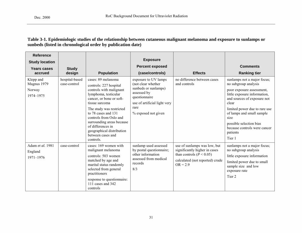
Do not quote or cite
RoC Background Document for Ultraviolet Radiation Dec. 2000
Table 3-1. Epidemiologic studies of the relationship between cutaneous malignant melanoma and exposure to sunlamps or sunbeds (listed in chronological order by publication date)
Reference Study location Exposure
Years cases Study Percent exposed Comments accrued design Population (case/controls) Effects Ranking tier
Klepp and hospital-based cases: 89 melanoma exposure to UV lamps no difference between cases sunlamps not a major focus; Magnus 1979 case-control controls: 227 hospital (not clear whether and controls no subgroup analysis
Norway controls with malignant sunbeds or sunlamps) poor exposure assessment,
1974–1975 lymphoma, testicular cancer, or bone or soft-
assessed by questionnaire
little exposure information, and sources of exposure not
tissue sarcoma use of artificial light very clear
The study was restricted rare limited power due to rare use to 78 cases and 131 % exposed not given of lamps and small sample controls from Oslo and size surrounding areas because possible selection bias of differences in because controls were cancer geographical distribution patients between cases and controls. Tier 1
Adam et al. 1981 case-control cases: 169 women with sunlamp used assessed use of sunlamps was low, but sunlamps not a major focus;
England
1971–1976
malignant melanoma
controls: 503 women matched by age and marital status randomly
by postal questionnaire; other information assessed from medical records
significantly higher in cases than controls (P < 0.05)
calculated (not reported) crude OR = 2.9
no subgroup analysis
little exposure information
limited power due to small sample size and low
selected from general 8/3 exposure rate practitioners
Tier 2 response to questionnaire: 111 cases and 342 controls
31

RoC Background Document for Ultraviolet Radiation Dec. 2000
Reference Study location Exposure
Years cases Study Percent exposed Comments accrued design Population (case/controls) Effects Ranking tier
Gallagher et al. population- cases: 595 newly exposure to sunlamps no association between sunlamps not a major focus; 1986 based case- confirmed including frequency and sunlamp use and melanoma subgroup analysis by sex and
Canada control controls: 595 age- and duration; assessed by (χ2, NS) body site, but nos. used for
1979–1981 sex-matched controls from insurance subscribers
interview with a standardized questionnaire
no association by gender or body site
risk estimates not reported, and OR not calculated
excluded lentigo maligna exposure characterized as moderate and relatively limited
% exposed not given
cases
Tier 2
Holman et al. population- cases: 511 preinvasive or exposure to sun and crude OR = 1.1 (0.6–1.8) sunlamps not a major focus; 1986 based case- invasive melanoma sunlamps assessed by no subgroup analysis because
Australia control controls: 511 sex- and structured questionnaire of small no. of exposed
1980–1982 age-matched controls from electoral rolls or student rolls of public schools
administered by nurse interviewers
9 overall
subjects
Tier 2
Elwood et al. hospital-based cases: 83 malignant home exposure to no association with risk sunlamps not a major focus; 1986 case-control melanoma identified from fluorescent lighting and calculated (not reported) crude no subgroup analysis
England pathology services of 2 the use of sunlamps OR = 1.3 little information on
1981–1984 hospitals
controls: 83 age-, sex-, and residence-matched hospital controls (in or out)
average exposure = 2.3 h
18/14
assessment of association; no risk estimate given
limited power due to small sample size and short duration of exposure
lentigo maligna melanoma excluded
Tier 3
32

RoC Background Document for Ultraviolet Radiation Dec. 2000
Reference Study location Exposure
Years cases Study Percent exposed Comments accrued design Population (case/controls) Effects Ranking tier
Holly et al. 1987 case-control cases: 121 consecutive exposure to use of melanoma patients similar to sunlamp use not a major
U.S. melanoma patients at clinic
tanning salon assessed by questionnaire
controls with respect to use of tanning salons
focus; no subgroup analysis; no information on risk
1984–1985 controls: 139 sex- and age-matched patients at same clinic (not dermatology)
% exposed not given estimate (none given)
very little exposure information.
lentigo maligna melanoma excluded
small sample size
Tier 1
Zanetti et al. 1988 population- cases: 208 histologically exposure to UVA lamps crude OR = 1.5 sunlamp use not a major
Italy based case- confirmed malignant assessed by adjusted OR = 0.9 (0.4–2.0) focus; no subgroup analysis
1984–1986 control melanoma from the
regional tumor registry
controls: 416 from National Social Service Registry
questionnaire
7/5 adjusted for age, hair color, skin reaction to the sun, sunburn in childhood, and education
Tier 2
33

RoC Background Document for Ultraviolet Radiation Dec. 2000
Reference Study location Exposure
Years cases Study Percent exposed Comments accrued design Population (case/controls) Effects Ranking tier
Osterlind et al. population- cases: 474 melanoma exposure to sunlamps sunbeds: crude OR = 0.7 (0.5– risk estimate for sunbeds 1988 based case- controls: 926 sex- and and sunbeds, including 1.0) given, but analysis by dose
Denmark control age-matched randomly no. of uses (< or > 10), sunlamps: use not related to not stated
1982–1985 selected from population registrar
assessed with structured questionnaire at home
risk and risk not related to no. of uses
sunlamp use evaluated by dose, but information on
interview analysis and risk estimates sunbeds: 14/18 not given
50% used < 10 times somewhat limited power for
sunlamps: 45/42 sunbeds due to low percentage of individuals exposed in the higher exposure group (~7%–8% used sunbeds > 10 times)
excluded lentigo maligna melanoma
Tier 3
Swerdlow et al. hospital-based cases: 180 malignant exposure to UV lamps ever use OR = 2.9 (1.3–6.4) sunlamps a major focus 1988 case-control melanoma from university and sunbeds, including exposure response for detailed exposure Scotland stratum depts. of dermatology and ever use, duration, age at increasing duration (P < 0.05) information and subtype
1979–1984 plastic surgery
controls: 197 hospital in-and out-patients with various nonmalignant diseases, stratum-matched
first exposure, and when exposure occurred (5 yr. before presentation) assessed by interview
21/8
greater risk for first use before age 30 (OR = 3.8)
greater risk for use > 5 years before presentation (OR = 9.1)
analyses
adj. for nevi, skin type, hair and eye color, and sun exposure
for age, sex, and city elevated risk for cancer on cells small after stratification
where treated legs and trunk by duration or exposure characteristics
elevated risk for superficial spreading melanoma and Tier 4
nodular melanoma
34

RoC Background Document for Ultraviolet Radiation Dec. 2000
Reference Study location Exposure
Years cases Study Percent exposed Comments accrued design Population (case/controls) Effects Ranking tier
MacKie et al. hospital-based cases: 280 (181 women exposure to artificial artificial sources of UV: sunlamps not a major focus; 1989 case-control and 99 men) identified sources of UV (classed M: crude OR = 2.6 (0.9– no subgroup analysis
Scotland from registry as modern sunbeds or 7.3), conditional regression
1989–1987 controls: 280 age- and sex- matched hospital patients with non-dermatological illness
older sunlamps); exposure to sunbeds 1 or 2 times/wk for at least 12 wk
artificial UV sources:
12/3
sunbed:
M: 8/1 F: 10/3
adj. OR = 1.3 (0.2–7.9)
F: crude OR = 1.5
sunbeds:
calculated ORs (by 2 x 2 table) from nos. of exposed cases and controls
M: crude OR = 8.6 F: crude OR = 3.8
method reported but details relating to matching not described
limited power due to small no. of exposed cases
adjusted ORs for nevi (total, atypical), freckling tendency, skin type, severe sunburn, tropical residence
Tier 2
Beitner et al. 1990 population- cases: 523 incident exposure in solariums no increased risk with frequent sunlamps not a major focus;
Sweden based case- malignant melanoma assessed by exposure to solariums no subgroup analysis
1978–1983 control controls: 505 age- and
sex-matched controls selected from population registry
questionnaire
% exposed not given no information given with respect to risk estimate or no. of individuals exposed
poor exposure assessment
Tier 1
35

RoC Background Document for Ultraviolet Radiation Dec. 2000
Reference Study location Exposure
Years cases Study Percent exposed Comments accrued design Population (case/controls) Effects Ranking tier
Walter et al. 1990 population- cases: 583 histologically exposure, including ever ever use (crude OR): detailed exposure
Ontario, Canada based case- confirmed use, year began, months M: 1.9 (1.2–3.0) information and subgroup
1984–1986 control controls: 608 randomly
selected from tax rolls, matched for sex, age, and municipality
of use, no. uses/wk, length of exposure, location, and parts of body exposed, assessed
F: 1.5 (0.2–2.1)
adj. did not change OR for either sex
analysis
adj. for age, nevus density, skin color, skin reaction to sun, and socioeconomic
by interview with questionnaire
M: 24/14
F: 28/21
exposure-response for cumulative min. of use (P < 0.01)
slightly greater risk for face, head, or neck than trunk; little risk for legs; greater risk for trunk in M than F
ORs for histol. type:
lentigo maligna and Hutchison’s melanotic freckle: M = 2.4, F = 3.1
superficial spreading and in situ: M = 1.90, F = 1.4
nodular: M = 1.7, F = 1.4
greater risk for home use
greater risk for first use before age 30
greater risk for 5 yr. since last use
status; adjusted analyses gave same effect as unadjusted analysis
recreational sun exposure a possible confounder
Tier 4
36

RoC Background Document for Ultraviolet Radiation Dec. 2000
Reference Study location Exposure
Years cases Study Percent exposed Comments accrued design Population (case/controls) Effects Ranking tier
Dunn-Lane 1993 hospital-based cases: 100 consecutive exposure to sunlamps 17% cases and 15% controls sunbeds not a major focus;
Ireland case-control patients at 7 hospitals and sunbeds assessed by used sunbeds; duration of use some indication that duration
1985–1986 controls: 100 sex- and age-matched orthopedic hospital controls with limb injuries excluding sports injuries
standard pre-coded questionnaire
17/15
similar
calculated (not reported) crude OR = 1.2
was considered, but risk estimates not given and details on duration of use not described
little exposure information
limited power due to small sample size
Tier 2
Garbe et al. 1993 case-control cases: 1,079 melanoma exposure to sunbeds adj. OR = 1.5 (0.9–2.4) for sunlamps not a major focus;
Germany patients from Central assessed by 885 cases and 705 controls no subgroup analysis
1983–1990 Malignant Melanoma Registry
controls: 778 outpatients from dermatology clinics excluding patients with previous UV treatment for skin disorders, skin cancer, or nevi
questionnaire and interview
8/7
with known information little exposure information
low percentages with exposure (7.7, 7.1)
adj. for no. of nevi, hair color, skin type, age, and participating center
Tier 2
37

RoC Background Document for Ultraviolet Radiation Dec. 2000
Reference Study location Exposure
Years cases Study Percent exposed Comments accrued design Population (case/controls) Effects Ranking tier
Autier et al. 1994 population- cases: 420 consecutive exposure assessed with ever use: crude OR = 1.0 (0.7– detailed exposure
Europe based case- malignant melanoma respect to ever use, 1.3) information and subgroup
1991 control patients
controls: 447 controls in the same municipality, randomly chosen by a quota sampling method, with no skin cancer history
location of exposure, type of machine, duration of exposure session, year first used, no. of sessions, reason for use by interview and questionnaire
26/27
for tanning purposes: crude OR for sunlamps = 1.8 (1.0–3.3)
OR for 10+ h exposure for tanning purposes:
first exposure before 1980: 2.1 (0.8–5.34)
experience of sunburn: 7.4 (1.7–32.3)
analysis; adj. for age, sex, hair color, and no. of holidays wk spent in sunny resort; overall OR given only as crude
insufficient follow-up for exposure occurring after 1980
Tier 4
Westerdahl et al. population- cases: 400 patients from exposure to sunbeds or adj. OR: detailed exposure 1994 based case- South Swedish Health sunlamps, including ever ever use: 1.3 (0.9–1.8) information (with respect to
Sweden control Care Region use and how often, >10 uses: 1.8 (1.0–3.2) dose); subgroup analysis
1988–1990 controls: 640 randomly selected from population registry matched by sex, age, and parish
assessed by comprehensive questionnaire
30/25
exposure response (P < 0.06)
greater risk age < 30 yr:
ever use: OR = 2.7 (0.7–9.8) use >10 times: OR = 7.7 (1– 64)
for use >10 times vs. none:
greater risk for trunk (OR = 4.2) than head or extremities (OR = 1.1)
adj. for history of sunburn, hair color, raised nevi, and history of frequent summer sunbathing
small cell numbers after stratifying by no. of uses and age
Tier 4
38

RoC Background Document for Ultraviolet Radiation Dec. 2000
Reference Study location Exposure
Years cases Study Percent exposed Comments accrued design Population (case/controls) Effects Ranking tier
Holly et al. 1995 population- cases: 452 women with ever use sunlamps OR for exposure category: some subgroup analysis on
U.S. based case- melanoma (medical or cosmetic); lower: 0.9 no. of uses but no definition
1981–1986 control controls: 930 age-matched
women in the same counties identified by random-digit dialing
how many times in life (excluding last 3 yr)
37/38
higher: 1.1
no difference or elevation in risk due different histologic types of melanoma
of lower and higher categories; subgroup analysis calculated for histologic type
both medical and cosmetic exposure included
Tier 3
Chen et al. 1998 population- cases: 624 newly sunlamp use assessed by OR for ever use: sunlamps major focus of
U.S. based case- diagnosed malignant nurse-interviewers with a crude = 1.3 (1.0–1.7) study; detailed exposure
1/15/87 control melanoma
controls: 512 sex- and age-matched community controls selected by
structured questionnaire and classified by type, year first used, and location, as well as
adj. = 1.13 (0.8–1.5)
no relationship between risk and total no. of uses
assessment, including attempt to define type of lamp used; detailed analysis
adj. for phenotype index (hair random-digit dialing information on potential age at first use < 25 yr: and eye color, skin type or
confounders adj. OR = 1.4 (0.9–2.1) tanning ability) and 23/19 no signif. increased risk for recreational sun exposure
any type of sunlamp insufficient follow-up time used > 2 types of lamp: for later exposures adj. OR = 3.5 (1.3–9) Tier 4 adj. OR for location:
home: 1.4 (1–2) commercial 0.8 (0.5–1.3)
used before 1970: adj. OR = 1.3 (0.8–2.1)
39

RoC Background Document for Ultraviolet Radiation Dec. 2000
Reference Study location Exposure
Years cases Study Percent exposed Comments accrued design Population (case/controls) Effects Ranking tier
Walter et al. 1999 population- cases: 583 newly exposure to sunlamp use, OR for ever use: good exposure assessment;
Ontario, Canada based case- diagnosed (1984–1986), including year, duration, crude = 1.6 (1.2–2.2) detailed analysis
1984–1986
reanalysis of Walter et al. 1990
control histologically confirmed
controls: 608 selected from property tax assessment and chosen to match the case
location, and part of body exposed, assessed by in-person interview with a structured questionnaire
adj. = 1.5 (1.2–2.1)
no difference in risk by body location
risk for lentigo maligna
adj. for sex, age, skin sun response
potential confounder is recreational sun exposure
distributions with respect to age, sex, and municipality of residence
26/18 highest: OR = 2.8 (1.4–5.3); risk signif. for superficial spreading and in situ: OR = 1.5 (1.1–2.0); elevated for all types
no difference in risk by skin reaction, but signif. elevated only for burners (larger sample size)
no difference by age at diagnosis
Tier 4
40

RoC Background Document for Ultraviolet Radiation Dec. 2000
Reference Study location Exposure
Years cases Study Percent exposed Comments accrued design Population (case/controls) Effects Ranking tier
Westerdahl et al. population- cases: 571 malignant exposure to sunbeds, OR for regular use: detailed exposure 2000 based case- melanoma from the including ever use, crude = 1.6 (1.1–2.4) information and analysis
Sweden control population-based regular use, exposure adj. = 1.8 (1.2–2.7) adj. for hair color, skin type,
1995–1997 Regional Tumor Registry in South Swedish Health Care Region
time, no. of times/wk, no. of wk/yr, location, season, age at first and
use at age < 35: OR = 2.3 use at age >35: OR = 1.6
raised nevi, no. of sun exposures
controls: 913 selected by random sampling and matched by sex, age, and
last use, assessed by comprehensive questionnaire
F: OR = 2.1 M: OR = 1.3
darker hair: OR = 2.3
not adj. for recreational sun exposure; however, controlling for nevi may take this into account, since they
parish 44/41 light hair: OR = 1.5
commercial: OR = 2.2 home: OR = 1.5
risk greater for use in winter; small sample size in summer, OR < 1
greater risk individuals aged < 36
risk greatest for lesions of extremities, then trunk; no risk for face; in M, no risk for upper extremities
exposure response up to 250 total uses or 15 uses/yr, after which the ORs decreased
test for trend, times/yr, 0.06, total uses, 0.26
are related to both sun exposure and skin type
Tier 4
41

Do not quote or cite
RoC Background Document for Ultraviolet Radiation Dec. 2000
Table 3-2. Recent epidemiologic studies of the relationship between cancer and medically related UV exposure
Reference Study
location
UVR treatment Type of cancer
Studydesign Population Exposure Effects
Adjustments Potential
confounders Stern et al. PUVA cohort 1,380 psoriasis patients psoralen 0.4– squamous-cell carcinoma: overall analysis not 1998 basal-cell and multicenter enrolled in 16-university 0.6 mg/kg orally, overall RR = 17.6 (15.6– adj. for other
U.S. squamous-cell
prospective center study
from 1/1/75 to 10/1/76
followed by UVA, usual dose 8–15 J/cm2 19.8); dose-related;
substantial risk at all doses
therapies; multivariate
carcinoma followed-up until 9/1/97
65% males
4 PUVA dose categories, based on no. of treatments: < 100, 100–159 160–
high dose adj. for other therapies: RR = 8.6 (4.9– 15.2)
analysis for different dose groups adj. for therapies as well as
1,042 basal-cell
1,422 squamous-cell
mean age 44
336, > 337
interview, documentation of PUVA therapy and
basal-cell carcinoma:
overall RR = 4.1 (3.7–4.6); dose-related; substantial risk only at highest dose
age, sex, area of residence, and anatomic site
no psoriasis PUVA Follow-up Study other treatments for
psoriasis high dose adj. for other therapies: RR = 4.7 (3.1–7.3)
controls; surveillance bias
reference group: PUVA < 100 treatments (low dose)
Stern et al. PUVA cohort follow-up until 2/29/96 two exposure groups 11 melanoma SEER incidence 1997 melanoma PUVA controls: U.S. population based on no. of RR = 2.3 (1.1–4.1) rates used for
U.S. Follow-up (SEER) treatments: 1975–1990: 4 melanoma
expected
Study low < 250 no psoriasis (above) high > 250 RR = 1.1 (0.3–2.9) controls;
1990–1996: 7 melanoma surveillance bias
RR = 5.4 (2.2–11.0)
42

Do not quote or cite
RoC Background Document for Ultraviolet Radiation Dec. 2000
Reference Study
location
UVR treatment Type of cancer
Studydesign Population Exposure Effects
Adjustments Potential
confounders Lindelof et al. PUVA cohort 4,799 patients treated information obtained entire cohort, RR: other therapies 1999 melanoma multicenter with PUVA at 11 centers from patient’s records melanoma: no psoriasis Sweden prospective 64% psoriasis patients
mean follow-up: M 15.9, F 16.2
subcohort of 1,867 followed 15–21 yr
at each center.
77% received oral PUVA.
UVA dose varied by disease; average dose for psoriasis patients 400–600 J/cm2
45 patients received > 400 treatments
537 patients received > 1,000 J/cm2
M: 1.1 (0.5–2.2, n = 8) F: 1.1 (0.4–2.3, n = 7)
squamous-cell carcinoma:
M: 5.6 (4.4–7.1, n = 68) F: 3.6 (2.1–5.8, n = 17)
subcohort, RR:
no excess melanoma
squamous-cell carcinoma:
M: 8.1 (6.1–10.6) F: 6.4 (3.3–11.2)
controls surveillance bias
Hannuksela- UVB cohort 5,687 psoriasis patients exposure assessed RR for UVB treatment: other therapies Svahn et al. squamous- nested case- from 1973–1984 from patients’ files squamous-cell carcinoma: small number of 2000 cell control PUVA Finnish Cancer percent exposed 1.6 (0.4–6.4) cases in nested Finland carcinoma
non-Hodgkin’s lymphoma
Registry
follow-up until 1/31/1995
nested study: 67 cases, 199 age- and sex-matched controls chosen from cohort using density sampling principle
(cases/controls):
squamous-cell carcinoma: 70/46
non-Hodgkin’s lymphoma: 16/47
laryngeal cancer: 55/38
non-Hodgkin’s lymphoma: 0.1 (0.0–0.8)
case-control study
43

Do not quote or cite
RoC Background Document for Ultraviolet Radiation Dec. 2000
Table 3-3. Recent epidemiologic studies of the relationship between cancer and occupational UV exposure
Reference UV exposure
Type of cancer Study design Population
Exposure Percent exposed
(cases/controls) Effects
Adjustments Potential
confounders or limitations
Walter et al. fluorescent lighting population-based same population interview: various occupational exp.: adj. for 1992
Sweden melanoma case-control used for sunbeds
(Walter et al. exposures to solar and nonsolar UVR,
M: OR = 1.47 (0.98–2.14) for exp.
socioeconomic status, sun exp.;
1999) residential and occup. 10 yr ago, dose- most results not use of fluorescent lamps, related for yr of altered by adj. for potential confounders cumulative risk factors (history
occup. use validated with exposure of sunburn,
employers, residential use validated by mail surveys
F: OR = 1.06 (0.76– 1.48) for exp. 10 yr ago, no dose-
socioeconomic status, occupational sun exp.)
occup. exposed 10 yr response fluorescent lighting
ago: domestic exp.: is ubiquitous
M: 77/70 F: 56/56
consistent risk in M but not F for
retrospective assessment
various indices of exposure
44

RoC Background Document for Ultraviolet Radiation Dec. 2000
Reference UV exposure
Type of cancer Study design Population
Exposure Percent exposed
(cases/controls) Effects
Adjustments Potential
confounders or limitations
Bajdik et al. nonsolar UVR population-based cases (1983–1984), Interview: job history, no increased risk of small no. of exposed 1996 basal-cell and case-control 180 squamous-cell outdoor exposure, from nonsolar UVR individuals except
Alberta, Canada squamous-cell carcinoma
226 basal-cell from Alberta Cancer Registry
406 aged-match controls from Alberta health insurance plan subscriber list
fluorescent lighting, other measures of UV exposure, confounders
welding:
squamous-cell carcinoma: 31/26 basal-cell carcinoma: 28/26
other exposures: < 10
exposures (e.g., fluorescent lights, welding torches, UV lamps)
for fluorescent lights and welding
insufficient follow-up
exposure misclassification
Holly et al. 1996 cccupational population-based 221 cases 1978– interview: potential welding: adj. for age, no. of
Western U.S. uveal melanoma case-control 1987 confounders and occup. adj. OR = 2.2 (1.3– nevi, eye color, and
(intraocular) 447 controls from history 3.5), no dose skin response
population within 18/11 relationship not adj. for solar 5-yr age group radiation exp.
white males
45

RoC Background Document for Ultraviolet Radiation Dec. 2000
46

Do not quote or cite
RoC Background Document for Ultraviolet Radiation Dec. 2000
4 Studies of Cancer in Experimental Animals
The IARC reviewed carcinogenicity studies of UVR in rats, mice, and hamsters. The animals were tested with broad-spectrum UVR or with discrete UVA, UVB, or UVC or a combination of these for carcinogenic effects on the skin and eye (IARC 1992; Appendix A).
4.1 Broad-spectrum UVR 4.1.1 Rats
The carcinogenic potential of UVR was recognized from the observation that daily irradiation of six albino rats with broad-spectrum UVR from a mercury-vapor lamp at a distance of 18 inches (46 cm) for one minute, three times a week, resulted in the formation of skin tumors (papillomas) in one rat (Findlay 1930, cited in IARC 1992). Six hundred rats were exposed to solar radiation for an average of five hours a day (exposure around solar noon in the summer was avoided). About 60% of the rats died from sunstroke. Of the 235 surviving rats, 70% developed tumors on the ears, eyes, nose, tail, neck, or paws. Squamous-cell carcinoma and spindle-cell sarcoma were the predominant tumor types. In complementary experiments, rats exposed to filtered sunlight did not develop tumors, but all 150 rats exposed to quartz mercury lamps developed tumors (types and sites unspecified) (Roffo 1934, cited in IARC 1992). Subsequent studies in which 2,000 white rats were exposed to sunlight yielded similar results (Roffo 1939, cited in IARC 1992). The IARC Working Group concluded that these studies provided adequate evidence of carcinogenicity in rats for UVR from sunlight.
In other studies, tumors (papillomas, squamous-cell carcinomas, and occasionally basal-cell carcinomas) were detected in rats (strain not specified) that were almost continuously exposed to broad-spectrum UVR from a quartz mercury lamp for 11 months (Putschar and Holz 1930, cited in IARC 1992). Squamous-cell carcinomas and, rarely, spindle-cell carcinomas and sarcomas, round-cell carcinomas, and basal-cell carcinomas of the skin were seen in 20 rats (strain unspecified) exposed for up to 10 months to broad-spectrum UVR from a mercury-vapor burner at a distance of 75 cm (Hueper 1942, cited in IARC 1992). Two of seven white rats exposed to UVR from a solar lamp, for two hours a day, six days a week, for a year or more, developed spindle-cell sarcomas of the eye (Huldschinsky 1933, cited in IARC 1992). Freeman and Knox (1964, cited in IARC 1992) exposed 66 pigmented and 12 unpigmented rats to UVR from mercury lamps, five days a week, for one year. The doses per session corresponded to approximately 1 MED. A total of 98 eye tumors developed. About two-thirds of the tumors were fibrosarcomas, and the rest were hemangioendotheliomas.
4.1.2 Mice
Daily irradiation of mice with broad-spectrum UVR from a mercury-vapor lamp at a distance of 18 inches (46 cm) for one minute, three times a week, resulted in the formation of skin papillomas within eight months (Findlay 1930, cited in IARC 1992). An unspecified number of mice exposed to sunlight developed squamous-cell carcinomas and spindle-cell sarcomas of the ear, eyes, paws, tail, and nose (Roffo 1939, cited in
47

Do not quote or cite
RoC Background Document for Ultraviolet Radiation Dec. 2000
IARC 1992). The IARC Working Group concluded that these studies provided adequate evidence of carcinogenicity in mice for UVR from sunlight.
A strain mice were exposed to broad-spectrum UVR at weekly doses of 3.6 to 43 × 107
ergs/cm2 (40 to 430 kJ/m2); 5% developed skin and eye tumors (spindle-cell sarcomas or fibrosarcomas, mostly in the cornea) and hemangioendotheliomas (Blum and Lippincott 1942, Lippincott and Blum 1943, Grady et al. 1943, all cited in IARC 1992). Of more than 600 A strain mice exposed to daily doses of broad-spectrum UVR at 0.32 to 8.6 × 10⁷ ergs/cm2 (3 to 86 kJ/m2) from unfiltered medium-pressure mercury arc lamps, over 90% developed skin tumors, mainly on the ears, the only site for which quantitative data were provided (Blum 1959, cited in IARC 1992).
More recent studies, mostly in mice, provide additional evidence for the carcinogenicity of broad-spectrum UVR (Emmett 1973, Urbach et al. 1974, Epstein 1978, 1985, Kripke and Sass 1978, WHO 1979, van der Leun 1984, Forbes et al. 1982, Staberg et al. 1983, Young et al. 1990, Menzies et al. 1991, all cited in IARC 1992).
4.1.3 Hamsters
Hemangioendotheliomas and fibrosarcomas developed in 14 eyes in a group of 19 hamsters (nine pigmented, 10 unpigmented) that were exposed to broad-spectrum UVR from mercury lamps at 50 cm from the skin, five days a week, for one year (Freeman and Knox 1964, cited in IARC 1992).
4.1.4 Guinea pigs
No tumors were found in the eyes of 17 guinea pigs that were exposed to broad-spectrum UVR from mercury lamps at 50 cm from the skin, five days a week, for one year (Freeman and Knox 1964, cited in IARC 1992).
4.1.5 Other species
Several researchers have reported skin and eye tumors in domestic animals (cows, goats, sheep, cats, dogs, horses, and swine) following exposure to sunlight (Emmett 1973, Dorn et al. 1971, Madewell et al. 1981, Nikula et al. 1992, all cited in IARC 1992).
Monodelphis domestica, a South American opossum that is unusually prone to photoreactivation, developed actinic keratoses and skin tumors (mostly fibrosarcomas and squamous-cell carcinomas) following exposure to broad-spectrum UVR from a Westinghouse FS-40 sunlamp (280 to 400 nm) (Ley 1985, Ley et al. 1987, both cited in IARC 1992). In a later study, 40 opossums (19 male and 21 female) were exposed to broad-spectrum UVR (FS-40 sunlamps, 280 to 400 nm) at a dose of 250 J/m2, three times weekly, for 70 weeks, and 29 control opossums (14 male and 15 female) were exposed to fluorescent lamps emitting primarily visible light (Kusewitt et al. 1991). Both groups of animals had their backs shaved and were housed under red lights to prevent photoreactivation. The UVR-exposed opossums developed a variety of hyperplastic and neoplastic skin lesions on the backs and on a single ear; 20 developed skin tumors (50%), and 13 (32.5%) had more than one tumor. Tumors included 25 papillomas, four keratoacanthomas, seven carcinomas in situ, three microinvasive squamous-cell
48

Do not quote or cite
RoC Background Document for Ultraviolet Radiation Dec. 2000
carcinomas, two invasive squamous-cell carcinomas, one basal-cell tumor, 10 dermal spindle-cell tumors, two benign melanomas, and one malignant melanoma. No skin tumors were observed in the control animals.
4.1.6 Action spectra
The action spectrum for tumor induction in SKH1 albino hairless mice was studied from a database containing information for approximately 1,100 mice treated with 14 different broadband UVR sources with spectral ranges from mainly 254 nm (from a Philips TUV germicidal lamp) to > 400 nm (from a Philips Xe3.0 fluorescent lamp) (de Gruijl et al. 1993). UVB at 293 nm was most effective in inducing tumors. However, because of a lack of data, the action spectrum for longer-wavelength UVA (340 to 400 nm) was much less well defined. A follow-up study showed that radiation from a custom-made Philips 365-nm source was carcinogenic in hairless mice but was a factor of 10-4 less effective than UVB at 293 nm. UVA radiation at 365 nm induced the same types of skin tumors as UVB exposure (mainly squamous-cell carcinomas and precursor lesions) (de Laat et al. 1997).
4.2 Primarily UVA Numerous experiments have been performed to assess the carcinogenicity of UVA (reviewed in IARC 1992). A large percentage of these studies, conducted primarily in hairless mice, did not detect tumors. The IARC Working Group noted that the doses of radiation (generally in the daily dose range of 160 kJ/m2) may have been too small, or exposure periods may have been too short. In other experiments, tumors clearly were induced by radiation purported to have been UVA, but the IARC Working Group noted that efforts to eliminate all UVB were likely insufficient. The studies reviewed below were considered to have controlled for the presence of UVB (IARC 1992).
4.2.1 Mice
Groups of 24 male and female SKH1 albino hairless mice were exposed to UVA from a bank of Philips TL40W/09 fluorescent tubes filtered through a 10-mm glass plate that strongly absorbed UVB. Animals were exposed 12 hours a day, seven days a week, for approximately one year. The daily dose was 220 kJ/m2. Most animals developed scratching lesions before they developed skin tumors. All animals had skin tumors, with a median time to appearance of 265 days. Larger lesions were examined microscopically (selection criteria not disclosed). Of the lesions examined, 60% were classified as squamous-cell carcinomas, 20% as benign tumors, and 20% as mild cellular and nuclear atypia. These lesions were similar to those observed in a parallel experiment with UVB, but the tumor latency period in the UVA-exposed animals was longer (van Weelden et al. 1986, 1988, cited in IARC 1992). However, residual UVB radiation was not believed to be responsible for the effect because more than 100,000 times the actual amount of residual UVB present would have been required to induce the observed tumor rate. Groups of 48 male and female SKH1 albino hairless mice were exposed to UVA (> 340 nm) at 220 kJ/m2, for two hours per day, seven days per week, for up to 400 days. Radiation was generated from mercury metal iodide lamps and passed through liquid filters. UVB was effectively eliminated from the radiation. Most of the animals developed skin tumors, and 31 exhibited tumors before any observed scratching. The
49

Do not quote or cite
RoC Background Document for Ultraviolet Radiation Dec. 2000
largest tumors (15/20) were examined microscopically and were classified as squamous-cell carcinomas (Sterenborg and van der Leun 1990, cited in IARC 1992).
In several studies, mice were exposed to UVR sources from which UVB was excluded so vigorously that shorter-wavelength UVA (315 to 340 nm) also was excluded; most of the animals developed squamous-cell carcinomas. In these experiments, exposure was mainly to wavelengths in the region of 340 to 400 nm (van Weelden et al. 1988, 1990, Sterenborg and van der Leun 1990, all cited in IARC 1992).
In one of these studies, when female SKH1 mice were exposed to filtered UVR (340 to 400 nm) at daily doses of 360 and 600 kJ/m2, 19/44 mice surviving at 18 weeks had skin tumors (mostly papillomas). At week 100, 22 surviving mice had 40 tumors, many of which were considered clinically to be squamous-cell carcinomas (it was not clear whether microscopic examination was used in classifying tumors) (Kligman et al. 1990, 1992, both cited in IARC 1992).
The carcinogenicity of short-wavelength UVA (315 to 340 nm) was investigated in a study using fluorescent tubes with peak emission near 330 nm and filtering UVB with glass. Groups of 24 male and female SKH1 mice were exposed to average daily doses of 20 or 56 kJ/m2, seven days per week, for 650 days. All mice in the high-dose group had multiple tumors, initially classified as mainly papillomas, but later as predominantly squamous-cell carcinomas. In the lower-dose group, three mice had skin tumors, all of which were papillomas (Kelfkens et al. 1991, cited in IARC 1992).
Bech-Thomsen (1997) investigated the carcinogenic effects of various UVA and UVR sources and their interactions in a series of studies with female C3H/Tif lightly pigmented hairless mice. In the first study (Bech-Thomsen et al. 1988), 200 mice were exposed to UVA (341 to 400 nm) from a filtered source at 150 to 200 kJ/m2, six days a week, for four weeks. No skin tumors were observed during the 57-week observation period (total dose = 4,050 kJ/m2). Among mice exposed to broad-spectrum UVR (UVA, UVB, and < 1% UVC) for 13 and 26 weeks, 35% and 88%, respectively, developed tumors by 57 weeks. Exposure to UVA (for four weeks at 4,200 KJ/m2) before exposure to broad-spectrum UVR (for 13 or 26 weeks) significantly delayed tumor development.
In subsequent studies, exposure of female C3H/Tif mice to UVA, alone or before or after exposure to broad-spectrum UVR, increased the incidence of tumors. In one study (Bech-Thomsen et al. 1991), mice were divided into 14 groups of 20 animals each, and three UVA sources, emitting varying amounts of UVB, were used either alone or before irradiation with simulated solar (broad-spectrum) UVR. All UVA exposures were for 20 minutes a day, five days a week, for 13 to 98 weeks. Exposure to broad-spectrum UVR was for 10 minutes a day, four days a week, for the lifetimes of the animals. One control group was not exposed to any UVR source, and one control group was exposed to broad-spectrum UVR only. All three UVA sources induced skin tumors. Of the 260 irradiated mice, 232 developed tumors; 230 developed multiple tumors that later fused by growth. Pre-irradiation with UVA sources with relatively high UVB outputs enhanced the carcinogenic effect of broad-spectrum UVR. The carcinogenic potential of UVA sources was directly related to their emission below 320 nm. In a follow-up study, Bech-Thomsen
50

Do not quote or cite
RoC Background Document for Ultraviolet Radiation Dec. 2000
et al. (1992) administered UVA radiation alone or after 12 weeks of exposure to broad-spectrum (simulated solar) UVR. This study demonstrated that a UVA source with a low carcinogenic potential could significantly increase the carcinogenic effect of broad-spectrum UVR.
Bech-Thomsen and Wulf (1993) also investigated whether the carcinogenic potential of UVA sources could be estimated from the International Commission on Illumination (CIE) human erythema action spectrum, which is used worldwide to assess the risk from UVR-emitting appliances used in the home. Two groups of 40 C3H/Tif mice were exposed to broad-spectrum UVR (with a UVB output of 16.7%) for 84 days. Subsequently, each group was exposed to one of two commercial UVA sources with different levels of UVB emissions (2.2% and 6.9%). After pre-irradiation with identical broad-spectrum UVR, exposure to the same erythemogenic dose from the differing UVA sources resulted in similar times to tumor development. An inverse relationship between the daily exposure dose and the tumor induction time was noted, whether the UVA was administered alone or after broad-spectrum UVR exposure. These researchers concluded that the CIE erythema action spectrum could be used to compare the carcinogenic potential of different UVR sources. The results of the Bech-Thomsen studies are summarized in Tables 4-1 and 4-2.
Table 4-1. Tumor incidences in female C3H/Tif mice exposed to UVA tanning sources with differing UVB emission levels
Daily dose (kJ/m2) Duration (weeks)
Tumor incidence Reference UVAa UVBb
121 8.9 41 20/20 Bech-Thomsen and Wulf 1993, Bech-Thomsen et al. 1991
81 6 59 21/21 Bech-Thomsen and Wulf 1993
245 5.5 75 19/20 Bech-Thomsen and Wulf 1993, Bech-Thomsen et al. 1991
162 3.6 86 13/20 Bech-Thomsen and Wulf 1993
289 0.6 98 6/20 Bech-Thomsen and Wulf 1993, Bech-Thomsen et al. 1991
21 0.5 88 1/22 Bech-Thomsen and Wulf 1993
199 0.4 97 1/20 Bech-Thomsen and Wulf 1993
82 6.1 47 20/20 Bech-Thomsen et al. 1992
163 3.7 74 13/20 Bech-Thomsen et al. 1992
199 0.4 85 1/20 Bech-Thomsen et al. 1992 a281–320 nm; b321–400 nm.
51

Do not quote or cite
RoC Background Document for Ultraviolet Radiation Dec. 2000
Table 4-2. Tumor incidences in female C3H/Tif mice exposed to broad-spectrum UVR and/or UVA
Total dose (kJ/m2)
Exposure regimen Duration (weeks)
Tumor incidence UVAa UVBb
4,050 0 UVA 4 wk 57 0/24
710 230 UVR 13 wk 54 8/23
4,760 230 UVA 4 wk, UVR 13 wk 57 0/25
1,410 460 UVR 26 wk 29 22/25
5,670 480 UVR 3 d, UVA 3.5 wk, UVR 26 wk
38 23/25
4,260 20 UVR 3 d, UVA 3.5 wk 57 0/25
Source: Bech-Thomsen et al. 1988 a321–400 nm; b281–320 nm.
4.2.2 Other species
4.2.2.1 Opossums
M. domestica developed non-melanoma skin tumors or melanocytic hyperplasia (a melanoma precursor lesion) following exposure to UVA (Ley 1997). Thirty dorsally shaved M. domestica were exposed three times a week for 81 weeks to 25,000 J/m2 of UVA radiation from filtered F40BLB fluorescent lamps (black lights). The incidences of non-melanoma skin tumors and melanocytic hyperplasia were 4% and 22%, respectively, in the exposed animals. These data suggest that the action spectra for the induction of melanoma and non-melanoma skin tumors are different.
4.2.2.2 Fish
Heavily pigmented backcross hybrids of the genus Xiphophorus (cross between platyfish and swordtails) are very sensitive to melanoma induction by UVR. Groups of six-day-old fish were irradiated with narrow-wavelength bands at 302, 313, 365, 405, and 436 nm and scored for melanomas four months later. Two groups of controls were used because the researchers realized that the initial control group was exposed to some ambient UVA and visible radiation. This could explain the high incidence of melanoma in the first control group. The second control group was kept under subdued yellow light for two months and had a much lower incidence of melanoma. The action spectrum (sensitivity per incident photon as a function of wavelength) for melanoma induction showed appreciable sensitivity at 365, 405, and probably 436 nm (Setlow et al. 1993). The tumor incidence for each wavelength is shown in Table 4-3.
52

Do not quote or cite
RoC Background Document for Ultraviolet Radiation Dec. 2000
Table 4-3. Incidences of melanoma in hybrid fish (Xiphophorus) exposed to various wavelengths of UVR
Wavelength (nm) No. of exposure
levels No. of fish No. of fish with melanoma
(%) Controla – 124 30 (24.2)
302 4 123 37 (30.1)
313 4 124 46 (37.1)
365 6 85 38 (44.7)
Controlb – 20 1 (5.0)
405 4 61 18 (29.5)
436 2 21 5 (23.8)
Source: Setlow et al. 1993 aControls were in ambient light in shaded greenhouse for the following irradiations: 313 nm, 7 of 9 at 302 nm, 7 of 9 at 365 nm, and 2 of 5 at 405 nm. bControls were in covered tanks for 2 months for the following irradiations: 436 nm and for 3 of 5 at 405 nm.
4.3 Primarily UVB 4.3.1 Rats
Skin-tumor induction was studied in a group of 40 (shaved) female NMR rats, eight to 10 weeks old at the initiation of the experiment. Animals were irradiated for 60 weeks (duration and frequency of exposures were not specified) at a distance of 37.5 cm from a commercial sunlamp emitting mainly UVB. Weekly doses of radiation were described as being 5.4 to 10.8 × 10⁴ J/m2. A total of 25 skin tumors, most of which were papillomas of the ears, developed in 16/40 animals (Stenbäck 1975, cited in IARC 1992).
4.3.2 Mice
Several studies have clearly indicated in albino mice a dose-response to UVB in the development of skin tumors. Forbes et al. (1981, cited in IARC 1992) demonstrated a dose-response relationship in the time to onset of skin tumors in SKH1 albino hairless mice exposed to UVB. Groups of 24 male and female mice, six to eight weeks old, were exposed to sunlamps emitting mainly UVB (< 1% below 280 nm; two-thirds from 280 to 320 nm, and one-third above 320 nm). Animals were irradiated five days per week, for up to 45 weeks. Although the duration of daily exposures was not stated, the daily dose of radiation was computed. Time to onset of skin tumors is summarized in Table 4-4.
53
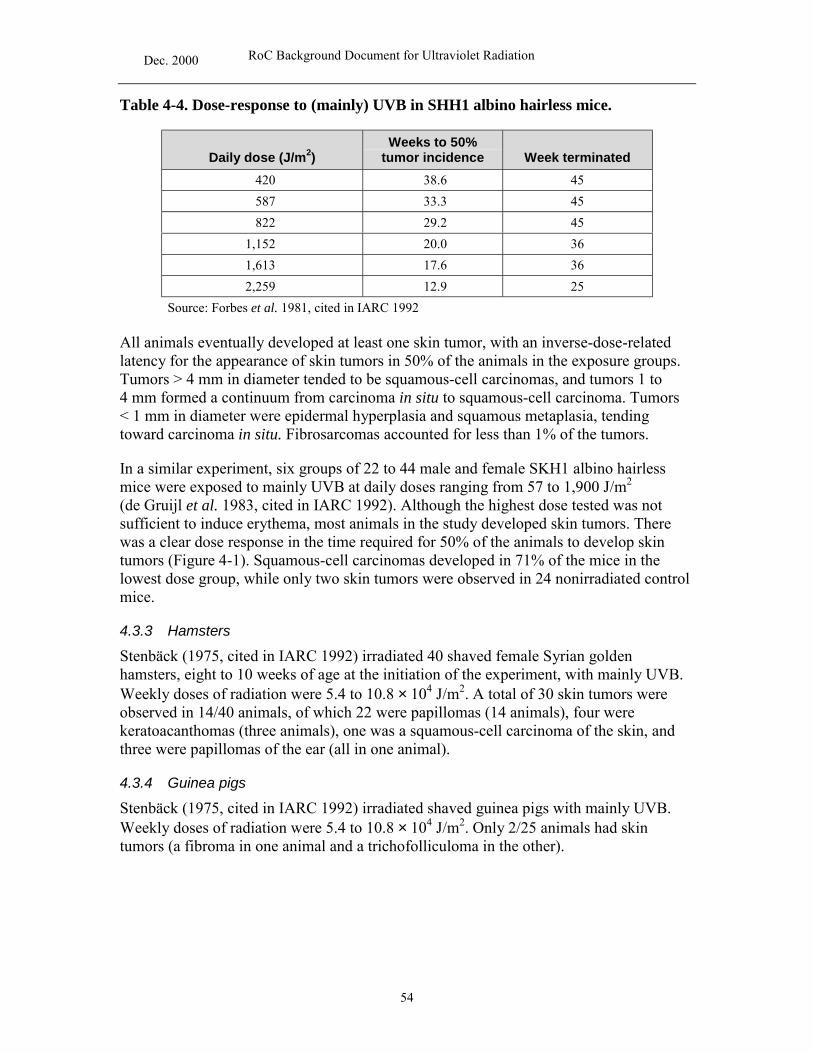
Do not quote or cite
RoC Background Document for Ultraviolet Radiation Dec. 2000
Table 4-4. Dose-response to (mainly) UVB in SHH1 albino hairless mice.
Daily dose (J/m2) Weeks to 50%
tumor incidence Week terminated 420 38.6 45
587 33.3 45
822 29.2 45
1,152 20.0 36
1,613 17.6 36
2,259 12.9 25
Source: Forbes et al. 1981, cited in IARC 1992
All animals eventually developed at least one skin tumor, with an inverse-dose-related latency for the appearance of skin tumors in 50% of the animals in the exposure groups. Tumors > 4 mm in diameter tended to be squamous-cell carcinomas, and tumors 1 to 4 mm formed a continuum from carcinoma in situ to squamous-cell carcinoma. Tumors < 1 mm in diameter were epidermal hyperplasia and squamous metaplasia, tending toward carcinoma in situ. Fibrosarcomas accounted for less than 1% of the tumors.
In a similar experiment, six groups of 22 to 44 male and female SKH1 albino hairless mice were exposed to mainly UVB at daily doses ranging from 57 to 1,900 J/m2
(de Gruijl et al. 1983, cited in IARC 1992). Although the highest dose tested was not sufficient to induce erythema, most animals in the study developed skin tumors. There was a clear dose response in the time required for 50% of the animals to develop skin tumors (Figure 4-1). Squamous-cell carcinomas developed in 71% of the mice in the lowest dose group, while only two skin tumors were observed in 24 nonirradiated control mice.
4.3.3 Hamsters
Stenbäck (1975, cited in IARC 1992) irradiated 40 shaved female Syrian golden hamsters, eight to 10 weeks of age at the initiation of the experiment, with mainly UVB. Weekly doses of radiation were 5.4 to 10.8 × 10⁴ J/m2. A total of 30 skin tumors were observed in 14/40 animals, of which 22 were papillomas (14 animals), four were keratoacanthomas (three animals), one was a squamous-cell carcinoma of the skin, and three were papillomas of the ear (all in one animal).
4.3.4 Guinea pigs
Stenbäck (1975, cited in IARC 1992) irradiated shaved guinea pigs with mainly UVB. Weekly doses of radiation were 5.4 to 10.8 × 10⁴ J/m2. Only 2/25 animals had skin tumors (a fibroma in one animal and a trichofolliculoma in the other).
54

Do not quote or cite
RoC Background Document for Ultraviolet Radiation Dec. 2000
Source: de Gruijl et al. 1983, cited in IARC 1992
Figure 4-1. Dose-effect relationship for the induction of < 1-mm skin tumors in hairless mice by exposure to UVB over a wide range of daily doses; tm = median induction time
4.3.5 Other species
4.3.5.1 Opossums
M. domestica developed actinic keratoses, fibrosarcomas, and squamous-cell carcinomas following exposure to a UVR sunlamp (280 to 400 nm). In another study, opossums were shaved and exposed three times per week for 70 weeks to 250 J/m2 of mainly UVB radiation with relative emissions of 0.04, 0.27, 0.69, 1.0, or 0.09 at wavelengths of 280, 290, 300, 313, or 360 mm, respectively. Melanomas were observed in 5/13 exposed animals and melanocytic hyperplasia in 8/13 exposed animals (Ley et al. 1989, cited in IARC 1992). In a subsequent study (Ley 1997), 30 dorsally shaved M. domestica were exposed three times a week for 81 weeks to 250 J/m2 of UV radiation from FS-40 sunlamps (approximately 150 J/m2 of UVB radiation). The incidences of non-melanoma skin tumors and melanocytic hyperplasia in exposed animals were 71% and 31%, respectively. Although the incidence of non-melanoma skin tumors was significantly higher than observed in opossums exposed to UVA, the incidence of melanocytic hyperplasia was similar to that in UVA-exposed animals (see Section 4.1.5).
4.3.5.2 Fish
Melanocytic neoplasms were induced in a group of 460 hybrid fish (Xiphophorus), following exposure to mainly UVB from FS-40 sunlamps. The sunlamps were filtered with acetate sheets transmitting > 290 nm or > 304 nm at various doses (150 or 300 J/m2
per day for > 290 nm; 850 or 1,700 J/m2 per day for > 304 nm) for 1 to 20 consecutive days. Melanocytic tumors were found in 19% to 40% of the exposed fish. Of 103 controls
55
Do not quote or cite
RoC Background Document for Ultraviolet Radiation Dec. 2000
from the two parent strains, 13% and 2% developed these tumors (Setlow et al. 1989, cited in IARC 1992).
4.4 Primarily UVC No studies were found in which animals were exposed solely to UVC. In the studies reviewed below, the source of UVC was a low-pressure mercury discharge germicidal lamp, which emitted 90% to 95% of its radiation at 254 nm, but also emitted significant amounts of UVB, UVA, and visible light.
4.4.1 Rats
Nine groups of six to 12 male CD-1 rats, 28 days of age, were shaved and exposed to varying doses of UVC from a germicidal lamp (Strickland et al. 1979, cited in IARC 1992). The dose range was 0.08 to 26.0 × 10⁴ J/m2. Exposure duration was not specified. Survival ranged from 75% to 92% in the experimental groups. Keratoacanthoma-like skin tumors developed at a yield that was approximately proportional to radiation throughout the dose range of 0.65 to 26.0 × 10⁴ J/m2. No tumors were observed at or below 0.32 × 10⁴ J/m2.
4.4.2 Mice
A group of 40 female C3H/HeNCrlBr mice was exposed to radiation from germicidal lamps at a weekly dose rate of 3 x 10⁴ J/m2. The duration of the experiment was not specified. Three animals died without tumors at experimental weeks 9, 43, and 63. All other animals had tumors, with 97% of the mice affected by 52 weeks. The median time to tumor onset was 43 weeks, and the mean number of tumors per tumor-bearing animal was 2.9. Microscopic examination revealed that of the 83 lesions initially considered to be tumors, 66 were squamous-cell carcinomas, 10 were proliferative squamous-cell lesions, and six were invasive fibrosarcomas (Lill 1983, cited in IARC 1992). The IARC Working Group noted that the 4% UVB content of the radiation source provided a weekly dose of 1,170 J/m2, which could not be excluded as a contributing factor in the induction of skin tumors (IARC 1992).
Groups of 24 male and female SKH1 albino hairless mice, 6 to 10 weeks of age, were exposed to UVC from germicidal lamps seven days per week, for 75 minutes per day, at a dose of 230, 1,460, or 7,000 J/m2. The highest dose applied was 60% lower during the initial seven days of the experiment. A total of 65 squamous-cell carcinomas of the skin were found. The numbers of animals with tumors were not reported, but the investigators noted that both the numbers of animals with tumors and the numbers of tumors per mouse were strongly dose-related (Sterenborg and van der Leun 1988, cited in IARC 1992). By comparing tumor incidences and onset times in their own UVC experiment to those from experiments with UVB, Sterenborg and van der Leun (1988, cited in IARC 1992) concluded that the UVB emitted from the germicidal lamp was insufficient to cause the tumors observed in their experiment. They estimated that the UVB present would require at least 850 days of exposure to induce skin tumors at the rate at which they had observed tumors after 161 days of exposure to the UVC. Further, they noted a qualitative difference between UVC- and UVB-induced skin tumors in mice, in that UVC-induced tumors were scattered more widely over the skin than were tumors
56
Do not quote or cite
RoC Background Document for Ultraviolet Radiation Dec. 2000
associated with UVB. Also, the dose-response curve was steeper in UVB-exposed mice than in mice exposed to the germicidal lamp radiation. The IARC Working Group noted that the observations given to exclude UVB as a causative factor in skin tumorigenesis did not rule out a possible interaction between the two types of radiation (IARC 1992).
4.5 Cancer development in human-mouse chimera models Several researchers investigated UV-induced skin cancers in human skin grafted to mice. Atillasoy et al. (1997) grafted white human skin onto 158 recombinase activating gene-1 (RAG-1) knockout mice. Mice were divided into four groups: control, a single administration of dimethyl(a)benzanthracene (DMBA), exposure to UVB (290 to 320 nm) at 500 J/m2 three times per week, or a combination of DMBA and UVB. Mice were examined three times a week, and all surviving mice were euthanized and autopsied after a median observation period of 10 months (range 3 to 16 months). About half of the grafts exposed to UVB (alone or with DMBA) developed milia, compared with 3% of DMBA-exposed grafts and none of the controls. Actinic keratoses were observed in 9% of the grafts exposed to UVB alone and 19% of the grafts exposed to DMBA plus UVB. Invasive squamous-cell carcinomas developed in 10% of the grafts exposed to DMBA plus UVB. None of the controls developed actinic keratoses or squamous-cell carcinomas. Melanocytic hyperplasia was found in 68% of the grafts exposed to UVB only and 77% of the grafts exposed to both UVB and DMBA. One human nodular-type malignant melanoma developed in a graft exposed to both DMBA and UVB (Atillasoy et al. 1998).
In a follow-up study (Sauter et al. 1998), 25 RAG-1 mice with human skin grafts received a single administration of DMBA followed by three weekly exposures to UVB (500 J/m2) for at least five months. Cysts, hyperplasia, precancers, or invasive cancers were seen in 24 of 25 exposed grafts, compared with none of the controls. Two squamous-cell carcinomas were observed. Of grafts exposed for seven or more months, 83% (15/18) developed squamous precancer or squamous-cell carcinoma of human origin, and 44% (8/18) developed melanocytic hyperplasia or melanoma. Direct correlations between p53 tumor suppressor gene expression and cell proliferation and the degree of histologic change were observed for both squamous epithelial and melanocytic cells.
Human skin was transplanted to severe combined immunodeficient mice and exposed to UVB (280 to 360 nm) at daily doses of at 7.3 × 10⁵ to 1.8 × 10⁶ J/m2 for two years (Nomura et al. 1997). Actinic keratoses developed in 77.8% (14/18) and squamous-cell carcinoma in 16.7% (3/18) of grafts exposed to UVB. None of the 15 control grafts developed actinic keratoses or squamous-cell carcinomas. The same p53 mutation at codon 242 (C TGC to C CGC) was observed in actinic keratoses and squamous-cell carcinomas, and double or triple mutations were observed in all skin cancers and three of eight actinic keratoses.
4.6 Summary Broad-spectrum UVR was carcinogenic to albino rats, inducing skin tumors (papilloma, squamous-cell carcinoma, spindle-cell sarcoma and carcinosarcoma, and basal-cell
57

Do not quote or cite
RoC Background Document for Ultraviolet Radiation Dec. 2000
carcinoma) and eye tumors (spindle-cell sarcoma and squamous-cell carcinoma). Broad-spectrum UVR induced skin or eye tumors (spindle-cell sarcoma or fibrosarcoma, mostly in the cornea) and hemangioendothelioma in mice and hamsters and caused skin tumors (mostly fibrosarcoma and squamous-cell carcinoma) in opossums. Broad-spectrum UVR also has been implicated in tumor development in domestic animals (cows, goats, sheep, cats, dogs, horses, and swine).
UVA induced skin tumors in mice (squamous-cell carcinoma and papilloma), opossums (melanocytic hyperplasia) and fish (melanoma). Prolonged UVB exposure caused skin tumors in rats (papilloma), mice (squamous-cell carcinoma, fibrosarcoma, papilloma, and keratoacanthoma), guinea pigs (fibroma and trichofolliculoma), opossums (melanocytic hyperplasia and melanoma), and fish (melanocytic neoplasms). Exposure of experimental animals to high doses of UVC caused skin tumors in rats (keratoacanthoma-like skin tumors) and mice (squamous-cell carcinoma and fibrosarcoma). Human skin grafts on mice also yielded skin tumors (squamous-cell carcinoma, actinic keratosis, melanocytic hyperplasia, and melanoma) following irradiation with UVB alone or after exposure to DMBA.
58

Do not quote or cite
RoC Background Document for Ultraviolet Radiation Dec. 2000
5 Genotoxicity
The IARC conducted an extensive review of the literature through 1991 on the genotoxicity of solar and ultraviolet radiation, to develop a better understanding of exposure to UVR, the intermediate biological responses, and their consequences, with emphasis on carcinogenesis (IARC 1992).
This section discusses pertinent genotoxicity information from the IARC review and from recent genotoxicity studies, focusing on UVR, including UVA, UVB, and UVC. It is important to recognize that many exogenously supplied photosensitizers, including some pharmaceuticals, can affect the biological response to UVR. In some cases, interactions with photosensitizers have therapeutic application; for example, UVA may be used in combination with furocoumarins to treat skin diseases or tumors (IARC 1992, Müller et al. 1998). However, UVR interactions with exogenous chemical agents are considered outside the scope of this document and are not addressed.
5.1 Methods for identifying and quantifying UVR-induced DNA lesions Griffiths et al. (1998) reviewed the measurement and significance of DNA lesions induced by UVR. UVR-induced DNA lesions and methods for identifying and quantifying them may be categorized as follows:
Single- and double-strand DNA breaks. UVR causes strand breakage interfering with inter- and intra-strand stabilization and inevitably resulting in some degree of α-helical unwinding. Several assays rely on this phenomenon and do not require DNA extraction, but are based on fluorescence labeling of DNA. Examples of such assays are the fluorescence-activated DNA unwinding assay, DNA sedimentation analysis, and the single-cell gel electrophoresis (or comet) assay (Griffiths et al. 1998).
Specific DNA sequences containing damage. UVR elicits antigenicity by altering DNA sequences through denaturation. Polyclonal and monoclonal antibodies have been raised against specific lesions that begin with thymine dimers. These antibodies have been used to recognize sequence-specific damage both on fixed section slides and in fluorescence-activated cell-sorter-type flow cytometry systems. For instance, Herbert et al. (1994, cited in Griffiths et al. 1998) developed a polyclonal antibody specific for a cyclobutane thymidine dimer with an adjacent 3′ or 5′ thymidine.
Specific DNA base lesions. Franklin and Haseltine (1984, cited in Griffiths et al. 1998) developed and demonstrated a high-performance liquid chromatographic (HPLC) assay that can separate and quantitate cyclobutane-type pyrimidine dimers and (6-4) photoproducts.
5.2 UVR-induced DNA photoproducts It is well documented that UVR induces mutations in both prokaryotic and eukaryotic cells, and any cell that is UV-irradiated will likely sustain DNA damage. UVA, UVB, and UVC have induced mutations in bacterial systems and cultured mammalian cells. In addition, UVA has induced mutations in yeast, and UVC has induced mutations in plants
59

Do not quote or cite
RoC Background Document for Ultraviolet Radiation Dec. 2000
and amphibians (IARC 1992). The type of damage induced depends upon the specific wavelength(s) applied and the competency of an affected cell to repair the damage without error. DNA is a major cellular chromophore absorbing UVR (mainly UVB); it responds to irradiation by yielding single-electron reactive intermediates and, depending on exposing wavelengths and energy produced, various identifiable photoproducts. All photoproducts are expected to have mutagenic potential; however, their specificity and potency vary (IARC 1992).
5.2.1 UVA-induced indirect DNA damage
Over 90% of the UV radiation reaching the surface of the earth is in the form of UVA. Upon absorption of UVA by cells and subsequent generation of activated oxygen, the energy is transferred to DNA. DNA poorly absorbs UVA; therefore, the induced genotoxic damage is due to absorption of photons by other endogenous chromophores (IARC 1992). Examples of endogenous chromophores within mammalian cells are riboflavin, porphyrins, quinones, and reduced nicotinamide cofactors (Griffiths et al. 1998).
UVA-excited endogenous photosensitizers produce a much lower level of base loss than does UVB (Cadet et al. 1992). The major DNA base lesions induced are 8-hydroxydeoxyguanosine (8-OHdG), produced from guanosine by the action of singlet oxygen; hydroxyhydroperoxides, indirectly generated from the radical cation of thymine under aerobic conditions; and pyrimidine photoproducts (however, their induction requires a six-fold greater energy input than UVC-induced lesions at a similar frequency). UVA does not induce formation of (6-4) photoproducts (Griffiths et al. 1998).
5.2.2 UVB-induced direct DNA damage
UVB photons directly cause the following major DNA base modifications: cyclobutane-type pyrimidine dimers, (6-4) photoproducts, the corresponding Dewar isomers, and thymine glycols. The pyrimidine dimers are five to 10 times more abundant than the other DNA base modifications. Depending upon the conditions of exposure, these pyrimidine dimers occur as cytosine-cytosine, thymine-thymine, or mixed dimers. The absorption spectra for cytosine and thymidine match the action spectrum for dimer formation and, in (6-4) photoproduct induction, the cytosines 5´ upstream of adjacent pyrimidines present perfect targets for such DNA damage (Griffiths et al. 1998).
UVB also is responsible for induction of DNA strand breaks. The incidence of DNA strand breaks increases as a function of increasing wavelength. Single-base lesions, mainly ring-saturated thymines known as thymine glycols, are also observed. Along with these, 8-OHdG adducts are induced over the dose range of 4 to 750 mJ/cm2 (Stewart et al. 1996, cited in Griffiths et al. 1998). UVB exposure also causes DNA-protein crosslinks, mostly affecting cysteine residues. At equivalent doses, UVB induces DNA-protein crosslinks at about one-tenth the frequency that UVA does (Griffiths et al. 1998).
5.2.3 Cellular mechanisms for minimizing UVR-induced DNA damage
Healthy eukaryotic cells can minimize UVR-induced DNA damage by several defense mechanisms, which interact to protect cells against toxic effects of UVR. These
60

Do not quote or cite
RoC Background Document for Ultraviolet Radiation Dec. 2000
mechanisms include production of antioxidant enzymes, production of detoxification enzymes, and repair of UVR-induced DNA lesions by means of direct reversal, base excision repair, nucleotide excision repair, transcription repair coupling, and mitochondrial repair of UV-induced lesions (Griffiths et al. 1998).
5.2.4 Cellular responses to UVR-induced DNA damage
Transcriptional activation of mammalian “early response genes” (e.g., c-fos and c-jun) is induced within minutes of UVR exposure. Early and secondary response genes also include genes mediating protein binding to DNA damage sites, cell proliferation control genes (e.g., growth arrest and DNA damage genes), genes coding for enzymes involved in signal transduction (e.g., protein kinase C) or for antioxidants (e.g., heme oxygenase), and the p53 tumor suppressor gene (Griffiths et al. 1998).
UVR-induced photoproducts have genotoxic consequences that vary depending on the particular exposure circumstances. In the survey below, genotoxic effects are classified according to the test system in which they were assessed. Data presented in IARC (1992) are summarized in Table 5-1. Studies that were not reviewed in IARC (1992) are discussed in the following text.
5.3 Prokaryotic systems 5.3.1 Induction of mutation in Salmonella typhimurium
UVC exposure unambiguously increased the frequencies of reverse gene mutations in several S. typhimurium tester strains, including repair-defective strains hisG46 and hisG428 (Cebula et al. 1995) and recA-uvrB (Hartmann et al. 1996, cited in Griffiths et al. 1998).
Table 5-1. Genetic and related effects of UVR exposure reviewed in IARC (1992)
Test system End point
Results (no. positive/no. studies)
UVA UVB UVC UVRa
Prokaryotic
Salmonella typhimurium mutation 1/1 1/1
Escherichia coli mutation 8/8 1/1 6/6
Escherichia coli DNA damage 5/5
Bacillus subtilis mutation 1/1
Lower eukaryotic
Saccharomyces cerevisiae
DNA damage or pyrimidine dimers
3/3 2/2 1/1
Saccharomyces cerevisiae
aneuploidy 1/1
Saccharomyces cerevisiae
mutation 2/2
61

RoC Background Document for Ultraviolet Radiation Dec. 2000
Test system End point
Results (no. positive/no. studies)
UVA UVB UVC UVRa
Plant
Wheat mutation 1/1
Unspecified plant cells DNA damage 1/1
Nicotiana tabacum unscheduled DNA synthesis
1/1
Chlamydomonas reinhardtii
pyrimidine dimers 1/1
Chlamydomonas reinhardtii
mutation 1/1
Tradescantia chromosomal aberrations
1/1 1/1
Nonmammalian eukaryotic
Drosophila melanogaster
DNA damage 1/1
ICR 2A frog cells DNA damage 1/1 2/2
ICR 2A frog cells SCE, chromosomal aberrations
1/1 2/2 1/1
A8W243 Xenopus frog cells
chromosomal aberrations
1/1
Fish (in vitro) DNA damage 1/1
Chick embryo fibroblasts SCE, chromosomal aberrations
2/2
Nonhuman mammalian in vitro
Chinese hamster ovary cells
DNA damage 2/2 1/1 1/1
Chinese hamster ovary cells
SCE, chromosomal aberrations
2/2 1/1 2/2
Chinese hamster ovary cells
mutation 3/3 2/2 3/3 2/2
Chinese hamster fibroblasts
chromosomal aberrations
2/2
Chinese hamster V79 lung cells
DNA damage 2/2 1/1 2/2
Chinese hamster V79 lung cells
mutation 2/2 4/4 4/4 3/3
62

RoC Background Document for Ultraviolet Radiation Dec. 2000
Test system End point
Results (no. positive/no. studies)
UVA UVB UVC UVRa
Chinese hamster V79 lung cells
SCE, chromosomal aberrations
3/3
Chinese hamster CHEF-125 cells
chromosomal aberrations
1/1
Syrian hamster embryo cells
cell transformation 1/2 1/1 3/3
C3H 10T1/2 mouse cells DNA damage 1/1
L5178Y mouse lymphoma cells
mutation 1/1 1/1 1/1 1/1
Mouse splenocytes micronuclei 1/1
New Zealand black mouse fetal fibroblasts
chromosomal aberrations
1/1
Mouse epidermal cells, embryo cells, fibroblasts, fibrosarcoma cells
cell transformation 7/7 6/6
Human in vitro
Fibroblasts DNA damage or pyrimidine dimers
4/4 4/4 8/8 4/4
Fibroblasts mutation 1/1 1/1 5/5 1/1
Fibroblasts micronuclei 1/1 1/1
Fibroblasts SCE, chromosomal aberrations
9/9 2/2
Fibroblasts cell transformation 1/1 3/3
Keratinocytes and melanocytes
DNA damage 1/1 1/1
Epithelial P3 cells DNA damage 1/1
Epithelial cells mutation 1/1 1/1 1/1
Teratoma or teratocarcinoma cells
DNA damage 3/3 3/3 3/3
Lymphoblastoid cells mutation 0/1 0/1 1/1
HeLa cells DNA damage 1/1
HeLa cells mutations 1/1
Melanoma cells SCE 1/1
Melanoma cells mutation 1/1
Melanoma cells micronuclei 1/1
63

RoC Background Document for Ultraviolet Radiation Dec. 2000
Test system End point
Results (no. positive/no. studies)
UVA UVB UVC UVRa
Lymphocytes mutation 2/2
Lymphocytes SCE, chromosomal aberrations
4/4
Nonhuman mammalian in vivo
Mouse skin DNA damage or pyrimidine dimers
3/3 1/1 1/1
Mouse skin fibroblasts cell transformation 1/1
Marsupial corneal cells DNA damage 2/2
Human in vivo
Epidermis or skin cells DNA damage or pyrimidine dimers
1/1 1/1 2/2
Cornea unscheduled DNA synthesis
1/1
Fibroblasts DNA damage 2/2 2/2
aIncludes solar, simulated solar, and sunlamp irradiation.
5.3.2 Induction of mutation in Saccharomyces cerevisiae
UVB and natural sunlight exposure increased the frequencies of pyrimidine dimer formation (Armstrong and Kunz 1992), single-base-pair substitution (Kunz and Armstrong 1998), and gene mutation (Armstrong and Kunz 1990) in S. cerevisiae. Natural sunlight and UVB induced similar G-C to A-T transitions; however, natural sunlight induced a higher percentage of G-C to T-A or C-G transversions. Dipyrimidine adducts likely were responsible for the transitions and are now recognized as a signature of sun exposure (Sarasin 1999). These data suggest that one type of DNA damage leads to most of the mutations associated with UVB exposure, whereas two different types of DNA damage may be involved in sunlight mutagenesis (Kunz and Armstrong 1998).
5.4 Plants and lower eukaryotic systems No additional genotoxicity studies in plant or eukaryotic systems were identified in the current literature.
5.5 Mammalian systems 5.5.1 Nonhuman mammalian in vitro assays
Oxidative damage in DNA is caused by UVB irradiation and results in the formation of a DNA adduct, 8-OHdG. Studies demonstrated a decrease in antioxidant enzyme defenses in SKH1 hairless albino mice after UVB radiation, implicating antioxidant status in protection against oxidative damage (Cameron and Pence 1992). A further study by this group examined mechanisms of UVB-induced DNA damage and subsequent modulation
64

Do not quote or cite
RoC Background Document for Ultraviolet Radiation Dec. 2000
by the antioxidants vitamin C (ascorbic acid), selenite, or Trolox (a water-soluble vitamin E analog). BALB/c MK-2 mouse keratinocytes were exposed to UVB at a dose range of 4 to 750 mJ/cm2. Adducts were measured via HPLC coupled with electrochemical and UV absorbency detection. Preincubation of the cells for two days with 0.4 or 0.8 µg/ml of ascorbic acid, 10 or 20 µg/ml of Trolox, and 5 or 12.5 µM selenite significantly decreased the number of 8-OHdG adducts per 105 deoxyguanines induced by UVB at 500 mJ/cm2. The results further elucidated mechanisms through which UVB altered DNA exposed ex vivo in cultured mouse skin cells and indicated that antioxidant nutrients might protect skin cells against UVB damage (Stewart et al. 1996, cited in Griffiths et al. 1998).
5.5.2 Human in vitro assays
Murata-Kamiya et al. (1995, cited in Griffiths et al. 1998) demonstrated that oxygen free radicals caused DNA base and sugar modifications and DNA strand breaks. They showed that a known mutagen, glyoxal, was produced by exposure of DNA to an oxygen-radical-forming system (5 mM ferrous sulfate–ethylenediaminetetraacetic acid, +37o C, 60 min). Glyoxal was produced with a 17-times-higher efficiency than 8-OhdG, with adduct formation at guanine sites. The authors predicted that this type of exposure of DNA to an oxygen-radical-forming system, with following glyoxal and guanine adduct formation, constituted one of the major types of UVA-induced DNA damage.
Mizuno et al. (1991, cited in Griffiths et al. 1998) conducted a study using a thymine dimer-specific monoclonal antibody (TDM-1), which was produced against mouse and human DNA after exposures of cells ex vivo to 313-nm UVB in the presence of acetophenone. When UVB-irradiated DNA was incubated with photolyase from E. coli and visible light, TDM-1 binding and the presence of thymine dimers were reduced. It was shown that TDM-1 binding to UVB-irradiated DNA was inhibited by photolyase, but not by 64M-1 antibody specific for (6-4) photoproducts. The authors concluded that the TDM-1 antibody had affinity for cyclobutane-type DNA thymine dimers. They measured, by competitive assessments with the two antibodies, the amount of each type of DNA damage in DNA extracted from UVB-irradiated mammalian cells. Repair experiments indicated that (6-4) photoproducts were excised from UVB-irradiated cellular DNA more rapidly than thymine dimers. Excision rates of both photoproducts were lower in mouse (NIH3T3) cells than in human fibroblasts.
Immunocytochemical methods were used to measure cyclobutane pyrimidine dimers, (6-4) photoproducts, and Dewar isomers in normal human mononuclear cells following ex vivo irradiation by natural sunlight or a UVB sunlamp (Clingen et al. 1995, cited in Griffiths et al. 1998). The induced photoproducts were detected following a 30- to 60-minute sunlight exposure, or with sunlamp irradiation as low as 50 to 100 J/m2. A dose-dependent increase in the binding of monoclonal antibodies specific for pyrimidine dimers, (6-4) photoproducts, and Dewar isomers was observed. The relative ratio of Dewar isomers to (6-4) photoproducts was much greater after exposure to natural sunlight than after exposure to broad-spectrum UVB. Use of the (6-4) monoclonal antibody indicated that binding sites increased slightly after a one-hour exposure to natural sunlight and remained relatively constant with further exposure. The authors
65

Do not quote or cite
RoC Background Document for Ultraviolet Radiation Dec. 2000
hypothesized that following irradiation with natural sunlight, most (6-4) photoproducts were converted into Dewar isomers, and that this conversion was likely caused by the UVA component. They concluded that the (6-4) photoproducts probably did not contribute directly to sunlight-induced carcinogenesis.
Human skin explants were studied with a [32P]-HPLC method for recognizing and measuring lesions (cyclobutane dimers, [6-4] photoproducts, and Dewar isomers) induced in DNA after exposure to UVA, UVB, or UVC (Bykov and Hemminki 1996). The experimental method was sensitive enough to detect the lesions at a UVB radiation dose of 10 J/m2. Dewar isomers were detected only at a high doses of UVB. The compounds were identified by their photochemical reactivity and by spiking with prepared standards. Treatment with nuclease P1 was used to identify the 5′-terminal nucleotide. UVA caused no detectable adducts .
5.5.3 Nonhuman mammalian in vivo assays
Formation of 8-OHdG adducts was evaluated in the epidermis of hairless mice after repeated exposure to UVB (Hattori et al. 1996, cited in Griffiths et al. 1998). Exposure of hairless mice to UVB at a dose of either 3.4 kJ/m2 (2 MED) or 16.8 kJ/m2 (10 MED), three times a week, for two weeks, induced a 2.5- or 6.1-fold increase, respectively, in the levels of 8-OHdG in DNA. An immunohistochemical method, using a monoclonal antibody specific for 8-OHdG, showed stronger and more extensive staining in the nuclei of UVB-irradiated epidermal cells than in those of nonirradiated cells. Western blots probed with antibodies against 4-hydroxy-2-nonenal-modified proteins confirmed the involvement of reactive oxygen species in the epidermal damage induced by chronic UVB exposure. The authors suggested that three pathways might regulate the formation of 8-OHdG after UVB exposure: photodynamic action, lipid peroxidation, and inflammation. They concluded that 8-OHdG might be active in sunlight-induced skin carcinogenesis.
5.5.4 Human in vivo assays
5.5.4.1 DNA damage and repair
DNA synthesis, measured by [3H] thymidine incorporation after lymphocyte activation, was studied in circulating leukocytes from patients with widespread psoriasis who were being treated with PUVA (oral 8-methoxypsoralen and high-intensity UVA) (Kraemer and Weinstein, 1977, cited in IARC 1992). Of 13 psoriasis patients treated with PUVA, seven demonstrated a significant (P < 0.05) reduction in lymphocyte incorporation of [3H]thymidine immediately after UVA treatment, compared with incorporation before UVA treatment. In addition to its therapeutic effects on epidermal cells, PUVA treatment affected circulating blood cells in some psoriasis patients. However, in 10 control subjects who received UVA alone, lymphocytes were capable of normal activation and DNA synthetic activity. This study raised the possibility of genotoxic effects in circulating lymphocytes. Strauss et al. (1979, 1980) observed induction of presumed mutations at the HPRT locus in lymphocytes in UVA-exposed patients, but not in the absence of psoralens. In another study, patients treated with PUVA, but not UVA alone, showed evidence of local and systemic impairment of the delayed cellular hypersensitivity component of the immune response, providing evidence for a possible
66

Do not quote or cite
RoC Background Document for Ultraviolet Radiation Dec. 2000
mechanism of tumor promotion in the skin (Bridges and Strauss 1980, Strauss 1982). Human studies evaluating PUVA treatment and cancer risk are reviewed in Section 3.
Irradiation of human buttock skin with 300-nm UVR in situ induced thymine dimers and (6-4) photoproducts (Chadwick et al. 1995, cited in Griffiths et al. 1998). Irradiation of human buttock skin with UVC (260 nm) immediately followed by UVA (320 nm) induced the Dewar isomers of the (6-4) lesions. All three lesions were detected in methanol-fixed paraffin sections through the use of specific monoclonal antibodies. The lesions were analyzed in an automated image analysis system, and the level of immuno-diaminobenzidine-peroxidase was measured in individual epidermal-cell nuclei. Staining patterns indicated a decrease with depth of about 2.5% per cell layer. Following irradiation with a shorter wavelength (260 nm), staining decreased rapidly with depth (39% per cell layer). The results showed effective penetration and damage induction by UVB in human skin after in vivo exposure.
Hori et al. (1992, cited in Griffiths et al. 1998) studied DNA extracted from a variety of human skin tumors and control tissues, including femoral skin and white blood cells, with an immunoblotting method using antibodies against UV-irradiated calf thymus DNA. The antibodies used were reactive to cyclobutane-type pyrimidine dimers. Immunoprecipitates were observed for facial actinic keratosis and keratosis-derived squamous-cell carcinoma specimens. Through the use of photoreactivation enzyme plus visible light, both immunoprecipitates were found to be specific for cyclobutane-type pyrimidine dimers. Immunofluorescence studies of actinic keratosis tissue showed that unremoved photodamage in DNA remained in the nucleus of actinic keratosis cells. The authors suggested that the tumor cells might be deficient in an enzyme required for repairing cyclobutane-type pyrimidine dimer damage.
Clingen et al. (1995, cited in Griffiths et al. 1998) used specific monoclonal antibodies in situ and a computer-assisted image analysis system to determine the relative induction of cyclobutane dimers, (6-4) photoproducts, and Dewar isomers in human mononuclear cells and fibroblasts following irradiation with UVC, broad-spectrum UVB, and narrow-spectrum UVB. DNA lesions were produced in different proportions, with broad-spectrum UVB inducing a greater combined yield of (6-4) photoproducts and Dewar isomers per cyclobutane dimer than UVC or narrow-spectrum UVB. Relative induction ratios of (6-4) photoproducts versus cyclobutane dimers were 0.15, 0.21, and 0.10 following irradiation with UVC or broad- or narrow-spectrum UVB, respectively.
5.5.4.2 Tumor suppressor and ras gene mutations
Brash et al. (1991, cited in IARC 1992) reported five C to T, four C to A, and three CC to TT mutations at various codons of the p53 tumor suppressor gene in 24 invasive squamous-cell carcinomas taken from sun-exposed skin; about 90% of squamous-cell carcinomas examined in this study contained p53 mutations. CC to TT transitions have not been found in any internal tumors, suggesting that sun exposure plays a role in p53 mutations. Pierceall et al. (1991, cited in IARC 1992) reported one C to T transition and one C to A transversion in 10 squamous-cell carcinomas examined. Ouhtit et al. (1997) investigated the frequency of p53 mutations in normal skin from Japanese patients. More mutations were found in skin samples taken from sites chronically exposed to the sun
67

Do not quote or cite
RoC Background Document for Ultraviolet Radiation Dec. 2000
than from covered sites. A recent study showed that 50% of mutations of the PTCH tumor suppressor gene found in basal-cell carcinomas were UVR-specific (Quinn 1997).
Melanomas from 37 patients with varying sun exposure were examined for N-ras mutations (van ’t Veer et al. 1989, cited in IARC 1992). N-ras mutations were found in tumors from seven patients who were continuously exposed to the sun. All mutations were base substitutions at TT or CC sites that are potential targets for UV photoproducts. In other studies, N-ras, Ki-ras, and Ha-ras base substitution mutations were found in melanomas, basal-cell carcinomas, and squamous-cell carcinomas (Sekiya et al. 1984, Corominas et al. 1989, Keijzer et al. 1989, Shukla et al. 1989, van der Schroeff et al. 1990, all cited in IARC 1992).
5.5.5 Other in vitro and in vivo end points
DNA 8-hydroxy-2′-deoxyguanosine is a mutation-prone (G-C to T-A transversion) DNA base-modified product generated by reactive oxygen species or photodynamic action. G-C to T-A transversions were observed in the p53 and ras genes of UVB-induced skin tumors from mice and in squamous- and basal-cell carcinomas from human skin exposed to sunlight (Hattori et al. 1996, cited in Griffiths et al. 1998).
5.5.6 Molecular epidemiological studies of DNA repair capacity
Wei et al. (1994) evaluated the relationship between DNA repair capacity and basal-cell carcinoma in 88 cases and 135 controls. Cases were Caucasian patients with histopathologically confirmed primary basal-cell carcinoma recruited from physician practices in the Baltimore area between 1987 and 1990. Controls were patients from the same physician practices who were cancer-free and were frequency-matched to cases by age. Cancer patients and controls provided a blood sample and completed a self-administered questionnaire that collected information with respect to demographics, family history, and potential confounders for basal-cell carcinoma. Lymphocytes were isolated from the blood. DNA repair was assessed with the host-cell reactivation assay, which measures the ability of lymphocytes from the participants to repair damaged DNA. Plasmids containing UVR-irradiated (0, 350, or 700 J/m2) chloramphenicol acetyl transferase (CAT) reporter genes were transfected into lymphocytes, and the ratio of CAT gene expression of irradiated plasmids to that of non-irradiated plasmids was calculated as the percentage of residual repair activity at a given UVR dose. The mean DNA repair capacity of all basal-cell carcinoma patients was 5% lower than that of controls, a difference of borderline significance. However, among subjects with red hair and skin type I, DNA repair capacity was significantly lower in cancer patients than in controls. Moreover, among subjects who reported frequent sunbathing, poor tanning ability, a history of multiple sunburns, exposure to chemicals, or multiple medical irradiations, the basal-cell carcinoma patients had significantly lower DNA repair capacity than the controls (P < 0.05), which suggested that DNA repair might be a susceptibility factor and the underlying molecular mechanism of sunlight-induced skin carcinogenesis in the general population.
Hall et al. (1994) used the host-cell reactivation assay to evaluate the relationship between DNA repair capacity and basal- or squamous-cell carcinoma in a population-
68

Do not quote or cite
RoC Background Document for Ultraviolet Radiation Dec. 2000
based case-control study. The study participants were residents of Australia between the ages of 40 and 64 who were listed on the electoral roll. They were invited to attend a skin cancer screening clinic, where they were examined by a dermatologist and interviewed. Cases were 87 individuals who had one or more skin cancers diagnosed at the survey or in the preceding year. Controls (86) were chosen by random sampling of the remaining survey attendees and matched by age and sex. DNA repair capacity was greater in subjects with skin cancer than in controls, but the difference was not statistically significant; for each 350-J/m2 increment in UV dose to the plasmids, repair capacity was greater by a factor of 1.07 (95% CI = 0.94 to 1.26) in subjects with basal-cell carcinoma and by a factor of 1.04 (95% CI = 0.85 to 1.26) in subjects with squamous-cell carcinoma.
5.6 Summary The IARC (1992) summarized genetic and related effects of UVR according to type (predominant wavelengths), test system, result (positive, negative, or conditional), and study reference (see Appendix A, Tables 32–35). Table 5-2 (updated from IARC 1992) summarizes genetic and related effects according to test system, UV irradiation type, and result.
5.6.1 UVA
UVA (315 to 400 nm) was genotoxic in prokaryotic and lower eukaryotic systems. Its biological effects are indirect and largely the result of energy transferred through active oxygen intermediates. In mammalian cell ex vivo exposure systems, UVA induced gene mutation, cytogenetic damage, and other forms of DNA damage. Few data are available on DNA damage in human skin and circulating blood from UVA in vivo exposures. The IARC (1992) cited twelve studies in prokaryotic systems; results were positive in nine for gene mutation and three for DNA damage. Of ten cited nonhuman mammalian in vitro studies, results were positive in two for DNA damage, six for gene mutation, and two for cytogenetic damage. Of 11 cited human in vitro studies, results were positive in eight for DNA damage and three for gene mutation. The one human in vivo study gave positive results. UVA radiation can induce cellular and viral gene expression. Based on the published literature, UVA (without exogenous photosensitizers) is a less potent genotoxic agent than UVB or UVC.
5.6.2 UVB
UVB (280 to 315 nm) was genotoxic in prokaryotic, lower eukaryotic, and plant systems. UVB photons are absorbed by DNA, and direct damage occurs through DNA base modifications. In mammalian cell ex vivo exposure systems, UVB induced gene mutation, cytogenetic damage, and other forms of DNA damage. In a number of studies, UVB caused DNA damage and gene mutation in human skin and circulating blood after in vivo exposure. IARC (1992) cited three studies in prokaryotic systems; two showed gene mutation, and one showed cytogenetic damage. Of 12 cited nonhuman mammalian in vitro studies, results were positive in three for DNA damage, seven for gene mutation, and two for cytogenetic damage. Of 11 cited human in vitro studies, results for gene mutation were positive in two studies and negative in one study; results were positive in two studies for cytogenetic damage and eight studies for DNA damage. Five animal in
69

Do not quote or cite
RoC Background Document for Ultraviolet Radiation Dec. 2000
vivo studies were cited, all with positive results. The two cited human in vivo studies both demonstrated DNA damage. UVB radiation can induce cellular and viral gene expression. Based on the published literature, UVB is a more potent genotoxic agent than UVA, but less potent than UVC.
5.6.3 UVC
UVC (100 to 280 nm) was genotoxic in prokaryotic, fungal, plant, and insect test systems. UVC photons are absorbed by DNA, and direct damage occurs through high-energy reactions. In mammalian cell ex vivo exposure systems, UVC induced gene mutation, cytogenetic damage, and other forms of DNA damage. In the few in vivo studies reviewed, UVC caused DNA damage and gene mutation in animal and human blood and skin. The IARC (1992) cited twenty-three studies in prokaryotic and lower eukaryotic systems; positive results were found in nine for gene mutation, two for cytogenetic damage, and 12 for DNA damage. Of 24 cited mammalian in vitro studies, two showed DNA damage, eight showed gene mutation, and 14 showed cytogenetic damage. Of 39 cited human in vitro studies, positive results were found in 14 for DNA damage, 11 for gene damage, and 14 for cytogenetic damage. The one cited animal in vivo study showed positive results for DNA damage, as did the two cited human in vivo studies. UVC radiation can induce cellular and viral gene expression. Based on the published literature, UVC is a more potent genotoxic agent than UVA or UVB.
70

Do not quote or cite
RoC Background Document for Ultraviolet Radiation Dec. 2000
Table 5-2. Genetic and related effects of UVA, UVB, and UVC exposure reviewed in IARC (1992)
Nonmammalian systems Mammalian systems Prokary-
otic Lower
eukaryotic Plant In vitro In vivo
D G D G A D G C
Animal cell Human cell Animal Human
D G S C T D G S M C T D G S M C A D S M C A
UVA
+ + 0 +1 + +1 +1 + + +1
UVB
+ + + +1 + + +1 + + + +1 + +
UVC
+ + + + +1 + +1 +1 +1 + + + + + + + +1 + + +1 +1
Source: Adapted from IARC 1992
A – aneuploidy; C – chromosomal aberrations; D - DNA damage; G – gene mutation; M – micronuclei; S – sister chromatid exchange; T – cell transformation
Consensus of IARC Working Group: + = positive for the specific end point and level of biological complexity.
+ 1 = positive, but only one valid study was available to the Working Group.
71

RoC Background Document for Ultraviolet Radiation Dec. 2000
72

Do not quote or cite
RoC Background Document for Ultraviolet Radiation Dec. 2000
6 Other Relevant Data
6.1 Absorption and transmission of UVR in biological tissues UVR may be transmitted, reflected, scattered, or absorbed by chromophores (biological molecules that absorb radiant energy) in tissue, such as the skin. Absorption of UVR depends on the wavelength of the UVR and the properties of the target chromophores. Absorption of UVR by a tissue chromophore is a prerequisite for any photochemical or photobiological effect; however, absorption does not necessarily have a biological consequence (IARC 1992, Gould et al. 1995). A molecule’s absorption spectrum (the range of wavelengths in which it absorbs UVR) differs from its action spectrum (the range of wavelengths in which biological responses are produced), but this range is the same in most instances (Gould et al. 1995). Measured transmission of UVR was maximal in the cornea at 380 nm, 80% in the aqueous humor at 400 nm, 90% in the lens at 320 nm, and 80% in the vitreous humor at 350 nm (Boettner and Wolter 1962, cited in IARC 1992). UV transmission at 300 to 400 nm in normal human lenses decreases with age (Lerman 1988, cited in IARC 1992).
Skin epidermis (the outer layer of the skin) can be divided into two regions based on function: an outermost, nonliving part called stratum corneum and an inner region of living cells (IARC 1992). In the skin, UVR is absorbed by the chromophores. The main chromophores present in the skin are melanin, DNA (λmax 260 nm at pH 4.5), urocanic acid (λmax 277 nm at pH 4.5), and the aromatic amino acids tryptophan (λmax 280 nm at pH 7) and tyrosine (λmax 275 nm at pH 7) (Morrison 1985, cited in IARC 1992). Urocanic acid, the deamination product of histidine, exists in two isomeric forms; the trans isomer is converted to cis upon UVR exposure. The amino acids tryptophan and tyrosine absorb UVR through the epidermis. Melanins are produced by melanocytes and are transferred to keratinocytes; they absorb broadly over the UVR spectrum (IARC 1992).
The depth to which UVR penetrates the human skin also is wavelength dependent. The atmosphere filters out UVC, the shortest wavelength produced by sunlight and the most potentially harmful to the genome, before it reaches the earth’s surface. Therefore, UVC plays only a minimal role in biological photochemical reactions. UVC produced by artificial sources and reaching the skin can penetrate only the epidermis. UVB has the potential to penetrate the epidermis and upper layer of the dermis, or papillary dermis. Although UVB makes up only 5% of the UV photons reaching the earth’s surface, it is the most biologically important component of sunlight. UVA, with the longest wavelength, reaches the deeper layer of the dermis, or reticular dermis (Table 6-1) (Gould et al. 1995, Farmer and Naylor 1996).
73

Do not quote or cite
RoC Background Document for Ultraviolet Radiation Dec. 2000
Table 6-1 Characteristics of UVR
Type of UVa Percent of solar radiation reaching earth’s surface
Wavelength(nm)
Depth of skin penetrance
UVA 6.3 320–400 papillary, reticular dermis
UVB 1.7 290–320 epidermis, papillary dermis
UVC 0 200–290 epidermis
Source: Gould et al. 1995 aWavelength classifications of UVA, UVB, and UVC are slightly different from the CIE designations.
6.2 Mechanisms of UV-induced skin cancer The frequency of skin cancer, including melanoma, has increased dramatically over the past 40 years and currently accounts for about 40% of all cancer in the United States (Whittaker 1996, Quinn 1997, Gilchrest et al. 1999). Although the reasons for this rapid increase in skin cancer are not completely understood, increased exposure to solar radiation and altered patterns of sun exposure are strongly implicated (Stary et al. 1997, Gilchrest et al. 1999). Aging is an important risk factor for skin cancer; however, even when age is excluded, UVR emerges as a primary factor in the three major types of skin cancer (squamous-cell carcinoma, basal-cell carcinoma, and melanoma) (Gilchrest et al. 1999).
Risk factors for non-melanoma skin cancer and melanoma are different (Ablett et al. 1998, Gilchrest et al. 1999). Squamous-cell and basal-cell carcinomas are most often found on areas of the body receiving maximum sun exposure (i.e., face, forearms, and backs of hands). In these cases, total cumulative UVR exposure is an important risk factor. Melanoma appears to be related to intense, intermittent UVR exposure. Several recent reviews and studies support UVR exposure as an important risk factor for melanoma (see Section 3 for a discussion of human studies on intermittent sun exposure and melanoma). Rünger (1999) suggested that UVA may play an important role in the pathogenesis of malignant melanoma. UVA induces melanoma in the platyfish-swordtail hybrid fish model and melanoma hyperplasia in the opossum Monodelphis domestica (see Section 4). Atillasoy et al. (1998) reported that chronic UVB irradiation, with or without an initiating carcinogen, could induce melanoma (see Section 4).
Human-mouse chimera models, in which human skin is grafted onto SCID/RAG mice, have been used to study etiological factors important in the genesis of human tumors (Satyamoorthy et al. 1999). UVB in combination with DMBA induced precancerous lesions and invasive squamous-cell carcinoma and melanoma (see Section 4.5).
UVR produces both direct and indirect damage to DNA that may alter gene expression and lead to mutations in protooncogenes and tumor suppressor genes. If unrepaired, these lesions can result in cancer. Other factors (e.g., immunological responses, antioxidant defenses, and genetic predisposition) also are important considerations (Streilein et al. 1994, Sarasin 1999). The evidence for DNA damage, DNA repair, and
74

Do not quote or cite
Wavelength Chromophore Process Damage
(nm)
visible
400
UVA
320
UVB
280
UVC
250
DNA strand breaks O2
o-Photosensitizer (P) Fenton Po- + O2 reaction oxidized bases
Type I hν 1P 3P Electron abstraction oxidized bases (8-oxoGua) O2
Type II 1O2(singlet oxygen)
P
8-oxoGua
8-oxoGua electron transfer
hν cytosine hydrates photohydration DNA bases
Pyr(6-4)Pyo + Dewar photoaddition dimerization Pyr<>Pyr
Thy-Ade
hν pyrimidine dimer (6-4) DNA
photoproduct
RoC Background Document for Ultraviolet Radiation Dec. 2000
immunosuppression as important mechanisms in UVR carcinogenesis is reviewed in the following sections.
6.2.1 DNA damage
UVR damage to biological systems occurs via phototoxic reactions that are either direct or mediated by photosensitizers in the target tissues (Cadet et al. 1997). In the skin, the effects of UVR are mediated by photosensitization reactions characterized by structural and functional changes in keratinocytes, melanocytes, Langerhans cells, and fibroblasts (Pathak 1996). The mechanism of UVR-induced DNA damage differs distinctly with wavelength (Cadet et al. 1997, Ito and Kawanishi 1997) (Figure 6-1). Damage to DNA by UVA proceeds indirectly via photosensitizers (non-DNA molecules) in photosensitization reactions, because DNA does not readily absorb UVA. In contrast, wavelengths shorter than 320 nm (UVB, UVC) directly photoactivate the DNA molecule to generate mainly pyrimidine photoproducts. Direct and indirect mechanisms of DNA damage are discussed below.
P= photosensitizer, hυ = radiation, Pyr = pyridine, Pyo = pyrimidone, Thy = thymidine, Ade = adenine, Gua = guanine.
Source: Cadet et al. 1997, Ito and Kawanishi 1997
Figure 6-1. Mechanisms of UV-induced DNA damage
75

Do not quote or cite
RoC Background Document for Ultraviolet Radiation Dec. 2000
6.2.1.1 Direct mechanisms
DNA is the primary cellular chromophore for UVB (Griffiths et al. 1998). The direct excitation of DNA bases by the UVB component of UVR gives rise, predominantly through oxygen-independent reactions, to three base modifications: cyclobutane-type pyrimidine dimers, pyrimidine (6-4) pyrimidone photoproducts, and related Dewar isomers (Cadet et al. 1997, Griffiths et al. 1998, Sarasin 1999). These mutagenic photoproducts result in C to T or CC to TT transitions that are always at pyrimidine– pyrimidine sequences and are now considered a signature of sun exposure (Sarasin 1999). In addition, UVB may cause some DNA strand breaks and single-base lesions. The yield of strand breaks increases with increasing wavelength, and single-base lesions are primarily thymine glycols (Griffiths et al. 1998).
Although UVB is more effective than UVA in generating direct DNA damage, UVA does induce some direct damage. Young et al. (1998) demonstrated that both UVA (320 to 360 nm) and UVB (300 nm) readily induced thymine dimers in both melanocytes and keratinocytes from human skin that was not exposed to sunlight. Furthermore, these data showed that thymine dimer levels in melanocytes were comparable to those observed in keratinocytes.
Like UVB, UVC causes direct excitation of DNA bases, through oxygen-independent reactions, leading mainly to formation of dimeric pyrimidine photolesions and relatively minor yields of DNA photoproducts that include the thymine-adenine photo-adducts, the “cytosine photohydrates” (Herrlich et al. 1994, Cadet and Vigny 1990, cited in Cadet et al. 1997), and a few purine decomposition products (Cadet et al. 1997).
Formation of 5,6-dihydroxydihydrothymine-type lesions (thymine glycols) in DNA following UVC irradiation also have been observed. It has been suggested that this photoproduct arises from the action of UVR-produced hydroxyl radicals (Hariharan and Cerruti 1976, 1977, cited in IARC 1992). Thymine compounds irradiated with UVC in the frozen state rapidly lose their absorption (Beukers et al. 1958, cited in IARC 1992); a dimer of thymine (two molecules linked by a cyclobutane ring involving the 5 and 6 carbon atoms) was shown to be responsible for the loss of absorption (Beukers and Berends 1960, Wulff and Fraenkel 1961, cited in IARC 1992). Continued irradiation leads to a wavelength-dependent equilibrium between dimer formation and dimer splitting to reform the monomer. Dimer formation is favored where the ratio of the dimer to monomer absorbency is relatively small (at wavelengths > 260 nm), whereas monomerization is favored at shorter wavelengths (around 240 nm), where the ratio is larger (Johns et al. 1962, cited in IARC 1992).
Pigmented mouse melanocytes, melan-b (brown) and melan-a (black), were more resistant than melan-c (albino) melanocytes to being killed by UVC or UVA, but were less resistant to being killed by UVB or UVA + UVB. In both the melanocytes and mouse melanoma cells, more pyrimidine dimer DNA damage was observed in pigmented cells than in nonpigmented cells. These results indicate that pigment does not protect against direct DNA damage in the form of pyrimidine dimers, nor does it necessarily protect against cell death (Hill et al. 1997).
76

Do not quote or cite
RoC Background Document for Ultraviolet Radiation Dec. 2000
6.2.1.2 Indirect mechanisms
In vitro experiments have firmly established that UVA is genotoxic by indirect mechanisms. Endogenous chromophores (photosensitizers) for UVA include riboflavin, porphyrins, quinones, tryptophan, and reduced nicotinamide cofactors (NADH and NADPH) (Ito and Kawanishi 1997, Cadet et al. 1997, Griffiths et al. 1998). The effects of exogenous photosensitizers, such as psoralens, porphyrins, coal tar, some antibiotics, and some nonsteroidal anti-inflammatory agents (Gould et al. 1995), are outside the scope of this document and are not discussed.
Following absorption of UVA, chromophores generate reactive oxygen species and radicals that can damage DNA (Griffiths et al. 1998). There are two competitive photosensitized reactions: type I reactions do not require oxygen and produce a radical intermediate via an electron transfer, whereas type II reactions require oxygen and produce singlet oxygen (1O2) (Ito and Kawanishi 1997).
Griffiths et al. (1998) reviewed indirect mechanisms of UVA-induced DNA damage. UVA interactions with photosensitizers in the target tissues promote the formation of three base lesions, as well as base loss (at a much lower level). One base lesion is 8-OHdG, the formation of which from guanosine appears to be mediated by singlet oxygen and is reported to be induced by UVA in mammalian cells at 10 times the rate of DNA strand breaks. Another base lesion is isomeric hydroxyhydroperoxides, produced through indirect generation of the radical cation of thymine in the presence of oxygen. The third base lesion is pyrimidine photoproducts; however, UVA generates this type of lesion much less efficiently than does UVB.
For both type I and type II mechanisms, 8-OHdG appears to be the major oxidation product of guanine in DNA (Ito and Kawanishi 1997). Peak et al. (1990, cited in Ito and Kawanishi 1997) reported the formation of H2O2 in human cells exposed to UVA. Neither O2
- nor H2O2 can cause DNA damage in aqueous solution. However, in the presence of metal ions, highly reactive species, such as the hydroxyl radical (OH) and metal-oxygen complexes, can be generated via metal-catalyzed reactions. Hydroxyl radicals generated from the Fenton reaction of iron with H2O2 may react with any of the bases and sugar moieties of DNA (Cadet et al. 1997, Ito and Kawanishi 1997).
6.2.2 DNA repair
Yarosh and Kripke (1996, cited in NTP 1997) found that UV-induced DNA photoproducts produced a variety of cellular responses contributing to skin cancer. Unrepaired DNA photoproducts cause the release of cytokines that contribute to tumor promotion, tumor progression, immunosuppression, and the induction of latent viruses. DNA repair enzymes are an important gene protection mechanism, because they can repair DNA photoproducts and block the carcinogenic responses triggered by cytokines. See Sections 3 and 5 for discussion of xeroderma pigmentosum patients and the role of DNA repair capacity in skin cancer.
77

Do not quote or cite
RoC Background Document for Ultraviolet Radiation Dec. 2000
6.2.3 Mutations
The photoproducts formed from UVR exposure as a result of DNA damage have varying mutagenic potentials. Cyclobutane-type thymine dimers, the major UVR photoproducts, are only weakly mutagenic (Banerjee et al. 1988, 1990, both cited in IARC 1992), whereas the less common (6-4) thymine-thymine photoproduct is highly mutagenic (LeClerc et al. 1991, cited in IARC 1992). UVR-induced cyclobutane dimer formation is directly involved in UVR carcinogenesis. Such dimers prevent gene transcription. Malignant transformation of the cell may result when the affected gene is a growth-regulating gene, such as an oncogene or tumor suppressor gene. DNA repair mechanisms include excision repair and photoreactivation. In the latter, the photoreactivating enzyme repairs UVR-induced cyclobutane dimers and (6-4) photoproducts; the enzyme is activated by UVA and visible light. Thus, photoreactivation repair of cyclobutane dimers effectively reduced the incidence of UVR-induced tumors in the opossum M. domestica (Ley et al. 1991, cited in Grabbe and Granstein 1994).
The mutagenicity also varies with the type of UVR. Peak et al. (1987, cited in Robert et al. 1996) found that the frequency of single-strand breaks per genome per lethal event was higher upon exposure of a human teratoma cell line to UVA than to UVB and/or UVC. This is consistent with the finding that UVA induces a greater proportion of rearrangements than UVB, 39% vs. 24%, possibly as a result of repair of single-strand breaks (Robert et al. 1996).
6.2.4 Tumor suppressor gene expression and mutation
Loss of p53 function is an important factor in multistep carcinogenesis. Burren et al. (1998) exposed human skin to sunlight and analyzed the skin for p53 expression and pyrimidine dimers. The exposed human skin showed increased levels of pyrimidine dimers and p53 protein expression. These effects varied according to the dose and wavelength of UVR. At equivalent biological doses, p53 expression was twice as high after exposure to simulated solar radiation than after exposure to UVA. At lower doses of UVA, expression of p53 was limited to the basal-cell keratinocytes; however, at higher doses, all layers of the epidermis were affected. The researchers found that even sub-erythemal doses of simulated solar radiation induced both pyrimidine dimers and p53 expression in human skin in situ (Burren et al. 1998).
Berg et al. (1996, cited in Griffiths et al. 1998) unequivocally demonstrated that constitutive p53 tumor suppression gene product alterations are an early event in the induction of skin cancer and are causally linked to UVB exposure. Sequencing data from a large number of skin tumors showed that p53 was mutated in over 90% of squamous-cell carcinomas (Brash et al. 1991, Ziegler et al, 1993, Wikonkal et al. 1997, cited in Wikonkal and Brash 1999). These p53 mutations were found in 74% of sun-exposed normal skin, compared with 5% in unexposed skin, indicating a strong association with sun exposure. The majority of the mutations were C to T transitions occurring at dipyrimidine sites, with single C to T transitions occurring in 70% of the cases and tandem CC to TT in 10% of the cases, suggesting a causal relationship between pyrimidine photoproducts and UVB carcinogenesis. The p53 tumor suppression gene product is involved in cell-cycle regulation and is responsible for initiating cell apoptosis.
78

Do not quote or cite
RoC Background Document for Ultraviolet Radiation Dec. 2000
Lack of p53 tumor suppressor gene product results in failure to arrest the cell cycle in G1
phase or to initiate the apoptotic pathway of cell death. Attempts by cells to replicate the damaged genome will result in accumulated mutations that will, in turn, contribute to genomic instability and reduced efficiency of DNA repair, leading to carcinogenesis (Hanawalt 1996). Although detection of p53 mutations in skin tumor cells suggests that p53 mutations are involved in some malignant melanomas, the role of p53 mutations in melanoma may not be as large as their roles in skin basal-cell carcinoma or squamous-cell carcinoma (Griffiths et al. 1998).
Sarasin (1999) reported that the PTCH tumor suppressor gene might have a role in skin cancer development. This gene is involved in signal transduction related to cell development and differentiation. Point mutations in PTCH were found in patients with Gorlin’s syndrome (nevoid basal-cell carcinoma syndrome), who have a high incidence of basal-cell carcinomas; in 30% to 60% of basal-cell carcinomas from DNA-repair-proficient individuals; and in 50% to 80% of basal-cell carcinomas from xeroderma pigmentosum patients.
6.2.5 Immunosuppression
Exposure to solar radiation and UVR has altered immune function in experimental animals and humans (IARC 1992). Studies of patients with DNA repair disorders such as xeroderma pigmentosum, cockayne syndrome, and sun-sensitive trichothiodystrophy have shown that DNA repair defects and elevated levels of sunlight-induced mutations in the skin are insufficient to explain the high incidence of skin cancer in xeroderma pigmentosum patients. Therefore, UVR-induced mutations in critical genes may be necessary but not sufficient for skin cancer (Bridges 1998). Immunosuppression has been suggested as a possibly important tumor-controlling mechanism (Quinn 1997, Bridges 1998, Sarasin 1999).
A study of mice with a defective XPA gene showed the full XP phenotype. These mice were hypersensitive to UVB and showed several immunological defects similar to those seen in human xeroderma pigmentosum patients (Bridges 1998). Quinn (1997) noted several other findings indicating that immunosuppression is related to skin cancer incidence: (1) immunosuppressed organ transplant recipients showed a marked increase in skin cancer, particularly squamous-cell carcinoma, (2) UVR decreased the ability to mount a delayed-type hypersensitivity response, and (3) mice exposed to low levels of UVR failed to reject highly immunogenic tumor cell lines.
UVB increases tumor necrosis factor, which may suppress the function of a neoplastic population of clonal T-cells in the skin, in a process mediated by urocanic acid and serving as an immune upregulator. Urocanic acid, one of the main chromophores present in the skin, exists in two isomeric forms, trans and cis. UVB converts trans-urocanic acid into cis-uracanic acid, which is reported to be immunosuppressive (Streilein 1993, Streilein et al. 1994, Herrmann et al. 1995). cis-Urocanic acid is thought to exert its immunosuppressive action by causing a local accumulation of tumor necrosis factor-α (Streilein et al. 1994), in turn preventing normal induction of contact hypersensitivity in the skin (Streilein 1993, Cadet et al. 1997). Pre-irradiation of mice with low doses of UVB (100 to 700 J/m2 of fluorescent sunlamp radiation daily for four hours) suppressed
79

Do not quote or cite
RoC Background Document for Ultraviolet Radiation Dec. 2000
the development of contact hypersensitivity to sensitizing chemicals (such as 2,4-dinitrofluorobenzene) subsequently applied to the irradiated skin (Toews et al. 1980, Elmets et al. 1983, cited in IARC 1992). Local suppression of contact hypersensitivity by UVB radiation also was observed in hamsters (Streilein and Bergstresser 1981, cited in IARC 1992).
UVB radiation decreases the alloactivating and antigen-presenting capacity of Langerhans cells and increases interleukin-2 and interleukin-6 production by human keratinocytes (Herrmann et al. 1995). In UV-irradiated skin cells, cell markers for Langerhans cells are diminished. In concert with and because of the resultant abrogation of the antigen-presenting function of Langerhans cells in these skin cells, suppressor T-cell activation and tolerance to antigen results in immunosuppression. Such immunosuppression has resulted in the growth of immunogenic neoplasms in mice and may facilitate the growth of human neoplasms (Baadsgaard 1991).
6.3 Initiation and promotion The evidence indicates that UVR is a complete carcinogen; that is, it both initiates and promotes carcinogenesis (Matsui and DeLeo 1991, IARC 1992, Soballe et al. 1996, Wikonkal and Brash 1999). The carcinogenic effects of UVR have been attributed largely to UVB, which has been reported to be at least 5,000 times more effective as a complete carcinogen than UVA (Forbes 1985, cited in Matsui and DeLeo 1991). However, in some animal studies, UVA administered alone hasinduced skin cancer (see Section 4.2).
Matsui and DeLeo (1991) reviewed the evidence that UVA acts as a classic promoter and discussed possible mechanisms. UVA was shown to promote squamous-cell carcinoma in albino hairless mice. A constant dose of UVA was least effective in inducing cancer, and a regimen of UVA plus UVB was most effective. Other studies indicated that UVA (320 to 400 nm) induced responses in vivo and in cultured mammalian cells similar to treatment with the phorbol ester tumor promoter 12-O-tetradecanoylphorbol-13-acetate. Current evidence indicates that UVA’s promotional effects are through modulation of protein kinase C, whereas UVB and UVC do not affect protein kinase C activity. UVA may also promote carcinogenesis through mechanisms involving reactive oxygen species (de Laat and de Gruijl 1996).
6.4 Summary UVA is the most abundant component of UVR that reaches the surface of the earth. Although UVB is partially filtered out by the atmosphere, it is the most biologically significant component of solar UVR reaching the earth’s surface, because it is absorbed by biologically critical targets in the skin, such as DNA. UVR may be transmitted, reflected, scattered, or absorbed by tissue chromophores in a wavelength- and chromophore-dependent manner. UVB and UVC induce damage to biological systems directly, whereas UVA-induced damage is indirect, mediated via endogenous photosensitizers in the target tissues in photodynamic or nonphotodynamic phototoxic reactions. These reactions result in damage to DNA (base mutations and dimerizations, strand breaks, and DNA-protein crosslinks for UVA; base dimerizations and strand breaks for UVB; and base dimerizations and glycol formation, strand breaks, and elevation of gene transcription for UVC). UVB causes skin cancer via mechanisms that
80

Do not quote or cite
RoC Background Document for Ultraviolet Radiation Dec. 2000
include DNA damage, immunosuppression, tumor promotion, and mutations in the p53 gene. There is some evidence that UVA, under certain conditions, may act as a complete carcinogen; however, there is more evidence that UVA acts as a tumor promoter. UVC radiation is filtered by the earth’s atmosphere and does not occur in sunlight. UVC is known to cause direct damage to DNA, as does UVB; therefore, its potential role in human carcinogenicity would result from exposure to artificial sources of UVR, such as germicidal lamps, rather than sunlight.
81

Do not quote or citeRoC Background Document for Ultraviolet Radiation Dec. 2000
82

Do not quote or cite
RoC Background Document for Ultraviolet Radiation Dec. 2000
7 References
1. Ablett,E., J.Pedley, P.A.Dannoy, R.A.Sturm P.G.Parsons. (1998). UVB-specific regulation of gene expression in human melanocytic cells: cell cycle effects and implication in the generation of melanoma. Mutat Res 422:31-41.
2. ACGIH. 1996. Threshold limit values for chemical substances and physical agents and biological exposure indicies. American Conference of Governmental Industrial Hygienists, Cincinnati, OH.
3. Adam,S.A., J.K.Sheaves, N.H.Wright, G.Mosser, R.W.Harris, and M.P.Vessey. (1981). A case-control study of the possible association between oral contraceptives and malignant melanoma. Br J Cancer 44:45-50.
4. Adami,J., G.Gridley, O.Nyren, M.Dosemeci, M.Linet, B,Glimelius, A.Ekbom, S.H.Zahm. (1999). Sunlight and non-Hodgkin's lymphoma: a population-based cohort study in Sweden. Int J Cancer 80:641-645.
5. Armstrong,B.K. and A.Kricker. (1996). Epidemiology of sun exposure and skin cancer. Cancer Surv 26:133-153.
6. Armstrong,J.D. and B.A.Kunz. (1992). Photoreactivation implicates cyclobutane dimers as the major promutagenic UVB lesions in yeast. Mutat Res 268:83-94.
7. Armstrong,J.D. and B.A.Kunz. (1990). Site and strand specificity of UVB mutagenesis in the SUP4-o gene of yeast. Proc Natl Acad Sci USA 87:9005-9009.
8. Atillasoy,E.S., R.Elenitsas, E.R.Sauter, P.W.Soballe, M.Herlyn. (1997). UVB induction of epithelial tumors in human skin using a RAG-1 mouse xenograft model. J Invest Dermatol 109:704-9.
9. Atillasoy,E.S., J.T.Seykora, P.W.Soballe, R.Elenitsas, M.Nesbit, D.E.Elder, K.T.Montone, E.Sauter, and M.Herlyn. (1998). UVB induces atypical melanocytic lesions and melanoma in human skin. Am J Pathol 152:1179-86.
10. Autier,P., M.Joarlette, F.Lejeune, D.Lienard, J.Andre and G.Achten. (1991). Cutaneous malignant melanoma and exposure to sunlamps and sunbeds: a descriptive study in Belguim. Melanoma Res 1:69-74.
11. Autier,P., J.F.Dore, F.Lejeune, K.F.Koelmel, O.Geffeler, P.Hille, J.P.Cesarini, D.Lienard, A.Liabeuf, M.Joarlette, P.Chemal, K.Hakim, A.Koeln, and U.R.Kleeberg. (1994). Cutaneous malignant melanoma and exposure to sunlamps or sunbeds: an EORTC multicenter case-control study in Belgium, France and Germany. EORTC Melanoma Cooperative Group. Int J Cancer 58:809-813.
12. Baadsgaard,O. (1991). In vivo ultraviolet irradiation of human skin results in profound perturbation of the immune system. Relevance to ultraviolet-induced skin cancer [published erratum appears in Arch Dermatol 1991 Sep;127(9):1368]. Arch Dermatol 127:99-109.
13. Bajdik,C.D., R.P.Gallagher, G.Astrakianakis, G.B.Hill, S.Fincham, and D.I.McLean. (1996). Non-solar ultraviolet radiation and the risk of basal and squamous cell skin cancer. Br J Cancer 73:1612-1614.
83

Do not quote or cite
RoC Background Document for Ultraviolet Radiation Dec. 2000
14. Banerjee,S.K., R.B.Christensen, C.W.Lawrence, and J.E.LeClerc. (1988). Frequency and spectrum of mutations produced by a single cis-syn thymine cyclobutane dimer in a single-stranded vector. Pro. Nat. Aca. Sci USA 85:8141-8145.
15. Banerjee,S.K., A.Borden, R.B.Christensen, J.E.LeClerc, and C.W.Lawrence. (1990). SOS-dependent replication past a single trans-syn T-T cyclobutane dimer gives a different mutation spectrum and increased error rate compared to replication past this lesion in uninduced cells. J Bacteriol. 172:2105-2112.
16. Bech T.N., H.C.Wulf, T.Poulsen, and K.Lundgren. (1988). Pretreatment with long-wave ultraviolet light inhibits ultraviolet-induced skin tumor development in hairless mice. Arch Dermatol 124:1215-1218.
17. Bech-Thomsen,N. (1997). Aspects of exposure to UVA tanning sources. Carcinogenic effect in mice and melanogenic effect in man and mice. Dan Med Bull 44:242-256.
18. Bech-Thomsen,N. and H.C.Wulf. (1993). Carcinogenic potential of fluorescent UV tanning sources can be estimated using the CIE erythema action spectrum. Int J Radiat Biol 64:445-450.
19. Bech-Thomsen,N., H.C.Wulf, T.Poulsen, F.G.Christensen, and K.Lundgren. (1991). Photocarcinogenesis in hairless mice induced by ultraviolet A tanning devices with or without subsequent solar-simulated ultraviolet irradiation. Photodermatol Photoimmunol Photomed 8:139-145.
20. Bech-Thomsen,N., T.Poulsen, F.G.Christensen, K.Lundgren, and H.C.Wulf. (1992). UVA tanning devices interact with solar-simulated UV radiation in skin tumor development in hairless mice. Arch Dermatol Res 284:353-357.
21. Beitner,H., S.E.Norell, U.Ringborg, G.Wennersten, and B.Mattson. (1990). Malignant melanoma: aetiological importance of individual pigmentation and sun exposure. Br J Dermatol 122:48-51.
22. Bentham,G. and A.Aase. (1996). Incidence of malignant melanoma of the skin in Norway, 1955-1989: associations with solar ultraviolet radiation, income and holidays abroad. Int J Epidemiol 25:1132-1138.
23. Beral,V, S.Evans, H.Shaw, and G.Milton. (1982). Malignant melanoma and exposure to fluorescent lighting at work. Lancet 2(8293):290-3.
24. Berg,R.J., H.J.van Kranen, H.G.Rebel, A.de Vries, W.A.van Vloten, C.F.Van Kreijl, J.C.van der Leun, and F.R.de Gruijl. (1996). Early p53 alterations in mouse skin carcinogenesis by UVB radiation: immunohistochemical detection of mutant p53 protein in clusters of preneoplastic epidermal cells. Proc Natl Acad Sci U.S.A. 93:274-278.
25. Beukers,R., J.Ylstra, and W.Berends. (1958). The effect of ultraviolet light on some components of nucleic acids. II. In rapidly frozen solutions. Recl Trav chlim Pays-Bas 77:729-732.
84

Do not quote or cite
RoC Background Document for Ultraviolet Radiation Dec. 2000
26. Beukers,R. and W.Berends. (1960). Isolation and identification of the irradiation product of thymidine. Biochim Biophys Acta 41:550-551.
27. Bhate,S.M., G.R.Sharpe, J.M.Marks, S.Shuster, and W.M.Ross. (1993). Prevalence of skin and other cancers in patients with psoriasis. Clin Exp Dermatol 18:401-4.
28. Blum,H.F. (1959) Carcinogenesis by Ultraviolet Light, Princeton, NJ:Princeton University Press.
29. Blum,H.F. and S.W.Lippincott. (1942). Carcinogenic effectiveness of ultraviolet radiation of wavelength 2537 ∆. J Natl Cancer Inst 3:211-216.
30. Boettner,E.A. and J.R.Wolter. (1962). Transmission of the ocular media. Invest Ophthalmol 1:776-783.
31. Brash,D.E., J.A.Rudolph, J.A.Simon, A.Lin, G.J.McKenna, H.P.Baden, A.J.Halperin, and J.Ponten. (1991). A role for sunlight in skin cancer: UV-induced p53 mutations in squamous cell carcinoma. Proc Natl Acad Sci USA 88:10124-10128.
32. Bridges,B.A. (1998). UV-induced mutations and skin cancer: how important is the link? Mutat Res 422:23-30.
33. Bridges,B. and G.Strauss. (1980). Possible hazards of photochemotherapy for psoriasis. Nature 283:523-524.
34. Bruynzeel,I., W.Bergman, H.M.Hartevelt, C.C.Kenter, V.van d, A.A.Schothorst, and D.Suurmond. (1991). “High single-dose” European PUVA regimen also causes an excess of non-melanoma skin cancer. Br J Dermatol 124:49-55.
35. Burren,R., C.Scaletta, E.Frenk, R.G.Panizzon, L.A.Applegate. (1998). Sunlight and carcinogenesis: expression of p53 and pyrimidine dimers in human skin following UVA I, UVA I + II and solar simulating radiations. Int J Cancer 76:201-6.
36. Bykov,V.J. and K.Hemminki. (1996). Assay of different photoproducts after UVA, B and C irradiation of DNA and human skin explants. Carcinogenesis 17:1949-1955.
37. Cadet,J. and P.Vigny. (1990). The photochemistry of nucleic acids. In: Bioorganic Photochemistry, H. Morrison, ed. (New York: Wiley and Sons), pp 1-272.
38. Cadet,J., C.Anselmino, T.Douki, and L.Voituriez. (1992). Photochemistry of nucleic acids in cells. J Photochem Photobiol B 15:277-298.
39. Cadet,J., M.Berger, T.Douki, B.Morin, S. Raoul, J.L.Ravanat, and S.Spinelli. (1997). Effects of UV and visible radiation on DNA-final base damage. Biol Chem 378:1275-1286.
40. Cameron,G.S. and B.C.Pence. (1992). Effects of multiple applications of tumor promoters and ultraviolet radiation on epidermal proliferation and antioxidant status. J Invest Dermatol 99:189-192.
85

Do not quote or cite
RoC Background Document for Ultraviolet Radiation Dec. 2000
41. Cebula,T.A., E.N.Henrikson, P.E.Hartman, and W.H.Biggley. (1995). Reversion profiles of coolwhite fluorescent light compared with far ultraviolet light: homologies and differences. Photochem Photobiol 61:353-359.
42. Chadwick,C.A., C.S.Potten, O.Nikaido, T.Matsunaga, C.Proby, and A.R.Young. (1995). The detection of cyclobutane thymine dimers, (6-4) photolesions and the Dewar photoisomers in sections of UV-irradiated human skin using specific antibodies, and the demonstration of depth penetration effects. J Photochem Photobiol B 28:163-170.
43. Chen,Y.T., R.Dubrow, T.Zheng, R.L.Barnhill, J.Fine, and M.Berwick. (1998). Sunlamp use and the risk of cutaneous malignant melanoma: a population- based case-control study in Connecticut, USA. Int J Epidemiol 27:758-765.
44. Cleaver,J.E. and K.Kraemer. (1989). Xeroderma pigmentosum. In The Metabolic Basis of Inherited Diseases II, C.R.Scriver, A.L.Beaudet, W.S.Sly, and D.Valle, eds. McGraw-Hill, New York. pp 2949-2971.
45. Clingen,P.H., C.F.Arlett, J.Cole, A.P.Waugh, J.E.Lowe, S.A.Harcourt, N.Hermanova, L.Roza, T.Mori, and O.Nikaido. (1995). Correlation of UVC and UVB cytotoxicity with the induction of specific photoproducts in T-lymphocytes and fibroblasts from normal human donors. Photochem Photobiol 61:163-170.
46. Corominas M, H.Kamino, J.Leon, and A.Pellicer. (1989). Oncogene activation in human benign tumors of the skin (keratoacanthomas): is H-ras involved in differentiation as well as proliferation? Proc Natl Acad Sci USA 86:6372-6376.
47. Currie C.L. and B.E. Monk. (2000). Welding and non-melanoma skin cancer. Clin Exp Dermatol 25(1):28-9.
48. Daya-Grosjean,L., N.Dumaz, and A.Sarasin. (1995). The specificity of p53 mutation spectra in sunlight induced human cancers. J Photochem Photobiol B 28:115-124.
49. de Gruijl,F.R., H.J.C.M.Sterenborg, P.D.Forbes, R.E.Davies, C.Cole, G.Kelfkens, H.van Weelden, H.Slaper, and J.C.van der Leun. (1993). Wavelength Dependence of Skin Cancer Induction by Ultraviolet Irradiation of Albino Hairless Mice. Cancer Research 53:53-60.
50. de Gruijl,F.R., J.B.Van Der Meer, and J.C.van der Leun. (1983). Dose-time dependency of tumor formation by chronic UV exposure. Photochem Photobiol 37:53-62.
51. de Laat,J.M., and F.R.de Gruijl. (1996). The role of UVA in the aetiology of non-melanoma skin cancer. Cancer Surv 26:173-91.
52. de Laat,A., J.C.van der Leun, and F.R.de Gruijl. (1997). Carcinogenesis induced by UVA (365-nm) radiation: the dose-time dependence of tumor formation in hairless mice. Carcinogenesis 18(5):1013-1020.
53. Dorn,C.R., D.O.Taylor, and R.Schneider. (1971). Sunlight exposure and risk of developing cutaneous and oral squamous cell carcinomas in white cats. J Natl Cancer Inst 46:1073-8.
86

Do not quote or cite
RoC Background Document for Ultraviolet Radiation Dec. 2000
54. Dubin,N., M.Moseson, and B.S.Pasternack. (1989). Sun exposure and malignant melanoma among susceptible individuals. Environ Health Perspect 81:139-151.
55. Dunn-Lane,J., B.Herity, M.J.Moriarty, and R.Conroy. (1993). A case control study of malignant melanoma. Irish Med J 86:57-59.
56. Elmets,C.A., P.R.Bergstresser, R.E.Tigelaar, P.J.Wood, and J.W.Streilein. (1983). Analysis of the mechanism of unresponsiveness produced by haptens painted on skin exposed to low dose ultraviolet radiation. J Exp Med 158:781-794.
57. Elwood,J.M. (1996). Melanoma and sun exposure. Semin Oncol 23:650-666.
58. Elwood,J.M. and J.Jopson. (1997). Melanoma and sun exposure: an overview of published studies. Int J Cancer 73:198-203.
59. Elwood J.M., C.Williamson, and P.J.Stapleton. (1986). Malignant melanoma in relation to moles, pigmentation, and exposure to fluorescent and other lighting sources. Br J Cancer 53:65-74.
60. Emmett,E.A. (1973). Ultraviolet radiation as a cause of skin tumors. CRC Crit Rev Toxicol 2:211-255.
61. English,D.R., B.K.Armstrong, A.Kricker, C.Fleming. (1997). Sunlight and cancer. Cancer Causes Control 8:271-283.
62. Epstein,J.H. (1978). Photocarcinogenesis: a review. Natl Cancer Inst Monogr 13-25.
63. Epstein,J.H. (1985). Animal models for studying photocarcinogenesis. In: Models in Dermatology, H.Maibach and N.Lowe, eds., Vol.2, (Basel, Karger), pp 303-312.
64. Farmer,K.C. and M.F.Naylor. (1996). Sun exposure, sunscreens, and skin cancer prevention: a year-round concern. Ann Pharmacother 30:662-673.
65. FDA. (1996). http://www.fda.gov/fdac/features/496_sun.html.
66. Findlay,G.M. (1930). Cutaneous papillomata in the rat following exposure to ultra-violet light. Lancet i:1229-1231.
67. Forbes,P.D., H.F.Blum, and R.E.Davies. (1981). Photocarcinogenesis in hairless mice: Dose-response and the influence of dose-delivery. Photochem Photobiol 34:361-365.
68. Forbes,P.D., R.E.Davies, F.Urbach, D.Berger, and C.Cole. (1982). Simulated stratospheric ozone depletion and increased ultraviolet radiation: effects on photocarcinogenesis in hairless mice. Cancer Res 42(7):2796-803.
69. Forbes P.D. (1985). UV-A and photocarcinogenesis. Photochem Photobiol 42:7S.
70. Forman,A.B., H.H.Roenigk Jr, W.A.Caro, and M.L.Magid. (1989). Long-term follow-up of skin cancer in the PUVA-48 cooperative study. Arch Dermatol 125:515-9.
87

Do not quote or cite
RoC Background Document for Ultraviolet Radiation Dec. 2000
71. Franklin,W.A. and W.A.Haseltine. (1984). Removal of UV light-induced pyrimidine-pyrimidone(6-4) products from Escherichia coli DNA requires the uvrA, uvrB, and urvC gene products. Proc Natl Acad Sci U.S.A. 81:3821-3824.
72. Freedman,D.M., S.H.Zahm, and M.Dosemeci. (1997). Residential and occupational exposure to sunlight and mortality from non-Hodgkin's lymphoma:composite (threefold) case-control study. BMJ 314:1451-1455.
73. Freeman,R.G. and J.M.Knox. (1964). Ultraviolet-induced corneal tumors in different species and strains of animals. J Invest Dermatol 90: 814-817.
74. Gallagher,R.P., J.M.Elwood, and G.B.Hill. (1986). Risk factors for cutaneous malignant melanoma: the western Canada melanoma study. Recent Results Cancer Res 102:38-55.
75. Garbe,C., J.Weiss, S.Krüger, E.Garbe, P.Büttner, J.Bertz, H.Hoffmeister, I.Guggenmoos-Holzmann, E.G.Jung, and C.E.Orfanos. (1993). The German melanoma registry and environmental risk factors implied. Recent Results Cancer Res 128:69-89.
76. Gasparro,F.P. (2000). Sunscreens, skin photobiology, and skin cancer: the need for UVA protection and evaluation of efficacy. Environ Health Perspect 108 (Suppl 1):71-8.
77. Gilchrest,B.A., M.S.Eller, A.C.Geller, and M.Yaar. (1999) The pathogenesis of melanoma induced by ultraviolet radiation. N Engl J Med 340:1341-8.
78. Gould,J.W., M.G.Mercurio, and C.A.Elmets. (1995). Cutaneous photosensitivity diseases induced by exogenous agents. J Am Acad Dermatol 33:551-573.
79. Grabbe,S., and R. D.Granstein. (1994). Mechanisms of ultraviolet radiation carcinogenesis. Chem. Immunol. 58:291-313.
80. Grady,H.G., H.F.Blum, and J.S.Kirby-Smith. (1943). Types of tumor induced by ultraviolet radiation and factors influencing their relative incidence. J Natl Cancer Inst 3:371-378.
81. Griffiths,H.R., P.Mistry, K.E.Herbert, and J.Lunec. (1998). Molecular and cellular effects of ultraviolet light-induced genotoxicity. Crit Rev Clin Lab Sci 35:189-237.
82. Hall,J., D.R.English, M.Artuso, B.K.Armstrong and M.Winter. (1994). DNA repair capacity as a risk factor for non-melanocytic skin cancer a molecular epidemiological study. Int J Cancer 58:179-184.
83. Hanawalt,P.C. (1996). Role of transcription-coupled DNA repair in susceptibility to environmental carcinogenesis. Environ Health Perspect 104(Suppl 3):547-551.
84. Hannuksela-Svahn,A., E.Pukkala, E.Läärä, K.Poikolainen, and J.Karvonen. (2000). Psoriasis, its treatment, and cancer in a cohort of Finnish patients. J Invest Dermatol 114:587-590.
88

Do not quote or cite
RoC Background Document for Ultraviolet Radiation Dec. 2000
85. Hariharan,P.V. and P.A.Cerutti. (1976). Excision of ultraviolet and gamma ray products of the 5,6-dihydroxy- dihydrothymine type by nuclear preperations of xeroderma pigmentosum cells. Biochim Biophys Acta 447:375-378.
86. Hariharan,P.V. and P.A.Cerutti. (1977). Formation of products of the 5,6-dihydroxydihydrothymine type by ultraviolet light in HeLa cells. Biochemistry 16:2391-2395.
87. Hartge,P., S.S.Devesa, D.Grauman, T.R.Fears, and J.F.J.Fraumeni. (1996). Non-Hodgkin's lymphoma and sunlight. J Natl Cancer Inst 88:298-300.
88. Hartmann,A., H.Blaszyk, and J.S.Cunninghamn. (1996). Overexpression and mutations of p53 in metastatic malignant melanoma. Int J Cancer 67:313-317.
89. Hattori,Y., C.Nishigori, T.Tanaka, K.Uchida, O.Nikaido, T.Osawa, H.Hiai, S.Imamura, and S.Toyokuni. (1996). 8-hydroxy-2'-deoxyguanosine is increased in epidermal cells of hairless mice after chronic ultraviolet B exposure [published erratum appears in J Invest Dermatol 1997 Feb;108(2):237]. J Invest Dermatol 107:733-737.
90. Herbert,K.E., N.Mistry, H.R.Griffiths, and J.Lunec. (1994). Immunochemical detection of sequence-specific modifications to DNA induced by UV light. Carcinogenesis 15:2517-2521.
91. Herrlich,P., C.Sachsenmaier, A.Radler-Pohl, S.Gebel, C.Blattner, and H.J.Rahmsdorf. (1994). The mammalian UV response: mechanism of DNA damage induced gene expression. Adv Enzyme Regul 34:381-395.
92. Herrmann,J.J., H.H.J.Roenigk, and H.Honigsmann. (1995). Ultraviolet radiation for treatment of cutaneous T-cell lymphoma. Hematol Oncol Clin North Am 9:1077-1088.
93. Hill,H.Z., W.Li, P.Xin, and D.L.Mitchell (1997). Melanin: A two edged sword? Pigment Cell Res 10:158-161.
94. Holly,E.A., J.W.Kelly, S.N.Shpall and S.Chiu. (1987). Number of melanocytic nevi as a major risk factor for malignant melanoma. J Am Acad Dermatol 17:459-468.
95. Holly,E.A., D.A.Aston, R.D.Cress, D.K.Ahn, and J.Kristiansen. (1995). Cutaneous melanoma in women. Am J Epidemiol 141:923-933.
96. Holly,E.A., D.A.Aston, D.K.Ahn, and A.H.Smith. (1996). Intraocular melanoma linked to occupations and chemical exposures. Epidemiology 7:55-61.
97. Holman,C.D.J., B.K.Armstrong, P.J.Heenan, J.B.Blackwell, F.J.Cumming, D.R.English, S.Holland, G.R.H.Kelsall, L.R.Matz, I.L.Rouse, A.Singh, R.E.J.Ten Seldam, J.D.Watt, and Z.Xu. (1986). The causes of malignant melanoma: results from the west Australian lions melanoma research project. Recent Results Cancer Res 102:18-37.
98. Hori,M., M.U.Udono, H.Yoshida, Y.Urata, K.Koike, and M.Ihara. (1992). Immunochemical detection of unrepaired cyclobutane-type pyrimidine dimers of DNAs extracted from human skin tumours. Arch Dermatol Res 284:283-289.
89

Do not quote or cite
RoC Background Document for Ultraviolet Radiation Dec. 2000
99. Hueper,W.C. (1942). Morphological aspects of experimental actinic and arsenic carcinomas in the skin of rats. Cancer Res 2:551-559.
100. Huldschinsky,K. (1933). Eye sarcoma in rats caused by abnormally long UV (Ger). Dtsch med Wochenschr 59:530-531.
101. IARC. (1982). IARC Monographs on the Evaluation of Carcinogenic Risks to Humans: Chemicals, Industrial Processes and Industries Associated with Cancer in Humans (Volumes 1 to 29). International Agency for Research on Cancer, World Health Organization, Lyon, France. Suppl 4.
102. IARC. (1987). IARC Monographs on the Evaluation of Carcinogenic Risks to Humans: Overall Evaluations of Carcinogenicity: An Updating of IARC Monographs Volumes 1 to 42. International Agency for Research on Cancer, World Health Organization, Lyon, France. Suppl 7.
103. IARC. (1992). IARC Monographs on the Evaluation of Carcinogenic Risks to Humans. Solar and Ultraviolet Radiation. International Agency for Research on Cancer, World Health Organization, Lyon, France 55:1-316.
104. Ito,K. and S.Kawanishi. (1997). Site-specific DNA damage induced by UVA radiation in the presence of endogenous photosensitizer. Biol Chem 378:1307-1312.
105. Jagger,J. (1985). Solar-UV Actions on Living Cells. Praeger, New York.
106. Johns,H.E., S.A.Rapaport, and M.Delbruck. (1962). Photochemistry of thymine dimers. J Mol Biol 4:104-114.
107. Keijzer,W., M.P.Mulder, J.C.Langeveld, E.M.Smit, J.L.Bos, D.Bootsma, J.H.Hoeijmakers. (1989). Establishment and characterization of a melanoma cell line from a xeroderma pigmentosum patient: activation of N-ras at a potential pyrimidine dimer site. Cancer Res 49:1229-35.
108. Kelfkens,G., F.R.de Gruijl, and J.C.van der Leun. (1991). Tumorigenesis by short-wave ultraviolet A: papillomas versus squamous cell carcinomas. Carcinogenesis 12:1377-1382.
109. Kim,Y.O., H.J.Chung, S.T.Chung, J.H.Kim, J.H.Park, K.S.Kil, and D.H.Cho. (1999). Phototoxicity of melatonin. Arch Pharm Res 22:143-50.
110. Klepp,O. and K.Magnus. (1979). Some environmental and bodily characteristics of melanoma patients. A case control study. Int J Cancer 23:482-486.
111. Kligman,L.H., M.J.Crosby, S.A.Miller, V.M.Hitchins, and J.Z.Beer. (1990). Skin cancer induction in hairless mice by long-wavelength UVA radiation: a process report (Abstract). Photochem Photobiol 51:18S-19S.
112. Klingman,L.H., M.J.Crosby, S.A.Miller, V.M.Hitchins, and J.Z.Beer. 1992. Carcinogenesis by long wavelength UVA and failure of reciprocity. In Biological Responses to Ultraviolet A Radiation F.Urbach ed. Valdenmar, Overland Park, KS, pp301-308.
90

Do not quote or cite
RoC Background Document for Ultraviolet Radiation Dec. 2000
113. Kraemer,K.H. and G.D.Weinstein. (1977). Decreased thymidine incorporation in circulating leukocytes after treatment of psoriasis with psoralen and long-wave ultraviolet light. J Invest Dermatol 69:211-214.
114. Kripke,M.L. and E.R.Sass, eds. (1978). International conference on ultraviolet carcinogenesis. (50). National Cancer Institute Monograph.
115. Kunz,B.A. and J.D.Armstrong. (1998). Differences in the mutational specificities of sunlight and UVB radiation suggest a role for transversion-inducing DNA damage in solar photocarcinogenesis. Mutat Res 422:77-83.
116. Kusewitt,L.A., L.A.Applegate, and R.D.Ley. (1991). Ultraviolet Radiation-induced Skin Tumors in a South American Opossum (Monodelphis domestica). Vet Pathol 28:55-65.
117. Larko,O. and G.Swanbeck. (1982). Is UVB treatment of psoriasis safe? A study of extensively UVB-treated psoriasis patients compared with a matched control group. Acta Derm Venereol 62:507-12.
118. LeClerc,J.E., A.Borden, and C.W.Lawrence. (1991). The thymine-thymine pyrimidine-pyrimidone (6-4) ultraviolet light photoproduct is highly mutagenic and specifically induces 3′-thymine-to-cytosine transitions in Escherichia coli. Proc. Natl. Acad. Sci. USA 88:9685-9689.
119. Lerman,S. (1988). Ocular phototoxicity [editorial]. N Engl J Med 319:1475-1477.
120. Ley,R.D. (1985). Photoreactivation of UV-Induced pyrimidine dimers and erythema in the marsupial Monodelphis domestica. Proc Natl Acad Sci USA 82:2409-2411.
121. Ley,R.D. (1997). Ultraviolet radiation A-induced precursors of cutaneous melanoma in Monodelphis domestica. Cancer Res 57:3682-4.
122. Ley,R.D., L.A.Applegate,T.D.Stuart, and R.J.M.Fry. (1987). UV radiation-induced skin tumors in Monodelphis domestica. Photodermatology 4:144-147.
123. Ley,R.D., L.A.Applegate, R.S.Padilla, and T.D.Stuart. (1989). Ultraviolet radiation-induced melanoma in Monodelphis domestica. Photochem Photobiol 50:1-5.
124. Ley,R.D., L.A.Applegate, R.J.M.Fry, and A.B.Sanchez. (1991). Photoreactivation of ultraviolet radiation-induced skin and eye tumors of Monodelphis domestica. Cancer Res. 51:6539-6542.
125. Lill,P.H. (1983). Latent period and antigenicity of murine tumors induced in C3H mice by short-wavelength ultraviolet radiation. J Invest Dermatol 81:342-346.
126. Lindelof B., B.Sigurgeirsson, E.Tegner, O.Larko, A.Johannesson, B.Berne, O.B.Christensen, T.Andersson, M.Torngren, L.Molin, et al. (1991). PUVA and cancer: a large-scale epidemiological study. Lancet 338(8759):91-3.
91

Do not quote or cite
RoC Background Document for Ultraviolet Radiation Dec. 2000
127. Lindelof,B., B.Sigurgeirsson, E.Tegner, O.Larko, A.Johannesson, B.Berne, B.Ljunggren, T.Andersson, L.Molin, E.Nylander-Lundqvist, and L.Emtestam. (1999). PUVA and cancer risk: the Swedish follow-up study. Br J Dermatol 141:108-12.
128. Lippincott,S.W. and H.F.Blum. (1943). Neoplasms and other lesions of the eye induced by ultraviolet radiation in strain A mice. J Natl Cancer Inst. 3:545-554.
129. MacKie,R.M., T.Freudenberger, and T.C.Aitchison. (1989). Personal risk-factor chart for cutaneous melanoma. Lancet 2:487-490.
130. Madewell,B.R., J.D.Conroy, E.M.Hodgkins. (1981). Sunlight-skin cancer association in the dog: a report of three cases. J Cutaneous Pathol 8:434-443.
131. Matsui,M.S. and V.A.DeLeo. (1991). Longwave ultraviolet radiation and promotion of skin cancer. Cancer Cells 3:8-12.
132. McMichael,A.J. and G.G.Giles. (1996). Have increases in solar ultraviolet exposure contributed to the rise in incidence of non-Hodgkin's lymphoma? Br J Cancer 73:945-950.
133. Maier,H., M.Schemper,, B.Ortel,, M.Binder, A.Tanew, and H.Honigsmann. (1996). Skin tumors in photochemotherapy for psoriasis: a single-center follow-up of 496 patients. Dermatology 193:185-91.
134. Menzies,S.W., G.E.Greenoak, V.E.Reeve, and C.H.Gallagher. (1991). Ultraviolet radiation-induced murine tumors produced in the absence of ultraviolet radiation-induced systemic tumor immunosuppression. Cancer Res. 51:2773-2779.
135. Mizuno,T., T.Matsunaga, M.Ihara, and O.Nikaido. (1991). Establishment of a monoclonal antibody recognizing cyclobutane-type thymine dimers in DNA: a comparative study with 64M-1 antibody specific for (6-4)photoproducts. Mutat Res 254:175-184.
136. Moan,J., A.Dahlback, and R.B.Setlow. (1999). Epidemiological support for an hypothesis for melanoma induction indicating a role for UVA radiation. Photochem Photobiol 70:243-7.
137. Morrison,H. (1985). Photochemistry and photobiology of urocanic acid. Photodermatology 2:158-165.
138. Morrison,W.L. (1992). Phototherapy and photochemotherapy. Adv Dermatol 7:255-270.
139. Muller,L., P.Kasper, B.Kersten, J.Zhang. (1998). Photochemical genotoxicity and photochemical carcinogenesis--two sides of a coin? Toxicol Lett;102-103:383-7.
140. Murata-Kamiya,N., H.Kamiya, N.Iwamoto, and H.Kasai. (1995). Formation of a mutagen, glyoxal, from DNA treated with oxygen free radicals. Carcinogenesis 16:2251-2253.
141. NASA. (2000). http://imagine.gsfc.nasa.gov/docs/science/ know_11/emspectrum.html
92
Do not quote or cite
RoC Background Document for Ultraviolet Radiation Dec. 2000
142. Newton,R., J.Ferlay, G.Reeves, V.Beral, D.M.Parkin. (1996). Effect of ambient solar ultraviolet radiation on incidence of squamous-cell carcinoma of the eye. Lancet 347(9013):1450-1.
143. Nikula,K.J., S.A.Benjamin, G.M.Angleton, W.J.Saunders, A.C.Lee. (1992). Ultraviolet radiation, solar dermatosis, and cutaneous neoplasia in beagle dogs. Radiat Res 129:11-8.
144. Nomura,T., H.Nakajima, T.Hongyo, E.Taniguchi, K.Fukuda, L.Y.Li, M.Kurooka, K.Sutoh, P.M.Hande, T.Kawaguchi, M.Ueda, H.Takatera. (1997). Induction of cancer, actinic keratosis, and specific p53 mutations by UVB light in human skin maintained in severe combined immunodeficient mice. Cancer Res 57:2081-4.
145. NTP. (1997). Report on Carcinogens. Solar Radiation and Exposure to Sunlamps or Sunbeds. U.S. DHHS, National Toxicology Program.
146. Østerland,A., M.A.Tucker, B.J.Stone, and O.M.Jensen. (1988). The Danish case-control study of cutaneous malignant melanoma. II. Importance of UV-light exposure. Int J Cancer 42:319-324.
147. Ouhtit,A., M.Ueda, H.Nakazawa, M.Ichihashi, N.Dumaz, A.Sarasin, and H.Yamasaki. (1997). Quantitative Detection of Ultraviolet-specific p53 Mutations in Normal Skin from Japanese Patients. Cancer Epidemiology, Biomarkers & Prevention 6:433-438.
148. Pasker-de Jong,P. C., G.Wielink,., P.G.van der Valk, and G.J.van der Wilt. (1999). Treatment with UV-B for psoriasis and nonmelanoma skin cancer: a systematic review of the literature. Arch Dermatol 135:834-40.
149. Pathak,M.A. (1996). Sunscreens: Progress and Perspectives on Photoprotection of Human Skin against UVB and UVA Radiation. The Journal of Dermatology 23:783-800.
150. Peak,M.J., C.A.Jones, B.A.Sedita, E.J.Dudek, D.R.Spitz, and J.G.Peak. (1990). Evidence that hydrogen peroxide generated by 365-nm UVA radiation is not important in mammalian cell killing. Radiat Res 123:220-223.
151. Peak,M.J., J.G.Peak, and B.A.Carnes. (1987). Induction of direct and indirect single-strand breaks in human cell DNA by far- and near-ultraviolet radiations: Action spectrum and mechanisms. Photochem. Photobiol. 45:381-387.
152. Phillips,R. (1983). Sources and Applications of Ultraviolet Radiation. Academic Press, London.
153. Pierceall,W.E., L.H.Goldberg, M.A.Tainsky, T.Mukhopadhyay, H.N.Ananthaswamy. (1991). Ras gene mutation and amplification in human nonmelanoma skin cancers. Mol Carcinog 4:196-202.
154. Pittelkow,M.R., H.O.Perry, S.A.Muller, W.Z.Maughan, P.C.O'Brien. (1981). Skin cancer in patients with psoriasis treated with coal tar. A 25-year follow-up study. Arch Dermatol 117:465-8.
155. Putschar,W. and F.Holtz (1930). Production of skin tumors in rats by long duration UV irradiation (Ger.). Z Krebsforsch. 33:219-260.
93

Do not quote or cite
RoC Background Document for Ultraviolet Radiation Dec. 2000
156. Quinn,A.G. (1997). Ultraviolet radiation and skin carcinogenesis. Br J Hosp Med 58:261-264.
157. Robert,C., B.Muel, A.Benoit, L.Dubertret, A.Sarasin, and A.Stary. (1996). Cell survival and shuttle vector mutagenesis induced by ultraviolet A and ultraviolet B radiation in a human cell line. J Invest Dermatol. 106:721-728.
158. RoC. (2000). 9th Report on Carcinogens. U.S. DHHS, National Toxicology Program.
159. Roffo,A.H. (1934). Cancer and the sun: carcinomas and sarcomas caused by the action of the sun in toto (Fr.). Bull Assoc fr Etude Cancer 23:590-616.
160. Roffo,A.H. (1939). Physico-chemical etiology of cancer (with special emphasis on the associaation with solar radiation) (Ger.). Strahlentherapie 66:238-350.
161. Rosenthal,F.S., S.K.West, B.Munoz, E.A.Emmett, P.T.Strickland, and H.R.Taylor. (1991). Ocular and facial skin exposure to ultraviolet radiation in sunlight: a personal exposure model with application to a worker population. Health Phys 61:77-86.
162. Runger,T.M. (1999). Role of UVA in the pathogenesis of melanoma and non-melanoma skin cancer. A short review. Photodermatol Photoimmunol Photomed 15:212-6.
163. Sarasin,A. (1999). The molecular pathways of ultraviolet-induced carcinogenesis. Mutat Res 428:5-10.
164. Satyamoorthy,K, F.Meier, M.Y.Hsu, C.Berking, and M.Herlyn. (1999). Human xenografts, human skin and skin reconstructs for studies in melanoma development and progression. Cancer Metastasis Rev 18:401-5.
165. Sauter,E.R., S.A.Klein, E.Atillasoy, K.T.Montone, T.Goodrow, R.L.Binder, J.T.Seykora, and M.Herlyn. (1998). Ultraviolet B-induced squamous epithelial and melanocytic cell changes in a xenograft model of cancer development in human skin. Mol Carcinog 23:168-174.
166. Seddon,J.M., E.S.Gragoudas, R.J.Glynn, K.M.Egan, D.M.Albert, and P.H.Blitzer. (1990). Host factors. UV radiation and risk of uveal melanoma. A case-control study. Arch Ophthalmol. 108:1274-1280.
167. Sekiya,T., M.Fushimi, H.Hori, S.Hrohashi, S.Nishimura, and T.Sugimura. (1984). Molecular cloning and the total nucleotide sequence of the human c-Ha-ras-1 gene activated in a melanoma from a Japanese patient. Proc Natl Acad Sci USA. 81:4771-4775.
168. Setlow,R.B., A.D.Woodhead, and E.Grist. (1989). Animal model for ultraviolet radiation-induced melanoma: platyfish-swordtail hybrid. Proc Natl Acad Sci USA. 86:8922-8926.
169. Setlow,R.B., E.Grist, K.Thompson, and A.D.Woodhead. (1993). Wavelengths effective in induction of malignant melanoma. Proc Natl Acad Sci USA 90:6666-6670.
94

Do not quote or cite
RoC Background Document for Ultraviolet Radiation Dec. 2000
170. Shukla,V.K., D.C.Hughes, L.E.Hughes, F.McCormick, and R.A.Padua. (1989). ras Mutations in human melanotic lesions: K-ras activation is a frequent and early event in melanoma development. Oncogene Res 5:121-127.
171. Siemiatycki,J. (1991). Risk Factors for Cancer in the Workplace. CRC Press, Boco Raton, FL.
172. Smith,K.C. (1989). The Science of Photobiology, K.C.Smith ed. Plenum, New York.
173. Soballe,P.W., K.T.Montone, K.Satyamoorthy, M.Nesbit, M.Herlyn. (1996) Carcinogenesis in human skin grafted to SCID mice. Cancer Res 56:757-764.
174. Staberg,B., H.C.Wulf, P.Klemp, T.Poulsen, and H.Brodthagen. (1983). The carcinogenic effect of UVA irradiation. J Invest Dermatol 81:517-519.
175. Stary,A., C.Robert, A.Sarasin. (1997). Deleterious effects of ultraviolet A radiation in human cells. Mutat Res 383:1-8.
176. Stenback,F. (1975). Studies on the modifying effect of ultraviolet radiation on chemical skin carcinogenesis. J Invest Dermatol 64:253-257.
177. Sterenborg,H.J. and J.C.van der Leun. (1988). Change in epidermal transmission due to UV-induced hyperplasia in hairless mice: a first approximation of the action spectrum. Photodermatol 5:71-82.
178. Sterenborg,H.J. and J.C.van der Leun. (1990). Tumorigenesis by a long wavelength UV-A source. Photochem Photobiol 51:325-330.
179. Stern,R.S. (1990). Genital tumors among men with psoriasis exposed to psoralens and ultraviolet A radiation (PUVA) and ultraviolet B radiation. The Photochemotherapy Follow-up Study. N Engl J Med 322:1093-1097.
180. Stern,R.S. and N.Laird. (1994). The carcinogenic risk of treatments for severe psoriasis. Cancer 73:2759-2764.
181. Stern,R.S., N.Laird, J.Melski, J.A.Parrish, T.B.Fitzpatrick, and H.L.Bleich. (1984). Cutaneous squamous-cell carcinoma in patients treated with PUVA. N Engl J Med 310:1156-61.
182. Stern,R.S., and R.Lange. (1988). Non-melanoma skin cancer occurring in patients treated with PUVA five to ten years after first treatment J Invest Dermatol 91:120-4.
183. Stern,R.S., E.J.Liebman, and L.Vakeva. (1998). Oral psoralen and ultraviolet-A light (PUVA) treatment of psoriasis and persistent risk of nonmelanoma skin cancer. PUVA Follow-up Study. J Natl Cancer Inst 90:1278-1284.
184. Stern,R.S., K.T.Nichols, and L.H.Vakeva. (1997). Malignant melanoma in patients treated for psoriasis with methoxsalen (psoralen) and ultraviolet A radiation (PUVA). The PUVA Follow-Up Study [see comments]. N Engl J Med 336:1041-1045.
185. Stern,R.S., L.A.Thibodeau, J.A.Parrish, and T.B.Fitzpatrick. (1979). Skin cancer after PUVA treatment for psoriasis [letter]. N Engl J Med 301:555.
95

Do not quote or cite
RoC Background Document for Ultraviolet Radiation Dec. 2000
186. Stern, R.S., S.Zierler, and J.A.Parrish. (1980). Skin carcinoma in patients with psoriasis treated with topical tar and artificial ultraviolet radiation. Lancet 1(8171):732-5.
187. Stewart,M.S., G.S.Cameron, and B.C.Pence. (1996). Antioxidant nutrients protect against UVB-induced oxidative damage to DNA of mouse keratinocytes in culture. J Invest Dermatol 106:1086-1089.
188. Strauss,G.H. (1982). PUVA: gambling with a system. Int J Dermatol 21:136-137.
189. Strauss,G.H., B.A.Bridges, M.Greaves, P.Hall-Smith, M.Price, and D.Vella-Briffa. (1980). Inhibition of delayed hypersensitivity reaction in skin (DNCB test) by 8-methoxypsoralen photochemotherapy. Possible basis for pseudo- promoting action in skin carcinogenesis? Lancet 2:556-559.
190. Strauss,G.H., R.J.Albertini, P.A.Krusinski, and R.D.Baughman. (1979). 6-Thioguanine resistant peripheral blood lymphocytes in humans following psoralen, long-wave ultraviolet light (PUVA) therapy. J Invest Dermatol 73:211-216.
191. Streilein,J.W. (1993). Sunlight and skin-associated lymphoid tissues (SALT): if UVB is the trigger and TNF alpha is its mediator, what is the message? J Invest Dermatol 100:47S-52S.
192. Streilein,J.W. and P.R.Bergstresser. (1981). Langerhans cell function dictates induction of contact hypersensitivity or unresponsiveness to DNFB in Syrian hamsters. J Invest Dermatol 77:272-277.
193. Streilein,J.W., J.R.Taylor, V.Vincek, I.Kurimoto, J.Richardson, C.Tie, J.P.Medema, and C.Golomb. (1994). Relationship between ultraviolet radiation-induced immunosuppression and carcinogenesis. J Invest Dermatol 103(Suppl 5):107S-111S.
194. Strickland,P.T., F.J.Burns, and R.E.Albert. (1979). Induction of skin tumors in the rat by single exposure to ultraviolet radiation. Photochem Photobiol 30:683-688.
195. Studniberg,H.M. and P.Weller. (1993). PUVA, UVB, psoriasis, and nonmelanoma skin cancer. J Am Acad Dermatol 29:1013-1022.
196. Swerdlow,A.J., J.S.English, R.M.MacKie, C.J.O'Doherty, J.A.Hunter, J.Clark, and D.J.Hole. (1988). Fluorescent lights, ultraviolet lamps, and risk of cutaneous melanoma [published erratum appears in BMJ 1988 Nov 5;297(6657):1172]. BMJ 297:647-650.
197. Swerdlow,A.J. and M.A.Weinstock. (1998). Do tanning lamps cause melanoma? An epidemiologic assessment. J Am Acad Dermatol 38:89-98.
198. Toews,G.B., P.R.Bergstresser, and J.W.Streilein. (1980). Epidermal Langerhans cell density determines whether contact hypersensitivity or unresponsiveness follows skin painting with DNFB. J Immunol 124:445-453.
96
Do not quote or cite
RoC Background Document for Ultraviolet Radiation Dec. 2000
199. Urbach,F., J.H.Epstein, and P.D.Forbes. (1974). Ultraviolet carcinogenesis: experimental, global,and genetic aspects. In Sunlight and Man. Normal and Abnormal Photobiological Responses. M.A.Pathak, L.C.Harber, M.Seiji, and A.Kukita, eds. University of Tokyo Press, Tokyo. pp. 259-283.
200. van der Leun,J.C. (1984), Yearly review: UV-carcinogenesis. Photochem Photobiol 39:861-868.
201. van der Schroeff,J.G., L.M.Evers, A.J.M.Boot, and J.L.Bos. (1990). Ras Oncogene mutations in basal cell carcinomas and squamous cell carcinomas of human skin. J Invest Dermatol. 94:423-425.
202. van’t Veer,L.J., B.M.T.Burgering, R.Versteeg, A.J.M.Boot, D.J.Ruiter, S.Osanto, P.I.Schrier, and J.L.Bos. (1989). Effects of ultraviolet B light on cutaneous immune responses of humans with deeply pigmented skin. J Invest Dermatol 97: 729-734.
203. van Weelden,H., F.R.de Gruijl, and J.C.van der Leun. (1986). Carcinogenesis by UVA, with an attempt to assess the carcinogenic risks of tanning with UVA and UVB. In The Biolgical Effects of UVA Radiation, F.Urbach and R.W.Gange, eds. Praeger , New York pp. 137-146.
204. van Weelden,H., F.R.de Gruijl, S.C.van der Putte, J.Toonstra, and J.C.van der Leun. (1988). The carcinogenic risks of modern tanning equipment: is UV-A safer than UV-B? Arch Dermatol Res 280:300-307.
205. van Weelden,H., S.C.van der Putte, J.Toonstra, and J.C.van der Leun. (1990). UVA-induced tumours in pigmented hairless mice and the carcinogenic risks of tanning with UVA. Arch Dermatol Res 282:289-294.
206. Vitasa,B.C., H.R.Tylor, P.T.Strickland, F.S.Rosenthal, S.West, H.Abbey, S.K.Ng, B.Munoz, and E.A.Emmett. (1990). Association of nonmelanoma skin cancer and actinic keratosis with cumulative solar ultraviolet exposure in Mryland watermen. Cancer 65:2811-2817.
207. Walter,S.D., W.D.King, and L.D.Marrett. (1999). Association of cutaneous malignant melanoma with intermittent exposure to ultraviolet radiation: results of a case-control study in Ontario, Canada. Int J Epidemiol 28:418-27.
208. Walter,S.D., L.D.Marrett, L.From, C.Hertzman, H.S.Shannon, and P.Roy. (1990). The association of cutaneous malignant melanoma with the use of sunbeds and sunlamps. Am J Epidemiol 131:232-43.
209. Walter,S.D., L.D.Marrett, H.S.Shannon, L.From, and C.Hertzman. (1992). The association of cutaneous malignant melanoma and fluorescent light exposure. Am J Epidemiol 135:749-62.
210. Wei,Q., G.M.Matanoski, E.R.Farmer, M.A.Hedayati, and L.Grossman. (1994). DNA repair and susceptivility to basal cell carcinoma: a case-control study. Am J Epidemiol 140:598-607.
97

Do not quote or cite
RoC Background Document for Ultraviolet Radiation Dec. 2000
211. Wei,Q., G.M.Matanoski, E.R.Farmer, M.A.Hedayati, and L.Grossman. (1995). DNA repair capacity for ultraviolet light-induced damage is reduced in peripheral lymphocytes from patients with basal cell carcinoma. J Invest Dermatol 104:933-936.
212. Westerdahl,J., C.Ingvar, A.Masback, N.Jonsson,., and H.Olsson, H. (2000). Risk of cutaneous malignant melanoma in relation to use of sunbeds: further evidence for UV-A carcinogenicity. Br J Cancer 82:1593-9.
213. Westerdahl,J., H.Olsson, A,Masback, C.Ingvar, N.Jonsson, L.Brandt, P.E.Jonsson, and T.Moller. (1994). Use of sunbeds or sunlamps and malignant melanoma in southern Sweden. Am J Epidemiol 140:691-699.
214. Whitmore,S.E. and W.L.Morison. (1997) Melanoma after PUVA therapy for psoriasis [letter; comment]. N Engl J Med 337:502-3.
215. WHO. (1979). Ultraviolet Radiation. In: Environmental Health Criteria 14 World Health Organization, Geneva.
216. Wikonkal,N.M., R.J.Berg, C.W.van Haselen, I.Horkay, E.Remenyik, A.Begany, J.Hunyadi, W.A.van Vloten, and F.R.de Gruijl. (1997). bcl-2 vs p53 protein expression and apoptotic rate in human nonmelanoma skin cancers. Arch Dermatol 133:599-602.
217. Wikonkal,N.M. and D.E.Brash. (1999). Ultraviolet radiation induced signature mutations in photocarcinogenesis. J Investig.Dermatol.Symp.Proc. 4:6-10.
218. Whittaker,S. (1996). Sun and skin cancer. British Journal of Hospital Medicine 56:515-518.
219. Wulff,D.L. and G.Fraenkel. (1961). On the nature of the thymine photoproduct. Biochim Biophys Acta 51:332-339.
220. Yarosh,D.B. and M.L.Kripke. (1996). DNA repair and cytokines in antimutagenesis and anticarcinogenesis. Mutat. Res. 350:255-260.
221. Young,A.R., S.L.Walker, J.S.Kinley, S.R.Plastow, D.Averbeckm, P.Morliere, and L.Dubertret. (1990). Phototumorigenesis studies of 5-methosypsoralen in bergamot oil: evaluation and modification of risk of human use in an albino mouse skin model. J Photochem Photobiol B Biol 7:231-250.
222. Young,A.R., C.S.Potten, O.Nikaido, P.G.Parsons, J.Boenders, J.M.Ramsden, C.A.Chadwick. (1998). Human melanocytes and keratinocytes exposed to UVB or UVA in vivo show comparable levels of thymine dimers. J Invest.Dermatol. 111:936-40.
223. Zanetti,R., S.Rosso, F.Faggiano, R.Roffino, S.Colonna, and G.Martina. (1988). A case-control study on cutaneous malignant melanoma in the province of Torino, Italy. Rev Epidem et Santé Publ 36:309-317.
224. Ziegler,A., D.J.Leffell, S.Kunala, H.W.Sharma, M.Gailani, J.A.Simon, A.J.Halperin, H.P.Baden, P.E.Shapiro, A.E.Bale, and D.Brash. (1993). Mutation hotspots due to sunlight in the p53 gene of nonmelanoma skin cancers. Proc Natl Acad Sci USA 90:4216-20.
98

Do not quote or cite
RoC Background Document for Ultraviolet Radiation Dec. 2000
Appendix A: IARC Monograph of Evaluation ofCarcinogenic Risks to Humans. Solar and UltravioletRadiation. Vol. 55. Lyon, France. World HealthOrganization. 1992. pp. 43-279.
99

Do not quote or citeRoC Background Document for Ultraviolet Radiation Dec. 2000
Appendix B: Profile for Solar Radiation and Exposure toSunlamps and Sunbeds. Report on Carcinogens, NinthEdition (2000)
101

RoC Background Document for Ultraviolet Radiation Dec. 2000
102

Do not quote or cite
RoC Background Document for Ultraviolet Radiation Dec. 2000
SOLAR RADIATION AND EXPOSURE TO SUNLAMPS OR SUNBEDS First listed in the Ninth Report on Carcinogens*
CARCINOGENICITY
Solar radiation is known to be a human carcinogen, based on sufficient evidence of carcinogenicity from studies in humans, which indicate a causal relationship between exposure to solar radiation and cutaneous malignant melanoma and non-melanocytic skin cancer. Some studies suggest that solar radiation may also be associated with melanoma of the eye and non-Hodgkin’s lymphoma (reviewed in IARC V.55, 1992).
Exposure to sunlamps or sunbeds is known to be a human carcinogen, based on sufficient evidence of carcinogenicity from studies in humans, which indicate a causal relationship between exposure to sunlamps or sunbeds and human cancer. Epidemiological studies have shown that exposure to sunlamps or sunbeds is associated with cutaneous malignant melanoma (Swerdlow et al., 1988; Walter et al., 1990; Autier et al., 1994; Westerdahl et al., 1994). Exposure-response relationships were observed for increasing duration of exposure, and effects were especially pronounced in individuals under 30 and those who experienced sunburn. Malignant melanoma of the eye is also associated with use of sunlamps. In contrast, there is little support for an association of exposure to sunlamps or sunbeds with non-melanocytic skin cancer (IARC V.55, 1992).
The evidence that solar radiation and exposure to sunlamps or sunbeds are human carcinogens is supported by experimental studies in laboratory animals, and studies demonstrating UV-induced DNA damage in human and animal cells. Sunlamps and sunbeds emit radiation primarily in the ultraviolet A (UVA) and ultraviolet B (UVB) portion of the spectrum. Numerous studies have shown that simulated solar radiation, broad spectrum UV radiation, UVA radiation, UVB radiation, and UVC radiation are carcinogenic in experimental animals. There is evidence for benign and malignant skin tumors and for tumors of the cornea and conjunctiva in mice, rats, and hamsters. UV radiation also causes a wide spectrum of DNA damage resulting in mutations and other genetic alterations in a variety of in vitro and in vivo assays for genotoxicity, including assays using human skin cells (IARC V.55, 1992).
PROPERTIES
Solar radiation from the sun includes most of the electromagnetic spectrum (IARC V.55, 1992). Of the bands within the optical radiation spectrum, UV light is the most energetic and biologically damaging. UV light is divided into UVA, UVB, and UVC. UVA is the most abundant of the three, representing 95% of the solar UV energy to hit the equator, and UVB represents the other 5%. The short wavelength UVC rays are
* there is no separate CAS registry number assigned to solar radiation and exposure to sunlamps or sunbeds.
103

Do not quote or cite
RoC Background Document for Ultraviolet Radiation Dec. 2000
absorbed by ozone, molecular oxygen, and water vapor in the upper atmosphere so that measurable amounts from solar radiation do not reach the earth’s surface (Farmer and Naylor, 1996).
Molecules that absorb UV and visible light contain moieties called chromophoric groups in which electrons are excited from the ground state to higher energy states. In returning to lower energy or ground states, the molecules generally re-emit light (Dyer, 1965). Molecules sensitive to UV light absorb and emit UV light at characteristic maximum wavelengths (λ), often expressed as λmax.
Photochemical and photobiological interactions occur when photons of optical radiation react with a photoreactive molecule, resulting in either a photochemically altered molecule or two dissociated molecules (Phillips, 1983; Smith, 1989; both cited by IARC V.55, 1992). To alter molecules, a sufficient amount of energy is required to alter a photoreactive chemical bond (breaking the original bond and/or forming new bonds).
UVB is considered to be the major cause of skin cancer despite its not penetrating the skin as deeply as UVA or reacting with the epidermis as vigorously as UVC. UVB’s reactivity with macromolecules combined with depth of penetration make it the biologically most potent portion of the UV spectrum, with respect to short-term and long-term effects. UVA, while possibly not as dangerous, also induces biological damage (Farmer and Naylor, 1996).
Photobiological reactions of concern for skin cancer risk due to UV light exposure are the reactions with the main chromophores of the epidermis— urocanic acid, DNA, tryptophan, tyrosine and the melanins. DNA photoproducts include pyrimidine dimers, pyrimidine-pyrimidone (6-4) photoproducts, thymine glycols, and DNA exhibiting cytosine and purine damage and other damage such as DNA strand breaks and cross-links and DNA-protein cross-links. The different DNA photoproducts have varying mutagenic potential (IARC V.55, 1992).
UV-induced DNA photoproducts produce a variety of cellular responses that contribute to skin cancer. Unrepaired DNA photoproducts may result in the release of cytokines that contribute to tumor promotion, tumor progression, immunosuppression, and the induction of latent viruses (Yarosh and Kripke, 1996; IARC V.55, 1992).
USE
Aside from the many benefits of sunlight/solar radiation, artificial sources of UVR are used for cosmetic tanning, promotion of polymerization reactions, laboratory and medical diagnostic practices and phototherapy, and numerous other applications (IARC V.55, 1992).
104

Do not quote or cite
RoC Background Document for Ultraviolet Radiation Dec. 2000
SOURCES
Ultraviolet light is naturally emitted by the sun and artificially from lamps such as tungsten-halogen lamps, gas discharge, arc, fluorescent, metal halide, and electrodeless lamps (IARC V.55, 1992) and lasers such as the 308-nm XeCl (xenon chloride) excimer and the 193-nm ArF (argon fluoride) excimer (Sterenborg et al., 1991).
The use of sunlamps and tanning beds is as a cosmetic source. The latter chiefly emit UVA (315-400 nm) although certain lamps that emitted considerable UVB and UVC radiation were more common before the mid-1970s (IARC V.55, 1992). However, UVB produces a better tan than UVA and recently, at least in the United States and United Kingdom, use of sunlamps with more UVB radiation has become widespread (Wright et al., 1997; cited by Swerdlow and Weinstock, 1998). Low-pressure mercury vapor lamps, sunlamps, and black-light lamps are considered to be low-intensity UV sources. High-intensity UV sources include high-pressure mercury vapor lamps, high-pressure xenon arcs, xenon-mercury arcs, plasma torches, and welding arcs. Three different UVA phosphors have been used in sunlamps sold in the United States over the past 20 years, producing emission spectra that peak at 340 nm, 350 nm, or 366 nm. Two modern U.S. sunlamps evaluated by the FDA emitted 99.0% and 95.7% UVA and the rest UVB radiation (<320 nm). A new high-pressure UVA sunbed with eighteen 1600-W filtered arc lamps emitted 99.9% UVA. An older-type sunlamp used more than 20 years ago (UVB/FS type) emitted 48.7% UVA (Miller et al., 1998).
EXPOSURE
The greatest source of human exposure to UVR is solar radiation; however, the exposure varies with the geographical location. With decreasing latitude or increasing altitude, there is greater exposure; for every 1000 feet above sea level, a 4% compounded increase in UVR exists. Decreases in the stratospheric ozone caused by chemicals generating free radicals increase UVR exposure. Heat, wind, humidity, pollutants, cloud cover, snow, season, and the time of day also affect UVR exposure (Consensus Development Panel, 1991).
Although use of sunscreen is known to protect from skin damage induced by UVR, sunscreen use has not become habitual by a large fraction of the U.S. population. For example, Newman et al. (1996) surveyed a random sample of persons in San Diego, a location with one of the highest incidences of skin cancer in the United States. Sunscreen was used only about 50% of the time on both face and body by tanners, about 40% of the time on the face, and 30% of the time on the body.
Most bulbs sold in the United States for use in sunbeds emit “substantial doses of both UVB and UVA” (Swerdlow and Weinstock, 1998, citing “personal communication from industry sources”). Many of the home and salon devices in the 1980s emitted both
105

Do not quote or cite
RoC Background Document for Ultraviolet Radiation Dec. 2000
UVA and UVB radiation, but current devices emit predominantly UVA (FTC, 1997; Sikes, 1998).
FDA scientists calculated that commonly used fluorescent sunlamps would deliver 0.3 to 1.2 times the annual UVA dose from the sun to a typical tanner requiring 20 sessions at 2 minimal erythemal doses (MED) per session. The common sunlamps would deliver to a frequent tanner (100 sessions at 4 MED/session) 1.2 to 4.7 times the UVA received annually from solar radiation. The frequent tanner would receive 12 times the annual UVA from solar radiation from the recently available high-pressure sunlamps (Miller et al., 1998).
In 1987, an American Academy of Dermatology (AAD) survey found that, although 96% of the U.S. population surveyed knew that sun exposure causes cancer, one-third of the adults responding develop tans. By 1987, the indoor tanning industry was one of the fastest growing in the United States (Sikes, 1998). Surveys of U.S. telephone book Yellow Pages found 11,000 indoor tanning facilities in 1986 and more than 18,000 facilities in 1988. About 11% of women and 6% of men were frequent patrons (Research Studies-SIS, 1989). New York State alone was estimated to have 1300 commercial tanning facilities in 1993 (Lillquist et al., 1994). By 1995, indoor tanning facilities were a $1 billion industry serving 1 million patrons a day (Guttman, 1995). About 1 to 2 million patrons visit tanning facilities as often as 100 times per year (Sikes, 1998).
A 1990 survey of 1,564 holders of drivers’ licenses residing in New York State outside of the New York City area, who were aged 17 to 74 years, were white, and had never had skin cancer, found that 21.5% of the respondents had ever used sun lamps (28.1% among those 16 to 24 years old) but that only 2.3% used sun lamps at least once a month. Ever users were more likely to be women, younger, and never married or divorced or separated (Lillquist et al., 1994). Surveys in the early 1990s of adolescents who had ever used tanning devices have found about twice as many girls as boys among the users (33% vs. 16% and 18.5% vs. 7.4%) (Banks et al., 1992; Mermelstein and Riesenberg, 1992; both cited by Lillquist et al., 1994).
Up to 25 million persons per year in North America are currently estimated to use sunbeds. Teenagers and young adults are prominent among users. A study of high school students in St. Paul, Minnesota, found that 34% had used commercial sunbeds at least 4 times in the past year. Fifty-nine percent of the users reported some skin injury. A 1995 U.S. survey found that commercial tanning salon patrons included 8% aged 16 to 19 years and 42% aged 20 to 29 years; 71% were female (Hurt and Freeman, undated; cited by Swerdlow and Weinstock, 1998).
Wisconsin dermatologists, ophthalmologists, and emergency room personnel reported treating 372 patients with ocular and/or dermal injuries from artificial tanning devices in a 12-month survey ca. 1990. Of these patients, 53% to 65% were exposed to tanning beds or booths and 17 to 35% were exposed to reflector bulb lamps. In the group of 155 emergency room patients with first or second degree skin burns from artificial
106

Do not quote or cite
RoC Background Document for Ultraviolet Radiation Dec. 2000
tanning, 58% were burned at tanning salons and 37% were burned at home (Garrett, 1990). Although FDA has mandated rules that require that tanning equipment labeling warn about overexposure, skin cancer, possible premature skin aging, and photosensitivity with certain cosmetics and medications, a Public Interest Research Group survey of 100 tanning salons in 8 states and the District of Columbia found 183 tanning devices without the required warnings (Cosmetic Insiders’ Report, 1991). Sikes (1998) stated, without attribution, that tanning devices caused 1,800 reported injuries in 1991, mostly in persons aged 15 to 24 years old. A survey of 31 tanning salons in 1989 in the greater Lansing, Michigan, area, population 450,000, found that 87% of the facilities offered their clients “tanning accelerators.” Respondents of five establishments stated that their tanning accelerators contained psoralens, but this could not be confirmed (Beyth et al., 1991).
Workers in many occupations, e.g., agricultural, construction, and road work laborers, spend a large component of their work day outdoors. Outdoor workers, therefore, are the largest occupational group exposed to solar UVR. Occupational exposure to artificial UVR occurs in industrial photo processes, principally UV curing of polymer inks, coatings, and circuit board photoresists; sterilization and disinfection; quality assurance in the food industry; medical and dental practices; and welding. Welders are the largest occupational group with artificial UVR exposure. However, only arc welding processes produce significant levels of UVR. UVR from welding operations is produced in broad bands whose intensities depend on factors such as electrode material, discharge current, and gases surrounding the arc (NIOSHa, 1972). [OSHA regulations require many protective measures to reduce UVR exposure of workers engaged in or working in the vicinity of arc welding operations.]
A study conducted on laboratory UV lasers such as those used in cornea shaping and coronary angioplasty showed that the relative risk may increase to a level comparable to that of individuals with an outdoor profession (Sterenborg et al., 1991).
Applying a mathematical power model based on human data, Lytle et al. (1992) suggested that there is an increased risk of squamous cell carcinoma (SCC) from exposure to UV-emitting fluorescent lamps. The estimates of annual incidence of new SCC, for indoor workers exposed to UV light, indicated that an exposure to typical fluorescent lighting (unfiltered by a clear acrylic prismatic diffuser) may add 3.9% (1.6%-12%) to the potential risk from solar UVR, thus resulting in an induction of an additional 1500 (600-4500) SCC per year in the United States. There is a small increased risk of SCC from exposure to UV-emitting fluorescent lamps, when compared to 110,000 SCC caused by solar exposure.
NIOSHa (1972) estimated that 211,000 workers in the manufacturing industries (Standard Industrial Codes [SICs] 19-39) were exposed to UVR; 49,000, in the transportation and communication industries (SICs 40-49); 17,000, in the wholesale, miscellaneous retail, and service stations categories (SICs 50, 59, 55); and 41,000, in the
107

Draft Do not quote or cite
RoC Background Document for Ultraviolet Radiation Dec. 2000
services industries (SICs 70-89). The sources considered were arc welding, air purifiers, and sanitizers.
REGULATIONS
The U.S. Food and Drug Administration (FDA) Center for Devices and Radiological Health (CDRH) have promulgated regulations concerning sunlamp products and UV lamps intended for use in sunlamp products. Manufacturers must notify CDRH of product defects and repair and replacement of defects. CDRH issues written notices and warnings in cases of noncompliance. Several performance requirements must be met by sunlamp products (21 CFR 1040.20), including irradiance ratio limits, a timer system, protective eyewear to be worn during product use, compatibility of lamps, and specific labels. The label should include the statement “DANGER—Ultraviolet radiation” and warn of the dangers of exposure and overexposure.
OSHA requires extensive UVR protective measures of employees engaged in or working adjacent to arc welding processes. Arc welding emits broad spectrum UVR. Workers should be protected from the UVR by screening, shields, or goggles. Employees in the vicinity of arc welding and cutting operations should be separated from them by shields, screens, curtains, or goggles. If possible, welders should be enclosed in individual booths. In inert-gas metal-arc welding UVR production is 5 to 30 times more intense than that produced by shielded metal-arc welding. OSHA-required protective measures in shipyard employment and marine terminals include filter lens goggles worn under welding helmets or hand shields and protective clothing that completely covers the skin to prevent UVR burns and other damage (OSHA, 1998a, 1998b, 1998c).
ACGIH (1996) has set various Threshold Limit Values (TLVs) for skin and ocular exposures. TLVs for occupational exposure are determined by these parameters:
1. “For the near UV spectral region (320 to 400 nm), total irradiance incident upon the unprotected eye should not exceed 1.0 mW/cm2 for periods greater than 103 seconds (approximately 16 minutes) and for exposure times less than 103 seconds should not exceed 1.0 J/cm2.”
2. Unprotected eye or skin exposure to UVR should not exceed 250 mJ/cm2 (180 nm) to 1.0x10⁵ mJ/cm2 (400nm) for an 8-hour period. The TLVs in the wavelength range 235 to 300 nm are 3.0 (at 270 nm) to 10 mJ/cm2.
3. Effective irradiance for broad band sources must be determined by using a weighting formula.
4. “For most white-light sources and all open arcs, the weighting of spectral irradiance between 200 and 315 nm should suffice to determine the effective irradiance. Only specialized UV sources designed to emit UV-A radiation would normally require spectral weighting from 315 to 400 nm.”
5. The permissible ultraviolet radiation exposure for unprotected eye and skin exposure may range from 0.1 µW/cm2 (8 hours/day) to 30000 µW/cm2 (0.1 sec/day).
108

Do not quote or cite
RoC Background Document for Ultraviolet Radiation Dec. 2000
6. “All of the preceding TLVs for UV energy apply to sources which subtend an angle less than 80°. Sources which subtend a greater angle need to be measured only over an angle of 80°.”
ACGIH (1996) added that even though conditioned (tanned) individuals may not be any more protected from skin cancer, they can tolerate skin exposure in excess of the TLV without erythemal effects. NIOSH criteria for a recommended standard for occupational exposure to UVR are practically identical to those given in ACGIH items 1 and 2 above (NIOSHa, 1972).
The Federal Trade Commission (FTC) investigates false, misleading, and deceptive advertising claims about sunlamps and tanning devices (FTC, 1997).
The American Medical Association passed a resolution in December 1994 that called for a ban of the use of suntan parlor equipment for nonmedical purposes. Dermatologists have urged the FDA to take action to discourage use of suntan parlors and suntan beds (Blalock, 1995). Currently, the FDA Center for Devices and Radiological Health and the Centers for Disease Control and Prevention (CDC) encourage avoidance of sunlamps and sunbeds (AAD, 1997). Although 27 states and municipalities had promulgated some regulations on indoor tanning facilities by late 1995, they are seldom enforced (Blalock, 1995). The American Academy of Dermatology’s Tanning Parlor Initiative provides a manual giving instructions on petitioning state, regional, and local governments on this issue and examples of regulatory legislation (Dermatology Times, 1990). Regulations are summarized in Volume II, Table A-35.
109