brain and cns tumors - the florida cancer data system … · source: american brain tumor...
TRANSCRIPT

1/17/2012
1
FCDS 2011/2012 Educational Webcast Series
January 19, 2012
Lynne Pearson, BHS, CTR, LHRM
Steven Peace, BS, CTR
Updated for 2012 Requirements and CSv02.03.02
Brain and CNS Tumors
2
Presentation Outline
Overview
Anatomy of the Human Brain
Multiple Primary and Histology Coding Rules
Collaborative Stage Data Collection System (CSv2)
C.S. Site Specific Factors
Treatment Options
Overview

1/17/2012
2
Brain tumors are:
4
Primary brain tumors - those that begin in the brain
and tend to stay in the brain - occur in people of all
ages, but they are statistically more frequent in
children and older adults.
Metastatic brain tumors – those that begin as a
cancer elsewhere in the body and spread to the brain
– are more common in adults than in children.
Source: American Brain Tumor Association Facts and Statistics http://abta.org
Brain tumors are:
5
the second leading cause of cancer-related deaths in children
(males and females) under age 20 (leukemia is the first)
the second leading cause of cancer-related deaths in males
ages 20-39
the fifth leading cause of cancer-related deaths in females
ages 20-39
Source: American Brain Tumor Association Facts and Statistics http://abta.org
Brain tumors are:
6
Usually described as intracranial neoplasms with varying
behaviors (benign, borderline, malignant)
Are frequently grouped in discussions, statistics, training,
treatment planning, and research to include pretty much any
structure within the cranium (including hormone secreting
ducts like the pineal and pituitary gland), the cranial nerves
(optic nerve, olfactory nerve, acoustic nerve), the lining of
the brain or meninges which also lines the rest of the central
nervous system’s critically important feature capable of
distributing chemically charged nerve impulses with
incredible speed and accuracy and a critical component of the
function of the central nervous system, the spinal cord.

1/17/2012
3
Intracranial vs. Extra-cranial
7
Brain tumors are classified as either intra- or extra-cranial and
both produce clinical/symptomatic effects that are similar in
terms of mass effect, hemorrhage, seizure activity, and edema
SEER Training Module
ALL Brain Tumors are Reportable
8
Public Law 107-260, the Benign Brain Tumor Cancer
Registries Amendment Act, [PDF-185KB] requires programs
participating in the National Program for Cancer Registries
(NPCR) to collect data on benign and borderline tumors of
the central nervous system in addition to the previously
required data on malignant tumors.
In addition to NPCR, the National Cancer Institute's (NCI)
Surveillance, Epidemiology and End Results (SEER) program
and the American College of Surgeons' (ACoS) Commission
on Cancer began requiring that these tumors be reported,
starting with cases diagnosed on January 1, 2004.
Brain tumors are ALL Reportable
9

1/17/2012
4
Brain and CNS Tumors – All Ages
10
2011 estimates in the United States
64,540 new cancer cases
This includes:
Malignant brain tumors (24,070)
Non-malignant brain tumors (40,470)
Source: American Brain Tumor Association Facts and Statistics http://abta.org
Brain and CNS Tumors - Children
11
Approximately 4,150 children younger than age 20 will be
diagnosed with primary brain tumors in 2011
2,960 will be less than 15 years of age
1,190 will be between the ages of 15 and 19
Giomas represent a high percentage of childhood tumors
55% of all tumors and 71% of malignant tumors in children age 0-14
39% if all tumors and 74% of malignant tumors in children age 15-19
Source: American Brain Tumor Association Facts and Statistics http://abta.org
12 Source: American Brain Tumor Association Facts and Statistics http://abta.org
Brain and CNS Tumors

1/17/2012
5
13
14
50% of childhood brain and
CNS tumors are infratentorial,
originating below the tentorium
20+% of childhood CNS
tumors are located in the sellar
or suprasellar region around the
sella turcica (the bone that
contains the pituitary gland)
Remainder of tumors occur in
spinal cord, brain stem, cranial
nerves, etc.
Childhood Brain Tumors
Tentorium - extension of the dura mater separating the cerebellum from the occipital lobes
Childhood Brain Tumors
15
Supratentorial - childhood Infratentorial - childhood • Craniopharyngiomas. • Cerebellar astrocytomas (usually high-grade).
• Diencephalic and hypothalamic gliomas. • Medulloblastomas (primitive neuroectodermal tumors).
• Germ cell tumors. • Ependymomas (low-grade or anaplastic).
• Low-grade astrocytomas. • Brain stem gliomas (high-grade or low-grade).
• Anaplastic astrocytomas. • Atypical teratoid tumors
• Glioblastoma multiforme.
• Mixed gliomas.
• Oligodendrogliomas.
• Primitive neuroectodermal tumors.
• Low-grade or anaplastic ependymomas.
• Meningiomas.
• Choroid plexus tumors.

1/17/2012
6
Pilocytic Astrocytoma
16
Synonyms include: Juvenile pilocytic astrocytoma Cystic cerebellar astrocytoma Juvenile pilomyxoid astrocytoma
Characteristics: Usually slow growing, well-circumscribed neoplasm Associated with the formation of a single (or multiple) cyst(s) Arise in cerebellum near brainstem Other common sites include hypothalamic region and optic chiasm May occur in cerebral hemispheres and spinal cord Associated with neurofibromatosis Type 1 (NF1) 10 year survival greater than 90% with total removal Not associated with recurrence with total removal WHO Grade I - benign
Pilocytic Astrocytoma
17
HOWEVER, when the ICD-O-3 was published, the behavior code for pilocytic astrocytoma downgraded from /3 (malignant behavior) to 1 (borderline behavior) as it still appears in the ICD-O-3 reference sitting on your desktop.
Registrars in the United States were in 2000 and continue to be instructed by our national standard setting agencies to assign the behavior code /3 to these tumors despite the WHO downgrade.
Rationale: To ensure complete reporting and data consistency, registrars should continue to assign the malignant behavior code (3) to pilocytic astrocytoma. This is the standard for all U.S. registries in all programs.
Confusing to researchers and public health studies since we reference ICD-O as our primary coding reference and ICD-O-3 has never published the U.S. change and does not assign a malignant behavior to this type of astrocytoma.
Causes and Risk Factors
No single risk factor accounting for the majority of brain tumors
has been identified even though
many environmental and genetic factors are being studied
18

1/17/2012
7
Causes and Risk Factors
ENVIRONMENTAL GENETIC
19
Many studies have examined
a wide spectrum of
environmental factors as a
cause for brain tumors. Of
the long list of factors
studied, only exposure to
ionizing radiation has
consistently been shown to
put one at increased risk for
developing a brain tumor.
There are a few rare genetic syndromes that involve brain tumors.
NF1 (NF1 gene) NF2 (NF2 gene) Turcots (APC gene) Gorlins (PTCH gene) Tuberous sclerosis
(TSC1 and TSC2 genes) Li-Fraumeni syndrome
(TP53 gene)
Range of tumors and symptoms
20
There are over 120 different types of brain/CNS tumors.
CNS tumors are associated with a range of symptoms and complications such as edema, seizures, endocrinopathy, fatigue, psychiatric disorder, venous thromboembolism that can seriously impact quality of life.
Symptoms depend very much on the size and location of the tumor. General symptoms include persistent headaches which tend to be worse with activity, at night or early in the morning, convulsions, vomiting, subtle changes in personality, memory, mental ability, drowsiness, lethargy.
SEER Training Modules
21
Range of tumors and symptoms

1/17/2012
8
Range of tumors and symptoms
22
Symptoms are often location specific or provide clues
Symptoms on the right side of the body may occur if the tumor is
located on the left side of the brain and vice-versa.
The speech center in most people is on the left side of the
brain. Symptoms of a tumor located here may include difficulty
saying correct words while still capable of understanding what
is being said.
If the tumor is located in the frontal lobe which controls
intellectual function, thought process, behavior and memory,
those activities may be affected.
Similarity to closed head injury victims (motorcycle crash).
SEER Training Modules
Midline Shift and Mass Effect
Source: Medscsape
23
• The bony cranium protects the brain from
outside impacts to the head. When swelling
occurs in the brain, there isn’t much
“give”.
• The swelling results in intracranial
pressure and can cause a number of effects
that begin to impact quality of life and
comfort for the patient.
• The easiest way to describe midline shift is
to bring to mind siting in a movie theater.
As soon as the person to one side of you
puts his elbow onto the shared armrest
between you, you tend to shift away.
Midline Shift and Mass Effect
Source: Medscsape
24
Midline is a central boundary separating
the left and right hemispheres.
Midline Shift – Tumor crosses the brain to
shift across the center line
Mass Effect is – Edema or swelling causes
the brain to shift across center line
Both create new symptoms at cross-over
Depends on the size and location of he
tumor and level of spread
Edema caused by many things
Either cause pushes midline out of
alignment

1/17/2012
9
The Brain is Incapable of Feeling Pain
25
Surgeons are able to cut living brains without fear of hurting their patients
However, symptoms from tumors and their effect within the cranial cavity on various functions of the brain is a different story, altogether.
Much is dependent upon tumor location and infiltration
Source: National Geographic, couretsy of Fred Hossler/Getty Images
Benign/Borderline/Malignant ???
26 Source: American Brain Tumor Association Facts and Statistics http://abta.org
Survival Trends
27
SEER data from 1995-2007 5-year relative survival
Males 34%
Females 38%
• Children age 0-19 have the highest 5-year relative
survival rate 72%
• The survival rate diminishes as age increases, down to
5% for persons age 75 and older

1/17/2012
10
28
Tumor-Specific Statistics
29
—Meningioma 34% of all primary brain tumors
—Glioma 31% of all primary brain tumors
(80% of all malignant brain tumors)
— Glioblastoma 17% of all primary brain tumors
(54% of all gliomas)
— Astrocytoma 7% of all primary brain tumors
— Oligodendroglioma 2% of all primary brain tumors
— Ependymoma 1% of all primary brain tumors
—Pituitary tumors 13% of all primary brain tumors
—Nerve sheath tumors 9% of all primary brain tumors
(ie: acoustic neuromas, schwannoma,
malignant peripheral nerve sheath
tumor) —Medulloblastoma/embryonal/and other tumors —of primitive (developmental) nerve origin
3% of all primary brain tumors
—Lymphoma 2-3% of all primary brain tumors
30

1/17/2012
11
31 Source: wikipedia.org and ccrcal.org
32
WHO Classification Groups
33
Tumors of Neuroepithelial Tissue
Tumors of Cranial and Paraspinal Nerves
Tumors of Meninges
Lymphomas and Hematopoietic Malignancies
Germ Cell Tumors
Tumors of the Sellar Region
Metastatic Tumors

1/17/2012
12
34
WHO Grade
35
Four categories of tumor
Grade I slow growing, non-malignant, associated with long- term survival – benign tumors
Grade II relatively slow-growing, sometimes recur as higher grade tumors, can be malignant or non-malignant (borderline malignant)
Grade III malignant and often recur as higher grade tumors
Grade IV reproduce rapidly and are very aggressive malignant tumors
WHO grade is not recorded as part of the histology
WHO grade is used by the clinician to plan treatment and predict prognosis
36

1/17/2012
13
ANATOMY OF THE HUMAN BRAIN
37 Source: National Geographic, couretsy of Fred Hossler/Getty Images
THE HUMAN BRAIN
38
The brain is the largest intracranial organ
The brain is a 3-pound mass of jelly-like fats and tissues
It is the most complex of all known living structures
The skull or cranium is bone that covers the brain
Up to one trillion nerve cells working together coordinate the physical actions and mental processes (voluntary and involuntary) that set humans apart from all other species
Source: CDC Data Collection of Primary CNS Tumors, NPCR Training Materials 2004
39
The CNS includes both intracranial sites (inside the cranium)
and extra-cranial sites (outside the cranium).
The pituitary gland, craniopharyngeal duct and pineal gland
are found inside, alongside brain tissue
Cranial nerves directly link to brain tissue
The spinal cord is part of the CNS though not intracranial
Any tumor that originates in the brain, spinal cord, the cranial
nerves, one of the glands/ducts within the cranium (pineal,
pituitary, craniopharyngeal), or the lining of the cranium
(meninges) is reportable regardless of behavior (benign,
borderline, or malignant).
Source: CDC Data Collection of Primary CNS Tumors, NPCR Training Materials 2004

1/17/2012
14
40 Source: University of Illinois
ICD-O Topography Codes (Anatomic Site)
41
42

1/17/2012
15
43
Ventricular System of the Brain
44
Source: solarnavigator.net/human_brain
Meninges and Brain Stem
45

1/17/2012
16
Cranial Nerves
46
Cranial Nerve Functions
47
Cranial Nerve: Major Functions:
I Olfactory smell
II Optic vision
III Oculomotor eyelid and eyeball movement
IV Trochlear turns eye downward and laterally, controls superior oblique muscles
V Trigeminal chewing, face & mouth touch & pain
VI Abducens turns eye laterally
VII Facial facial expressions, taste, tears, saliva
VIII Vestibulocochlear Also referred to as Auditory Nerve: hearing, equilibrium sensation
IX Glossopharyngeal Taste, senses carotid blood pressure
X Vagus aortic blood pressure, heart rate, stimulates digestive organs, taste
XI Spinal Accessory controls trapezius & sternocleidomastoid muscles, controls swallowing
XII Hypoglossal controls tongue movements
Characteristics of Brain Tumors
48
Start in the brain and grow steadily there.
Very rarely spread to other organs through the bloodstream.
Are named for the cells from which they arise, each having a certain function essential to normal physiological functioning of the brain. For example:
Gliomas arise from glial cells which support the CNS. Astrocytomas arise from astrocytes
Ependymomas arise from ependymal cells which line the ventricles (fluid filled spaces within the brain) or central canal of the spinal cord.
Oligodendrogliomas arise from oligodentdrocyte cells which make up the fatty substance called myolin that covers nerves like electrical insulation.
Brain Stem Gliomas arise in the lowest part of the brain.

1/17/2012
17
Characteristics of Brain Tumors
49 Source: medicalgeek.com/indian-post-graduate-exams
Histologic Type - Glioma
50
Most common category of primary brain tumors. They begin in glial
cells (supporting cells of the CNS)
Often spread into surrounding brain tissue along nerve fibers invading the
spaces between nearby normal brain cells. Some invade the surrounding brain
more than others.
Difficulty obtaining complete surgical removal. MRI scans show the largest
part of the glioma, but cannot reliably show areas of the brain where tumor
cells have invaded. Aggressive efforts to remove small numbers of tumor cells
within the brain could cause loss of neurologic function.
When it is not possible to remove the entire glioma, post-op radiation therapy
and chemotherapy may be advised.
Even with maximum safe resection followed by radiation and chemotherapy,
gliomas can grow back.
Glioma – 3 Main Histologic Types
51
1. Astrocytoma: In adults most often arise in the cerebrum. In
children they occur in the brain stem, cerebrum and
cerebellum. Rarely in brain stem in adults. Felt to be most
aggressive of brain tumors.
Grade I and II astrocytomas are low-grade astrocytomas.
Grade III astrocytoma is an “anaplastic astrocytoma”.
Grade IV astrocytoma is a “glioblastoma multiforme”.
1/17/2012
18
52
2. Oligodroglioma: Rare tumor that usually occurs in the
cerebrum, grows slowly and usually does not spread
into surrounding brain tissue like astrocytoma does.
Most common in middle-aged adults.
3. Ependymoma: Most commonly arise in children and
young adults. They are also seen with neurofibromatosis
Type II. (which we will discuss in a bit)
Glioma – 3 Main Histologic Types
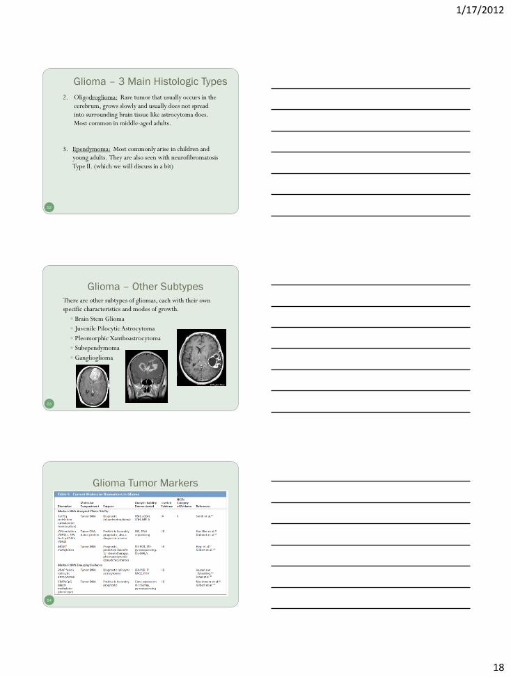
Glioma – Other Subtypes
53
There are other subtypes of gliomas, each with their own
specific characteristics and modes of growth.
Brain Stem Glioma
Juvenile Pilocytic Astrocytoma
Pleomorphic Xanthoastrocytoma
Subependymoma
Ganglioglioma
Glioma Tumor Markers
54
1/17/2012
19
Non-Glial Tumors
55
Medulloblastoma: Usually arises in the cerebrum,
is the most common brain tumor in children, and
is sometimes called a “primitive neuroectodermal
tumor” or PNET.
Meningioma: Arises from the meninges which are
the outside coverings of the brain between the
skull and the brain itself. It usually presses on the
brain, but does not invade it and often grows
slowly.
Non-Glial Tumors
56
Schwannoma: Arises from Schwann cells present
in certain nerves, including those that control
balance and hearing.
A common site is the vestibular nerve which
carries signals from the inner ear to the brain stem.
Tumors in this location are called “acoustic
neuromas” (a.k.a. vestibular schwannoma), and
occur most often in adults.
Non-Glial Tumors
57
Craniopharyngioma: Grows at the base of the brain,
arises from the tissue connecting the brain and the
pituitary gland and occurs in both adults and
children.
Pituitary Adenoma: Arises from the pituitary gland
and may cause compression of the optic nerves
causing vision problems. Some produce excessive
amounts of hormones that can disrupt the body’s
metabolism. Roswell Park Cancer Insitute

1/17/2012
20
Observing Migration of Glioma Cells
58
Seeing Cancer Cells Migrate
Seeing Cancer Cells Migrate
Source: Case Western Reserve University School of Medicine, public release 8/25/11
Neurofibromatosis
59
The neurofibromatoses (NF) are a group of genetic disorders
which cause tumors to grow along nerves and can also affect the
development of non-nervous tissues such as bones and skin.
Neurofibromatosis Type I (NF-I), also known as Peripheral NF
and historically as von Recklinghausen Disease
Occurs in 1:4,000 births
Multiple cafe-au-lait spots (not reportable)
Many, many neurofibromas on or under the skin (not reportable)
Enlargement and deformation of bones and curvature of the spine
Tumors may develop in brain, on cranial nerves, or the spinal cord
Neurofibromatosis Foundation
NF Type I: First documented photo 1871
60
Source Credit: Dr. Stanley B. Burns
http://www.cbsnews.com/2300-204_162-10007019-6.html#ixzz1clEzAchI

1/17/2012
21
Other Manifestions of NF Type I
Lisch nodules on the eye Café-au-lait spots on skin
61
Melanocytic hemartomas Discolored birth marks
Medscape Source: Dermnet.com; Dermatologic Manifestations of NF Type I
Neurofibromatosis Type II
62
Neurofibromatosis Type II (NFII), also known as Multiple Inherited Schwannomas, Meningiomas and Ependymomas (MISME) or Bilateral Acoustic Neurofibromatosis (BAN ).
Is a genetically inherited diseasecaused by mutations of the "Merlin" gene, which appears to influences the form and movement of cells
Primary manifestation is a development of non-malignant brain tumors in the region of the cranial nerves, frequently bilaterally. The eighth cranial nerve is the auditory-vestibular nerve which transmits sensory information from the inner ear to the brain and is commonly affected.
Source: California Ear Institute
Acoustic Neuroma/Schwannoma
63 Source: http://thrivingwithneurofibromatosis.blogspot.com

1/17/2012
22
Multiple Primary Rules
Histology Coding Rules
64
Different Rules for Benign and Malignant
65
Sequence Numbering for Brain Tumors
66
Malignant primary brain and CNS tumors are assigned Sequence Codes in the range 00-35 Sequence Chronologically 00-35 Only count malignant tumors in the sequence If only one malignant tumor occurs, it is coded 00 If subsequent (multiple) primary malignant and/or in situ neoplasms,
the sequence number for the first tumor begins at 01, the sequence number for the second primary tumor is 02, and so forth.
Non-malignant primary brain and CNS tumors are assigned Sequence Codes in the range 60-87. Sequence Chronologically 60-87 Only count benign/borderline or reportable by agreement neoplasms
in the sequence If only one non-malignant tumor occurs, it is coded 60. If subsequent (multiple) non-malignant neoplasms are diagnosed, the
first tumor should be sequenced as 61, the second 62 and so forth.

1/17/2012
23
Benign and Borderline Tumor Rules
67
68
When multiple tumors are present registrars should identify and document specific characteristics for MPH Rules Text
Date of Diagnosis (Timing is not used to determine number of abstracts or primary neoplasms to abstract)
Method and Details of Diagnosis (some are never resected)
Location of Tumor
Laterality
Histologic Type – refer to Chart 1
Tumor Behavior
Multiple Meningioma’s (meningiomatosis)
Neurofibromatosis Characteristics (when applicable)
Benign and Borderline Tumor Rules
Malignant Tumor Rules
69

1/17/2012
24
70
Malignant Tumor Rules
71
When multiple tumors are present registrars should identify and document specific characteristics for MPH Rules Text Date of Diagnosis (Timing is not used to determine number of abstracts
or primary neoplasms to abstract) Method and Details of Diagnosis (most attempt resection) Location of Tumor (not spread or invasion – but bulk of tumor) Histologic Type – refer to Chart 1 and/or Chart 2 Tumor Behavior Variations or Combinations of One or More Glial Tumors Over Lifetime –
astrocytoma, glioblastoma, ependymoma, or oligodendroglioma Special rules for determining # abstracts
Special rules for determining whether or not is mixed glioma
Note: Recurrence, progression, or any reappearance of histologies on the same branch in Chart 1 or Chart 2 is always the same disease process.
Malignant Tumor Rules
Report /Sequence All Tumors Over Lifetime
72
REMINDER: Sequence numbers for malignant neoplasms and for benign, borderline, and other reportable-by-agreement cases are assigned over a lifetime.
Therefore,
IF A PATIENT WAS DIAGNOSED WITH A NON-MALIGNANT CNS
NEOPLASM BEFORE REPORTING WAS REQUIRED (January 1, 2004),
THE NEW (SECOND) NEOPLASM SHOULD BE ASSIGNED SEQUENCE NUMBER 62
AND THE FIRST NEOPLASM (Seq 61) IS REPORTABLE AS A HISTORICAL CASE TO FCDS.
(An abstract/accession is not be required by CoC or SEER but is by FCDS)
Any benign and/or borderline brain or CNS tumor(s) diagnosed before January 1, 2004 ARE REPORTABLE TO FCDS as historical cases when accompanied by another reportable primary.
FCDS Data Acquisition Manual and CDC Data Collection of Primary Central Nervous System Tumors

1/17/2012
25
73
*2013*
2013
74
75

1/17/2012
26
76
77
78

1/17/2012
27
Treatment
Surgical Option(s)
80
Decisions regarding aggressiveness of surgery for primary
brain lesions are complex and depend on the: Age and performance status of the patient
Proximity to “eloquent” areas of the brain
Feasibility of decreasing the mass effect with aggressive surgery
Resectability of the tumor (including the number and location of lesions)
In patients with recurrent disease, the time since the last surgery
Surgical options include: Stereotactic biopsy
Open biopsy or debulking procedure
Subtotal resection
Maximal safe resection
Craniotomy
81
Any bony opening that is cut into the skull.
Source: Mayfield Clinic

1/17/2012
28
Craniotomy Procedure
Source: MedlinePlus/US National Library of Medicine, NIH 82
A section of the skull, (called a bone flap) is removed to access the brain underneath.
Typically the bone flap is replaced.
If the flap is not replaced, the procedure is called a craniectomy
Surgeon has drawn the cutline circle
around the tumor location
83 Source: The Alien-a set on Flickr www.flickr.com/photos/woodcreeper/sets/598206
Surgeon has cut the scalp and pulled it back to
expose the skull over the tumor
84 Source: The Alien-a set on Flickr www.flickr.com/photos/woodcreeper/sets/598206

1/17/2012
29
The skull is removed revealing the dura layer
under which is the brain and tumor
85 Source: The Alien-a set on Flickr www.flickr.com/photos/woodcreeper/sets/598206
Here you see the circular cut through the dura
layer with the brain and tumor exposed.
86 Source: The Alien-a set on Flickr www.flickr.com/photos/woodcreeper/sets/598206
Meningioma Resected
87 Source: The Alien-a set on Flickr www.flickr.com/photos/woodcreeper/sets/598206

1/17/2012
30
Pre- and Post-Operative Imaging
88
Pre-op tumor is outlined in red
Post-operative MRI shows complete resection of the tumor
Source: Desert Spine and Neurosurgical Institute
Surgery Codes
89
Surgery Codes
90
1/17/2012
31
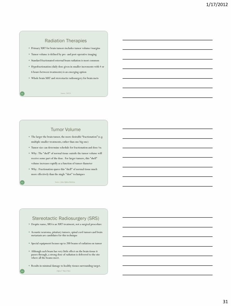
Radiation Therapies
91
Primary XRT for brain tumors includes tumor volume/margins
Tumor volume is defined by pre- and post-operative imaging
Standard fractionated external beam radiation is most common
Hypofractionation (daily dose given in smaller increments with 4 or
6 hours between treatments) is an emerging option
Whole brain XRT and stereotactic radiosurgery for brain mets
Source: NCCN
Tumor Volume
92
The larger the brain tumor, the more desirable “fractionation” (e.g.
multiple smaller treatments, rather than one big one)
Tumor size can determine schedule for fractionation and dose/tx
Why: The "shell" of normal tissue outside the tumor volume will
receive some part of the dose. For larger tumors, this "shell"
volume increases rapidly as a function of tumor diameter
Why: Fractionation spares this "shell" of normal tissue much
more effectively than the single "shot" techniques
Source: Johns Hpkins Medicine
Stereotactic Radiosurgery (SRS)
93
Despite name, SRS is an XRT treatment, not a surgical procedure
Acoustic neuroma, pituitary tumors, spinal cord tumors and brain metastasis are candidates for this technique
Special equipment focuses up to 200 beams of radiation on tumor
Although each beam has very little effect on the brain tissue it passes through, a strong dose of radiation is delivered to the site where all the beams meet.
Results in minimal damage to healthy tissues surrounding target.
Zdpirce” Mayo Clinic

1/17/2012
32
94 Source: San Diego Gamma Knife Center
Chemotherapy
95
Chemotherapy is not an effective initial treatment for low-grade
brain tumors. Why? Because most standard chemo agents have a
hard time passing into the brain because of how the brain protects
itself (the blood-brain barrier)
Not all types of brain tumors respond to chemotherapy
In general, chemotherapy for brain tumors is usually administered
following surgery or radiation therapy
Participation in clinical trials should be encouraged
Blood Brain Barrier
96
Composed of special cells that make up brain’s blood vessels
Selectively prevents substances from entering the blood and brain,
only allowing essential molecules such as amino acids, oxygen,
glucose and water through
Adenosine, a molecule produced by the body, seems to modulate
the entry of large molecules into the brain
When adenosine receptors are activated on cells that comprise the
blood-brain barrier, a gateway into the barrier can be established
Science Daily Source: September 13, 2001

1/17/2012
33
Approved Chemotherapy Agents
97
Carmustine (BCNU) – IV or dissolvable wafers placed surgically
Temozolomide (Temodar) – oral
Lomustine (CCNU) – oral
Carboplatin
Cisplatin
Etoposide
Irinotecan
Vincristine
Procarbazine (Matulane) – oral
Methotrexate - oral, by injection or intrathecally
NCCN Treatment Guidelines
Infiltrative Low-Grade Glioma
99
Best management strategy has yet to be defined
Small tumor samples can provide a lower histologic grade
Rationale: Needle biopsies are often performed when lesions are in deep or critical regions of the brain, but can be misleading because gliomas often have varying degrees of cellularity, mitosis, or necrosis from one region to another
General recommendation is to first attempt as complete an excision of tumor as possible (based on postsurgical MRI verification) without compromising function
No consensus exists regarding proper timing of postoperative external beam radiation
Chemotherapy is not a traditional upfront treatment modality

1/17/2012
34
100
When possible, maximal safe resection
If gross total resection is achieved, some patients may be observed without adjuvant therapy. However, close follow-up is essential as over half of patients will eventually progress
These tumors behave aggressively in patients over 40 years old
Adjuvant radiation or chemotherapy is recommended
If stereotactic biopsy, open biopsy, or other subtotal excision was done, immediate fractionated external beam RT or chemotherapy should be given
Infiltrative Low-Grade Glioma
Anaplastic Glioma and Glioblastoma
101
Whenever possible, major tumor removal should be performed
If glioblastoma is confirmed, options include radiation, chemotherapy, best supportive care, chemoradiation only if carmustine wafer was implanted
If high-grade glioma is confirmed, BCNU wafer is an option
In patients with good Karnofsky score (70 or above) options include fractionated external beam radiation therapy, chemotherapy or chemoradiation in the context of a clinical trial
In patients with poor Karnofsky score (below 70) management may include radiation, chemotherapy or best supportive care
Intracranial Ependymoma
102
Whenever possible, maximal safe resection should be attempted
Adjuvant treatment depends on the extent of surgical resection,
histology and staging by cranial spinal MRI and CSF cytology
CSF dissemination occurs in up to 15% of intracranial ependymomas
If MRI spine /CSF reveal disease, craniospinal radiation is mandatory
If gross total resection with negative spinal MRI and CSF, adjuvant
regional fractionated EBRT or observation may be considered

1/17/2012
35
Medulloblastoma and PNET (supratentorial)
103
MRI is the gold standard to assess PNET
Maximal safe resection is recommended when possible
Average Risk Patients: craniospinal radiation alone or
concurrent chemoradiation followed by chemotherapy are
both options
High Risk Patients: patients with large cell or anaplastic
medulloblastoma, supratentorial PNET, disease
dissemination, unresectable tumors, or residual tumors over
1.5cm post-surgery are high risk and should undergo
radiation followed by chemotherapy
Primary CNS Lymphoma
104
Treatment to be initiated as immediately following diagnosis
Treatment options depend on patient overall health and age
For healthier patients a high-dose methotrexate regimen
RT after systemic treatment depends on the responsiveness
of the disease to the chemotherapy
However, one or both may increase neurotoxicity, especially
in patients older than 60 years of age
Primary Spinal Cord Tumors
105
MRI is the gold standard for diagnosis of spinal cord lesions
Asymptomatic patients may be observed or resected
Symptomatic patients should undergo some form of surgery
Maximal safe resection should be attempted
Post-operative adjuvant radiation is not recommended
However, if symptoms persist after incomplete resection or
biopsy, radiation should be administered

1/17/2012
36
Meningioma
106
Meningiomas are typically diagnosed by CT or MRI imaging
Biopsy may be considered for confirmation
Options stratified by presence/absence of symptoms and tumor size
Most asymptomatic patients with small tumors (<30mm) may just
be observed. If neurological impairment is imminent, surgery (if
accessible) or radiotherapy (EBRT OR SRS) is feasible
Asymptomatic tumors >30mm can be either resected or observed
Meningioma
107
Symptomatic disease requires active treatment by surgery if possible
Non-surgical candidates should undergo radiation
All patients with surgically resected grade III meningiomas (even
after gross total resection) should receive adjuvant radiation for local
control regardless of tumor size and symptom status
Additional Resources
108
NCCN Evidence Based Treatment Guidelines, nccn.org, 2011
Collaborative Stage Data Collection System, AJCC, 2010
Multiple Primary and Histology Coding Rules, SEER 2007
The 2007 WHO Classification of Tumours of the Central Nervous
System, David N. Louis, Hiroko Ohgaki, Otmar D. Wiestler, Webster K.
Cavenee, Peter C. Burger, Anne Jouvet, Bernd W. Scheithauer and Paul
Kleihues, World Health Organization, Lyon, France, 2007
Data collection of primary central nervous system tumors.
National Program of Cancer Registries Training Materials.
Department of Health and Human Services, Centers for Disease Control and
Prevention. Atlanta, Georgia, 2004.

1/17/2012
37
109