brain activation and pupil response during covert ... · brain activation and pupil response during...
TRANSCRIPT

Brain activation and pupil response during covertperformance of the Stroop Color Word task
GREGORY G. BROWN,1 SANDRA S. KINDERMANN,1 GREG J. SIEGLE,1 ERIC GRANHOLM,1
ERIC C. WONG,1,2 and RICHARD B. BUXTON2
1Psychiatry Department,2Radiology Department, University of California San Diego andVA San Diego Healthcare System
(Received November 25, 1997;Revised June 22, 1998;Accepted June 25, 1998)
Abstract
Patterns of brain activation associated with covert performance of the Stroop Color–Word task were studied inyoung, healthy, adult volunteers using blood oxygen level dependent (BOLD) functional magnetic resonanceimaging (fMRI). Comparisons of the incongruous Stroop condition were made with both color naming and wordreading baselines. Areas of the left and right anterior cingulate, the right precuneus, and the left pars opercularisdisplayed larger BOLD signal responses during the incongruous Stroop condition than during baseline conditions.Activation of BOLD signals in these areas was highly repeatable. In a second experiment, pupil diameter was usedto assess cognitive load in 7 individuals studied during overt and covert performance of both Stroop and colornaming conditions. Cognitive load was similar in overt and covert response conditions. Results from the BOLDstudy indicate that brain regions participating in selective visual attention and in the selection of motor programsinvolved in speech were activated more by the Stroop task than by the baseline tasks. The neural substrate involvedin the resolution of the perceptual and motor conflicts elicited by the Stroop Color–Word task does not appear to bea single brain region. Rather, a network of brain regions is implicated, with separate regions within this systemsupporting distinct functions. (JINS, 1999,5, 308–319.)
Keywords: Stroop Color–Word Interference Test, Functional magnetic resonance imaging, Selective attention
INTRODUCTION
Since Stroop’s original paper (1935), the Stroop Color–Word task has been used to study interference and attentionin hundreds of studies (MacLeod, 1991). Experiment 2 de-scribed in Stroop’s 1935 paper is the most widely repeatedof the three experiments he reported. In that experimentStroop presented five color words: “RED,” “GREEN,”“BLUE,” “BROWN,” and “PURPLE,” in ink colors thatwere incongruous with the word (e.g., the word “RED” pre-sented in blue ink). Stroop asked participants in the exper-iment to name the color of the ink. He found that reactiontimes were on the average 74% longer when naming theink color of these incongruous words than when namingthe ink color of color squares.
Brain systems activated by the Stroop Color–Word taskhave been investigated with a variety of functional imaging
methods. In Table 1 we present a summary of brain areasdifferentially activated when individuals name the inkcolor of incongruous color words. Although most studiesreported lateral or polar frontal lobe activation, the specificareas of frontal activation varied widely across studies.Among the relatively circumscribed areas of activation re-ported in Table 1, the anterior cingulate (AC) is the mostconsistently reported area of Stroop activation. Even stud-ies reporting activation of AC cortex do not always de-scribe the specific location of activation. For example, Larrueet al. (1994) reported only that the incongruous Stroop con-dition activated the right superior mesial frontal region. Otherauthors do report the Talairach–Tournoux (1988) coordi-nates of AC activation (Bench et al., 1993; Pardo et al., 1990;Taylor et al., 1994). However, in each case, location of func-tional activity was determined solely from images obtainedfrom positron emission tomography (PET). When localiz-ing areas of activation from functional images alone, ana-tomical landmarks visible on high resolution structural brainimages cannot be used to confirm the localization. Further-more, because the Talairach–Tournoux transformation ad-
Reprint requests to: Gregory G. Brown, Psychology Service (116B),VASDHS, 3350 La Jolla Village Drive, San Diego, CA 92161. E-mail:[email protected].
Journal of the International Neuropsychological Society(1999),5, 308–319.Copyright © 1999 INS. Published by Cambridge University Press. Printed in the USA.
308

justs only for gross differences in brain shape and volume,it does not standardize across normal variation in gyral andsulcal anatomy. To increase the precision of localization,recent investigators have mapped functional images ontohigh resolution structural images (Mazziotta & Koslow,1987).
Because the anterior cingulate is a multifunctional organ,interpreting the functional significance of AC activation willdepend upon the specific localization of the activation. Intheir review, Devinsky et al. (1995) proposed a division ofAC cortex into affective areas and cognitive areas. They fur-ther divide the cognitive area into regions involved in mo-tor, nociceptive, and information processing functions. Evenwithin a single domain of behavior there may be multiplerepresentation of functions that are localized in different sub-regions of the AC cortex depending upon the specific be-havior. In animal studies, multiple areas of motor controlhave been reported within the AC cortex or cingulate sulcus(Picard & Strick, 1996). Additionally, the adjacent supple-mentary motor area (SMA) and pre-SMA cortex are in-volved in motor behaviors (Goldberg, 1985; Picard & Strick,1996). Because the cingulate sulcus is difficult to resolveon functional images, previous functional studies of brainactivation produced by the Stroop paradigm would have dif-ficulty separating the AC cortex from adjacent SMA andpre-SMA. Furthermore, it is difficult from functional im-ages alone to determine rigorously the location of areas offunctional activation in relation to the cingulate sulcus, whereimportant cingulate motor systems reside. One aim of thepresent study is to provide a more specific localization ofStroop activation within the AC than has been possible inprevious studies.
In the neuropsychology literature, the longer responsetimes to the incongruous Stroop stimuli are often inter-preted as measures of response inhibition or selective vi-sual attention (Lezak, 1995). The selective attention account
of the Stroop effect assumes that the longer reaction timesare due to stimulus–stimulus competition between the in-congruous color and lexical features (Hock & Egeth, 1970;Kornblum et al., 1990; Taylor et al., 1994). As children learnto generalize the ability to read across types of scripts, theylearn to attend to lexical features rather than script featuresof words. Many children are capable of distinguishing na-tive or foreign words from word-like scribbling by age 4years, regardless of whether the words are written in upperor lower cases or in printed or cursive letters (Gibson, 1970).Under the attention hypothesis, attending to the lexical fea-tures of words is highly automatic, whereas attending to inkcolor is not (MacLeod, 1991). Control processes utilizingattention are required to suppress the automatic bias to at-tend to lexical features and shift the focus of attention to-wards color features, as required by the incongruous Stroopcondition, lengthening response times.
Whereas the selective attention hypothesis places the lo-cus of the Stroop effect in the perceptual system, the re-sponse inhibition account places the locus in the responsesystem. According to the response inhibition account, in-compatible motor programs are elicited by the incongruousStroop stimuli (Morton & Chambers, 1973). Because onlyone word can be spoken at a time, the two activated motorprograms must be reconciled before a word is emitted. Thisreconciliation lengthens response times, especially when themotor program that is suppressed is prepotent, as in the in-congruous Stroop condition.
A second aim of this study is to contribute to a resolutionof the selective-attention–response-inhibition controversy.To the extent that brain systems involved in selective visualattention are differentially activated during the Stroop task,our results would support the selective attention hypoth-esis. To the extent that brain systems involved in motor pro-gramming are differentially activated by the Stroop task, aresponse inhibition hypothesis would be supported.
Table 1. Brain areas activated by Stroop tasks
Author(s)Anteriorcingulate
Supplementarymotor
Basalganglia Thalamus
Frontalcortex
Temporalcortex
Occipitalcortex
Parietalcortex
Bench et al. (1993)1 No No No No Yes No No YesBench et al. (1993)2 Yes No No No Yes No No NoCarter et al. (1995) Yes No No Yes Yes No Yes YesCarter et al. (1997) Yes No No No Yes Yes No NoGeorge et al. (1994) Yes No Yes Yes Yes No Yes YesKhorram-Sefat et al. (1996) No No Yes No Yes Yes Yes NoLarrue et al. (1994) ? No No No Yes? No No NoPantelis et al. (1996) Yes No No Yes No No Yes YesPardo et al. (1990) Yes Yes Yes No No Yes Yes NoPeterson et al. (1996) Yes No Yes No Yes Yes No NoTaylor et al. (1997)1,a Yes0No ? No No Yes Yes No YesTaylor et al. (1997)2 No No Yes No Yes Yes Yes Yes
1Experiment 1.2Experiment 2.aAnterior cingulate activation appeared during one of two conditions.Note. A question mark identifies possible activation.
Brain activation during covert Stroop task 309
Theories of Stroop phenomena often implicitly general-ize results from overt Stroop performance to circumstanceswhere attentional competition and response conflicts are re-solved covertly. Even though covert resolution of atten-tional or response interference is typical of extralaboratorybehavior, we found very little data available on covert per-formance of the Stroop Color Word task. Among the neuro-imaging papers listed in Table 1, only the fMRI study byKhorram-Sefat et al. (1996), reported in a conference ab-stract, investigated brain activation during covert perfor-mance of the Stroop Task. In an automated search of thePsychologicalAbstract database, none of the more than 1,000published papers on Stroop effects listed the terms “co-vert,” “implicit,” or “tacit” in the title, abstract, or keywordlist. A third goal of the present study is to determine whetherthe anterior cingulate, the brain region most commonly ac-tivated in functional neuroimaging studies of the Stroop ef-fect studied during overt performance, is activated duringcovert processing of the incongruous Stroop stimuli. Con-firming activation of the AC during covert Stroop perfor-mance would strengthen the view that the AC is centrallyinvolved in brain activities that represent a general behav-ioral system adapted to resolve processing conflicts.
Both error data and response times indicate that color nam-ing of the incongruous Stroop stimuli is more difficult thannaming color blocks or reading color-neutral words (Mac-Leod, 1991). The greater difficulty of the incongruous Stroopcondition is conventionally attributed to the additional cog-nitive processes involved in the incongruous condition com-pared with baseline conditions (Taylor et al., 1997). Thatthe incongruous condition involves cognitive activities notinvolved in the baseline condition is a fundamental assump-tion of the subtraction logic underlying most functional im-aging studies of the Stroop effect. Recently, investigatorshave used autonomic measurements to monitor the greatereffortfulness of the incongruous Stroop condition com-pared with typical control conditions. Typically, partici-pants experience greater autonomic responsiveness in theStroop condition compared with control conditions (Hoshika-wa & Yamamoto, 1997; Renaud & Blondin, 1997). Auto-nomic nervous system measurements provide investigatorswith methods to monitor the comparative effortfulness ofprocessing incongruous Stroop stimuli and control stimuliin overt and covert response conditions.
Because the greater effortfulness of the incongruousStroop condition compared with baseline might interact withovert–covert response conditions, interpretation of resultsfrom functional neuroimaging studies of covert Stroop per-formance is limited by the paucity of studies comparing overtand covert response conditions. Covert performance couldlead to either more or less activation on neuroimaging stud-ies compared with overt performance. If individuals werecovertly responding only to the Stroop condition and ignor-ing the baseline stimuli, then a greater number of areas ofactivation or more intense areas of activation might appearcompared with overt performance. If covert performanceomitted a critical stage of processing Stroop stimuli, then
covert Stroop performance might be less effortful and leadto less intense brain activation than overt performance. Afourth goal of our study was to determine whether the greatercognitive demand of the incongruous Stroop condition wascomparable during covert and overt performance. Becausepupil diameter is a well-validated measure of cognitive ef-fort or load (Beatty, 1982; Granholm et al., 1996), we usedpupilometric methods to compare overt and covert Stroopperformance.
EXPERIMENT 1
Brain areas activated by the incongruous Stroop conditionwere studied on three trials. To test the hypothesis that ar-eas within the anterior cingulate would activate during im-plicit performance of the Stroop Color–Word task, allparticipants responded covertly. The first trial contrastedbrain regions activated during the Stroop incongruous con-dition with regions activated during color naming. The colornaming task controlled for activation associated with pro-cessing a color stimulus, for activation associated with thelexical access of color words, and for activation of the finalmotor pathways activated by the implicit articulation of colorword responses (Taylor et al., 1997). The second trial con-trasted brain regions activated during the incongruous Stroopcondition with regions activated during reading of color-neutral words. Because participants in the experiment mightread the color words during the incongruous Stroop condi-tion, some areas activated more by processing of the incon-gruous Stroop stimuli than by color naming could reflectthe effects of reading. The color-neutral words used as abaseline in Trial 2 controlled for potential reading effects.To determine the stability of Trial 1 findings, Trial 3 repli-cated Trial 1.
METHODS
Research Participants
Eight well-motivated, healthy, adult volunteers (5 female,7 right-handed) below 55 years of age were studied.
Behavioral Activation Tasks
Participants were scanned during three separate study trials,each lasting 360 s. Trials consisted of 4.5 cycles of alter-nating baseline and experimental conditions. Each condi-tion lasted 40 s yielding an 80-s cycle time with an unpairedbaseline task following the 4th cycle.
Participants participated in three trials. On Trial 1 partici-pants named the color of red, blue, or green squares duringthe baseline condition. During the experimental condition,they named the script color of the words “RED,” “BLUE,”or “GREEN” printed in a color incongruous with the colorword. Stimuli covered a 28 to 38 visual angle. On Trial 2participants read the animal names “TIGER,” “DOG,” and“BEAR” during the baseline condition. The experimental
310 G.G. Brown et al.
condition employed the same incongruous stimuli used inthe experimental condition of Trial 1. Trial 3 was a replica-tion of Trial 1. Images from the three trials were collectedover about a 30-min period.
Stimuli were back-projected by an overhead projector ontoa screen placed at the foot of the magnet. Participants useda mirror attached to a head coil to view the stimuli, whosepresentation was controlled by a PC computer containing aDigitry, Inc. Cognitive Testing Station (Edgecomb, MA).Subjects covertly named or read. More specifically, they weretold to speak loudly enough that they could subjectively heartheir response yet not move their lips, jaw, or tongue. Allparticipants reported that they understood this instruction.We performed an exit interview to determine whether par-ticipants attempted to follow this covert response instruc-tion and to determine whether they attempted to respond toall classes of stimuli. All participants reported complyingwith the task instructions. Prior to fMRI study, participantswere pretested in an overt response condition to determinea presentation rate at which they would make errors on 10to 20% of the incongruous, Stroop trials (M 6 SD: ontime5 3216 81 ms; off time5 3146 90 ms). During thefMRI study, incongruous and baseline stimuli were pre-sented at these tailored presentation rates to correct forbetween-participant differences in Stroop ability. During thepretest, incongruous Stroop stimuli and color blocks werepresented following the 4.5 cycle procedure described above.Participants responded verbally.
Image Acquisition
Participants were scanned in a 1.5 T GE Signa scannerequipped with an inserted three-axis balanced torque headgradient coil designed for rapid switching (Wong et al.,1992). Manual shimming further enhanced the signal-to-noise within the anterior cingulate. A midsagittal localizerslice was obtained to determine landmarks for eight axialslices (5 mm thick, 1 mm interslice gap) for functional im-aging. Echo planar, gradient recalled (TR5 2,500 ms,TE5 40 ms, FOV5 24 cm, 3.2 slices0s) axial images com-posed of 643 64 3.75 mm square pixels were acquired con-tinuously. Over the 365-s experiment, 146 images of eachslice were usually acquired. The pulse sequence weightedthe echo planar images (EPI) for blood oxygen level depen-dent (BOLD) contrast (Bandettini et al., 1995; Ogawaet al., 1990). Axial images were obtained. The EPI sliceswere selected to include the anterior cingulate gyrus andportions of the frontal, parietal, temporal, and occipital lobes.In addition to EPI images, 124 2.0 mm contiguous anatom-ical images were collected with a 3D SPGR protocol (TR580 ms, TE5 60 ms, flip angle 408, acquisition time: 12 min,19 s).
Postscanning Image Processing
We used a 4.5 cycle square wave to model the time courseof the fMRI signal response to alterations in behavioral con-
ditions. To account for the known time course of the cere-brovascular response to alterations in neuronal activity(Bandettini et al., 1995), the slopes of the square-wave stepswere modeled with 7.5 s rise and settling times to yield areference vector.
Images from 7 of the 8 participants were of sufficient qual-ity to justify further image analysis.We used theAFNI (Analy-sis of Functional NeuroImages) 2.11 package to analyze theimages (Cox, 1997). The image analysis for all three trialsinvolved the following steps. A 2D (coregistration) motionattenuation algorithm was used to reduce the effects of mo-tion. Next, time course data in each pixel were correlatedwith the reference vector using the following linear model(Cox, 1996, p. 22):Xi 5 a{r i 1 b{i 1 a 1 ei . The first termof the model represents the linear relationship between thereference vectorr and the intensity of the MR signal for theith occurrence of pixelX among the 146 replications ofeach image. The parametera is the intensity of signal changeassociated with the transition from one behavioral state toanother, namely, from control to Stroop conditions in ourstudy. This change-intensity value is adjusted for the otherterms in the model, which generally represent artifacts tobe controlled. In our analysis we added a term to removelinear trends over time (b{i ). The third term,a, representsthe mean signal intensity. The error term,ei , is assumedto be normally distributed with a mean of zero and con-stant variance,s2, over pixel replications. We report analy-ses ofa values, taken to represent change-intensity valuesadjusted for linear trends. After calculatinga for eachpixel, the eight slices were combined into a functional 3Dbrick that contained change-intensity information in each84.38 mm3 voxel. Similarly, the 124 slices obtained fromthe SPGR protocol were combined into an anatomical 3Dbrick. Because the BOLD and anatomical images were ac-quired in the same image session, we were able to use theknown offset of the center of the functional images fromthe center of the anatomical images to overlay the func-tional brick onto the anatomical brick. We then used stan-dard anatomical landmarks to transform the coordinates ofthe functional and anatomical bricks into Talairach–Tournouxcoordinates (1988). An anatomist verified the landmarks.This step also involved reslicing the functional 3D bricksinto cubes, 3.5 mm long on each side (42.875 mm3).
Because all the questions described in the Introductionfocused on neurobehavioral systems that underlie the in-creased processing demands of the incongruous Stroopcondition, we restricted our analysis to focus on areas ofincreased BOLD response during processing of incongru-ous stimuli. Areas of increased BOLD response to the ex-perimental condition compared with the baseline conditionswere identified by the change-intensity variable. The firststep in identifying areas of significantly greater activationduring the experimental condition involved performing one-samplet tests for Trial 1 (df 5 6, p 5 .025) on the intensitysignal contained in each voxel. Next, we used aclusterthresholdto reduce the number of false positive valuescaused by multiple statistical tests. On this step adjacent
Brain activation during covert Stroop task 311

voxels withp values falling below .025 had to form a con-nected region with a minimum volume of 253 mm3 beforeany voxel within the region was designated as significantlyactivated. Using the AlphaSims program (Ward, 1997), weran 1,000 Monte Carlo simulations to estimate the proba-bility that voxel clusters of various sizes would occur bychance given: a block size roughly equal to the intracranialportion of the functional images (about 323 32 3 16 re-sliced voxels), a single voxelp value of .025, an adjacencydistance of 1 voxel width (i.e., 3.5 mm), and no spatial fil-tering. Under these assumptions, less than 5% of voxel clus-ters activated by chance were as large or larger than sixresliced voxels; that is, 253 mm3. The cluster threshold iden-tified regions of intereston Trial 1 that guided our analysisof Trials 2 and 3. Only voxels found significantly activatedatp5 .05 on Trials 2 or 3 that overlapped with regions foundactivated by the cluster threshold criterion on Trial 1 were
ultimately consider to be activated voxels. To summarize,activated voxels had to pass the cluster threshold on Trial 1and the region of interest threshold on Trials 2 or 3.
The image analysis strategy described above was devel-oped to identify regions commonly activated in all threetrials involving the incongruous Stroop stimuli, while pro-tecting against Type I errors. We believe this analysis isappropriate to identify robustly the core brain systems in-volved in the Stroop effect. However, the strategy wouldfail to identify brain regions differentially activated by thethree tasks. Although identifying regions uniquely acti-vated by one of the three study tasks was not a primary goalof the study, we performed an exploratory analysis of im-ages to identify brain regions that were not consistently ac-tivated. The exploratory analysis simply involved specifyingregions that met the cluster threshold but not the region ofinterest threshold.
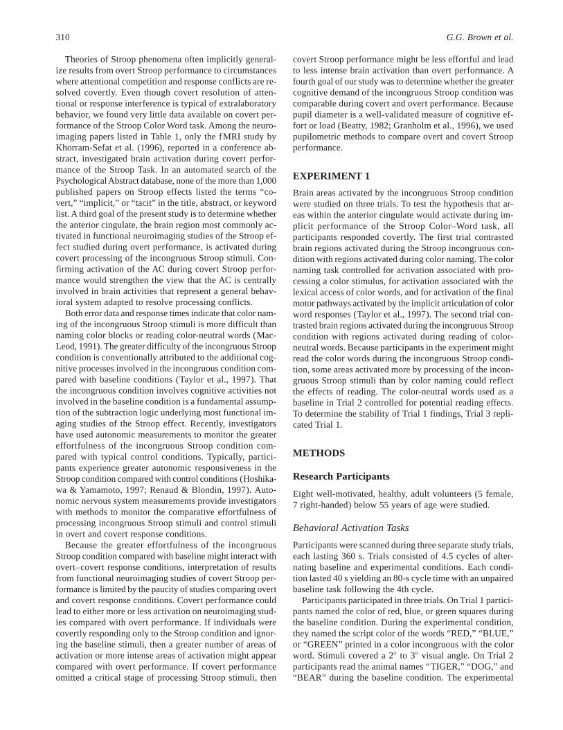
Fig. 1. A. Trial 1. Area of left anterior cingulate activated on the initial trial comparing the incongruous stimuluscondition with the color naming baseline. Trial 3. Replication of this area of activation on Trial 3. Note. The cross-hairsare placed in the same Talairach & Tournoux location on Trials 1 and 3. B. Areas of left and right anterior cingulateactivation on Trials 1, 2, and 3 combined. Color scale shows activation in arbitrary units with bright yellow represent-ing the greatest area of activation and pink representing the smallest.
312 G.G. Brown et al.

RESULTS
Behavioral Data
All participants were studied during overt performance ofthe Stroop Color–Word task outside the magnet to tailor thedifficulty level of the task to a participant’s ability. For eachparticipant, a presentation rate was chosen that produced anerror rate of about 10% on trials involving incongruousStroop stimuli. When naming colored blocks, all partici-pants responded with 100% accuracy at these presentationrates. The larger error rate when participants were namingthe text color of the incongruous stimuli compared with colornaming of colored blocks confirmed the existence of theStroop effect for the presentation times used in the presentstudy.
Imaging Findings
On Trial 1, participants named colored squares in the base-line condition and performed the incongruous color–wordtask in the experimental condition. Areas of activation re-flected regions where the BOLD signal intensity was greaterwhen participants named the text color of incongruous colorwords than when naming the color of colored squares. Onlyfour areas met both the Cluster Threshold on Trial 1 and theRegion of Interest Criterion on Trial 3. On Trial 1, two ofthese areas were bilaterally located in the anterior cingulatewith the right hemisphere centroid (Brodmann area 32)located at Talairach coordinates,X (right: 1, left: 2) 5 7.8,Y (anterior:1, posterior:2) 5 23.2,Z (superior:1, infe-rior: 2) 5 35.0, and the left hemisphere centroid located atX 5 23.7 Y5 13.9, andZ 5 35.2. The left anterior cingu-late site spanned the cingulate sulcus involving both areas24 and 32. On Trial 3, the pixel at the center of the righthemisphere area of activation on Trial 1 was again found tobe significantly activated [t~5! 5 4.49,p5 .006], as was thepixel at the center of the left hemisphere area of activationon Trial 1 [t~5! 5 5.43,p5 .003]. Figure 1 shows the iden-tical Talairach coordinates of the areas of left cingulate ac-tivation on Trials 1 and 3. Another anterior area of activationon Trial 1 was located in the pars opercularis (Brodmann’sarea 44) of the left hemisphere (centroid in Talairach coor-dinates:X 5 241.3,Y 5 15.9,Z 5 11.7). The pixel at thecenter of the pars opercularis activation on Trial 1 was alsoactivated on Trial 3 [t~5! 5 2.92,p 5 .03]. An area of acti-vation in the right precuneus region, anterior and superiorto the parietal-occipital sulcus at the border of Brodmann’sareas 19 and 39 of the right hemisphere, was also observedon Trial 1 (centroid in Talairach coordinates:X5 21.5,Y5263.4, andZ 5 34.8). The center of this region signifi-cantly activated on Trial 3 [t~5! 5 6.15,p 5 .002].
On Trial 2, participants read animal words during the base-line condition to contrast brain activation elicited by read-ing with activation elicited by the incongruous color–wordcondition. All four brain regions that showed greater acti-vation during the incongruous condition on Trials 1 and 3
overlapped with areas showing significantly greater activa-tion during the incongruous condition on Trial 2 [left cin-gulate:t~4! 5 6.89,p 5 .002; right cingulate:t~4! 5 4.97,p 5 .008; precuneus region–right hemisphere:t~4! 5 3.47,p 5 .03; pars opercularis–left hemisphere:t~4! 5 3.49,p 5.025]. Left and right hemisphere composite images of pix-els activated in the anterior cingulate region on Trials 1, 2,and 3 are presented in Figure 1. Across the three trials, ar-eas of significant anterior cingulate activation fell into Brod-mann’s areas 24 or 32 or in the cingulate sulcus separatingarea 24 from area 32.
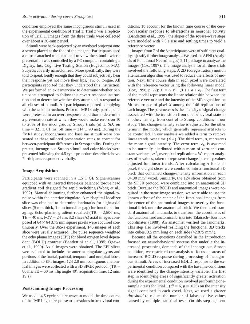
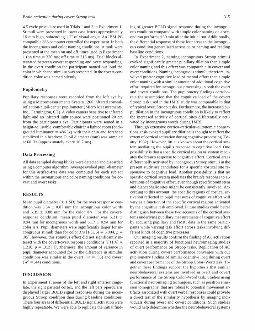
As mentioned above, there were several areas that metthe cluster threshold on Trial 1 yet fell short of the region ofinterest criterion on Trial 3. Most of these areas were in whitematter, as are shown in Figures 2 and 3. Perhaps the mostinteresting areas of signal response that occurred on onlyone trial were the apparent left lenticular activation seen onFigure 2, the apparent left parietal signal seen on Figure 3,and a large area of apparent signal seen in the left thalamuson Trial 2 (not pictured). Finally, no brain regions showedmore activation during color naming or reading conditionsthan during the incongruous color–word condition.
EXPERIMENT 2
Naming the script color of the incongruous Stroop stimuliinvolves a greater cognitive load than simple color naming.Experiment 2 tests the hypothesis that the differential cog-nitive load is comparable between overt and covert re-sponse conditions. This test was accomplished by recordingpupillary dilation responses during performance of covertand overt versions of the Stroop Color–Word task. Numer-ous studies have documented the covariance of task-evokedpupillary dilation with variations of processing load or cog-nitive effort (Beatty, 1982).
METHODS
Research Participants
After providing informed consent, 7 healthy undergraduateand graduate students (4 male, 3 female), who did not par-ticipate in Experiment 1, were studied. All participants wereright-handed, with a mean age of 26.36 2.9 years and meaneducation of 18.76 2.4 years. All participants were screenedfor normal or corrected-to-normal vision using a Snellen wallchart.
Stroop Color–Word task
Pupil diameter measured while participants named the textcolor of incongruous color words was compared with pupildiameter measured while participants named the text colorof strings ofX’s varying from three to five characters long.We presentedX’s in the baseline condition rather than col-ored blocks to equate luminance in the experimental andcontrol conditions. Otherwise the task was identical to the
Brain activation during covert Stroop task 313

Fig. 2. Activation of left pars opercularis (in cross-hairs) on Trial1. The three other areas of apparent activation on the axial viewdid not appear on the subsequent replication trial (Trial 3). Noticethe left anterior cingulate activation on the coronal view. Colorscale shows activation in arbitrary units with bright yellow repre-senting the greatest area of activation and pink representing thesmallest.
Fig. 3. Right precuneus activation (in cross-hairs). The axial slicealso shows areas of left and right cingulate activation. The remain-ing apparent areas of activation on the axial slice did not appearon the replication trial. Color scale shows activation in arbitraryunits with bright yellow representing the greatest area of activa-tion and pink representing the smallest.
314 G.G. Brown et al.
4.5 cycle procedure used in Trials 1 and 3 in Experiment 1.Stimuli were presented in lower case letters approximately16 mm high, subtending 1.28 of visual angle. An IBM PCcompatible 386 computer controlled the experiment. In boththe incongruous and color naming conditions, stimuli werepresented at the mean on and off times used in Experiment1 (on time5 320 ms; off time5 315 ms). Trial blocks al-ternated between covert responding and overt responding.In the overt condition the participant named out loud thecolor in which the stimulus was presented. In the covert con-dition color was named silently.
Pupilometry
Pupillary responses were recorded from the left eye byusing a Micromeasurements System 1200 infrared corneal-reflection-pupil-center pupilometer (Micro Measurements,Inc., Farmington, CT). A video camera sensitive to infraredlight and an infrared light source were positioned 20 cmfrom the participant’s eye. Participants were seated in aheight-adjustable, comfortable chair in a lighted room (back-ground luminance5 486 lx) with their chin and foreheadstabilized in a headrest. Pupil diameter (mm) was sampledat 60 Hz (approximately every 16.7 ms).
Data Processing
All data sampled during blinks were detected and discardedusing a computer algorithm. Average evoked pupil-diameterfor this artifact-free data was computed for each subjectwithin the incongruous and color naming conditions for co-vert and overt tasks.
RESULTS
Mean pupil diameter (6 1 SD) for the overt-response con-dition was 5.546 0.87 mm for incongruous color wordsand 5.356 0.88 mm for the colorX’s. For the covert-response condition, mean pupil diameter was 5.3160.94 mm for incongruous words and 5.176 0.94 mm forcolor X’s. Pupil diameters were significantly larger for in-congruous stimuli than for colorX’s [F~1,6! 5 6.004,p 5.05], however, this stimulus effect did not significantly in-teract with the covert-overt response conditions [F~1,6! 51.218,p 5 .312]. Furthermore, the amount of variance inpupil diameter accounted for by the difference in stimulusconditions was similar in the overt (h2 5 .53) and covert(h2 5 .44) conditions.
DISCUSSION
In Experiment 1, areas of the left and right anterior cingu-late, the right parietal cortex, and the left pars opercularisdisplayed larger BOLD signal responses during the incon-gruous Stroop condition than during baseline conditions.These four areas of differential BOLD signal activation werehighly repeatable. We were able to replicate the initial find-
ing of greater BOLD signal response during the incongru-ous condition compared with simple color naming on a sec-ond run performed 30 min after the initial run. Additionally,the differential response of these four areas to the incongru-ous condition generalized across color naming and readingbaseline conditions.
In Experiment 2, naming incongruous Stroop stimulievoked significantly greater pupillary dilation than simplecolor naming and this effect was comparable in covert andovert conditions. Naming incongruous stimuli, therefore, in-volved greater cognitive load or mental effort than simplecolor naming with a similar amount of additional cognitiveeffort required for incongruous processing in both the overtand covert conditions. The pupilometry findings corrobo-rate the assumption that the cognitive load of the covertStroop task used in the fMRI study was comparable to thatof typical overt Stroop tasks. Furthermore, the increased pu-pil dilation in the incongruous condition is likely to reflectthe increased activity of cortical sites differentially acti-vated by incongruous words during fMRI.
Through extensive cortico–reticular–autonomic connec-tions, task-evoked pupillary dilation is thought to reflect theextent of cortical activation during cognitive processing (Be-atty, 1982). However, little is known about the cortical sys-tem mediating the pupil’s response to cognitive load. Onepossibility is that a specific cortical region or system medi-ates the brain’s response to cognitive effort. Cortical areasdifferentially activated by incongruous Stroop stimuli in thepresent study are candidates for a specific cortical site re-sponsive to cognitive load. Another possibility is that nospecific cortical system mediates the brain’s response to al-terations of cognitive effort, even though specific brain stemand diencephalic sites might be consistently involved. Ac-cording to this account, the specific regions of cortical ac-tivation reflected in pupil measures of cognitive effort willvary as a function of the specific cortical regions activatedby the cognitive task employed. Future studies could betterdistinguish between these two accounts of the cortical sys-tems underlying pupillary measurements of cognitive effortby acquiring pupillary and fMRI data in the same partici-pants while varying task effort across tasks involving dif-ferent kinds of cognitive processes.
Our imaging results confirm the finding of AC activationreported in a majority of functional neuroimaging studiesof overt performance on Stroop tasks. Replication of ACactivation during covert performance converges with ourpupilometry finding of similar cognitive load during overtand covert performance of the Stroop Color–Word task. To-gether these findings support the hypothesis that similarneurobehavioral systems are involved in overt and covertperformance of the Stroop Color–Word task. Studies usingfunctional neuroimaging techniques, such as positron emis-sion tomography, that are robust to potential movement ar-tifacts associated with overt verbal responses could providea direct test of the similarity hypothesis by imaging indi-viduals during overt and covert conditions. Such studieswould help determine whether the neurobehavioral systems
Brain activation during covert Stroop task 315

involved in overt Stroop performance are generally in-volved in the resolution of processing conflicts.
When the definition of activation was liberalized by drop-ping the region of interest criterion, several areas appearedto show increased BOLD response to the incongruous con-dition. Activation in these areas might have been artifac-tual. However, motion artifacts are most prominent near theedge of the brain; none of the areas of apparent activationwas near the periphery of the brain. Furthermore, imageswere reregistered to minimize motion artifacts. Drainingveins can be a source of large BOLD signal distal from ar-eas of neuronal activation. However, because neural and ve-nous compartments of the brain are stable features of cerebralanatomy, vein–brain artifacts would likely be present on bothTrial 1 and Trial 3. The areas of increased BOLD responseobserved only on the first trial might have been valid re-sponses that habituated with repetition. In support of thehabituation hypothesis, two studies found habituation ef-fects on PET functional images during repeated Stroop runs(Bench et al., 1993; Pantelis et al., 1996). However both ofthese studies found an attenuation of the functional signalin the anterior cingulate, an area that showed robust activa-tion across replications in our study. Finally, these apparentregions of activation might have been false positives inef-fectively filtered by the clustering threshold. In support ofthe false-positive account, most areas activated only on onetrial were in white matter, which typically responds withminimal activation to behavioral manipulations. When theregion of interest criterion was added to our definition ofactivation, none of the activated regions was located in whitematter.
The large left-thalamic activation (900 mm3) on Trial 2might have resulted from any of the processes discussedabove. However, it also might have been induced by themore verbal nature of the baseline (reading rather than colornaming) used on Trial 2. Possibly the reading baseline taskincreased demands for verbal resources in the left hemi-sphere and potentiated the verbal component of the incon-gruous Stroop condition. That the reading baseline mighthave activated more left hemisphere sites than the color-naming baseline was supported by the apparent activationof left parietal cortex and left caudate sites on Trial 2 butnot on Trial 1. Furthermore, priming of attention resourcesspecific to either hemisphere has been described in a vari-ety of studies (see Kinsbourne, 1975, for a review). Of thefunctional brain imaging studies of the Stroop effect thatwe reviewed, only the Larrue et al. (1994) study used a read-ing baseline to contrast with the incongruous condition. Al-though no thalamic activation was reported, the method offunctional imaging used, SPECT, is less sensitive to changesin thalamic activity than other functional imaging methods(Roland, 1993, p. 431). The contribution of the thalamus tothe neural substrate involved in the Stroop effect remains tobe resolved by future studies.
The areas of the left and right cingulate that showed anincreased BOLD response during the incongruous condi-tion are involved in premotor functions related to program-
ming facial movements (Picard & Strick, 1996). In humans,the area forms part of a rostral cingulate zone (RCZ) acti-vated during complex speech, eye, and finger movements(Picard & Strick, 1996). Although the caudal cingulate zone(CCZ) posterior to the anterior commissure plane has directconnections to the spinal cord, the RCZ has a low densityof corticospinal neurons (He et al., 1995). Furthermore, inmonkeys neurons in areas analogous to human CCZ re-spond rapidly after sensory stimulation, whereas neurons inareas analogous to human RCZ precede self-paced move-ments by 0.5 to 2.0 s, implying a premotor function (Shimaet al., 1991).
Although the available human and animal data support apremotor function for the RCZ, how this function is char-acterized varies across investigators. Some describe the RCZpremotor area as a center of willful activity, voluntary move-ment, motor preparation, response selection, complex mo-tor activity, internal planning, or attention to action. Goldberg(1985, p. 575) has argued that anterior cingulate cortex con-tributes to all voluntary action by altering the excitatorythresholds needed to elicit specific motor plans and by con-trolling the intensity or amplitude of motor responses. Wewill return to Goldberg’s ideas below.
We also observed consistent activation in the pars oper-cularis of the left hemisphere, near the area identified byTaylor et al. (1997) as the site that reflected processing spe-cific to the Stroop task. Both neuropsychological studies offocal lesion patients and functional imaging studies haveexamined the function of this brain region. After reviewingthe functional brain imaging literature, Roland (1993,pp. 285–286) concluded that the left pars opercularis andpars triangularis form the core of the left inferior frontalcenter participating in language production. Petersen et al.(1988) found that the pars opercularis, in particular, partici-pates in single word production when words are presentedvisually or aurally. Lesions in the pars triangularis produceaphemia, a disorder in the timing and coordination of themotor programs involved in articulation (Schiff et al., 1983).Voice onset time studies of patients with left hemispherelesions confirm the importance of anterior areas in the im-plementation of articulation (Blumstein et al., 1980). Theobserved activation of the pars opercularis when subjectsare performing the incongruous Stroop condition is proba-bly due to its role in selecting motor programs involved inspeech and implementing their articulation. Our findings in-dicate that the process of selecting motor programs occurseven when speech is covert and seems potentiated when adominant speech program is inhibited as the appropriatespeech program is selected.
An area of right parietal cortex (RtPC) was the fourtharea of significant activation observed. The differential ac-tivation of the RtPC in the incongruous Stroop condition iscompatible with the contribution of the parietal cortex toattention. The critical involvement of the inferior parietalcortex in attending to stimuli has been established by singlecell recording studies (Goldberg & Robinson, 1977; Lynch,1980), by functional neuroimaging studies (Pardo et al.,
316 G.G. Brown et al.

1991; Reivich et al., 1983), and by human lesion studies(Friedland & Weinstein, 1977; Heilman & Van Den Abell,1980). That the right parietal cortex showed a larger BOLDsignal change than the left is consistent with previous neuro-imaging (Pardo et al., 1991; Reivich et al., 1983) and hu-man lesion studies (Friedland & Weinstein, 1977; Heilman& Van Den Abell, 1980) of attention. Parietal activation wasspecifically observed in the precuneus, a brain region in-volved in saccade generation (Luna et al. 1998; Petit et al.,1996). The activation of a brain region involved in visualscanning during processing of incongruous Stroop stimulistrengthens the hypothesis that brain mechanisms involvedin visual attention contribute to the resolution of processingconflicts on Stroop Color–Word tasks.
Both the selective attention and response selection hy-potheses are supported by our findings. In our study, brainregions participating in selective visual attention and in theselection of motor programs involved in speech were acti-vated more by the Stroop task than by the baseline tasks.Thus, the neural substrate involved in the resolution of theperceptual and motor conflicts elicited by the Stroop taskdoes not appear to encompass a single brain region. Rathera network of brain regions appears to be implicated. None-theless, the separate regions within this system seem tosupport distinct functions. We envision a network whosecomponents are integrated by the following scheme. Be-cause a mature reader must learn to attend to the lexical fea-tures of words across different types of scripts, individualsare biased to attend to the lexical features of the incongru-ous stimuli rather than to the color features. Shifting atten-tion to script features activates neuronal pools in the rightparietal lobe involved in selective visual attention. How-ever, because the lexical features of the incongruous Stroopstimuli are automatically activated, two codes, one repre-senting lexical features, the other representing color fea-tures, are activated for further processing. The contributionof the premotor anterior cingulate system is to bias the net-work towards selecting the motor program associated withstimulus color. Following Goldberg’s (1985) conjecture,the anterior cingulate biases the network by altering thethreshold required to elicit the motor speech program thatgoverns the articulation of a color name. This account iscompatible with evidence that the AC cortex appears to makea nonspecific contribution to processing incongruous Stroopstimuli, whenever it is activated (Taylor et al., 1997). Al-though the cingulate activity biases the system towards se-lection of the motor representation of the color name, thetendency for the lexical features to activate the motor rep-resentation of the word name is not entirely suppressed. Con-sequently, two motor speech programs would tend to beactivated, with a bias towards activation of the color name,that is, towards the name of the script color. The final stepin resolving the conflict would involve programming thesequence of articulation associated with the code that ex-ceeds its threshold first. The competitive activation of speechcodes increases neuronal activity in the pars opercularis.Within this system there is no one location where inhibition
of the Stroop color–word response occurs. Rather, the sys-tem activates the color name code and progressively pro-motes the likelihood of its motor expression.
The hypothesis that a brain system rather than one brainsite is involved in the resolution of processing conflicts in-duced by the Stroop Color–Word task helps explain the trou-blesome finding that no single brain area is consistentlyactivated in functional neuroimaging studies of the Stroopeffect. Rather than a single region activated by the Strooptask, a variety of regions are activated depending upon ex-perimental variables such as presentation rate, practice, andtype of baseline task. To confirm the neural system hypoth-esis of the Stroop effect, the differential activation of sys-tem components needs to be related to manipulations of taskvariables. Furthermore, the pattern of brain activation wefound needs to be generalized beyond the limitations of thepresent study. In particular, because our functional imagesonly encompassed part of the brain, regions not imaged mightbe involved in processing Stroop stimuli. Whole brain stud-ies at higher field strengths could comprehensively imageneural systems subserving Stroop activation at 2 to 3 mmresolution. Additionally, the extent to which our findingsare dependent upon the particular presentation time usedneeds further study. Finally, imaging studies directly com-paring patterns of brain activation during overt and covertprocessing of Stroop stimuli need to be performed. Speci-fying behavioral conditions associated with componentsof the neural system subserving Stroop effects is likely toadvance cognitive theories of both attention and responsecompetition.
ACKNOWLEDGMENTS
The study was supported by grants from National Institute of Men-tal Health (P30 MH49671 and T32 MH19934) and from the Vet-erans Administration (VISN 22 Mental Illness Research, Educationand Clinical Center Grant). We wish to acknowledge Laura Sy-monds for verifying Talairach–Tournoux landmarks on the func-tional images.
REFERENCES
Bandettini, P.A., Wong, E.C., Binder J.R., Rao, S.M., Jesmanowicz,A., Aaron, E.A., Lowry T.F., Forster, H.V., Hinks, R.S., & Hyde,J.S. (1995). Functional MRI using the BOLD approach: Dy-namic characteristics and data analysis methods. In D. LeBihan(Ed.), Diffusion and perfusion: Magnetic resonance imaging(pp. 335–362). New York: Raven Press.
Beatty, J. (1982). Task-evoked pupillary responses, processing load,and the structure of processing resources.Psychological Bul-letin, 91, 276–292.
Bench, C.J., Frith, C.D., Grasby, P.M. Friston, K.J., Paulesu, E.,Frackowiak, R.S.J., & Dolan, R.J. (1993). Investigations of thefunctional anatomy of attention using the Stroop test.Neuro-psychologia, 31, 907–922.
Blumstein, S.E., Cooper, W.E., Goodglass, H., Statlender, S., &Gottlieb, J. (1980). Production deficits in aphasia: A voice-onset time analysis.Brain and Language, 9, 153–170.
Brain activation during covert Stroop task 317

Carter, C.S., Mintun, M., & Cohen, J.D. (1995). Interference andfacilitation effects during selective attention: An H2O
15O PETstudy of Stroop task performance.Neuroimage, 2, 264–272.
Carter, C.S., Mintun, M., Nichols, T., & Cohen, J.D. (1997). An-terior cingulate gyrus dysfunction and selective attention def-icits in schizophrenia: [15O]H2O PET study during single trialStroop task performance.American Journal of Psychiatry, 154,1670–1675.
Cox, R.W. (1996).Analysis of functional neuroimages: Version 2.00.Unpublished manuscript, Biophysics Research Institute, Med-ical College of Wisconsin.
Cox, R.W. (1997). AFNI: Software for analysis and visualizationof functional magnetic resonance neuroimages.Computers andBiomedical Research, 29, 162–173.
Devinsky, O., Morrell, M.J., & Vogt, B.A. (1995). Contributionsof the anterior cingulate to behavior.Brain, 118, 279–306.
Friedland, R.P. & Weinstein, E.A. (1977). Hemi-inattention andhemisphere specialization: Introduction and historical review.In E. Weinstein & R.P. Friedland (Eds.),Advances in neurol-ogy. Volume 18: Hemi-inattention and hemisphere specializa-tion (pp. 1–31). New York: Raven Press.
George, M.S., Ketter, T.A., Parekh, P.I., Rosinksy, N., Ring, H.,Casey B.J., Trimble, M.R., Horwitz, B., Herscovitch, P., & Post,R.M. (1994). Regional brain activity when selecting a re-sponse despite interference: An H2O
15O PET study of theStroop and an emotional Stroop.Human Brain Mapping, 1, 194–209.
Gibson, E. (1970). The ontogeny of reading.American Psycholo-gist, 25, 136–143.
Goldberg, G. (1985). Supplementary motor area structure and func-tion: Review and hypotheses.Behavioral and Brain Sciences,8, 567–616.
Goldberg, M.E. & Robinson, D.C. (1977). Visual responses of neu-rons in monkey inferior parietal lobule. The physiological sub-strate of attention and neglect.Neurology, 27, 350.
Granholm, E., Asarnow, R.F., Sarkin, A.J., & Dykes, K.L. (1996).Pupillary responses index cognitive resource limitations.Psy-chophysiology, 33, 457–461.
He, S-Q., Dum, R.P., & Strick, P.I. (1995). Topographic organiza-tion of corticospinal projections from the frontal lobe: Motorareas on the medial surface of the hemisphere.Journal of Neuro-science, 15, 3284–3306.
Heilman, K.M. & Van Den Abell, T. (1980). Right hemisphere dom-inance for attention: The mechanisms underlying hemisphericasymmetries of inattention (neglect).Neurology, 30, 327–330.
Hock, H.S. & Egeth, H. (1970). Verbal interference with encodingin a perceptual classification task.Journal of Experimental Psy-chology, 83, 299–303.
Hoshikawa, Y. & Yamamoto, Y. (1997). Effects of Stroop color–word conflict test on the autonomic nervous system responses.American Journal of Physiology, 272, H1113–H1121.
Kinsbourne, M. (1975). The mechanism of hemispheric control ofthe lateral gradient of attention. In P.M.A. Rabbitt & S. Dornic(Eds.),Attention and performance V(pp. 81–97). London: Ac-ademic Press.
Kornblum, S., Hasbroucz, T., & Osman, A. (1990). Dimensionaloverlap: Cognitive basis for stimulus-response compatibility—A model and taxonomy.Psychological Review, 97, 253–270.
Khorram-Sefat, D., Russ, M., & Hacker, H. (1996). Functional MRIof the Stroop Task.Neuroimage, 3, S189.
Larrue, V., Celsis, P., Bes, A., & Marc-Vergnes, J.P. (1994). Thefunctional anatomy of attention in humans: Cerebral blood flow
changes induced by reading, naming, and the Stroop effect.Jour-nal of Cerebral Blood Flow and Metabolism, 14, 958–962.
Lezak, M.D. (1995).Neuropsychological assessment(3rd ed.). NewYork: Oxford University Press.
Luna, B., Thulborn, K.R., Strojwas, M.H., McCurtain, B.J., Berman,R.A., Genovese, C.R., & Sweeney, J.A. (1998). Dorsal corticalregions subserving visually guided saccades in humans: AnfMRI study. Cerebral Cortex, 8, 40–47.
Lynch, J.C. (1980). The functional organization of posterior pari-etal association cortex.Behavioral and Brain Science, 3, 485–534.
MacLeod, C.M. (1991). Half a century of research on the Stroopeffect: An integrative review.Psychological Bulletin, 109, 163–203.
Mazziotta, J.C. & Koslow, S.H. (1987). Assessment of goals andobstacles in data acquisition and analysis from emission to-mography: Report of a series of international workshops.Jour-nal of Cerebral Blood Flow and Metabolism, 7, S1–S31.
Morton, J. & Chambers, S.M. (1973). Selective attention to wordsand colours.Quarterly Journal of Experimental Psychology,25, 387–397.
Ogawa, S., Lee, T-M., Nayak, A.S., & Glynn, P. (1990). Oxygen-ation sensitive contrast in magnetic resonance image of rodentbrain at high magnetic fields.Magnetic Resonance in Medi-cine, 14, 68–78.
Pantelis, C., Egan, G., Pipingas, A., Maruff, P., O’Keefe, G.,Velakoulis, D., Collinson, S., & Chua, P. (1996). Practice de-pendent alterations in activation of the anterior cingulate cor-tex during the Stroop task: A positron emission tomographystudy.Neuroimage, 3, S193.
Pardo, J.V., Fox, P.T., & Raichle, M.E. (1991). Localization of ahuman system for sustained attention by positron emission to-mography.Nature, 349, 61–64.
Pardo, J.V., Pardo, P.J., Janer, K.W., & Raichle, M.E. (1990). Theanterior cingulate cortex mediates processing selection in theStroop attentional conflict paradigm.Proceedings of the Na-tional Academy of Science, USA, 87, 256–259.
Peterson, B., Anderson, A., Skudlarski, P., Zhang, H., & Gore, J.(1996). An FMRI study of the Stroop effect.NeuroImage, 3,S195.
Petersen, S.E., Fox, P.T., Posner, M.I., Mintun, M., & Raichle, M.E.(1988). Positron emission tomographic studies on the corticalanatomy of single-word processing.Nature, 331, 585–589.
Petit, L., Orssaud, C., Tzourio, N., Crivello, F., Berthoz, A., &Mazoyer, B. (1996). Functional anatomy of a prelearned se-quence of horizontal saccades in humans.Journal of Neurosci-ence, 16, 3714–3726.
Picard, N. & Strick, P.L. (1996). Motor areas of the medial wall:A review of their location and functional activation.CerebralCortex, 6, 342–353.
Reivich, M., Gur, R., & Alavi, A. (1983). Positron emission tomo-graphic studies of sensory stimuli, cognitive processes and anx-iety. Human Neurobiology, 2, 25–33.
Renaud, P. & Blondin, J.-P. (1997). The stress of Stroop perfor-mance: Physiological and emotional responses to color-wordinterference, task pacing, and pacing speed.International Jour-nal of Psychophysiology, 27, 87–97.
Roland, P.E. (1993).Brain activation. New York: John Wiley &Sons.
Schiff, H.B., Alexander, M.P., Naeser, M.A., & Galaburda, A.M.(1983). Aphemia: Clinical–anatomic correlations.Archives ofNeurology, 40, 720–727.
318 G.G. Brown et al.

Shima, M.H., Inase, M., Alzawa, H., & Tanji, J. (1991). Twomovement-related foci in the primate cingulate cortex ob-served in signal-triggered and self-paced forelimb movements.Journal of Neurophysiology, 65, 188–202.
Stroop, J.R. (1935). Studies of interference in serial verbal reac-tions.Journal of Experimental Psychology, 18, 643–662.
Talairach, J. & Tournoux, P. (1988).Co-planar stereotaxic atlas ofthe human brain. New York: Thieme Medical Publishers.
Taylor, S.F., Kornblum, S., Lauber, E.J., Minoshima, S., & Koeppe,R.A. (1997). Isolation of specific interference processing in theStroop task: PET activation studies.Neuroimage, 6, 81–92.
Taylor, S.F., Kornblum, S., Minoshima, S., Oliver, L.M., & Koeppe,R. (1994). Changes in medial cortical blood flow with astimulus-response compatibility task.Neuropsychologia, 32,249–255.
Ward, B.D. (1997).Simultaneous inference for FMRI data. Bio-physics Research Institute, Medical College of Wisconsin, Mil-waukee, WI.
Wong, E.C., Bandettini, P.A., & Hyde, J.S. (1992). Echo-planarimaging of the human brain using a three axis local gradientcoil. Abstracts of the Proceedings of the 11th Annual Meetingof the Society of Magnetic Resonance in Medicine, 1, 105.
Brain activation during covert Stroop task 319