botanica marina archimer
TRANSCRIPT

Ple
ase
note
that
this
is a
n au
thor
-pro
duce
d P
DF
of a
n ar
ticle
acc
epte
d fo
r pub
licat
ion
follo
win
g pe
er re
view
. The
def
initi
ve p
ublis
her-
auth
entic
ated
ver
sion
is a
vaila
ble
on th
e pu
blis
her W
eb s
ite
1
Botanica Marina February 2012, Volume 55, Issue 1, Pages 59–71 http://dx.doi.org/10.1515/bot-2011-121 © 2009–2011 by Walter de Gruyter GmbH & Co. KG
Archimer http://archimer.ifremer.fr
Three Dinophyceae from Clipperton Island lagoon (eastern Pacific Ocean), including a description of Peridiniopsis cristata var. tubulifera
var. nov.
Alain Couté1 , Catherine Perrette2 , Nicolas Chomérat3, * 1 Département Régulations, Développement et Diversité Moléculaire (RDDM), Muséum National d’Histoire Naturelle, UMR 7245, 57 Rue Cuvier – Case 39, 75005 Paris, France 2 Eau-Céans, 1 Rue de la Prairie, 94440 Santeny, France 3 Ifremer, Laboratoire Environnement et Ressources Finistère Bretagne Nord – Station de Concarneau, Place de la Croix, 29900 Concarneau, France *: Corresponding author : Nicolas Chomérat, email address : [email protected]
Abstract : Clipperton Island is a small French coral atoll in the eastern Pacific Ocean, which has been rarely investigated because of its remote location and difficult access. There is little scientific information on this ecosystem and only a few microalgae have been reported from the lagoon. To date, only one dinoflagellate taxon, Peridiniopsis cristata, is known to inhabit the lagoon. During an expedition in 2005 to study the lagoon and the surrounding oceanic waters of Clipperton Island, a further investigation of the lagoon phytoplankton was undertaken. In addition to P. cristata, which was still abundant in the upper water layer, two other thecate dinoflagellates were recorded. One dinoflagellate was identified as Durinskia baltica, while the other had the same thecal plate arrangement as P. cristata but was slightly smaller, (27) 30–40 μm long, 26–32 μm wide, and its plates were unornamented and devoid of prominent lists. Hence, the latter taxon is described as P. cristata var. tubulifera since it represents an extreme variation in the ornamentation of the nominate variety. This study provides a description and detailed illustrations of the three taxa found in this peculiar tropical marine ecosystem Keywords : dinoflagellates ; lagoon ; morphology ; phytoplankton ; taxonomy

3
Introduction
Clipperton Island is a deserted small coral atoll located southeast of Socorro Island, as
part of the Revillagigedo Archipelago in Eastern Pacific Ocean. This island has been
first registered in 1711 by the captains of two French vessels, and is now an overseas
possession of France (Sachet 1960). This small island has had a chaotic history (Sachet
1962a, Reyes Bonilla et al. 1999) and has got the reputation of being one of the most
obscure, isolated and unpleasant places on earth (Jean-Baptiste et al. 2009). The island
has been inhabited on some occasions and an exhaustive historical sketch is given by
Sachet (1960, 1962a). Phosphate guano was mined on the island until 1917, and there
are signs that human impacts have affected the inland ecosystem already naturally
damaged (Jost 2003).
Owing to its situation on the way of the North Equatorial Current, and its
frequent exposure to the Countercurrent, this is a place where swells meet, and in
addition with the fringing reef of about one hundred meters wide, the access and landing
from the Ocean is very difficult and only possible during calm weather (Jost 2003).
Therefore, it has been the subject of relatively little scientific investigation (Charpy et
al. 2010), and most studies in the past decade concern coral reefs and invertebrate fauna
(Carricart Ganivet and Reyes Bonilla 1999, Monniot 2007, Salvat et al. 2008,
Sanvicente-Añorve et al. 2010).
Clipperton atoll is the only coral one in the east equatorial Pacific Ocean, and
contrary to most Indo-Pacific atolls, which can exchange water with the surrounding
ocean through ‘passes’ in the atoll rim, the lagoon has been locked form the ocean for
about 150 years (Sachet 1962a). Consequently, it possesses very peculiar
biogeochemical characteristics with slightly brackish surface waters resulting from its

4
isolation and heavy rainfall, and a deeper layer of salty and anoxic water with hydrogen
sulphide, and deeper, methane (Charpy et al. 2009).
There were very scarce investigations on microalgae from the Clipperton lagoon
and only a handful of data exists, as a result of rare sampling conducted during
scientific or military expeditions. Only one species of dinoflagellates has been
documented to date from this lagoon, first described as Glenodinium cristatum by
Balech (1961) and later transferred to the genus Peridiniopsis Lemmermann by
Bourrelly (1968). The morphological characteristics of this apparently endemic taxon
have later been further studied with scanning electron microscopy (SEM) by Ricard and
Bourrelly (1982), with only a few available photomicrographs.
During the recent Clipperton expedition (Étienne 2005), sampling of the lagoon
allowed to further investigate the phytoplankton of this closed atoll environment. It
revealed a higher diversity than previously recorded, with 16 taxa of Cyanophyta, 21
Chlorophyta, 3 Dinophyta, 2 Euglenophyta and only 1 taxon of Heterokontophyta
(Charpy et al. 2010). Among dinoflagellates, two unidentified taxa were reported in
addition to the endemic P. cristata (Balech) Bourrelly (Couté et al. 2009, Charpy et al.
2010). The examination of the thecal morphology of these two taxa revealed that one
species belongs to Durinskia baltica (Levander) Carty et Cox, while the other possesses
all the characters of the genus Peridiniopsis without, however, be assigned to any
known species. Thus, the aim of this taxonomic study is to provide a description and
detailed SEM illustrations of the three thecate dinoflagellates found in Clipperton
lagoon, including a new variety of P. cristata.

5
Material and methods
Sampling area
Clipperton Island is the only coral atoll lost in the eastern equatorial Pacific Ocean
(10°18′N and 109°13′W) at ca. 945 km southeast from the Socorro Island in the
Revillagigedo Archipelago and 1300 km southwest from the western Mexican coasts
(Figure 1). The surface area of the island is 8.9 km2, with only 1.7 km2 emerged. The
ring-shaped atoll is elliptical with a circumference of 11.8 km (Figure 1C), enclosing a
lagoon of 7.2 km2 (Jost 2003) with a marked stratification at about 10 m. The surface
water lagoon is brackish, with the deep waters up to 30 m showing a salinity close to
that of the surrounding ocean and high concentration of hydrogen sulphide, while in the
deepest parts (>40 m), this gas is replaced by methane. Historical descriptions of
Clipperton Island indicate that the lagoon became isolated from the ocean between 1839
and 1858 (Jost 2003, Jean-Baptiste et al. 2009). When no connexion exists between the
ocean and the lagoon, a freshwater or slightly brackish lens formed at the surface of the
saline oceanic waters due mainly to the high precipitation rate largely exceeding the
evaporation one (Niaussat 1986).
The lagoon is highly eutrophic (Carsin et al. 1985) and the nutrient input by
seabird droppings is high but dissolved nitrogen and phosphorus concentrations in water
remain low owing to the high nutrient uptake by autotrophic phytoplankton cells
(Charpy et al. 2010). Obvious events of hypoxia and anoxia are responsible for the
disappearance of biological communities that formerly prospered, thus only remain
abundant cyanobacteria and unicellular organisms which can cope with such extreme
conditions (Reyes Bonilla et al. 1999). The main part of the lagoon is 2–5 m deep (Jost
2005) and is colonized by dense macrophyte beds (Najas marina var. latifolia A. Braun,
Potamogeton pectinatus L. and Ruppia maritima L.) covered by epiphytic filamentous

6
cyanobacteria (Couté and Garrouste 2009). The lagoon has three deep basins (>20 m),
including a spot known as Trou-Sans-Fond with acidic waters at its base (Charpy et al.
2010). This hole corresponds to a former volcanic pipe of 200 m in diameter, with a
depth believed to exceed 90 m but actually measured at 37 m during the Clipperton
expedition (Charpy et al. 2009). However, the deepest spot of this basin may have been
overlooked or could have been filled with organic matter (Charpy et al. 2009). The
maximum depth (45 m) has been found in the basin located at the east of the atoll.
Sampling
Samples were only collected at surface with a plankton net (20 µm mesh) at several
locations in the lagoon covering almost all the area during the J.-L. Étienne’s expedition
in February 2005 (Figure 1C, small arrows). The average surface water salinity (0–10
m) measured during the expedition was 5.7 psu (Charpy et al. 2009).
Living subsamples were observed at the base camp immediately after collecting and
later fixed using neutral formaldehyde (4–5% final concentration) for further
morphological studies.
Microscopical examination
Live phytoplankton samples were examined on the atoll using a Zeiss Axioscop 2+
(Carl Zeiss, Oberkochen, Germany) microscope but micrographs of fixed cells were
taken when back in the laboratory (in Paris) using an Axioscop microscope fitted with a
digital camera. For SEM, cells were isolated individually using a micropipette and
prepared according to Couté (2002). They were dehydrated in a graded series of ethanol
solutions (15%, 30%, 50%, 70%, 90%, 95%, and absolute) and then critical-point dried
with a EMS 850 (EMS, Fort Washington, USA). Then they were prepared according to

7
Chomérat and Couté (2008). Observations were made with a JSM–840A (Jeol Ltd,
Tokyo, Japan) microscope at the Muséum National d’Histoire Naturelle (NMNH) in
Paris, France.
Measurements were taken on the SEM micrographs. Line drawings were made from
SEM micrographs.
Results
Only three dinoflagellate taxa belonging to the order Peridiniales were recorded in the
lagoon during the 2005 Clipperton expedition.
Durinskia baltica (Levander) Carty et Cox (Figures 2, 5–11, 34–40)
Basionym Glenodinium balticum Levander 1894, p. 52.
Reference Carty and Cox 1986, p. 200, figs. 11–14.
Synonyms Glenodinium cinctum sensu Levander 1892, p. 407, figs. 1–4; Peridinium
balticum (Levander) Lemmermann 1910, p. 657.
Description Cells are almost spherical, 21–22 µm long and 21–24 µm wide, and
slightly flattened with the dorsiventral length varying from 19 to 22 µm. The cingulum
is median and slightly descending (Figure 5). Epitheca and hypotheca are smooth and
roughly equal in size (Figures 2, 5, 6, 34, 35).
The thecal surface is smooth with numerous and dispersed circular pores, with a
thecal plate formula being: Po, X, 4′, 2a, 6′′, 5c, 4s, 5′′′, 2′′′′ (Table 1). The epitheca is

8
composed of the apical pore complex (APC) and 12 plates. Two plates form the APC, a
small and elongated quadrangular X plate and a Po plate (Figures 9, 39). The apical
series comprises four plates unequal in size. The ventral 1′ plate is five-sided, because
of the large contact with the sulcal area (Figures 5, 34). Plates 2′ and 4′ are six-sided, the
latter being the largest of the apical series, while plate 3′ is four-sided, almost
rectangular in shape (Figures 7, 36). The two intercalary plates are unequal in size. Plate
1a is rather small and pentagonal, located on the left lateral side of the cell, while plate
2a is hexagonal and extends on almost all the dorsal part of the cell (Figures 6, 7, 36).
Precingular plates 1′′, 4′′ and 6′′ are four-sided while plates 2′′, 3′′ and 5′′ are five-sided
(Figures 5–7, 11).
The cingulum comprises five plates among which C1 is the smallest of the series
(Figures 10, 34, 38). The sulcus is short and comprises four visible plates (Figures 5, 10,
38). Plate Sd forms a short sulcal list (Figure 10). Plate Ss, in contact with the first
cingular plate C1, is elongated and partially hidden by the list. Plate Sm is the smallest
while Sp is the largest of the series (Figures 10, 38).
The hypotheca is composed of five postcingular and two antapical plates almost
equal in size (Figures 8, 37). The postcingular plate 3′′′ is the smaller of the series and
quadrangular as the others, except plate 2′′′ which is pentagonal (Figure 37).
A variation in the epithecal pattern has been observed in some cells. In most
specimens, the plates 1a and 2a are contiguous (Figures 6, 7) but on others, they are
found to be shortly separated by an extra edge of the apical 3′ plate (Figures 11, arrow,
40, arrowhead). In the latter, the 1a plate is usually four-sided (Figures 11, 40) rather
than five-sided (Figures 7, 39). In addition, plates 2a and 3′ are five-sided, excluding the
small indentation by the APC (Figure 11), whereas in the most common epithecal
pattern, the former is hexagonal (Figure 6) and the latter four-sided (Figure 7).

9
This species is photosynthetic and brownish in colour (Figure 2).
It is illustrated and referred as to Dinophyceae Peridiniales sp. “Dino1” in Couté
et al. (2009) and Charpy et al. (2010).
Peridiniopsis cristata (Balech) Bourrelly var. cristata (Figures 3, 12–17, 25, 26, 28,
29, 32, 33, 41–45)
Basionym Glenodinium cristatum Balech 1961, p. 47, fig. 1.
As a nomenclatural note, Peridiniopsis being of a feminine gender (Sournia 1986), the
combination P. cristatum proposed by Bourrelly (1968) is incorrect. Therefore, we
make the correction here to cristata without any modification of the authority (McNeill
et al. 2006).
Description Cells are (37)43–57 µm long, including the antapical “crest”, and
(31)33–41 µm wide. They are slightly compressed dorsiventrally with a depth of 31–37
µm. The epitheca is conical to pyriform while the hypotheca is hemispherical (Figures
3, 12, 13, 16, 17, 25, 26, 41–43). The epitheca is higher than the hypotheca (Figures 12,
13), but the latter possesses an antapical membranous extension resembling a crest
(Figures 12, 13, 16, 17, 25, 26, 41–43).
The thecal plate formula is Po, X?, 3′, 1a, 6′′, 6c, 5s(?), 5′′′, 2′′′′ (Table 1). The
epitheca is symmetrical in respect to the sagittal plane and is composed of an apical
pore complex and 10 plates, three apical, one intercalary and six precingular plates. Due
to the strong ornamentation and the presence of a dorsal membranous crest near the
apical pore, only the Po plate of the APC can be observed (Figure 32) and the
10
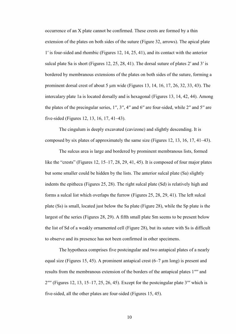
occurrence of an X plate cannot be confirmed. These crests are formed by a thin
extension of the plates on both sides of the suture (Figure 32, arrows). The apical plate
1′ is four-sided and rhombic (Figures 12, 14, 25, 41), and its contact with the anterior
sulcal plate Sa is short (Figures 12, 25, 28, 41). The dorsal suture of plates 2′ and 3′ is
bordered by membranous extensions of the plates on both sides of the suture, forming a
prominent dorsal crest of about 5 µm wide (Figures 13, 14, 16, 17, 26, 32, 33, 43). The
intercalary plate 1a is located dorsally and is hexagonal (Figures 13, 14, 42, 44). Among
the plates of the precingular series, 1′′, 3′′, 4′′ and 6′′ are four-sided, while 2′′ and 5′′ are
five-sided (Figures 12, 13, 16, 17, 41–43).
The cingulum is deeply excavated (cavizone) and slightly descending. It is
composed by six plates of approximately the same size (Figures 12, 13, 16, 17, 41–43).
The sulcus area is large and bordered by prominent membranous lists, formed
like the “crests” (Figures 12, 15–17, 28, 29, 41, 45). It is composed of four major plates
but some smaller could be hidden by the lists. The anterior sulcal plate (Sa) slightly
indents the epitheca (Figures 25, 28). The right sulcal plate (Sd) is relatively high and
forms a sulcal list which overlaps the furrow (Figures 25, 28, 29, 41). The left sulcal
plate (Ss) is small, located just below the Sa plate (Figure 28), while the Sp plate is the
largest of the series (Figures 28, 29). A fifth small plate Sm seems to be present below
the list of Sd of a weakly ornamented cell (Figure 28), but its suture with Ss is difficult
to observe and its presence has not been confirmed in other specimens.
The hypotheca comprises five postcingular and two antapical plates of a nearly
equal size (Figures 15, 45). A prominent antapical crest (6–7 µm long) is present and
results from the membranous extension of the borders of the antapical plates 1′′′′ and
2′′′′ (Figures 12, 13, 15–17, 25, 26, 45). Except for the postcingular plate 3′′′ which is
five-sided, all the other plates are four-sided (Figures 15, 45).

11
The thecal ornamentation is quite variable, probably in relation with the age of
the cells. On newly divided cells with a thin theca, the plates are verrucose with
numerous small warts lying between thecal pores (Figures 25, 28). On older cells, the
warts are thicker (Figure 26) and merge to form reticulations and areolae surrounding
the thecal pores (Figure 29). The crests are smooth or ornamented by small warts but
never have reticulations (Figures 16, 17, 26, 33, 43).
This species is photosynthetic and brownish in colour (Figure 3).
Peridiniopsis cristata var. tubulifera Couté, Perrette et Chomérat var. nov. (Figures
4, 18–24, 27, 30, 31, 46–51)
Diagnosis Organismus unicellularis, photosyntheticus, cum stigmate et theca.
Cellulae pyriformes leviter compressae dorso-ventraliter in laterale visu. Longitudo:
(27)30–40 µm; latitudo: 26–32 µm. Formula thecae laminarum: Po, X, 3′, 1a, 6′′, 6c, 5s,
5′′′, 2′′′′. Epitheca late conica, ad apicem colliformis. Lamina X minima et apicalis porus
cum parva propria tubuliformi apicali fauce sublatus. Hypotheca rotundata et brevior
quam epitheca. Cingulum paulo excavatum et leviter descendens. Sulcus brevus et ad
antapicem latior. Thecae laminae laeves et cum circularibus poris ornatae.
Description Unicellular photosynthetic organism with a theca and a stigma. Cells
pyriform, slightly compressed dorsiventrally in lateral view. Length: (27)30–40 µm;
width: 26–32 µm. Thecal plate formula: Po, X, 3′, 1a, 6′′, 6c, 5s, 5′′′, 2′′′′. Epitheca
widely conical, forming a neck at the apex. Plate X small and apical pore with a
prominent rim, forming a characteristic apical tubular structure. Hypotheca
hemispherical and shorter than epitheca. Cingulum moderately excavated and slightly

12
descending. Sulcus short widening at the antapex. Thecal plates smooth and with
circular pores dispersed.
Holotype SEM stub no. 124.2005.AC deposited at the RDDM Department
(Cryptogamy building) of the Muséum National d’Histoire Naturelle (MNHN) of Paris,
France. Holotype specimen illustrated in Fig. 18.
Type locality Brackish surface water (0.5 m), lagoon of Clipperton Island (10°18’N
109°13’W), Eastern Pacific Ocean. Collected with a plankton net by A. Couté in
February 2005.
Etymology The specific epithet comes from tubulus and adjectival suffix –fer,
meaning bearing a small tube.
Morphology Cells are roughly pyriform, (27)30–40 µm long, 26–32 µm wide and 22–
26 µm thick. The epitheca is broadly conical, forming a well defined neck at the apex
(apical horn) and the hypotheca is hemispherical. The outline of the cells reminds a fig-
fruit shape and is very similar to the shape of the nominate variety excluding the crests
(Figures 18, 19, 46, 47). Some more rounded cells are found (Figures 4, 24) and may
correspond to cyst-forming cells.
The thecal plate formula is Po, X, 3′, 1a, 6′′, 6c, 5s, 5′′′, 2′′′′ (Table 1). The
epitheca is nearly symmetrical in respect to the sagittal plane and is composed of 12
plates, including the pore plate Po, the canal plate X, three apical, one intercalary and
six precingular plates. The plates of the apical pore complex are located on the

13
prominent neck formed by the apical plates (Figure 30). The X plate is very small (0.8
µm long by 0.4 µm wide) and four-sided while Po is rounded (Figure 30). The tubular
rim bordering the apical pore is high and prominent (Figures 31, 50). The apical plate 1′
is four-sided and rhombic (Figures 18, 22, 24, 46), sometimes five-sided, depending on
the length of the suture with the anterior sulcal plate Sa. The intercalary plate 1a is
located dorsally and is six-sided (Figures 19, 22, 47, 48). Among the plates of the
precingular series, 1′′, 3′′ and 4′′ are quadrangular while 2′′, 5′′ and 6′′ are pentagonal
(Figures 22, 48).
The cingulum is slightly excavated and descending about half its width (Figures
18, 24, 46). It is composed by six plates nearly equal in size (Figures 18–21, 46, 47).
The sutures of cingular plates are not facing those of the pre- and postcingular plates
and are shifted (Figures 19–21).
The sulcus is short and broad towards the antapex. It is composed of five plates.
The anterior sulcal plate (Sa) slightly indents the epitheca (Figures 18, 24, 27, 46, 51).
At its posterior end, Sa forms a small incurved list (Figures 24, 27, 46). The right sulcal
plate (Sd) is relatively high and forms a developed sulcal list which borders the furrow
(Figures 18, 27, 46, 51). The left sulcal plate (Ss) is small, contacts shortly the first
cingular plate C1 and is located just above the small plate Sm (Figures 27, 51). The Sm
plate borders the flagellar pore that is partially obscured from the view by the right
sulcal list. Both of these plates are deeply excavated in the sulcus. The posterior sulcal
plate (Sp) is the largest of the sulcal plates (Figures 18, 23, 24, 27, 46, 49, 51). It is
broad and nearly reaches the antapex of the cell (Figure 24, 46).
The hypotheca comprises five postcingular and two antapical plates (Figures 23,
49). Except for the plate 3′′′ which is five-sided, all other postcingular plates are four-
sided. The two antapical plates have almost the same size (Figures 23, 49)

14
The plates are smooth with circular thecal pores, including those of the sulcal
series. A row of pores borders the cingulum on the epitheca and hypotheca and on each
side of the cingulum (Figures 19, 21).
This species is photosynthetic and brownish in colour (Fig. 4).
It is illustrated and referred as to Dinophyceae Peridiniales sp. “Dino 2” in Couté
et al. (2009) and Charpy et al. (2010).
Discussion
Phytoplankton studies of the Clipperton lagoon are very scarce (Charpy et al. 2010).
The first mention of microalgae in the lagoon was made by Taylor (1939) who reported
only 13 taxa of microalgae belonging to Cyanophyceae and Chlorophyceae. Later,
Sachet (1962b) reported a total of 24 species of Cyanophyceae, 10 species of
Chlorophyceae and one unidentified species of Dinophyceae (Glenodinium sp.), which
was later described by Balech (1961) as Glenodinium cristatum. Since the
inconsistencies regarding the accurate circumscription of the genus Glenodinium
Ehrenberg, Bourrelly (1968) transferred G. cristatum to the genus Peridiniopsis mainly
based on its thecal characteristics. Peridiniopsis cristata has been again recorded in
March 1980 and studied by Ricard and Bourrelly (1982) who first used SEM to further
investigate the morphology of this dinoflagellate. It is remarkable that this species, still
present in recent samples, has never been reported elsewhere in any other tropical
regions, and appears as endemic to Clipperton lagoon (Ricard and Bourrelly 1982),
which is very unusual for a planktonic dinoflagellate. Surprisingly, Popovský and
Pfiester (1990) mentioned its occurrence in saline ponds of France but this statement
without any reference is inconsistent with the mention by Ricard and Bourrelly (1982),
and actually very doubtful. Interestingly, Iltis and Couté (1984) found a resembling

15
taxon in the Lake Poopó at an altitude of 3686 m in Bolivia, but with some
morphological differences and they described it as P. cristata var. boliviensis
(incorrectly named P. cristatum var. boliviense). Cells of this variety are more rounded
and the apical crest (i.e. the list between plates 2′ and 3′) is absent or hardly developed
which contrasts with the type of the nominate variety from Clipperton lagoon. In the
description of P. cristata var. boliviensis, the photomicrograph (Iltis and Couté 1984, pl.
3, fig. 8) probably does not correspond to the same taxon since the biconical shape and
the acuminate antapex do not remind the outline observed in SEM (Iltis and Couté
1984, pl. 3, figs 1–7). The thecal arrangement and ornamentation in the nominate
variety and P. cristata var. boliviensis are rather similar.
From the original description of Peridiniopsis cristata var. cristata (Balech
1961) and its reinvestigation by Ricard and Bourrelly (1982), this study revealed that
the Clipperton lagoon specimens are unambiguously the same taxon. In contrast with
Balech (1961) who identified four sulcal plates, Bourrelly (1985) found a fifth small
sulcal plate Sm located below Ss. In our study, Sm has been putatively identified on a
young cell and was probably hidden by the sulcal list in most well-developed
specimens. In addition, Ricard and Bourrelly (1982) reported a pore on the upper right
corner of the C2 plate of the nominate variety which has not been previously mentioned
by Balech (1961) in his detailed description. During our investigations of P. cristata
var. cristata from the type locality, no specimen was found to possess this peculiar
feature. For this reason, and since no other dinoflagellate is known to possess a pore on
the C2 plate, we consider that this pore or hole may result from an artefact or the activity
of a parasitic organism. Thus, in our opinion, this criterion is probably of no
taxonomical value and should not be further considered.

16
In spite of a very different ornamentation, it is striking that the thecal
arrangement of the new variety Peridiniopsis cristata var. tubulifera is very similar to
that of the nominate variety and they have the same plate formula. The cells of the new
variety tubulifera are smaller than the ones of the nominate variety but their size ranges
overlap when the length of the latter is considered without the lists. The major
difference between these two taxa is the total absence of ornamentation and lists on the
theca of P. cristata var. tubulifera while the nominate variety is usually ornamented. In
addition, the apex of this new variety is peculiar, because of the presence of the tubular
rim structure, not seen in P. cristata var. cristata. This structure may be present in the
latter but hidden by the prominent apical crest surrounding the apical pore. This tubular
rim is typical of the Peridiniaceae (Toriumi and Dodge 1993) but it is generally less
developed and not as high as in P. cristata var. tubulifera where it is particularly
prominent. In some other genera, the plates of the apical series can form a rather similar
tubular neck or horn, as in Protoperidinium tuba (Schiller) Balech (Schiller 1931–1937)
or in the fossil dinoflagellates Gonyaulax jurassica Deflandre and Rhynchodiniopsis
aptiana Deflandre (Grassé 1952).
Since no other major morphological difference than the ornamentation was
found to distinguish P. cristata var. tubulifera from the nominate variety, we consider
that they very likely belong to the same species but correspond to different varieties
with an extreme variation in the ornamentation. For several species in cultures, it has
been demonstrated that ornamentation is highly variable and cannot be a taxonomic
criterion for identification (e.g. Craveiro et al. 2009). A variable ornamentation has been
reported in cells of P. cristata var. cristata, ranging from verrucose to strongly
reticulate, but all cells possessed apical and antapical lists (= “crests”), whereas these
lists were always absent in P. cristata var. tubulifera. No specimen with a transitional

17
ornamentation and rudimentary lists has been found in the samples. For P. cristata var.
boliviensis, Iltis and Couté (1984) observed only specimens with developed crests and
there is no mention of cells with a different ornamentation.
Bourrelly (1968, 1985) and Popovský and Pfiester (1990) placed P. cristata var.
cristata in the group borgei of Peridiniopsis, which is defined by an epithecal plate
pattern of 3′, 1a and 6′′. This definition encompasses widely different and unrelated
species, some being now transferred to another genus (Boltovskoy 1999), but the
resemblance of P. cristata var. cristata and P. cristata var. tubulifera with P. borgei
Lemmermann is conspicuous. They are in the same size range and are similarly shaped
but P. borgei possesses a large central vacuole and a dorsal starch-envelopped pyrenoid
(Calado and Moestrup 2002), which have not been observed in living specimens of the
taxa from Clipperton lagoon.
From an ecological point of view, species in the genus Peridiniopsis are
generally found in freshwater environments and it is a common genus in freshwater
plankton (Bourrelly 1985, Popovský and Pfiester 1990, Lewis and Dodge 2002), but
some taxa are described from brackish waters such as P. salina Trigueros (Trigueros
2000) and P. cristata var. cristata (Balech 1961). The new variety P. cristata var.
tubulifera can also be placed in the group of brackish species as it is co-occurring with
P. cristata var. cristata in Clipperton lagoon. In addition, although the bolivian variety
P. cristata var. boliviensis was described from an inland mountain lake that does not
receive seawater, it can be considered as a brackish form since the salinity in Lake
Poopó was 7.5 psu (Iltis and Couté 1984) and thus slightly higher than the salinity
observed in the upper water layer of Clipperton lagoon.
Durinskia baltica has not been previously recorded from Clipperton lagoon and
this is the first report of this species to date. This taxon has had a long and complex

18
taxonomical history before Carty and Cox (1986) erected the genus Durinskia for
dinoflagellates with the plate formula Po, X, 4′, 2a, 6′′, 5c, 4s, 5′′′, 2′′′′ to accommodate
Peridinium balticum (Levander) Lemmermann. Although it has been described with
thecal plates with numerous transversally oriented pores, P. dybowskii Woloszyńska has
been considered to be conspecific with D. baltica which does not have this feature
(Schiller 1931–1937, Popovský and Pfiester 1990). However, this is questionable since
these species seem to have very different habitats, P. dybowskii being reported from
humus rich habitats with low pH (Hansen and Flaim 2007) while D. baltica occurs
generally in brackish waters. The recent combination, Durinskia occulata (Stein)
Hansen et Flaim, proposed by Hansen and Flaim (2007) for a species described from the
Moldau River by Stein (1883) and found again in an Italian lake, corresponds to a
morphologically close taxon with the same thecal plate arrangement than D. baltica.
Durinskia occulata has a more globular shape compared to the more pronounced dorso-
ventral flattening of both P. dybowskii and D. baltica (Hansen and Flaim 2007). The
present material is somewhat smaller than the dimensions given for D. occulata from
Ampola Lake, i.e. 28–34 µm in length and width (Hansen and Flaim 2007) but it
presents similarities in the ornamentation and thecal plate arrangement. Since D.
occulata has only been recorded in freshwater habitats and its tolerance to brackish
waters has not yet been demonstrated, we consider it as a freshwater species separate
from D. baltica which is commonly found in brackish waters, and even in some marine
environments (Carty and Cox 1986). Moreover, the present material was in the same
size range than given by Levander (1894) for Glenodinium balticum, i.e. 22–30 µm.
Consequently, we consider that this material fits morphologically and ecologically more
with D. baltica than with D. occulata. Nevertheless, further investigations using

19
molecular data are necessary to conclude about these taxa with the same thecal plate
pattern which may be synonyms.
In a clonal culture of this species, Chesnick and Cox (1985) observed some
variations in the plate pattern and the number of plates in some thecal series can differ
from the typical organization. However, the thecal variation of the 3′ and 2a plates that
we observed, and the resulting disconnection of plates 1a and 2a was not reported by
these authors and appears as a new type of variation for this species. Interestingly,
variations of the thecal plate pattern are not rare in dinoflagellates from brackish waters,
as reported also for Protoperidinium bolmonense Chomérat et Couté (Chomérat and
Couté 2008).
In conclusion, this study brings new data on the diversity of planktonic
dinoflagellates in Clipperton lagoon. The presence of Peridiniopsis cristata var. cristata
described in 1961 from this lagoon and endemic of this area is remarkable. The
knowledge of this ecosystem is still to be improved and, as sampling is only made from
time to time during expeditions, the plankton dynamics is not understood yet. For
several years, P. cristata var. cristata was considered as the unique dinoflagellate living
in this lagoon but this study revealed that other taxa are present. Whether P. cristata
var. tubulifera constitutes a particular morph in the life cycle of P. cristata var. cristata
or belongs to a different species has not been demonstrated and remains to be verified.
In addition, further studies should manage to better understand their population
dynamics and their seasonality. In addition, in order to better understand their evolution
in this isolated ecosystem, their molecular phylogenetic relationships with other
dinoflagellates from distant locations should be investigated. In particular, it is
necessary to clarify the relationships between P. cristata var. cristata and its two
varieties boliviensis and tubulifera.

20
Acknowledgements
The authors are very grateful to Dr. Jean-Louis Étienne for the invitation of A. Couté to
participate to the Clipperton expedition in 2005 and for the organization and facilities
on the atoll. Total Foundation is acknowledged for its support and funding of the
transportation to Clipperton Island and Carl Zeiss (Oberkochen, Germany) for lending
the Axioscop 2+ differential interference contrast microscope during the expedition.

21
References
Balech, E. 1961. Glenodinium cristatum, sp. nov. (Dinoflagellata). Neotropica 7: 47–
51.
Boltovskoy, A. 1999. The genus Glochidinium gen. nov., with two species: G.
penardiforme comb. nov. and G. platygaster sp. nov. (Peridiniaceae). Grana 38: 98–
107.
Bourrelly, P. 1968. Notes sur les Péridiniens d'eau douce. Protistologica 4: 5–14.
Bourrelly, P. 1985. Les algues d'eau douce. Initiation à la systématique. Tome 3. Les
algues bleues et rouges. Les Eugléniens, Péridiniens et Cryptomonadines. N. Boubée,
Paris. pp. 606.
Calado, A.J. and Ø. Moestrup. 2002. Ultrastructural study of the type species of
Peridiniopsis, Peridiniopsis borgei (Dinophyceae), with special reference to the
peduncle and flagellar apparatus. Phycologia 41: 567–584.
Carricart Ganivet, J.P. and H. Reyes Bonilla. 1999. New and previous records of
scleractinian corals from Clipperton Atoll, Eastern Pacific. Pac. sci. 53: 370–375.
Carsin, J.-L., F. Bourrouilh-Le Jan, R.C. Murphy, R. Taxit and P.-M. Niaussat. 1985.
The natural eutrophication of the waters of the Clipperton lagoon: equipments, methods,
results, discussions. In: (C. Gabrie, J.L. Toffart and B. Salvat eds.) Proceedings of the

22
fifth international coral reef congress. Antenne Muséum–École Pratique des Hautes
Études, Moorea. pp. 359–364.
Carty, S. and E.R. Cox. 1986. Kansodinium gen. nov. and Durinskia gen. nov.: two
genera of freshwater dinoflagellates (Pyrrhophyta). Phycologia 25: 197–204.
Charpy, L., M. Rodier and G. Sarazin. 2009. Biogéochimie du lagon. In: (L. Charpy,
ed.) Clipperton, environnement et biodiversité d'un microcosme océanique (Patrimoines
naturels 68). Muséum national d'histoire naturelle – Institut de recherche pour le
développement, Paris, Marseille. pp. 67–79.
Charpy, L., M. Rodier, A. Couté, C. Perrette-Gallet and C. Bley-Loëz. 2010.
Clipperton, a possible future for atoll lagoons. Coral Reefs 29: 771–783.
Chesnick, J.M. and E.R. Cox. 1985. Thecal plate tabulation and variation in Peridinium
balticum (Pyrrhophyta: Peridiniales). Trans. Am. Microsc. Soc. 104: 387–394.
Chomérat, N. and A. Couté. 2008. Protoperidinium bolmonense sp. nov. (Peridiniales,
Dinophyceae), a small dinoflagellate from a brackish hypereutrophic lagoon (South of
France). Phycologia 47: 392–403.
Couté, A. 2002. Biologie et microscopie électronique à balayage. Mém. SEF 6: 31–44.
Couté, A. and R. Garrouste. 2009. Un état des lieux de la flore et de la végétation
terrestres et dulçaquicoles. In: (L. Charpy, ed.) Clipperton, environnement et

23
biodiversité d'un microcosme océanique (Patrimoines naturels 68). Muséum national
d'histoire naturelle – Institut de recherche pour le développement, Paris, Marseille. pp.
279–296.
Couté, A., C. Loez-Bley and C. Perrette-Gallet. 2009. Les micro-algues. In: (L. Charpy,
ed.) Clipperton, environnement et biodiversité d'un microcosme océanique (Patrimoines
Naturels 68). Muséum national d'histoire naturelle – Institut de recherche pour le
développement, Paris, Marseille. pp. 93–110.
Craveiro, S.C., A.J. Calado, N. Daugbjerg and Ø. Moestrup. 2009. Ultrastructure and
LSU rDNA–based revision of Peridinium group palatinum (Dinophyceae) with the
description of Palatinus gen. nov. J. Phycol. 45: 1175–1194.
Étienne, J.-L. 2005. The Clipperton expedition.
http://www.jeanlouisetienne.com/clipperton. Last accessed 10 February 2011.
Grassé, P.-P. 1952. Traité de zoologie. Anatomie, systématique, biologie. Tome I.
Phylogénie. Protozoaires : généralités, flagellés. Masson et Cie, Paris. pp. 1071.
Hansen, G. and G. Flaim. 2007. Dinoflagellates of the Trentino Province, Italy. J.
Limnol. 66: 107–141.
Iltis, A. and A. Couté. 1984. Péridiniales (Algae, Pyrrhophyta) de Bolivie. Rev.
hydrobiol. trop. 17: 279–286.

24
Jean-Baptiste, P., É. Fourré, J.-L. Charlou, J.-P. Donval and T. Corrège. 2009. Gaining
insight into Clipperton's lagoon hydrology using tritium. Estuar. coast. shelf sci. 83: 39–
46.
Jost, C.-H. 2003. Clipperton - Île de La Passion : une aire française du Pacifique à
protéger ! In: (J.M. Lebigre and P.M. Decoudras eds.) Les aires protégées insulaires et
littorales tropicales, Actes du Colloque dymset, transcultures, sepanrit, Nouméa, 30-31
octobre 2001. Collection "Îles et Archipels" 32. CRET, Presses universitaires,
Bordeaux, pp. 223–243.
Jost, C.-H. 2005. Risques environnementaux et enjeux à Clipperton (Pacifique français).
Cybergeo 314: 1–15.
Lefèvre, M. 1932. Monographie des espèces d'eau douce du genre Peridinium. Arch.
bot. Mém. Caen 2: 1–208.
Lemmermann, E. 1910. Kryptogamenflora der Mark Brandenburg. Algen I
(Schizophyceen, Flagellaten, Peridineen). Verlag von Gebrüder Borntraeger, Leipzig.
pp. 712.
Levander, K.M. 1892. Notiz über die Täfelung der Schalenmembran des Glenodinium
cinctum Ehrbg. Zool. Anz. 15: 405–408.

25
Levander, K.M. 1894. Materialen zur Kenntniss der Wasserfauna in der Umgebung von
Helsingfors, mit besonderer Berücksichtigung der Meeresfauna I. Protozoa. Acta Soc.
fauna flora Fenn. 13: 1–115.
Lewis, J. and J.D. Dodge. 2002. Phylum Pyrrhophyta (Dinoflagellates). In: (D.M. John,
B.A. Whitton and A.J. Brook, eds.) The freshwater algal flora of the British Isles.
Cambridge University Press, Cambridge. pp. 186–207.
McNeill, J., F.R. Barrie, H.M. Burdet, V. Demoulin, D.L. Hawksworth, K. Marhold,
D.H. Nicolson, J. Prado, P.C. Silva, J.E. Skog, J.H. Wiersema and N.J. Turland. 2006.
International code of botanical nomenclature (Vienna code). Regnum Vegetabile 146.
A.R.G. Gantner Verlag KG, Ruggell. pp. 568.
Monniot, F. 2007. Some ascidians (Tunicata) from the Clipperton Island. Cah. biol.
mar. 48: 303–310.
Niaussat, P.M. 1986. Le lagon et l'atoll de Clipperton. Collection Travaux et mémoires
de l'académie des sciences d'Outre-mer, 2e édition. Académie des sciences d'Outre-mer
/ Institut du Pacifique, Paris. pp. 189.
Popovský, J. and L.A. Pfiester. 1990. Dinophyceae (Dinoflagellida). In: (H. Ettl, J.
Gerloff, H. Heynig and D. Mollenhauer, eds.) Süßwasserflora von Mitteleuropa Volume
6. Gustav Fischer Verlag, Jena, Stuttgart. pp. 272.

26
Reyes Bonilla, H., J.P. Carricart Ganivet, V. Solis Weiss and A. Granados Barba. 1999.
El atolón de Clipperton. Aspectos históricos y ecológicos. Cienc. desarro. 149: 16–23.
Ricard, M. and P. Bourrelly. 1982. Quelques algues microscopiques du lagon de l'atoll
de Clipperton (Pacifique Tropical Nord). Cryptog. Algol. 3: 25–31.
Sachet, M.-H. 1960. Histoire de l'île de Clipperton. Cah. Pac. 2: 3–32.
Sachet, M.-H. 1962a. Geography and land ecology of Clipperton Island. Atoll Res. Bull.
86: 1–115.
Sachet, M.-H. 1962b. Flora and vegetation of the Clipperton Island. Proc. Calif. acad.
sci. 31: 249–307.
Salvat, B., M. Adjeroud and L. Charpy. 2008. Les récifs coralliens de Clipperton. Terre
vie 63: 179–187.
Sanvicente-Añorve, L., M. Hermoso-Salazar, V. Solis Weiss and I.H. Salgado Ugarte.
2010. Carapace relative growth of Trapezia Latreille ,1828 (Decapoda, Brachyura),
crabs that are symbionts of hard corals, from Clipperton atoll and the Revillagigedo
Islands: ecological and zoogeographical implications. Crustaceana 83: 1371–1383.
Schiller, J. 1931–1937. Dinoflagellatae (Peridinidae) in monographischer Behandlung.
In: (L. Rabenhorst, ed.) Kryptogamenflora von Deutschland, Österreich und der

27
Schweiz. Akademische Verlagsgesellschaft, Leipzig. 10(3): Teil 1 (1-3) (1931-1933): 1–
617. Teil 2 (1-4) (1935-1937): 1–590.
Sournia, A. 1986. Atlas du phytoplancton marin. 1 : Introduction, cyanophycées,
dictyochophycées, dinophycées et raphidophycées. Éditions du Centre National de la
Recherche Scientifique, Paris. pp. 219.
Stein, F.R. 1883. Der Organismus der Arthrodelen Flagellaten. II Hälfte. Wilhelm
Engelmann Verlag, Leipzig. pp. 30.
Taylor, W.M.R. 1939. Algae collected on the Presidential Cruise of 1938. Smithson.
misc. collect. 98: 1–18.
Toriumi, S. and J.D. Dodge. 1993. Thecal apex structure in the Peridiniaceae
(Dinophyceae). Eur. J. Phycol. 28: 39–45.
Trigueros, J.M. 2000. Peridiniopsis salina (Peridiniales, Dinophyceae), a new species
of brackish dinoflagellate from Urdaibai estuary, North Spain. Phycologia 39: 126–133.

28
Figure legends:
Figure 1 Location of Clipperton Island (A) off Central America, and (B) more
precisely in the eastern equatorial part of Pacific Ocean, (C) Clipperton Island and
lagoon. Small arrows indicate net sampling collection.

29
Figures 2–4 Thecate dinoflagellates from Clipperton Island lagoon fixed in
formaldehyde, LM.
(2) Durinskia baltica. (3) Peridiniopsis cristata var. cristata. (4) Peridiniopsis cristata
var. tubulifera var. nov.
Scale bars=5 µm (Figures 2, 4); 10 µm (Figure 3).

30
Figures 5–11 Durinskia baltica, SEM.
(5) Ventral view. (6) Dorsal view. (7) Apical view showing the arrangement of thecal
plates. Note that the two anterior intercalary plates 1a and 2a are contiguous (arrow). (8)
Antapical view. (9) Detail of the apical pore complex (APC). (10) Detail of the sulcal
area. (11) Variation of the epithecal plate pattern on a specimen with the two anterior
intercalary plates 1a and 2a disconnected (arrow).
Scale bars=10 µm (Figures 5–8); 5 µm (Figure 11); 2 µm (Figures 9–10).

31
Figures 12–17 Peridiniopsis cristata var. cristata, SEM.
(12) Ventral view. (13) Dorsal view. (14) Apical view. (15) Antapical view. (16) Left
lateral view. (17) Right lateral view.
Scale bars=10 µm.

32
Figures 18–23 Peridiniopsis cristata var. tubulifera, SEM.
(18) Ventral view. (19) Dorsal view. (20) Right lateral view. (21) Left lateral view. (22)
Apical view. (23) Antapical view.
Scale bars=10 µm.

33
Figures 24–33 Comparison of Peridiniopsis cristata var. tubulifera with the
nominate variety, SEM.
(24) Ventral view of a rounded cell of var. tubulifera, note the absence of
ornamentation. (25) Ventral view of a cell of var. cristata with moderate verrucose
ornamentation. (26) Cell of var. cristata with strong ornamentation. Note that the C2
plate is without the pore reported by Ricard and Bourrelly (1982) (arrowhead). (27)
Detail of the sulcal area of var. tubulifera. (28) Detail of the sulcal area of a moderately

34
ornamented cell of var. cristata with smooth sulcal plates. (29) Detail of the sulcal area
of a strongly ornamented cell of var. cristata with sulcal plates reticulate, some platelets
are hidden by the list of Sd. (30) Apical pore complex of var. tubulifera. (31) Lateral
view of the prominent tubular rim (arrow) surrounding the apical pore in var. tubulifera.
(32) Apical pore complex of var. cristata, with membranous extensions (crests)
surrounding the pore (arrows). (33) Lateral view of the dorsal crest of var. cristata.
Scale bars=10 µm (Figures 24–26); 5 µm (Figures 27–29); 2 µm (Figures 32–33); 1 µm
(Figures 30–31).

35
Figures 34–51 Drawings of the three thecate dinoflagellates from Clipperton
Island lagoon.
(34–40) Durinskia baltica. (34) Ventral view. (35) Dorsal view. (36) Apical view. (37)
Antapical view. (38) Detail of the sulcal area. (39) Detailed part of the epitheca and
apical pore complex. (40) Variation of the epithecal plate pattern, with an extra edge of
the apical 3′ plate (arrowhead) and resulting in four-sided intercalary 1a plate and five-
sided 3′ and 2a plates.

36
(41–45) Peridiniopsis cristata var. cristata.
(41) Ventral view. (42) Dorsal view. (43) Right lateral view. (44) Apical view. (45)
Antapical view.
(46–51) Peridiniopsis cristata var. tubulifera. (46) Ventral view. (47) Dorsal view. (48)
Apical view. (49) Antapical view. (50) Detail of the apex and tubular rim in lateral
view. (51) Detail of the sulcal area.
Scale bars=A/10 µm (Figures 34–37, 41–49); B/5 µm (Figures 38–40); C/1 µm (Figure
50); D/2 µm (Figure 51).

37