boronic acid paper mbs paper rev clean 5-18-16
TRANSCRIPT
UCLAUCLA Previously Published Works
TitleProtein Complexation and pH Dependent Release Using Boronic Acid Containing PEG-Polypeptide Copolymers.
Permalinkhttps://escholarship.org/uc/item/2cz577cw
JournalMacromolecular bioscience, 17(1)
ISSN1616-5187
AuthorsNegri, Graciela EDeming, Timothy J
Publication Date2017
DOI10.1002/mabi.201600136 Peer reviewed
eScholarship.org Powered by the California Digital LibraryUniversity of California

- 1 -
Article Type: Communication Protein complexation and pH dependent release using boronic acid containing PEG-polypeptide copolymersa Graciela E. Negri,1 Timothy J. Deming1,2,* ––––––––– G. E. Negri, Prof. T. J. Deming 1 Department of Chemistry and Biochemistry, University of California, Los Angeles, 607 Charles E Young Dr. E, Los Angeles, CA 90095-1600, USA 2 Department of Bioengineering, University of California, Los Angeles, 5121 Engineering 5, Los Angeles, CA 90095-1600, USA Fax: (+1) 310-794-5956; E-mail: [email protected] –––––––––
Abstract
New poly(L-lysine)-b-poly(ethylene glycol) copolypeptides have been prepared, where the
side-chain amine groups of lysine residues are modified to contain ortho-amine substituted
phenylboronic acid, i.e. Wulff-type phenylboronic acid (WBA), groups to improve their pH
responsive, carbohydrate binding properties. These block copolymers form nanoscale
complexes with glycosylated proteins that are stable at physiological pH, yet dissociate and
release the glycoproteins under acidic conditions, similar to those found in endosomal and
lysosomal compartments within cells. These results suggest that WBA modified polypeptide
copolymers are promising for further development as degradable carriers for intracellular
protein delivery.
a Supporting Information is available online from the Wiley Online Library or from the author.

- 2 -
FIGURE FOR ToC_ABSTRACT
Block copolymers containing Wulff-type phenylboronic acid groups form nanoscale complexes with glycosylated proteins that are stable at physiological pH, yet dissociate and release the glycoproteins under acidic conditions, similar to those found in endosomal and lysosomal compartments within cells. These results suggest that these polypeptide copolymers are promising for further development as degradable carriers for intracellular protein delivery.
pH Responsive Protein Complexation
- 3 -
1. Introduction
Many biological therapeutics, such as for enzyme-based therapies, require a carrier to reduce
their clearance by the immune system as well as for delivery into cells or cellular
compartments where they are needed [1]. Stimulus responsive carriers, such as those that
respond to pH or redox, are especially desirable as they can be a means to effect release of
therapeutic cargos upon cellular uptake [2]. For these reasons, the reversible and pH
responsive diol-binding characteristics of phenylboronic acid (PBA) groups have been used in
many carriers proposed for delivery of glycosylated therapeutics [3]. Recent efforts in this
area have focused on PBA groups that have been modified with tethered OH and NR2 groups
[4], which adjust the properties of diol-PBA complexes so that they have increased stability at
neutral pH, and can yet release their cargos at more acidic pH as found in endosomal and/or
lysosomal compartments.
Although some PBA-containing polypeptides have been reported for development as
therapeutic carriers, these have primarily utilized simple PBA groups, which form only weak
complexes with carbohydrate diol groups at physiological pH [5]. Here, we have prepared
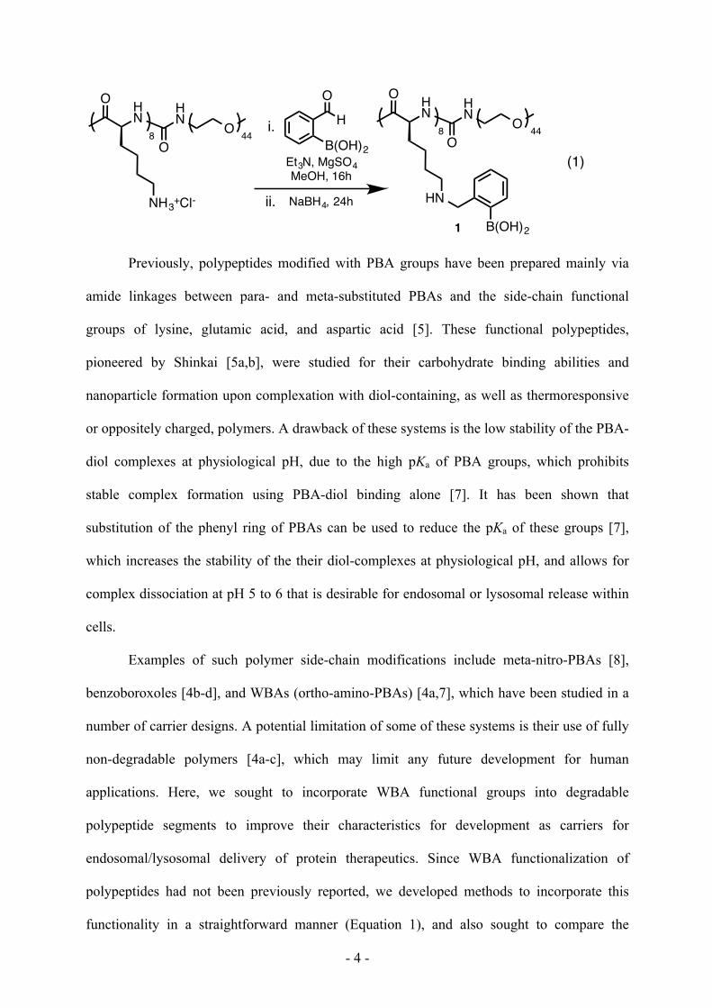
new poly(L-lysine)-b-poly(ethylene glycol), K8PEG44, copolypeptides, where the side-chain
amine groups of lysine residues have been modified to contain ortho-substituted amine PBA,
i.e. Wulff-type phenylboronic acid (WBA) [6], groups (KWBA8PEG44, 1, Equation 1) to
improve their pH responsive diol binding properties [4a,7]. These block copolymers were
found to form nanoscale complexes with glycosylated proteins that were stable at
physiological pH, yet were found to dissociate and release the glycoproteins at acidic
conditions (pH 5.0), similar to those found in endosomal and lysosomal compartments.
- 4 -
Previously, polypeptides modified with PBA groups have been prepared mainly via
amide linkages between para- and meta-substituted PBAs and the side-chain functional
groups of lysine, glutamic acid, and aspartic acid [5]. These functional polypeptides,
pioneered by Shinkai [5a,b], were studied for their carbohydrate binding abilities and
nanoparticle formation upon complexation with diol-containing, as well as thermoresponsive
or oppositely charged, polymers. A drawback of these systems is the low stability of the PBA-
diol complexes at physiological pH, due to the high pKa of PBA groups, which prohibits
stable complex formation using PBA-diol binding alone [7]. It has been shown that
substitution of the phenyl ring of PBAs can be used to reduce the pKa of these groups [7],
which increases the stability of the their diol-complexes at physiological pH, and allows for
complex dissociation at pH 5 to 6 that is desirable for endosomal or lysosomal release within
cells.
Examples of such polymer side-chain modifications include meta-nitro-PBAs [8],
benzoboroxoles [4b-d], and WBAs (ortho-amino-PBAs) [4a,7], which have been studied in a
number of carrier designs. A potential limitation of some of these systems is their use of fully
non-degradable polymers [4a-c], which may limit any future development for human
applications. Here, we sought to incorporate WBA functional groups into degradable
polypeptide segments to improve their characteristics for development as carriers for
endosomal/lysosomal delivery of protein therapeutics. Since WBA functionalization of
polypeptides had not been previously reported, we developed methods to incorporate this
functionality in a straightforward manner (Equation 1), and also sought to compare the
HN
O
NH3+Cl-
O
HN O8 44
HN
O
HN
O
HN O8 44
B(OH)2
H
O
B(OH)2Et3N, MgSO4MeOH, 16h
i.
ii. NaBH4, 24h
1
(1)

- 5 -
properties of the resulting polymers to the more widely studied PBA analog. To encourage
robust protein complexation, high densities of WBA or PBA groups on the polypeptide
segments were desired.
2. Experimental Section
2.1. Materials and Methods.
Unless otherwise stated, copolymer functionalization reactions were performed in glass vials,
under ambient pressure. Room temperature reactions were performed at ca. 22 °C. Hexanes
and THF were deoxygenated by sparging with N2, then dried by passing through alumina
columns. Commercial anhydrous DMF was used as received. All other solvents were used as
received from Fisher Scientific. Alizarin Red S, horseradish peroxidase, bovine serum
albumin, and ABTS were purchased from Sigma Aldrich. Alpha-L-iduronidase was provided
by the laboratory of Dr. Patricia Dickson (LA Biomed). 2-formylphenylboronic acid and 4-
carboxyphenylboronic acid were purchased from Combi-Blocks Inc. (San Diego, CA). N-
Carboxybenzyloxy-L-lysine-N-carboxyanhydride (Cbz-Lys NCA) was prepared by a
previously reported procedure [9]. Isocyanate-terminated, 2000 kDa poly(ethylene glycol)
(PEG44-NCO) was prepared by a previously reported procedure [10]. Dialysis was performed
using deionized water (18.2 MΩ-cm) prepared by passing in-house deionized water through a
Millipore Milli-Q Biocel A10 unit. Regenerated cellulose dialysis tubing was from Spectrum
Labs (Rancho Dominguez, CA). In all other experiments, in-house deionized water was used.
NMR spectra were recorded on a Bruker AV400 instrument with chemical shifts reported
relative to the solvent signal. Dynamic light scattering was conducted with a Malvern
Zetasizer Nano ZS model Zen 3600 (Malvern Instruments Inc., Westborough, MA).
Fluorimetry was conducted on a QuantaMaster 40 Spectrofluorometer (Photon Technology
International Inc., Birmingham, NJ). UV/Vis spectroscopy was conducted using an
Agilent/HP diode-array 8453 UV-Visible spectrophotometer. Tandem gel permeation
chromatography/light scattering (GPC/LS) was performed on a SSI Accuflow Series III liquid
- 6 -
chromatograph pump equipped with a Wyatt DAWN EOS light scattering (LS) and Optilab
rEX refractive index (RI) detectors. Separations were achieved using xStream H2O 500 Å
Jordi Labs 5 μm mixed bed column with 0.5 wt% potassium trifluoroacetate in HFiP as the
eluent at 60 °C. All GPC/LS samples were prepared at concentrations of 10 mg/mL.
2.2. Experimental Procedures
Poly(L-lysine hydrochloride)8-b-poly(ethylene glycol)44, K8PEG44. This copolymer was
prepared by a procedure similar to methods previously reported [9]. In a N2 filled glove box,
Cbz-Lys NCA (570 mg, 1.9 mmol) was dissolved in anhydrous DMF (50 mg/mL). To the
stirred solution was added (PMe3)4Co (67 µL of a 50 mg/mL solution in 1:1 DMF/THF) via
syringe. After 2 h an aliquot (20 µL) was removed for analysis by FTIR, which confirmed that
all of the NCA had been consumed. To the reaction mixture was then added PEG44-NCO (480
mg, 2.6 eq. per Co). The mixture was stirred for an additional 16 h and then removed from the
glove box. The majority of the solvent was removed under reduced pressure, and the polymer
was then precipitated by addition of water (15 mL) to the concentrated solution (ca. 4 mL)
followed by additional washing with water (3 x 15 mL), then was lyophilized to dryness. The
crude polymer product (ca. 1.9 mmol lysine residues) was dissolved in TFA (25 mL), and the
mixture cooled in an ice bath. To this solution was added 33% HBr/AcOH (1.7 mL, 9.2
mmol) and the reaction was stirred for 5 h, followed by solvent removal using rotary
evaporation. The product was transferred into 2000 Da MWCO dialysis tubing and dialyzed
against 3 mM HCl(aq) for 24 h, followed by Milli-Q water for 48 h with changes of dialysate
(4 times daily). The product was isolated by lyophilization to yield a white powder (380 mg,
85% yield). Poly(L-lysine) segment length was determined by comparing 1H NMR
integrations of the 3.03 ppm resonance from lysine residues (-CH2NH2) to the 3.73-3.80 ppm
resonances from PEG repeats (-CH2CH2O-). 1H NMR (400 MHz, D2O, 25 °C): δ 4.34 (t, 1H),
3.73-3.80 (m, 176H), 3.40 (s, 3H), 3.03 (t, 2H), 1.40-1.81 (m, 6H). GPC/LS analysis of
K8PEG44 gave Mw/Mn = 1.13 (see SI, Figure S1).

- 7 -
Poly(Nε-(2-boronobenzyl)-L-lysine)8-b-poly(ethylene glycol)44, KWBA8PEG44 (1). K8PEG44
(15 mg, 0.030 mmol lysine residues) and 2-formylphenylboronic acid (6.0 mg, 0.036 mmol)
were mixed in a round bottom flask and dissolved in MeOH (0.5 mL). To this solution was
added triethylamine (42 µL, 0.30 mmol) and MgSO4 (ca. 3-5 mg). The reaction was stirred
for 16 h at room temperature under N2, sodium borohydride (2 mg, 0.061 mmol) was then
added, and the reaction was stirred for another 24 h. The solution was transferred into 2000
Da MWCO dialysis tubing and dialyzed against 3 mM HCl(aq) for 24 h, followed by Milli-Q
water for 48 h with changes of dialysate (4 times daily). The product was isolated by
lyophilization to yield a white powder (19 mg, 92% yield). 1H NMR (400 MHz, CDCl3,
25 °C): δ 8.33 (d, 1H), 8.25 (d, 1H), 8.12 (d, 1H), 7.89 (d, 1H), 2.97 (s, 2H), 2.90 (s, 2H).
4-Carboxyphenylboronic acid succinimidyl ester. Prepared using a previously reported
procedure [11]. 4-Carboxyphenylboronic acid (250 mg, 1.5 mmol) and N-hydroxysuccinimide
(210 mg, 1.8 mmol) were mixed in a round bottom flask and dissolved in DMF (15 mL). To
the resulting solution was added EDC-HCl (410 mg, 2.1 mmol). The reaction was stirred for
24 h at room temperature under N2. The solution was diluted with water (30 mL) and the
product was extracted with EtOAc (3 times 20 mL). The organic layer was washed with
water, then brine, and then dried over MgSO4. The filtered solution was then concentrated and
the product crystallized under vacuum overnight (330 mg, 83% yield). The product was used
without further purification. 1H NMR (400 MHz, CDCl3, 25 °C): δ 7.38 (s, 1H), 6.75-7.17 (m,
3H), 3.92 (t, 1H), 3.26-3.68 (m, 181H), 2.44 (t, 2H), 1.10-1.78 (m, 6H).
Poly(Nε-(4-boronobenzoyl)-L-lysine)8-b-poly(ethylene glycol)44, KPBA8PEG44 (2).
K8PEG44 (8.0 mg, 0.019 mmol lysine residues) was dispersed in MeOH in a round bottom
flask and 4-carboxyphenylboronic acid succinimidyl ester (26 mg, 5 eq. per lysine residue)
was added. To this mixture was added triethylamine (14 µL) and the reaction was stirred for
16 h at room temperature. The solution was transferred into 2000 Da MWCO dialysis tubing
and dialyzed against 3 mM HCl(aq) for 24 h, followed by Milli-Q water for 24 h with changes
- 8 -
of dialysate (4 times daily). The product was isolated by lyophilization to yield a white
powder (7.4 mg, 73%). 1H NMR (400 MHz, D2O w/ NaOD, 25 °C): δ 7.29-7.51 (br s, 4H),
4.00 (t, 1H), 3.26-3.60 (m, 179H), 3.13 (t, 2H), 1.10-1.78 (m, 6H).
Alizarin Red S (ARS) Fluorescence Assay. ARS displays a significant shift in wavelength
and intensity of visible light absorption upon complexation with aromatic boronic acids [12].
Copolymer 1 was combined with ARS in 100 mM pH 7.4 HEPES buffer at final
concentrations of 1.0 mM (1) and 0.1 mM (ARS). These solutions were prepared in the
presence of varying concentrations of catechol (0 to 100 mM) to compete with ARS for
complexation of boronic acid groups in 1. Fluorescence emission spectra for the different
mixtures were collected upon excitation at 480 nm.
Characterization of Copolymer-Protein Assembly. Complexation between boronic acid-
containing copolymers (1 or 2) and proteins (HRP, BSA, or IDUA) was detected using
dynamic light scattering. All precursor solutions, buffers and Milli Q water were first passed
through 0.2 µm pore size Whatman Nucleopore Track-etched polycarbonate membranes to
remove extraneous dust prior to mixing. Samples were prepared in disposable microcuvettes
at final volumes of 450 µL. Microcuvettes were rinsed three times with filtered Milli Q water,
then ethanol, and dried upside down to prevent contamination by dust. Copolymer and protein
solutions were initially analyzed separately to confirm minimal self-aggregation, as
demonstrated by total scattering intensities below 100 kcps, in 125 mM pH 7.4 HEPES, pH
6.5 HEPES, and pH 5.0 citrate buffers. Initial analyses were carried out at the following
concentrations: 1, HRP and IDUA at 0.10 mg/mL, and BSA at 0.050 mg/mL and 2 at 0.033
mg/mL due to aggregation seen in these samples at higher concentrations. For preparation of
sample mixtures, stock solutions of polymers and proteins were prepared at 0.2 mg/mL then
combined at a ca. 10 to 1 ratio of boronic acid to monosaccharide groups, to yield samples
with final polymer concentrations of 0.025 mg/mL. Due to the tendency of BSA to aggregate
in buffer, the 10 to 1 mixtures were performed to yield samples for this protein with final

- 9 -
polymer concentrations of 0.0125 mg/mL. Since BSA is non-glycosylated, a mass ratio of
copolymer to protein similar to that for HRP analysis was used. Complexation was confirmed
by observation of scattering intensities above 150 kcps and a shift in the number distribution
to larger diameters. Number distributions were used to enable better visualization of data for
non-aggregating molecules.
HRP Activity Assay using ABTS. 2-2’-Azino-bis(3-ethylbenzothiazoline-6-sulfonic acid)
diammonium salt (ABTS) was used as a colorimetric probe to assay peroxidase activity of
HRP [13]. The activity of free HRP was analyzed by preparing the enzyme-ABTS solution
with final concentrations of 0.033 units/mL (HRP) and 8.7 mM (ABTS), in 125 mM pH 7.4
HEPES buffer. Upon addition of 0.30% w/w hydrogen peroxide in water (0.10 mL, final
conc. 0.01% w/w), UV-vis spectra were collected every 10 s for a total time of 120 s. To test
the activity of HRP when complexed with 1, the copolymer solution was added to the HRP-
ABTS solution as described above for HRP (final copolymer concentration: 0.0022 mg/mL).
ABTS incubated with H2O2 final conc. (0.01% w/w) but without HRP was found to show no
significant changes in absorbance over time. The absorbance data at 405 nm were normalized
by setting absorbance to zero at the beginning of each experiment. All data were collected at
25 °C.
3. Results and Discussion
In initial studies, we attempted to prepare conjugates of either meta-nitro-PBAs or WBAs to
the side-chain amine groups in poly(L-lysine). At high degrees of functionalization, the meta-
nitro-PBA modified poly(L-lysine) samples possessed minimal water solubility, and the
WBA modified poly(L-lysine) samples only possessed reasonable water solubility at short
chain lengths. Based on these results, we designed and prepared block copolymer 1, which
contains a 2 kDa PEG segment attached to a short, WBA functionalized, oligo(L-lysine)
segment (Equation 1). The PEG segments were used as biocompatible, non-interacting, water
- 10 -
solubilizing chains to limit aggregation of WBA-diol complexes [2]. To prepare the WBA
conjugates, ortho-formyl-PBA was attached to lysine side-chains via reductive amination
[5c,7c,14], which directly introduced the ancillary ortho-aminomethyl functionality,
characteristic of WBAs, into the PBA groups. Previous studies have shown that the secondary
aminomethyl-WBA groups, as used here, behave similarly to tertiary aminomethyl-WBA
groups that are also of interest [7]. The resulting WBA-containing copolymer 1 was found to
be water soluble over a range in pH from 3.0 to 9.0, and was thus suitable for diol-binding
studies. We also prepared the para-amide linked PBA analog of 1, (KPBA8PEG44, 2, see SI,
Scheme S1) for use as a control polymer in complexation experiments.
To verify that copolymer 1 is capable of binding specifically to diol groups, we
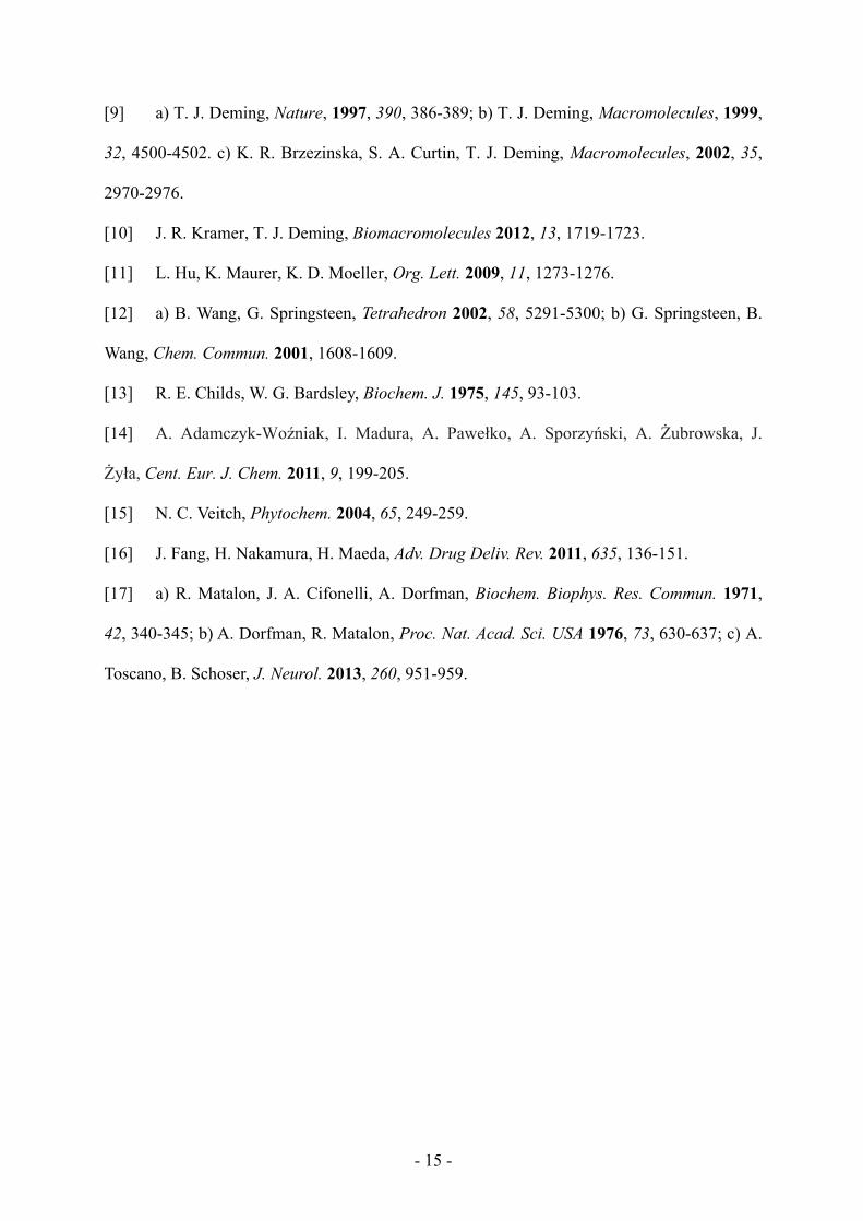
performed initial complexation studies using Alizarin Red S (ARS). Since its optical
properties change upon complexation of its catechol group to aryl boronic acids, ARS has
been used to detect binding of PBAs to diols via UV/Vis or fluorescence spectroscopy [12].
When 1 was added to ARS in aqueous solution, a color change from red to yellow-orange was
observed. Fluorimetry was used to further probe the specificity of the interaction between
ARS and 1 (Figure 1). To an aqueous solution of ARS (0.1 mM) and 1 (1.0 mM) at pH 7.4
was added increasing amounts of catechol (0 to 100 mM, see SI, Figure S2), which was used
to compete with ARS for complexation with 1. The fluorescence of the ARS-1 complex
decreased with increasing catechol concentration, as would be expected for conversion of
ARS-1 into catechol-1 complexes in the presence of excess catechol (Figure 1, Figure S2).
These studies confirmed that copolymer 1 was capable of reversibly binding diol-containing
molecules.
To evaluate the ability of copolymer 1 to form complexes with a glycosylated protein,
our initial studies utilized horseradish peroxidase (HRP), a readily available, glycosylated
enzyme with a total carbohydrate content of 18 to 22% [15]. Complex formation between
HRP and 1 was studied by dynamic light scattering (DLS) analysis of mixtures of 1 and HRP
- 11 -
(ca. 10 boronic acid groups per monosaccharide) in aqueous buffers ranging from pH 7.4 to
pH 5.0 (Figure 2). DLS analysis showed that 1 and HRP, separately in solution, did not show
any aggregation behavior in the range of pH studied. When solutions of 1 and HRP were
mixed, their aggregation into multimeric complexes of ca. 25 to 65 nm average diameter was
found to occur at both pH 7.4 and pH 6.5. At pH 5.0, no aggregation was seen in mixtures of
1 and HRP, showing that complexation between 1 and HRP is pH-dependent and that no
complexation occurs at this pH (Figure 2). The exact ratio of 1 to HRP in the complexes was
not be determined due to the unknown and variable degree and type of glycosylation in this
protein [15].
The complexes between 1 and HRP at pH 6.5 and 7.4 were stable over time in buffer
(48 hours, 22 ºC), and possessed average diameters that would be desirable for bloodstream
circulation and passive targeting via the enhanced permeation and retention (EPR) effect [16].
To determine if the HRP complexed with 1 remained in active form, an assay using the
chromogenic HRP substrate 2-2’-azino-bis(3-ethylbenzothiazoline-6-sulfonic acid)
diammonium salt (ABTS) was conducted and showed that complexed HRP did not lose
activity relative to free HRP (see SI, Figure S3) [13]. As an additional control to confirm that
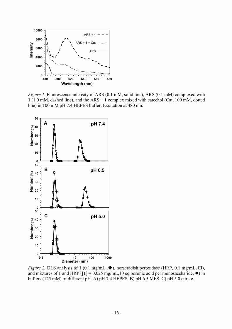
aggregation of 1 with HRP involved glycosylation sites on HRP, we also studied the ability of
copolymer 1 to form complexes with a non-glycosylated protein, bovine serum albumin
(BSA). At pH 7.4, no complex aggregates were observed by DLS to form between 1 and BSA
(Figure 3a), which suggests that the presence of surface carbohydrates on a protein are
necessary for strong complexation with 1 under these conditions. Further, we also observed
that addition of catechol to complexes of 1 and HRP resulted in dissociation of the complexes,
due to the preferential binding of catechol to WBA groups in 1 (see SI, Figure S4) [12]. To
confirm that pH responsive complexation of HRP with 1 is due to its WBA groups, we also
studied the ability of PBA containing copolymer 2 to form complexes with HRP. Copolymer
2 was found by DLS analysis to be unable to form complex aggregates with HRP even at pH
- 12 -
7.4, which is likely due to the high pKa of the PBA groups (Figure 3b) [7]. These control
experiments show that complexes of 1 and HRP do not denature the enzyme, are likely due to
complex formation between WBA and surface carbohydrate groups, and that the structure of
the WBA group greatly enhances complex stability at physiological pH, and allows for HRP
release at pH 5.0.
To show the results obtained above for 1 and HRP are not specific to that protein, and
also show an example with therapeutic potential, we studied the ability of copolymer 1 to
form complexes with alpha-L-iduronidase (IDUA), a glycosylated enzyme used for enzyme
replacement therapy for the treatment of mucopolysaccharidosis I [17]. Complex formation
between IDUA and 1 was studied using DLS analysis of mixtures of 1 and IDUA (ca. 10
boronic acid groups per monosaccharide) in aqueous buffers ranging from pH 7.4 to pH 5.0
(Figure 4). Similar to results seen with HRP, DLS analysis showed that 1 and IDUA,
separately in solution, did not show any aggregation behavior in the range of pH studied.
When solutions of 1 and IDUA were mixed, their aggregation into complexes of ca. 50 to 125
nm average diameter was found to occur at both pH 7.4 and pH 6.5. At pH 5.0, no
aggregation was seen in mixtures of 1 and IDUA, showing that complexation between 1 and
IDUA is pH-dependent and that no complexation occurs at this pH (Figure 4). These results
are similar to those obtained using HRP, and suggest that copolymer 1 has potential for pH
dependent complex assembly with a variety of different glycosylated proteins.
4. Conclusions
We have described the synthesis of a new WBA functionalized polypeptide-PEG copolymer,
1. While traditional PBA functionalized polypeptides and copolymers have been prepared in
the past [4,5], 1 displays the ability to bind strongly to catechols and glycosylated proteins at
physiological pH. The structural features of the WBA groups also allow for pH-dependent
complexation of glycosylated enzymes. The nanoscale dimensions of the complexes, and the
physiological relevance of the pH transition between complexed and non-complexed proteins,

- 13 -
suggest 1 is suitable for further development as a carrier for intracellular delivery of
glycosylated enzymes.
Supporting Information
Supporting Information is available from the Wiley Online Library or from the author.
Abbreviations α-amino acid N-carboxyanhydride (NCA), poly(ethylene glycol) (PEG),
trifluoroacetic acid (TFA), horseradish peroxidase (HRP), bovine serum albumin (BSA), 2-2’-
azino-bis(3-ethylbenzothiazoline-6-sulfonic acid) diammonium salt (ABTS), molecular
weight cutoff (MWCO), Alizarin Red S (ARS), 2-[4-(2-hydroxyethyl)piperazin-1-
yl]ethanesulfonic acid (HEPES), 2-(N-morpholino)ethanesulfonic acid (MES), 2-amino-2-
hydroxymethyl-propane-1,3-diol (TRIS), N-(3-dimethylaminopropyl)-N’-ethylcarbodiimide
hydrochloride (EDC-HCl), alpha-L-iduronidase (IDUA).
Acknowledgements: This work was supported by the NSF under award No. DMR 1308081
Received: Month XX, XXXX; Revised: Month XX, XXXX; Published online:
((For PPP, use “Accepted: Month XX, XXXX” instead of “Published online”)); DOI:
10.1002/mabi.((insert number))
Keywords: (boronic acid, polypeptide, block copolymer, protein delivery)
[1] a) E. Beutler, Trends Biochem. Sci. 1981, 6, 95-97; b) R. Vaishiya, V. Khurana, S.
Patel, A. K. Mitra, Expert Opin. Drug Deliv. 2015, 12, 415-440.
- 14 -
[2] a) S. Mura, J. Nicolas, P. Couvreur, Nat. Mater. 2013, 12, 991-1003; b) F. D. Jochum,
P. Theato, Chem. Soc. Rev. 2013, 42, 7468-7483.
[3] a) J. N. Cambre, B. S. Sumerlin, Polymer 2011, 52, 4631-4643; b) W. L. A. Brooks, B.
S. Sumerlin, Chem. Rev. 2016, 116, 1375-1397.
[4] a) K. T. Kim, J. J. L. M. Cornelissen, R. J. M. Nolte, J. C. M. van Hest, J. Amer. Chem.
Soc. 2009, 131, 13908-13909; b) J. I. Jay, B. E. Lai, D. G. Myszka, A. Mahalingam, K.
Langheinrich, D. F. Katz, P. F. Kiser, Mol. Pharmaceutics 2010, 7, 116-129; c) A.
Mahalingam, A. R. Geonnotti, J. Balzarini, P. F. Kiser, Mol. Pharmaceutics 2011, 8, 2465-
2475; d) G. A. Ellis, M. J. Palte, R. T. Raines, J. Amer. Chem. Soc. 2012, 134, 3631-3634.
[5] a) T. Kimura, S. Arimori, M. Takeuchi, T. Nagasaki, S. Shinkai, J. Chem. Soc. Perkin
Trans. 2 1995, 1889–1894; H. Kobayashi, K. Nakashima, E. Ohshima, Y. Hisaeda, I.
Hamachi, S. Shinkai, J. Chem. Soc. Perkin Trans. 2 2000, 997-1002; c) N. D. Winblade I. D.
Nikolic, A. S. Hoffman, J. A. Hubbell, Biomacromolecules 2000, 1, 523-533; d) L. Zhao, J.
Ding, C. Xiao, P. He, Z. Tang, X. Pang, X. Zhuanga, X. Chen, J. Mater. Chem. 2012, 22,
12319-12328; e) G. Liu, R. Ma, J. Ren, Z. Li, H. Zhang, Z. Zhang, Y. An, L. Shi, Soft Matter
2013, 9, 1636-1644; f) J. Ren, Y. Zhang, J. Zhang, H. Gao, G. Liu, R. Ma, Y. An, D. Kong, L.
Shi, Biomacromolecules 2013, 14, 3434-3443.
[6] a) G. Wulff, Pure & Appl. Chem. 1982, 54, 2093-2102; b) M. Lauer, G. Wulff, J.
Organomet. Chem. 1983, 256, 1-9.
[7] a) S. L. Wiskur, J. L. Lavigne, H. Ait-Haddou, V. Lynch, Y. H. Chiu, J. W. Canary, J.
W.; E. V. Anslyn, Org. Lett. 2001, 3, 1311-1314; b) L. I. Bosch, T. M. Fyles, T. D. James,
Tetrahedron 2004, 60, 11175-11190; c) N. Y. Edwards, T. W. Sager, J. T. McDevitt, E. V.
Anslyn, J. Amer. Chem. Soc. 2007, 129, 13575-13583.
[8] Y. Li, W. Xiao, K. Xiao, L. Berti, J. Luo, H. P. Tseng, G. Fung, K. S. Lam, Angew.
Chem. Int. Ed. 2012, 51, 2864-2869.
- 15 -
[9] a) T. J. Deming, Nature, 1997, 390, 386-389; b) T. J. Deming, Macromolecules, 1999,
32, 4500-4502. c) K. R. Brzezinska, S. A. Curtin, T. J. Deming, Macromolecules, 2002, 35,
2970-2976.
[10] J. R. Kramer, T. J. Deming, Biomacromolecules 2012, 13, 1719-1723.
[11] L. Hu, K. Maurer, K. D. Moeller, Org. Lett. 2009, 11, 1273-1276.
[12] a) B. Wang, G. Springsteen, Tetrahedron 2002, 58, 5291-5300; b) G. Springsteen, B.
Wang, Chem. Commun. 2001, 1608-1609.
[13] R. E. Childs, W. G. Bardsley, Biochem. J. 1975, 145, 93-103.
[14] A. Adamczyk-Woźniak, I. Madura, A. Pawełko, A. Sporzyński, A. Żubrowska, J.
Żyła, Cent. Eur. J. Chem. 2011, 9, 199-205.
[15] N. C. Veitch, Phytochem. 2004, 65, 249-259.
[16] J. Fang, H. Nakamura, H. Maeda, Adv. Drug Deliv. Rev. 2011, 635, 136-151.
[17] a) R. Matalon, J. A. Cifonelli, A. Dorfman, Biochem. Biophys. Res. Commun. 1971,
42, 340-345; b) A. Dorfman, R. Matalon, Proc. Nat. Acad. Sci. USA 1976, 73, 630-637; c) A.
Toscano, B. Schoser, J. Neurol. 2013, 260, 951-959.
- 16 -
Figure 1. Fluorescence intensity of ARS (0.1 mM, solid line), ARS (0.1 mM) complexed with 1 (1.0 mM, dashed line), and the ARS + 1 complex mixed with catechol (Cat, 100 mM, dotted line) in 100 mM pH 7.4 HEPES buffer. Excitation at 480 nm.
Figure 2. DLS analysis of 1 (0.1 mg/mL, u), horseradish peroxidase (HRP, 0.1 mg/mL, o), and mixtures of 1 and HRP ([1] = 0.025 mg/mL,10 eq boronic acid per monosaccharide, l) in buffers (125 mM) of different pH. A) pH 7.4 HEPES. B) pH 6.5 MES. C) pH 5.0 citrate.
0
2000
4000
6000
8000
10000
480 500 520 540 560 580
Inte
nsity
Wavelength (nm)
ARS + 1
ARS + 1 + Cat
ARS
0
10
20
30
40
50
Num
ber (
%) pH 7.4 A
0
10
20
30
40
50
Num
ber (
%) pH 6.5 B
0
10
20
30
40
50
0.1 1 10 100 1000
Num
ber (
%)
Diameter (nm)
pH 5.0 C
- 17 -
Figure 3. DLS analysis of control complexation experiments using a non-glycosylated protein, bovine serum albumin (BSA), or the PBA based copolymer 2. A) 1 (0.1 mg/mL, u), BSA (0.05 mg/mL, o) and mixture of 1 and BSA at the same mass ratio used for HRP ([1] = 0.0125 mg/mL, l). B) 2 (0.033 mg/mL) (u), HRP (0.1 mg/mL) (o) and mixture of 2 and HRP ([2] = 0.025 mg/mL, 10 eq boronic acid per monosaccharide, l).
0
10
20
30
40
50 N
umbe
r (%
) pH 7.4 A
B
0
10
20
30
40
50
0.1 1 10 100 1000
Num
ber (
%)
Diameter (nm)
pH 7.4 B

- 18 -
Figure 4. DLS analysis of 1 (0.1 mg/mL, u), alpha-L-iduronidase (IDUA, 0.1 mg/mL, o), and mixtures of 1 and IDUA ([1] = 0.025 mg/mL, 10 eq boronic acid per monosaccharide, l) in buffers (125 mM) of different pH. A) pH 7.4 HEPES. B) pH 6.5 MES. C) pH 5.0 citrate.
0
10
20
30
40
50 N
umbe
r (%
) pH 7.4 A
0
10
20
30
40
50
Num
ber (
%) pH 6.5 B
0
10
20
30
40
50
0.1 1 10 100 1000
Num
ber (
%)
Diameter (nm)
pH 5.0 C
- 19 -
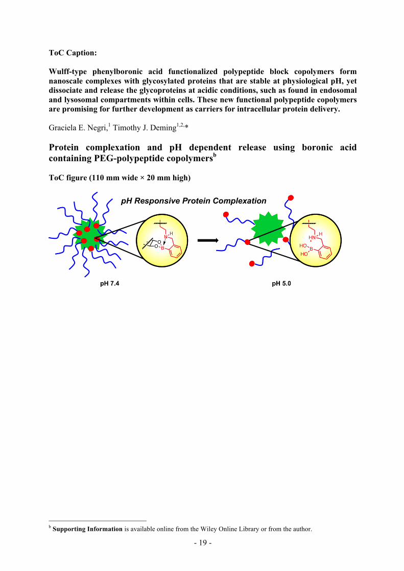
ToC Caption: Wulff-type phenylboronic acid functionalized polypeptide block copolymers form nanoscale complexes with glycosylated proteins that are stable at physiological pH, yet dissociate and release the glycoproteins at acidic conditions, such as found in endosomal and lysosomal compartments within cells. These new functional polypeptide copolymers are promising for further development as carriers for intracellular protein delivery. Graciela E. Negri,1 Timothy J. Deming1,2,* Protein complexation and pH dependent release using boronic acid containing PEG-polypeptide copolymersb ToC figure (110 mm wide × 20 mm high)
b Supporting Information is available online from the Wiley Online Library or from the author.
pH Responsive Protein Complexation