born blue - spa blue anesthesia and chd kristine faust, crna, ... –2-3 million estimated by the...
TRANSCRIPT

Born Blue
Anesthesia and CHD
Kristine Faust, CRNA, MS, MBA, DNAP

Disclosures

Disclosures
• None to Report

Objectives
• Review all congenital defects in which the patient is “blue”
• Describe physiology of the single ventricle patient
• Identify the 3 stages of repair for the single ventricle patient
• Describe the anesthesia implications of each stage

Congenital Heart Disease (CHD)
• Most common birth defect
• Occurs in 1 out of 115 – 150 live births
• 40, 000 births per year
• 4,800 babies with CCHD
(Critical Congenital Heart Disease)
• 1/13 infant deaths due to CHD
• CHD mortality decreased 33 % from 1996 – 2006
March of dimes website, 2014
American Heart Association

Adult Congenital Heart Disease
• 1.3 million patients alive today with CHD
–2-3 million estimated by the adult congenital
heart association
American Heart Association website, 2014
Adult Congenital heart association website,
2014

Health Care Admissions
Opotowsky, 2009

Health Care Costs
• Costs increased 127% (1998 to 2005)
• Individual admissions $19,000 to $43,000
• Estimated national costs increased from $691 million to $3.16 billion
Opotowsky, 2009

Causes of CHD
• Mom with History
–Diabetes
–Lupus
–Rubella
–Obesity
–Phenylketonuria (PKU)
• Other contributing factors
–Smoking
–Alcohol March of dimes website, 2014

Causes of CHD
• 30% of CHD patients also have a chromosomal defect
–Downs
–Turners
–Noonan
–Velocardiocraniofacial syndrome (VCF)
–Allagille syndrome
March of dimes website, 2014

Genetics and CHD
• Chromosomal abnormalities 10%
–2/3-trisomy 21
–1/3-others
• Trisomy 13
• Trisomy 18
• Turners syndrome
• 22q11.2 deletion-conotruncal defects(TOF, aortic
arch, truncus, VSD)
CDC website

Genetics and CHD
• Other 90% no specific genetic association
–Environmental
• Rubella, ethanol, lithium, maternal diabetes, folate
deficiency, obesity, smoking
CDC Website

Most Common Types of CHD
• Ventricular Septal Defect (14 -16%)
• Atrial Septal Defect (4 -10%)
• Tetralogy of Fallot (9 -14%)
• Coarctation of the Aorta (8 -11%)
• Transposition of the Great Vessels (10 -11%)
• Single ventricle (4 -8%)
American Heart Association
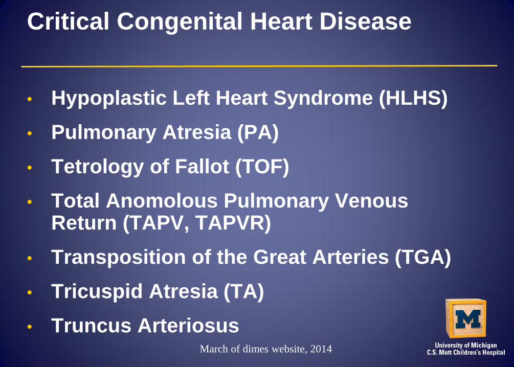
Critical Congenital Heart Disease
• Hypoplastic Left Heart Syndrome (HLHS)
• Pulmonary Atresia (PA)
• Tetrology of Fallot (TOF)
• Total Anomolous Pulmonary Venous Return (TAPV, TAPVR)
• Transposition of the Great Arteries (TGA)
• Tricuspid Atresia (TA)
• Truncus Arteriosus March of dimes website, 2014
Anesthesia and Uncommon Diseases, pg 76,
2012

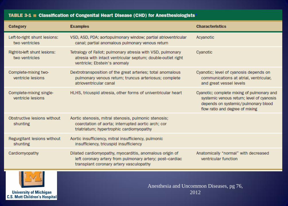
Left to Right Shunts
• VSD, ASD, AP window, partial AV canal, PAPVR
• Acyanotic
Anesthesia and
Uncommon Diseases,
2012

Right to Left Shunts
• TOF, PA with VSD, PA with IVS, DORV, Ebsteins’ anomaly
• Cyanotic
Anesthesia and Uncommon Diseases, 2012

Complete Mixing Lesions
• DTGA, TAPVR, TA, Complete AV canal
• Cyanotic, level depends of communications

Obstructive Lesions without shunting
• AS, MS, PS, Coarctation, IAA, Cortriatrium, hypertrophic cardiomyopathy
Anesthesia and Uncommon Diseases, 2012
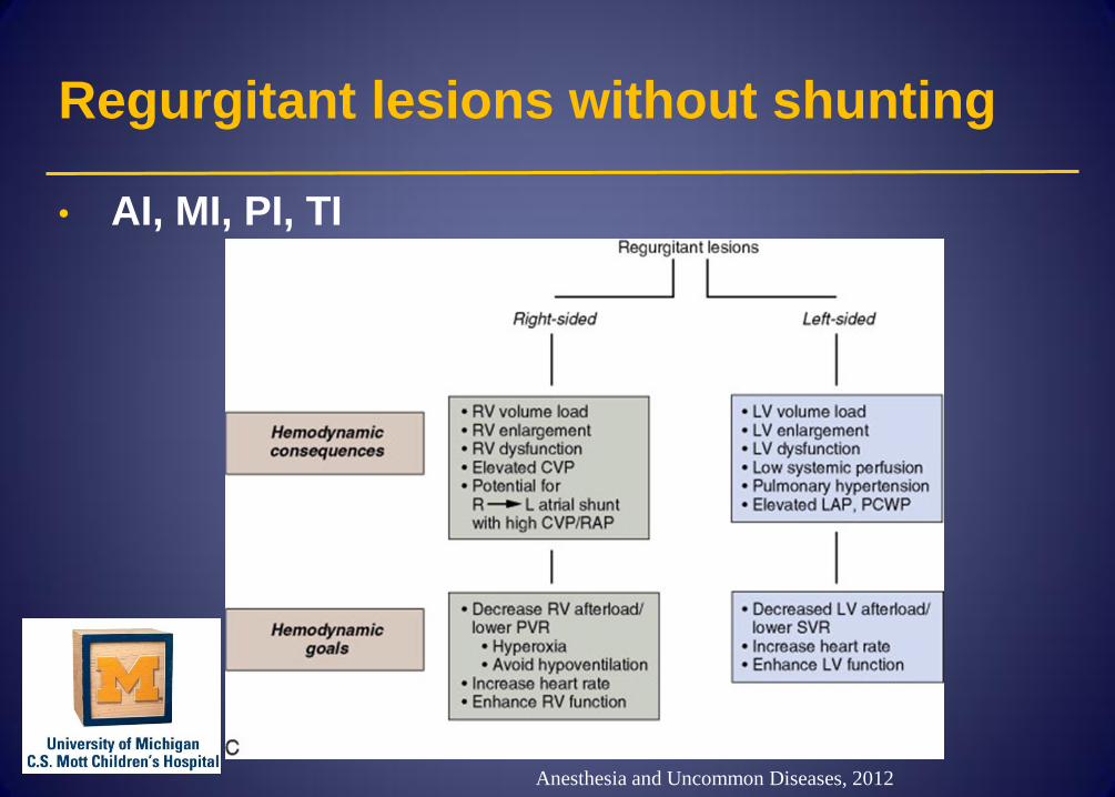
Regurgitant lesions without shunting
• AI, MI, PI, TI
Anesthesia and Uncommon Diseases, 2012

Cardiomyopathy
• Dilated cardiomyopathy, myocarditis, anomalous LCA from pulmonary artery, post transplant cardiomyopathy
• Anatomically “normal” with decreased function

HLHS
• Fourth most common congenital heart defect
• 7.5% of newborns
• Some genetic inheritance?
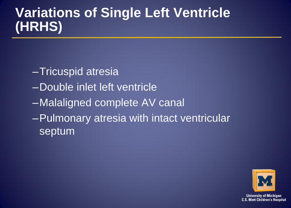
Variations of Single Left Ventricle (HRHS)
–Tricuspid atresia
–Double inlet left ventricle
–Malaligned complete AV canal
–Pulmonary atresia with intact ventricular
septum

Variations of Single Right Ventricle (HLHS)
–Mitral valve atresia
• Hypoplastic left heart syndrome (HLHS)
• Double outlet right ventricle
–Aortic valve atresia –HLHS
–Large VSD and normal LV
–Malaligned complete AV canal
–Heterotaxy syndromes-pulmonary stenosis or atresia

Two ventricles with potential single ventricle physiology
–Tetrology of Fallot with pulmonary atresia
–Truncus arteriosis
–Total anomalous pulmonary venous connection
(TAPVR)
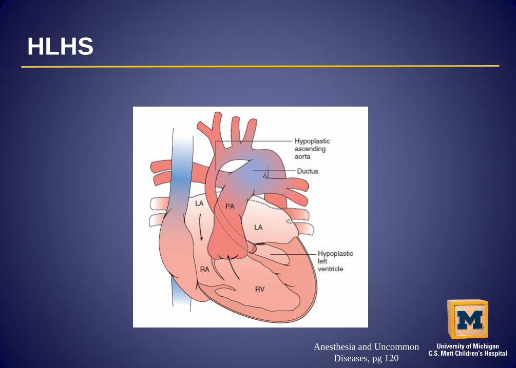
HLHS
Anesthesia and Uncommon
Diseases, pg 120

History of CHD surgery
• 1939: Robert Gross ligates a PDA, Boston Children's
• 1944: Alfred Blalock performs BT shunt for TOF, Johns Hopkins
• 1953: John Gibbon uses CPB to close an ASD, University of Pennsylvania
• 1960: Dwight Harken replaces Aortic Valve, Brigham and Women's Hospital

History of CHD surgery
• 1967: Christian Barnard performs first heart transplant, South Africa
• 1975: Adib Domingos Jatene performs arterial switch for d-TGA, Brazil
• 1977: William Norwood performs Norwood for HLHS, CHOP
• 1985: LL Bailey performs first successful neonatal heart transplant, Loma Linda, CA

History CHD at Mott
• Bove arrived 1987-began work on HLHS
• 850 CHD operations per year (about 50 Norwoods)
• Overall 95% survival rate
• 2nd largest Congenital Heart Center in the US

HLHS
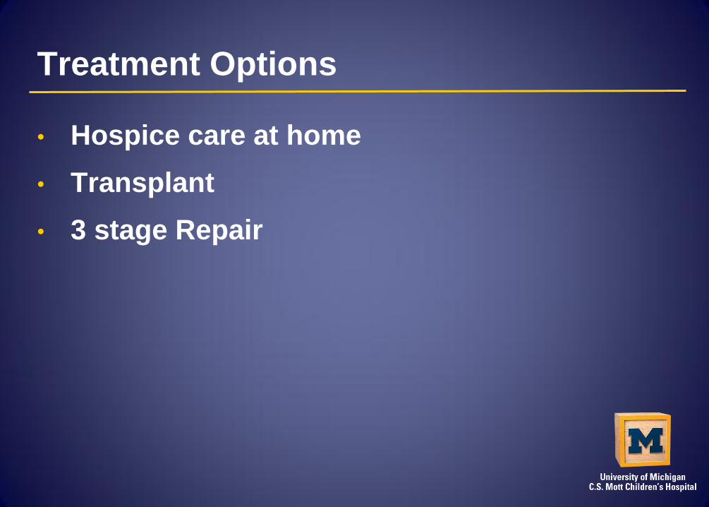
Treatment Options
• Hospice care at home
• Transplant
• 3 stage Repair

Stages of Single Ventricle Repair
• Norwood
–Hybrid
• Bidirectional Glenn or Hemi-Fontan
• Fontan

Classic Norwood
Anesthesia and Uncommon Diseases, pg
121

Norwood

Norwood BTS AP

Norwood BTS Lateral

Sano Shunt Variation
Anesthesia and Uncommon
Diseases, pg 121

Sano Shunt Variation

Sano AP view

Sano lateral view

MRI Pictures

Central AP

Central lateral

Hybrid
Anesthesia and Uncommon Diseases, pg 121

Hybrid

Bidirectional Glenn Anatomy
Andropoulos, 467

Bidirectional Glenn Anatomy
Anesthesia and
Uncommon
Diseases, pg 123

BDG AP

BDG lateral

Left Glenn AP

Left Glenn lateral

Hemi-Fontan Anatomy
Field guide 264

Hemi-Fontan Anatomy

Hemi-Fontan Anatomy

Hemi-Fontan Anatomy
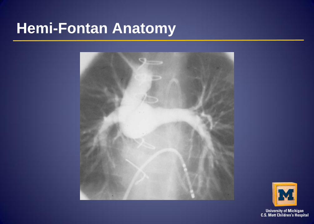
Hemi-Fontan Anatomy

Hemi AP

Hemi lateral

Fontan Anatomy
Anesthesia and
Uncommon
Diseases, pg 119

Fontan (lateral tunnel)
Anesthesia and
Uncommon
Diseases, pg 119
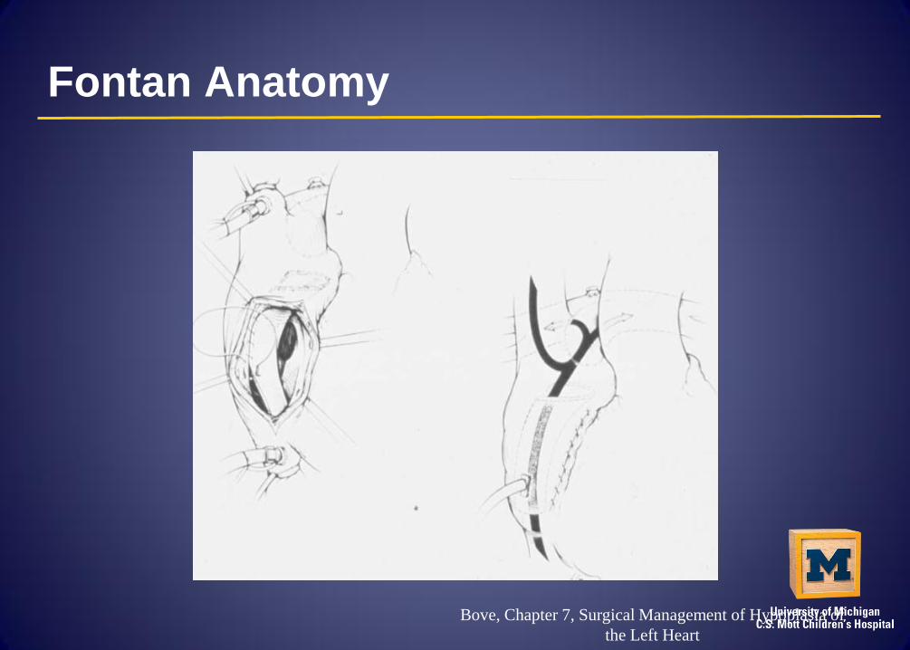
Fontan Anatomy
Bove, Chapter 7, Surgical Management of Hypoplasia of
the Left Heart

Fontan Anatomy
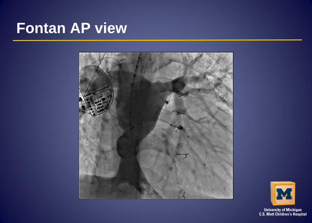
Fontan AP view

Fontan lateral


HLHS Physiology
Balancing the parallel circulations
Goals
• good cardiac output
• normal end-organ function
• normal systemic oxygen delivery
• normal blood flow

Normal or “Series”

Parallel or “Balanced” Norwood and Hemifontan Stage

HLHS (after Fontan completion)

Anesthesia and Uncommon Diseases,
pg. 122, 2012

Andropoulos, 458
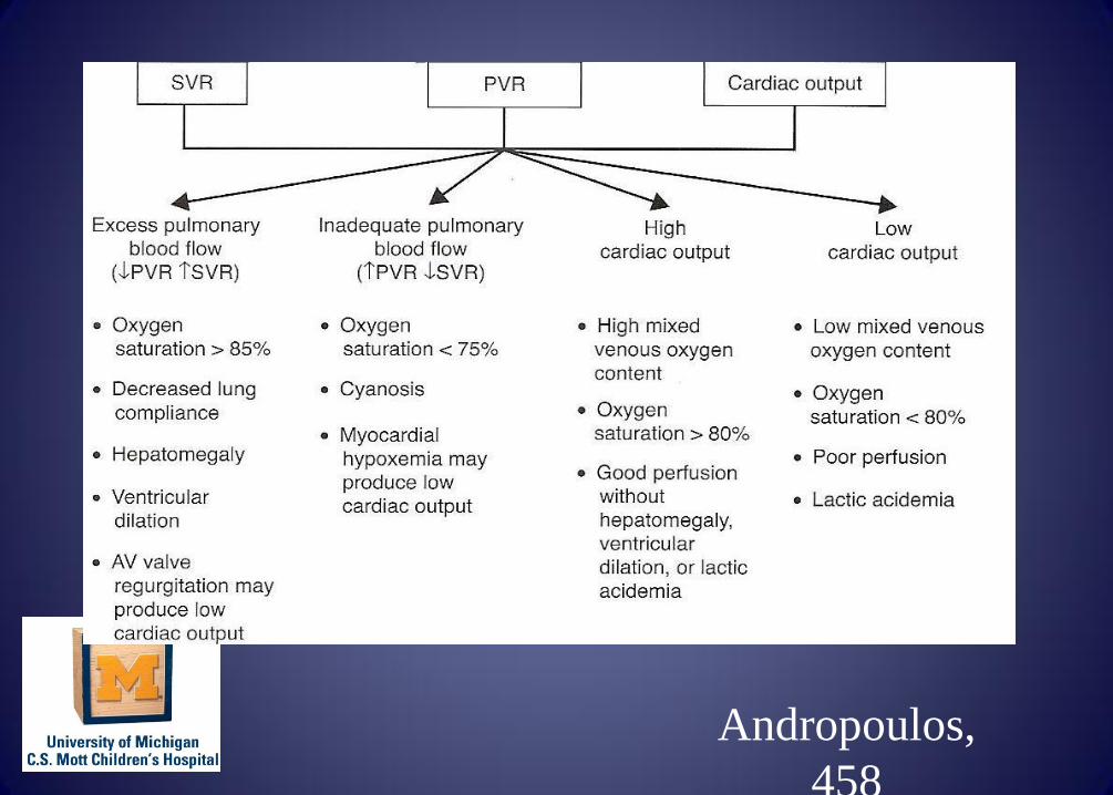
Andropoulos,
458

Anesthetic Planning
• Understand hemodynamic consequences of patient's lesion and state of repair.
• Construct a set of hemodynamic goals for each patient.

Anesthetic Planning
• Plan anesthetic agents and techniques, ventilatory management, and inotropic/vasoactive drug support based on these goals.
• Although no anesthetic agent or technique is contraindicated, avoid agents or doses counter to hemodynamic goals, and use agents that promote these goals

Preoperative Assessment
• History
–Cardiac lesion
–Cyanotic
–One or two ventricles
–Septated, any intra atrial or ventricular
communication
–Surgery or cath lab procedures
–Palliated vs corrected
–Ventricular function

Preoperative Assessment
• History (cont)
–Coronary anatomy
–Outflow tract obstruction
–Exercise tolerance, feeding
–Medical therapy

Preoperative Assessment
• Physical
–General appearance
–BP
–Cyanosis, clubbing
–Tachypnea, retractions
–Peripheral pulses, perfusion
–Precordium, heart sounds, murmurs
–Hepatomegaly, JVD
–Diaphoresis
–Adequacy of veins, arterial pulses

Preoperative Assessment
• Chest X-ray
–Heart size and configuration
–Pulmonary vasculature
–Pulmonary parenchymal disease

Preoperative Assessment
• EKG
–Rhythm
–Rate
–ST segments
–Axis deviation

Preoperative Assessment
• Hemoglobin
–Normal, low, high for age and gender

Preoperative Assessment
• Oxygen saturation
–What is normal for this patient
–Any recent changes
–On home O2

Preoperative Assessment
• Echo
–Cardiac anatomy, residual defects, ventricular
function
–Outflow tract obstruction
–Valvar regurgitation
–Atrial/ventricular communication

Preoperative Assessment
• CT
–Anatomy of extra cardiac structures
• Aorta, pulmonary, arteries, veins

Preoperative Assessment
• Cardiac MRI
–Anatomy of intra and extra cardiac structures
–Ventricular function
–Qp/Qs
Preoperative Assessment
• Cardiac Cath
–Detailed anatomy
–Hemodynamics
–SVR/PVR, Qp/Qs
–PVR reactivity

Norwood Physiology
Anesthesia and
Uncommon
Diseases, pg 121

Anesthesia and Uncommon Diseases, pg 121

Norwood Anesthesia
–Preserve balance SVR and PVR
–Minimize myocardial depression
–Extreme caution hypotension
–HCT >40-45
–Narcotic, inhalational or combination
• NO List
–Sat greater than 75-85%(not good)
–NO hyperventilate
–NO 100% oxygen

Factors Affecting Hemodynamics of the HLHS Patient Manipulations to Increase PVR
Decrease inspired concentration O2
Hypoventilation (PCO2 40-50)
PEEP
Manipulations to Decrease PVR
Increased inspired concentration O2
Hyperventilation (PCO2 20-25)
Inotropic support
Stokes, 2005

Bidirectional Glenn or Hemifontan Physiology
Anesthesia and
Uncommon
Diseases, pg 123

Hemi-Fontan Physiology
• Caution to NOT hyperventilate
–Decreases cerebral flow
–Decreases pulmonary flow
• NOT use 100% oxygen
• Narcotic, inhalational or combination

Hemi-Fontan and Anesthesia
• Baseline Sats
• Exercise tolerance (SOB, dusky with feeds?)
• Echo, EKG, labs, cath, chest x-ray
• Other congenital issues
• SVC syndrome
• IV access

Anesthesia and Uncommon Diseases, pg. 122, 2012

Fontan Physiology
Anesthesia and
Uncommon
Diseases, pg 119

Anesthesia and
Uncommon Diseases,
pg 123

Fontan Physiology
• Adequate preload
• Low pulmonary vascular resistance to preserve pulmonary flow
• Maintaining sinus rhythm
• Minimizing myocardial depression
• Spontaneous ventilation

Fontan and Anesthesia
• Baseline Saturation
• Exercise tolerance
• Echo, EKG, labs, cath, chest x-ray
• Other congenital issues
• Premed
• Narcotic, inhalational or combination

Fontan Long-term Issues
• Cardiovascular
–Arrhythmias
–Heart failure
–Thromboembolism
• Pulmonary
–Plastic Bronchitis

Fontan Long-term issues
• Protein Losing Enteropathy
–Hypoablbuminemia
– Intestinal protein loss
–Edema
–Ascites
– Immune deficiency

Fontan
• Optimal Fontan physiology
–CVP of 10-15mmHg
–Pulmonary artery pressure of 10-15mmHg
–Left atrial (LA) pressure of 5-10 mmHg
• Transpulmonary gradient
Postoperative
• Pain control
• Need for overnight stay
• Need for special monitoring
• Need for ICU bed

Celermajer, 2013
Summary
• Understand the patient’s anatomy/physiology including shunts (Draw it out!)
• Understand how anesthetics and vasoactive substances affect the patient’s cardiovascular physiology
• Understand the effects of ventilation and oxygenation on the patient’s cardiovascular physiology

References
• Anesthesia and Uncommon Diseases
• Anesthesia for Congenital Heart Disease
• CDC, AHA, ACHA, March of Dimes websites