bmc neuroscience biomed central - unifr.ch central page 1 of 12 (page number not for citation...
TRANSCRIPT

BioMed CentralBMC Neuroscience
ss
Open AcceResearch articleA case of polymicrogyria in macaque monkey: impact on anatomy and function of the motor systemEric Schmidlin*†1, Christophe Jouffrais†1,2, Patrick Freund1, Patrizia Wannier-Morino1, Marie-Laure Beaud1, Eric M Rouiller1 and Thierry Wannier1Address: 1Unit of Physiology and Program in Neurosciences, Department of Medicine, Faculty of Sciences, University of Fribourg, Chemin du Musée 5, CH-1700 Fribourg, Switzerland and 2IRIT, Université de Toulouse and CNRS, 133 route de Narbonne, 31062 Toulouse cedex 9, France
Email: Eric Schmidlin* - [email protected]; Christophe Jouffrais - [email protected]; Patrick Freund - [email protected]; Patrizia Wannier-Morino - [email protected]; Marie-Laure Beaud - [email protected]; Eric M Rouiller - [email protected]; Thierry Wannier - [email protected]
* Corresponding author †Equal contributors
AbstractBackground: Polymicrogyria is a malformation of the cerebral cortex often resulting in epilepsyor mental retardation. It remains unclear whether this pathology affects the structure and functionof the corticospinal (CS) system. The anatomy and histology of the brain of one macaque monkeyexhibiting a spontaneous polymicrogyria (PMG monkey) were examined and compared to the brainof normal monkeys. The CS tract was labelled by injecting a neuronal tracer (BDA) unilaterally ina region where low intensity electrical microstimulation elicited contralateral hand movements(presumably the primary motor cortex in the PMG monkey).
Results: The examination of the brain showed a large number of microgyri at macro- andmicroscopic levels, covering mainly the frontoparietal regions. The layered cortical organizationwas locally disrupted and the number of SMI-32 stained pyramidal neurons in the cortical layer IIIof the presumed motor cortex was reduced. We compared the distribution of labelled CS axonsin the PMG monkey at spinal cervical level C5. The cumulated length of CS axon arbors in the spinalgrey matter was not significantly different in the PMG monkey. In the red nucleus, numerousneurons presented large vesicles. We also assessed its motor performances by comparing itscapacity to execute a complex reach and grasp behavioral task. The PMG monkey exhibited anincrease of reaction time without any modification of other motor parameters, an observation inline with a normal CS tract organisation.
Conclusion: In spite of substantial cortical malformations in the frontal and parietal lobes, thePMG monkey exhibits surprisingly normal structure and function of the corticospinal system.
BackgroundPolymicrogyria is a developmental malformation of thecerebral cortex, characterized by multiple small gyri withabnormal cortical lamination [1]. PMG can be unilateral
or bilateral and its extent varies from focal PMG in other-wise normal brain to diffuse PMG with multiple otherbrain abnormalities. The spectrum of clinical manifesta-tions ranges from normal individuals, with only selective
Published: 23 December 2009
BMC Neuroscience 2009, 10:155 doi:10.1186/1471-2202-10-155
Received: 10 February 2009Accepted: 23 December 2009
This article is available from: http://www.biomedcentral.com/1471-2202/10/155
© 2009 Schmidlin et al; licensee BioMed Central Ltd. This is an Open Access article distributed under the terms of the Creative Commons Attribution License (http://creativecommons.org/licenses/by/2.0), which permits unrestricted use, distribution, and reproduction in any medium, provided the original work is properly cited.
Page 1 of 12(page number not for citation purposes)
BMC Neuroscience 2009, 10:155 http://www.biomedcentral.com/1471-2202/10/155
impairment of cognitive function [2] and no or easily con-trolled epilepsy, to patients with severe encephalopathyand intractable epilepsy [3]. Motor and cognitive deficitssuch as a delay in development [4], or congenital contrac-tures [5] are commonly described in patients sufferingfrom PMG. Microscopically, two histological types ofPMG were recognized: a simplified four layered form andan unlayered form [6]. The two types of PMG may coexistin contiguous cortical areas [7]. Recent report providesevidence that PMG areas are functional [8].
The present report describes a case of spontaneouslyoccurring PMG in a macaque monkey for which tracing ofcorticospinal projections had been obtained. Moreover,the animal was involved in a study on the mechanisms ofbimanual coordination, and its PMG was discovered aftersacrifice. The first goal of the present report was to presentin more details the general morphological traits of thePMG brain. More specifically, we sought to establishwhich brain regions and how the laminar pattern of thecerebral cortex have been affected by the PMG. In humanpatients with a unilateral PMG, the CS tract originatingfrom the affected hemisphere presented an altered struc-ture in DTI and fMRI investigations [9]. The second aim ofthe present case report in monkeys was to evaluatewhether the cortical malformations affected the character-istics of the corticospinal projections. For this purpose,the anterograde tracer Biotinylated Dextran Amine (BDA)was injected unilaterally in the electrophysiologicallyidentified hand representation of the presumed primarymotor cortex. Finally, the motor capacity of the PGMmacaque was compared with that of a normal macaquemonkey, both trained to perform the same motor task,namely a modified version of the so-called "reach andgrasp drawer" task [10].
ResultsThe PMG monkey described in this study is the only caseof cortical malformation ever observed in our laboratory.
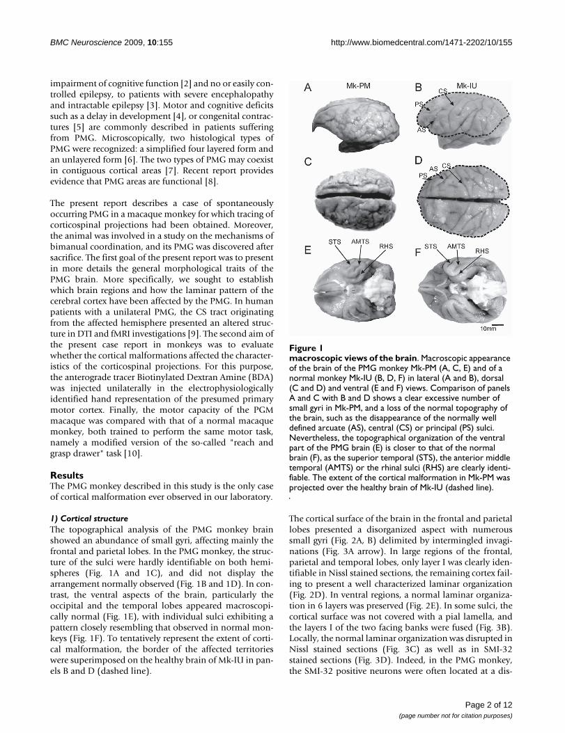
1) Cortical structureThe topographical analysis of the PMG monkey brainshowed an abundance of small gyri, affecting mainly thefrontal and parietal lobes. In the PMG monkey, the struc-ture of the sulci were hardly identifiable on both hemi-spheres (Fig. 1A and 1C), and did not display thearrangement normally observed (Fig. 1B and 1D). In con-trast, the ventral aspects of the brain, particularly theoccipital and the temporal lobes appeared macroscopi-cally normal (Fig. 1E), with individual sulci exhibiting apattern closely resembling that observed in normal mon-keys (Fig. 1F). To tentatively represent the extent of corti-cal malformation, the border of the affected territorieswere superimposed on the healthy brain of Mk-IU in pan-els B and D (dashed line).
The cortical surface of the brain in the frontal and parietallobes presented a disorganized aspect with numeroussmall gyri (Fig. 2A, B) delimited by intermingled invagi-nations (Fig. 3A arrow). In large regions of the frontal,parietal and temporal lobes, only layer I was clearly iden-tifiable in Nissl stained sections, the remaining cortex fail-ing to present a well characterized laminar organization(Fig. 2D). In ventral regions, a normal laminar organiza-tion in 6 layers was preserved (Fig. 2E). In some sulci, thecortical surface was not covered with a pial lamella, andthe layers I of the two facing banks were fused (Fig. 3B).Locally, the normal laminar organization was disrupted inNissl stained sections (Fig. 3C) as well as in SMI-32stained sections (Fig. 3D). Indeed, in the PMG monkey,the SMI-32 positive neurons were often located at a dis-
macroscopic views of the brainFigure 1macroscopic views of the brain. Macroscopic appearance of the brain of the PMG monkey Mk-PM (A, C, E) and of a normal monkey Mk-IU (B, D, F) in lateral (A and B), dorsal (C and D) and ventral (E and F) views. Comparison of panels A and C with B and D shows a clear excessive number of small gyri in Mk-PM, and a loss of the normal topography of the brain, such as the disappearance of the normally well defined arcuate (AS), central (CS) or principal (PS) sulci. Nevertheless, the topographical organization of the ventral part of the PMG brain (E) is closer to that of the normal brain (F), as the superior temporal (STS), the anterior middle temporal (AMTS) or the rhinal sulci (RHS) are clearly identi-fiable. The extent of the cortical malformation in Mk-PM was projected over the healthy brain of Mk-IU (dashed line).
Page 2 of 12(page number not for citation purposes)

BMC Neuroscience 2009, 10:155 http://www.biomedcentral.com/1471-2202/10/155
Page 3 of 12(page number not for citation purposes)
Reconstructions of frontal Nissl-stained sections in PMG monkeyFigure 2Reconstructions of frontal Nissl-stained sections in PMG monkey. Panels A and B: Location of cortical malforma-tion on coronal sections in the right hemisphere at two rostrocaudal levels. Sub-cortical structures, such as the thalamus (Thal.), the lateral geniculate nucleus (LGN), the putamen (Put) or the corpus callosum (CC) are visible and they do not present conspicuous abnormalities, but the lateral ventricle (L.V.) is enlarged, including two enlarged photo micrograph of the cortex showing the microscopic cortical ectopic sulci (#), where the layer I is clearly distinguishable. Light grey: unlayered cor-tex. Dark grey: normally layered cortex: Squared: subcortical nuclei Panel C: Photograph of the cerebral cortex in Mk-PM with the position of the sections depicted in panels A and B. Panels D and E: The cortical organization in layers is lost in the parietal lobe (D) but normal in the inferior temporal lobe (E). Panel F: Diagram showing the distribution of the number of sulci per cortical length (number of sulcus per 10 mm) in the PMG monkey (black) and in the three control monkeys, Mk-IU, Mk-I2 and Mk-IZ (grey). Diamonds represent the results obtained in the frontoparietal region and bars in the ventro-temporal region. N.s is for non-statistically significant. Panel G: Photomicrograph of the Corpus Callosum (CC) in the right hemisphere of Mk-PM showing large quantity of BDA stained fibers. Scale bar: 500 microns.

BMC Neuroscience 2009, 10:155 http://www.biomedcentral.com/1471-2202/10/155
tance from the surface that varied over a short rostro-cau-dal interval, and were not always oriented perpendicularlyto the brain surface (Fig. 3D).
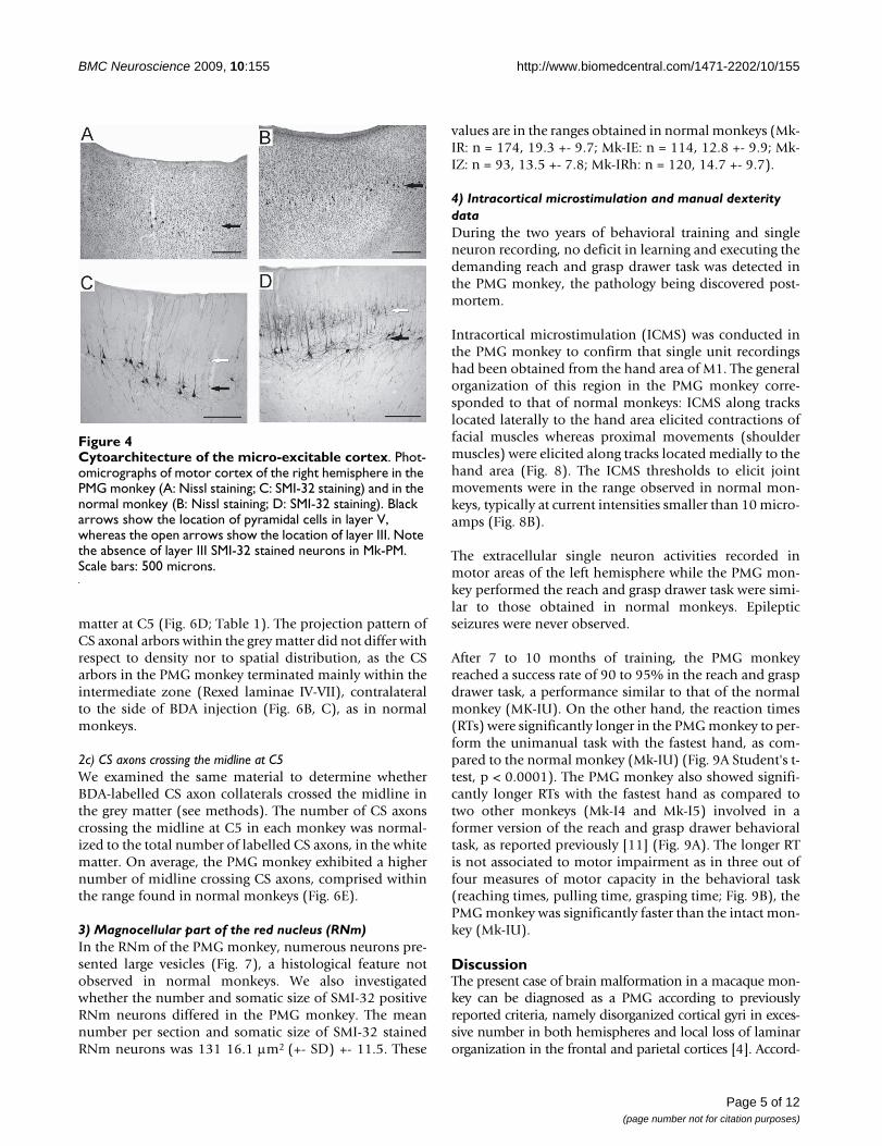
In the cortical region where ICMS elicited movements ofthe hand and where the laminar organization was identi-fiable, large pyramidal neurons were observed in Nissland in SMI-32 stained material (Fig. 4A and 4C, blackarrows) at a depth corresponding to layer V in normalmonkeys (Fig. 4B and 4D, black arrows). However, in nor-mal monkeys, numerous layer III pyramidal neurons werealso SMI-32 positive (Fig. 4D, white arrow), whereas onlyfew were recognizable in the PMG monkey (Fig. 4C).
When compared to normal monkeys (Mk-IU, Mk-I2 andMk-IZ), the frequency of sulci measured on coronal sec-tions in the fronto-parietal as well as in the ventro-tempo-ral cortical regions was significantly higher in the PMGmonkey (p < 0.05, Mann and Whitney with Bonferronicorrection for multiple comparisons). Furthermore, in thePMG monkey, the frequency of sulci in the fronto-parietal
cortex was nearly twice as high than that of the ventro-temporal region (Fig. 2F; p < 0.05, Mann-Whitney). Nostatistically significant difference was observed in thethree normal monkeys. These differences do not reflectchanges in the volume of the cortex, as at a comparablerostro-caudal position, the measured distance betweenthe corpus callosum and the external part of the lateral fis-sure is similar among all animals.
In contrast to the few SMI-32 positive neurons detected inlayer III of the presumed M1 area of the PMG-monkey,injections of BDA in this cortical region in the left hemi-sphere stained a large number of fibers in the corpus cal-losum (Fig. 2G) and several retrogradely labelled neuronswere found in the right hemisphere in the frontal lobe.This observation suggests that a significant number ofpyramidal neurons in lamina III are present, but do notexpress the neurofilament recognized by the SMI-32 anti-body. As the cortical structure of the PMG brain is also dis-turbed in the contralateral side, it was not possible toassess the exact areas where projections were terminatingand where callosal neurons were stained.
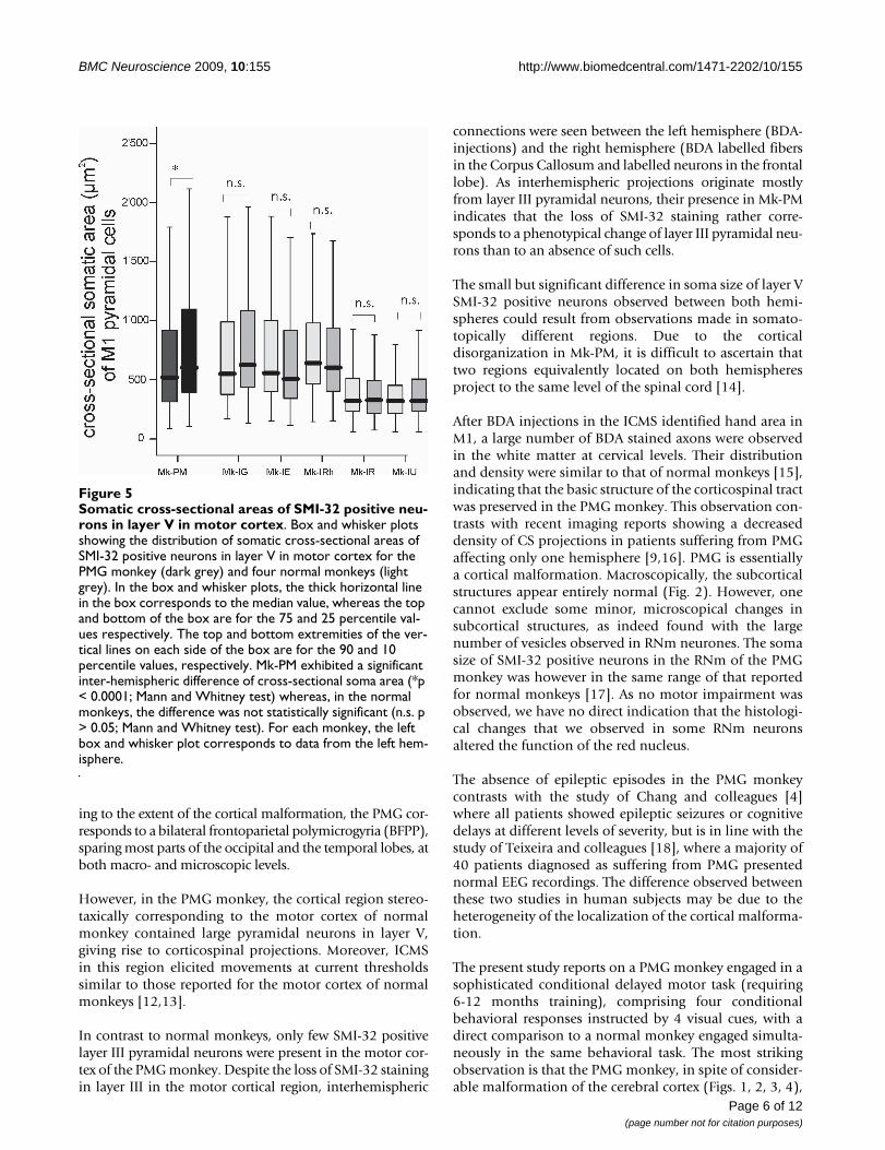
The analysis of the cross-sectional area of SMI-32 positivepyramidal cells in the putative layer V of M1 showed a sta-tistically significant difference of somatic size between theleft and the right hemispheres of the PMG monkey, a dif-ference that was not observed in normal monkeys (Fig. 5).However, the somatic size of SMI-32 stained pyramidalcells in the PMG monkey is comprised in the range foundin normal monkeys. As M1 is not well defined in the PMGmonkey, it is difficult to ascertain that the measures weredone on neurons placed in equivalent regions. The rela-tive number of SMI-32 positive cells in layer V was not dif-ferent among all monkeys.
2) The corticospinal (CS) projection2a) Crossed versus uncrossed CS projectionsIn normal macaque monkeys, after a unilateral BDA injec-tion in M1, 85-95% of the CS fibers were found in theopposite dorsolateral funiculus whereas 5-15% were inthe ipsilateral dorsolateral and ventral funiculi (Fig. 6B).In the PMG monkey, BDA injections were placed in a cor-tical territory where ICMS elicited contralateral handmovements (Fig. 6A). This territory was later found tocontain large pyramidal neurons. The proportions of BDAlabelled CS axons in the contralateral and ipsilateral cervi-cal white matter of the PMG monkey were 95% and 5%,respectively (Fig. 6C), thus comprised within the rangefound in normal monkeys.
2b) Density of CS axonal arbors in the grey matterThe macroscopic structure and general histology of thespinal cord of the PMG monkey was normal. When com-pared to three normal monkeys, the PMG monkey exhib-ited a comparable CS arborization density in the grey
Abnormalities in cortical organizationFigure 3Abnormalities in cortical organization. Panel A: Phot-omicrograph of a Nissl stained section demonstrating the dis-organization of the microgyrial cortex. The layered organization is irregular and anomalous, with the formation of small islets (arrow). Panel B: Photomicrograph showing the lack of lumen and of pia between two facing portions of cortex in a sulcus, resulting in the fusion of both layers I in a unique strip of white matter (black arrow). Panel C: Phot-omicrograph of a Nissl stained section showing the absence of well defined laminar organization. Panel D: Photomicro-graph of a SMI-32 stained section adjacent to the section pre-sented in panel C. Note that the SMI-32 positive neurons occupy positions in the cortex which vary strongly with regard of its distance to the cortex surface (black arrows). White arrows in panels C and D indicate the same blood vessels. Scale bars: 500 microns.
Page 4 of 12(page number not for citation purposes)
BMC Neuroscience 2009, 10:155 http://www.biomedcentral.com/1471-2202/10/155
matter at C5 (Fig. 6D; Table 1). The projection pattern ofCS axonal arbors within the grey matter did not differ withrespect to density nor to spatial distribution, as the CSarbors in the PMG monkey terminated mainly within theintermediate zone (Rexed laminae IV-VII), contralateralto the side of BDA injection (Fig. 6B, C), as in normalmonkeys.
2c) CS axons crossing the midline at C5We examined the same material to determine whetherBDA-labelled CS axon collaterals crossed the midline inthe grey matter (see methods). The number of CS axonscrossing the midline at C5 in each monkey was normal-ized to the total number of labelled CS axons, in the whitematter. On average, the PMG monkey exhibited a highernumber of midline crossing CS axons, comprised withinthe range found in normal monkeys (Fig. 6E).
3) Magnocellular part of the red nucleus (RNm)In the RNm of the PMG monkey, numerous neurons pre-sented large vesicles (Fig. 7), a histological feature notobserved in normal monkeys. We also investigatedwhether the number and somatic size of SMI-32 positiveRNm neurons differed in the PMG monkey. The meannumber per section and somatic size of SMI-32 stainedRNm neurons was 131 16.1 μm2 (+- SD) +- 11.5. These
values are in the ranges obtained in normal monkeys (Mk-IR: n = 174, 19.3 +- 9.7; Mk-IE: n = 114, 12.8 +- 9.9; Mk-IZ: n = 93, 13.5 +- 7.8; Mk-IRh: n = 120, 14.7 +- 9.7).
4) Intracortical microstimulation and manual dexterity dataDuring the two years of behavioral training and singleneuron recording, no deficit in learning and executing thedemanding reach and grasp drawer task was detected inthe PMG monkey, the pathology being discovered post-mortem.
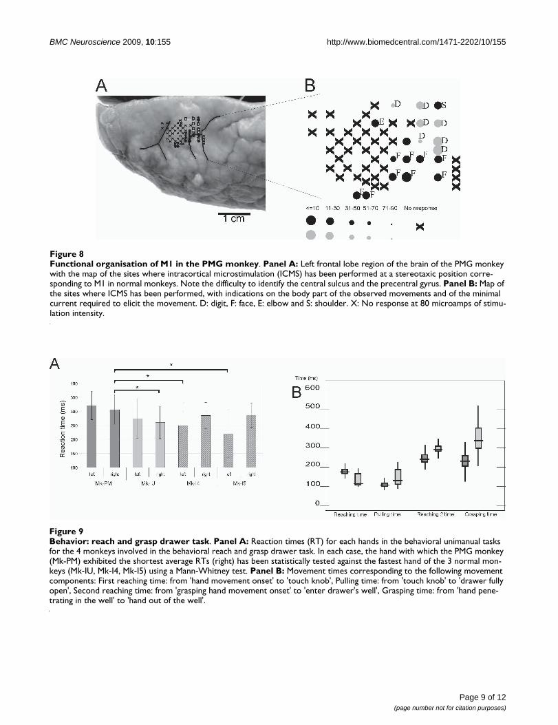
Intracortical microstimulation (ICMS) was conducted inthe PMG monkey to confirm that single unit recordingshad been obtained from the hand area of M1. The generalorganization of this region in the PMG monkey corre-sponded to that of normal monkeys: ICMS along trackslocated laterally to the hand area elicited contractions offacial muscles whereas proximal movements (shouldermuscles) were elicited along tracks located medially to thehand area (Fig. 8). The ICMS thresholds to elicit jointmovements were in the range observed in normal mon-keys, typically at current intensities smaller than 10 micro-amps (Fig. 8B).
The extracellular single neuron activities recorded inmotor areas of the left hemisphere while the PMG mon-key performed the reach and grasp drawer task were simi-lar to those obtained in normal monkeys. Epilepticseizures were never observed.
After 7 to 10 months of training, the PMG monkeyreached a success rate of 90 to 95% in the reach and graspdrawer task, a performance similar to that of the normalmonkey (MK-IU). On the other hand, the reaction times(RTs) were significantly longer in the PMG monkey to per-form the unimanual task with the fastest hand, as com-pared to the normal monkey (Mk-IU) (Fig. 9A Student's t-test, p < 0.0001). The PMG monkey also showed signifi-cantly longer RTs with the fastest hand as compared totwo other monkeys (Mk-I4 and Mk-I5) involved in aformer version of the reach and grasp drawer behavioraltask, as reported previously [11] (Fig. 9A). The longer RTis not associated to motor impairment as in three out offour measures of motor capacity in the behavioral task(reaching times, pulling time, grasping time; Fig. 9B), thePMG monkey was significantly faster than the intact mon-key (Mk-IU).
DiscussionThe present case of brain malformation in a macaque mon-key can be diagnosed as a PMG according to previouslyreported criteria, namely disorganized cortical gyri in exces-sive number in both hemispheres and local loss of laminarorganization in the frontal and parietal cortices [4]. Accord-
Cytoarchitecture of the micro-excitable cortexFigure 4Cytoarchitecture of the micro-excitable cortex. Phot-omicrographs of motor cortex of the right hemisphere in the PMG monkey (A: Nissl staining; C: SMI-32 staining) and in the normal monkey (B: Nissl staining; D: SMI-32 staining). Black arrows show the location of pyramidal cells in layer V, whereas the open arrows show the location of layer III. Note the absence of layer III SMI-32 stained neurons in Mk-PM. Scale bars: 500 microns.
Page 5 of 12(page number not for citation purposes)
BMC Neuroscience 2009, 10:155 http://www.biomedcentral.com/1471-2202/10/155
Page 6 of 12
ing to the extent of the cortical malformation, the PMG cor-responds to a bilateral frontoparietal polymicrogyria (BFPP),sparing most parts of the occipital and the temporal lobes, atboth macro- and microscopic levels.
However, in the PMG monkey, the cortical region stereo-taxically corresponding to the motor cortex of normalmonkey contained large pyramidal neurons in layer V,giving rise to corticospinal projections. Moreover, ICMSin this region elicited movements at current thresholdssimilar to those reported for the motor cortex of normalmonkeys [12,13].
In contrast to normal monkeys, only few SMI-32 positivelayer III pyramidal neurons were present in the motor cor-tex of the PMG monkey. Despite the loss of SMI-32 stainingin layer III in the motor cortical region, interhemispheric
connections were seen between the left hemisphere (BDA-injections) and the right hemisphere (BDA labelled fibersin the Corpus Callosum and labelled neurons in the frontallobe). As interhemispheric projections originate mostlyfrom layer III pyramidal neurons, their presence in Mk-PMindicates that the loss of SMI-32 staining rather corre-sponds to a phenotypical change of layer III pyramidal neu-rons than to an absence of such cells.
The small but significant difference in soma size of layer VSMI-32 positive neurons observed between both hemi-spheres could result from observations made in somato-topically different regions. Due to the corticaldisorganization in Mk-PM, it is difficult to ascertain thattwo regions equivalently located on both hemispheresproject to the same level of the spinal cord [14].
After BDA injections in the ICMS identified hand area inM1, a large number of BDA stained axons were observedin the white matter at cervical levels. Their distributionand density were similar to that of normal monkeys [15],indicating that the basic structure of the corticospinal tractwas preserved in the PMG monkey. This observation con-trasts with recent imaging reports showing a decreaseddensity of CS projections in patients suffering from PMGaffecting only one hemisphere [9,16]. PMG is essentiallya cortical malformation. Macroscopically, the subcorticalstructures appear entirely normal (Fig. 2). However, onecannot exclude some minor, microscopical changes insubcortical structures, as indeed found with the largenumber of vesicles observed in RNm neurones. The somasize of SMI-32 positive neurons in the RNm of the PMGmonkey was however in the same range of that reportedfor normal monkeys [17]. As no motor impairment wasobserved, we have no direct indication that the histologi-cal changes that we observed in some RNm neuronsaltered the function of the red nucleus.
The absence of epileptic episodes in the PMG monkeycontrasts with the study of Chang and colleagues [4]where all patients showed epileptic seizures or cognitivedelays at different levels of severity, but is in line with thestudy of Teixeira and colleagues [18], where a majority of40 patients diagnosed as suffering from PMG presentednormal EEG recordings. The difference observed betweenthese two studies in human subjects may be due to theheterogeneity of the localization of the cortical malforma-tion.
The present study reports on a PMG monkey engaged in asophisticated conditional delayed motor task (requiring6-12 months training), comprising four conditionalbehavioral responses instructed by 4 visual cues, with adirect comparison to a normal monkey engaged simulta-neously in the same behavioral task. The most strikingobservation is that the PMG monkey, in spite of consider-able malformation of the cerebral cortex (Figs. 1, 2, 3, 4),
Somatic cross-sectional areas of SMI-32 positive neurons in layer V in motor cortexFigure 5Somatic cross-sectional areas of SMI-32 positive neu-rons in layer V in motor cortex. Box and whisker plots showing the distribution of somatic cross-sectional areas of SMI-32 positive neurons in layer V in motor cortex for the PMG monkey (dark grey) and four normal monkeys (light grey). In the box and whisker plots, the thick horizontal line in the box corresponds to the median value, whereas the top and bottom of the box are for the 75 and 25 percentile val-ues respectively. The top and bottom extremities of the ver-tical lines on each side of the box are for the 90 and 10 percentile values, respectively. Mk-PM exhibited a significant inter-hemispheric difference of cross-sectional soma area (*p < 0.0001; Mann and Whitney test) whereas, in the normal monkeys, the difference was not statistically significant (n.s. p > 0.05; Mann and Whitney test). For each monkey, the left box and whisker plot corresponds to data from the left hem-isphere.
(page number not for citation purposes)

BMC Neuroscience 2009, 10:155 http://www.biomedcentral.com/1471-2202/10/155
performs apparently as well as the normal monkey, bothin terms of training curve and stabilized motor perform-ance after training. However, the behavioral data derivedfrom the reach and grasp drawer task showed that thePMG monkey (Mk-PM) had significantly longer RTs thanthe normal monkeys (Fig. 9A). Nevertheless, after initia-tion of the movement sequence, the time intervalsbetween different movement components of the overallmotor response did not show any systematic variation(longer or shorter). Indeed, the first reaching time (inter-val between movement onset and drawer knob grasping)in the unimanual task was longer in the PMG monkey butthe pulling time (opening of the drawer), the secondreaching time and the grasping time were slightly shorter
(Fig. 9B). This observation of "normal" motor control inthe PMG monkey is in line with a generally normal organ-ization of its corticospinal tract (Fig. 6). Furthermore,"normal" motor control in the PMG monkey is also con-sistent with the electrophysiological data, namely thepresence of low (normal) threshold ICMS effects observedin the presumed hand area of the primary motor cortex(Fig. 8). The latter ICMS data are also coherent with a nor-mal density and appearance of large pyramidal neurons inlayer V in the presumed motor cortex in the PMG monkey,as seen in SMI-32 staining (Fig. 4).
The significantly prolonged RTs in the PMG monkey maybe associated to a deficit of attention. It has been shown
Corticospinal projections in PMG monkeyFigure 6Corticospinal projections in PMG monkey. Panel A: Site of BDA injection in M1 hand area of the left hemisphere (arrow), close to identified layer V pyramidal neurons. Panels B and C: Reconstructions of BDA stained corticospinal (CS) fibers in coronal sections of the cervical spinal cord at the C5 level, as a result of BDA injection in the right motor cortex of a normal monkey (B) and in the left motor cortex of the PMG monkey (C); scale bar 1 mm. For better visual comparison of both reconstructions, the reconstruction in panel C was drawn with the left spinal side on the right side of the drawing. Grey dots indicate the location and distribution of the CS axons in the white matter. In both monkeys, most fibers were found in the dor-solateral funiculus (DLF) contralateral to the injection site, the rest running along the dorsolateral and ventral funiculi ipsilateral to the injection site. In comparison to normal monkeys, slightly fewer CS fibers were found in the grey matter ipsilateral to the injection side in the PMG monkey. Panel D: Normalized cumulated axonal arbor length of corticospinal projections in the cer-vical grey matter in the PMG monkey (black) and in three normal monkeys (grey). Panel E: Number of midline crossing CS fibers at cervical level C5. The number of fibers crossing the midline was normalized by dividing it by the total number of labelled CS fibers present in the white matter (see methods for detail).
Page 7 of 12(page number not for citation purposes)

BMC Neuroscience 2009, 10:155 http://www.biomedcentral.com/1471-2202/10/155
that, in a delayed conditional task instructed with visualcue signals and requiring discrimination of a specific stim-ulus among irrelevant distracters, attention is under thecontrol of top-down inputs from the lateral prefrontal cor-tex onto visual cortical areas [19]. The authors found a sig-nificant increase of RTs in the task after lesion of thelateral prefrontal cortex. In the present case, as the PMGinvolved the frontal lobe, the lateral prefrontal cortex maybe affected, leading to a deficit of attention. Along thisline, the disorganization of some cortical layers, and thedecrease of the density of SMI-32 positive neurons in layerIII (Fig. 4C), suggests that some cortico-cortical interac-tions may be abnormal in the PMG monkey.
ConclusionsOverall, these data suggest that the PMG pathology mayhave affected some cortico-cortical connections (crucialfor attention), but not the corticospinal projection as indi-cated by normal motor control in a well trained behavio-ral task.
MethodsAnimalsThe data were derived from eleven young adults (2-9 yearsold) macaque monkeys (Macaca mulatta or fascicularis, ofeither sex, weighing from 3.0 to 9.0 kg, see Table 2). Mon-keys Mk-IG, Mk-IE, Mk-IRh, Mk-IR, and Mk-IZ wereinvolved in previously published studies [17,20]. Surgicalprocedures and animal care were conducted in accordancewith the Guide for the Care and Use of Laboratory Ani-mals (ISBN 0-309-05377-3; 1996) and approved by local(Swiss) veterinary authorities. Details on the sacrifice ofthe animals at the end of the experiments and on histolog-ical processing are available in Additional file 1.
Behavioral experimentsThe PMG monkey (Mk-PM) and a normal monkey (Mk-IU) were enrolled in a conditional delayed bimanual dex-terity task (see additional files 1 and 2 and additional Fig.S1), corresponding to a modified and more complex ver-sion of the so-called "reach and grasp drawer task" [10,21-23]. In order to locate the hand representation of the pri-mary motor cortex (M1), an intracortical microstimula-tion (ICMS) mapping was performed, as described indetail earlier [13,24-28].
Tracing experimentsIn the PMG monkey and three normal monkeys, a crani-otomy provided access to the cerebral cortex, allowinginjections of the tracer BDA at physiologically defined lociin the M1 hand area of one hemisphere. Under propofolanaesthesia (Disoprivan, 3 mg/kg/h, i.v.), the craniotomywas performed in those four monkeys to expose the stere-
Table 1: Quantitative anatomical data for the CS tract tracing.
Monkey Mk-PM PMG Mk-I1 Intact Mk-I2 Intact Mk-IZ Intact
Volume of BDAInjected in M1 in μl
24 10 22.5 25.5
Number of BDAInjection sites
13 7 15 17
Survival time(in days) cumulated
41 21 45 51
Number of BDA-labelled CS axons at C5 in white matter 821 3133 1394 1884
% of uncrossed CS axons at C5 14.9 11.2 14.5 8.9
Normalized axon arbor length at C5 per section 10.71 6.88 3.58 17.32
Normalized number of axonal arbors crossing midline at C5 0.00268 0.00121 0.000717 0.00584
Survival time: number of days separating the injection of BDA in the contralesional M1 and the day of sacrifice of the animal.
Abnormal vesicles in RNm neuronsFigure 7Abnormal vesicles in RNm neurons. SMI-32 staining of the red nucleus pars magnocellularis (RNm) in the PMG monkey. The general structure of the nucleus is comparable to that observed in normal monkeys (A), but abnormal accu-mulation of vesicles were observed in SMI-32 positive RNm neurons (B). Scale bars: (A) 200 μm; (B) 50 μm.
Page 8 of 12(page number not for citation purposes)
BMC Neuroscience 2009, 10:155 http://www.biomedcentral.com/1471-2202/10/155
Page 9 of 12(page number not for citation purposes)
Functional organisation of M1 in the PMG monkeyFigure 8Functional organisation of M1 in the PMG monkey. Panel A: Left frontal lobe region of the brain of the PMG monkey with the map of the sites where intracortical microstimulation (ICMS) has been performed at a stereotaxic position corre-sponding to M1 in normal monkeys. Note the difficulty to identify the central sulcus and the precentral gyrus. Panel B: Map of the sites where ICMS has been performed, with indications on the body part of the observed movements and of the minimal current required to elicit the movement. D: digit, F: face, E: elbow and S: shoulder. X: No response at 80 microamps of stimu-lation intensity.
Behavior: reach and grasp drawer taskFigure 9Behavior: reach and grasp drawer task. Panel A: Reaction times (RT) for each hands in the behavioral unimanual tasks for the 4 monkeys involved in the behavioral reach and grasp drawer task. In each case, the hand with which the PMG monkey (Mk-PM) exhibited the shortest average RTs (right) has been statistically tested against the fastest hand of the 3 normal mon-keys (Mk-IU, Mk-I4, Mk-I5) using a Mann-Whitney test. Panel B: Movement times corresponding to the following movement components: First reaching time: from 'hand movement onset' to 'touch knob', Pulling time: from 'touch knob' to 'drawer fully open', Second reaching time: from 'grasping hand movement onset' to 'enter drawer's well', Grasping time: from 'hand pene-trating in the well' to 'hand out of the well'.

BMC Neuroscience 2009, 10:155 http://www.biomedcentral.com/1471-2202/10/155
otaxic area corresponding to the motor cortex. In intactmonkeys, injections of BDA were placed in the rostralbank of the central sulcus, following the central sulcusgoing from lateral (hand representation) to medial (legrepresentation). In the PMG monkey, the BDA injectionswere performed at side where ICMS elicited hand move-ments. Based on our previous experience of tracing the CStract with BDA in monkeys, the survival time after BDAinjection was set to three to four weeks [25].
General morphologyTo compare the frequency of sulci in the brain of the PMGmonkey with that of normal monkeys, we counted thenumber of sulci in the frontoparietal and ventro-temporallobes in the PMG monkey and in three control monkeys(Mk-IU, Mk-I2 and Mk-IZ), divided by the measuredlength of the corresponding cortical surface. The measure-ments were made on 5 coronal sections at 40× magnifica-tion, regularly distributed between the rostral end of theNucleus Caudatus and the rostral end of the Lateral Genic-ulate Nucleus (LGN). On the rostral sections, where thelateral fissure is not present, the measure was obtainedfrom the region delimited by the Corpus Callosum andthe angle between the lateral and the ventral walls of thefrontal lobe. A depression of the cortical surface was con-sidered as a sulcus when we could identify a clear invagi-nation of the layer I (Fig. 2A).
Measurement of CS axonal arborizationAt cervical level C5, in the grey matter, the presence ofBDA labelled CS axonal arbors was investigated on fivecoronal sections at 400× magnification. Using Neuroluc-ida® software, each BDA-labelled axonal segmentobserved in the grey matter was traced and the cumulatedaxonal arbor length was then computed. As the number ofBDA labelled CS axons varied across monkeys, the meas-ures were normalized to the total number of BDA-labelledCS axons counted in the white matter at C5 level on threecoronal sections (Table 2). Furthermore, on the five sec-tions at C5 level, the numbers of CS fibers crossing themidline were counted, as previously reported [29]. Fornormalization, their cumulated number was divided bythe total number of CS axons present at C5 level in thewhite matter.
Authors' contributionsES sampled the behavioral data, performed the analysis ofthe BDA staining in the cervical cord, as well as the mac-roscopical analysis of the sulci in the brain. He also per-formed most of the statistical analysis of the data and partof the behavioral training. CJ was responsible for thebehavioral training and electrophysiological recording inthe monkeys as well as the analysis and the interpretationof the behavioral data. PF contributed to the BDA projec-tions analysis wrote the first draft of the paper. PW carriedout the morphological analysis of the cells and of their
Table 2: List of monkeys included in the present study with identification code.
Mk-PM Mk-I1 Mk-I2 Mk-IZ Mk-IU Mk-IR Mk-IE Mk-IRh Mk-IG Mk-I4 Mk-I5
PMG Intact Intact Intact Intact Intact Intact Intact Intact Intact Intact
Species mul. fasc. fasc. fasc mul. mul. mul. mul. mul. fasc. fasc
Age at sacrifice (years)
5 3.75 7.75 8 9 5 5 6.5 8 2 2.5
Study BIM + RT + SF
SCI study SCI study +SF
SCI study + RN + SF
Cortex BIM + RT + SF
Cortex + RN
Cortex+ RN
Cortex+ RN
Cortex RT RT
ICMS Yes No No No Yes No No No No Yes Yes
Monkey Mk-PM exhibited a brain with PMG whereas the other 10 monkeys had a brain with normal appearance.Under species, "mul." is for macaca mulatta whereas "fasc." is for macaca fascicularis.Under ICMS (intracortical microstimulation), "Yes" refers to the four monkeys subjected to ICMS sessions in M1.BIM + RT: Bimanual reach and grasp task study: assessment of motor performance in the present reach and grasp drawer task (see additional file 1 Fig. S1) with measures of RT's.RT: Assessment of RT in a former version of the reach and grasp drawer task[11];SF: Measurement of the Sulcus frequency in the cortex.RN: Red Nucleus investigation study [17]; Cortex: Cortical anatomical investigation study [20];SCI: Spinal cord injury study: monkeys used as control intact monkeys in previous studies on spinal cord injury, used here for comparison.Numbers in brackets correspond to the reference number in the manuscript.
Page 10 of 12(page number not for citation purposes)
BMC Neuroscience 2009, 10:155 http://www.biomedcentral.com/1471-2202/10/155
distribution in the Red Nucleus. MLB made the morpho-logical analysis of the cortical neurons in the presumedmotor cortex. EMR participated to the design and thecoordination of the study, the interpretation of the data,the surgical, histological and behavioral procedures. TWparticipated to the design of the study, the analysis of thehistological data, the coordination of the comments andthe replies to the reviewers received from the co-authors.All authors read and approved the final manuscript.
Additional material
AcknowledgementsThe authors wish to thank the technical assistance of Georgette Fischer, Véronique Moret, Françoise Tinguely, Christiane Marti, Monika Bennefeld and Christine Roulin (histology and behavioral evaluations), Josef Corpa-taux, Bernard Bapst, Laurent Bossy and Bernard Morandi (animal house keeping), André Gaillard (mechanics), Bernard Aebischer (electronics), Laurent Monney (informatics).
Grant Sponsors: Swiss National Science Foundation, grants No 31-61857.00, 310000-110005 (EMR, 4038043918/2 (PNR-38), 3100A0-104061/1 and 310000-118357/1 (TW); Novartis Foundation; The Swiss National Science Foundation Centre of Competence in Research (NCCR) on "Neural plasticity and repair" and the Christopher Reeves Foundation (Spinal Cord Consortium, Springfield, N.J.).
References1. Crome L: Microgyria. J Pathol Bacteriol 1952, 64:479-495.2. Galaburda AM, Sherman GF, Rosen GD, Aboitiz F, Geschwind N:
Developmental dyslexia: four consecutive patients with cor-tical anomalies. Ann Neurol 1985, 18:222-233.
3. Guerrini R, Dravet C, Raybaud C, Roger J, Bureau M, Battaglia A, LivetMO, Colicchio G, Robain O: Neurological findings and seizureoutcome in children with bilateral opercular macrogyric-likechanges detected by MRI. Dev Med Child Neurol 1992,34:694-705.
4. Chang BS, Piao X, Giannini C, Cascino GD, Scheffer I, Woods CG,Topcu M, Tezcan K, Bodell A, Leventer RJ, Barkovich AJ, Grant PE,Walsh CA: Bilateral generalized polymicrogyria (BGP): a dis-tinct syndrome of cortical malformation. Neurology 2004,62:1722-1728.
5. Clark M, Pitt M, Neville BG: Lower motor neuron involvementin perisylvian polymicrogyria. Dev Med Child Neurol 2006,48:842-846.
6. Adamsbaum C, Robain O, Cohen PA, Delalande O, Fohlen M, KalifaG: Focal cortical dysplasia and hemimegalencephaly: histo-
logical and neuroimaging correlations. Pediatr Radiol 1998,28:583-590.
7. Harding B, Copp AJ: Malformations. Greenfield's Neuropathology1997:397-533.
8. Janszky J, Ebner A, Kruse B, Mertens M, Jokeit H, Seitz RJ, Witte OW,Tuxhorn I, Woermann FG: Functional organization of the brainwith malformations of cortical development. Ann Neurol 2003,53:759-767.
9. Munakata M, Onuma A, Takeo K, Oishi T, Haginoya K, Iinuma K:Morphofunctional organization in three patients with unilat-eral polymicrogyria: combined use of diffusion tensor imag-ing and functional magnetic resonance imaging. Brain Dev2006, 28:405-409.
10. Kazennikov O, Wicki U, Corboz M, Hyland B, Palmeri A, Rouiller EM,Wiesendanger M: Temporal structure of a bimanual goal-directed movement sequence in monkeys. Eur J Neurosci 1994,6:203-210.
11. Kermadi I, Liu Y, Tempini A, Calciati E, Rouiller EM: Neuronal activ-ity in the primate supplementary motor area and the pri-mary motor cortex in relation to spatio-temporal bimanualcoordination. Somatosens Mot Res 1998, 15:287-308.
12. Asanuma H, Rosen I: Functional role of afferent inputs to themonkey motor cortex. Brain Res 1972, 40:3-5.
13. Schmidlin E, Wannier T, Bloch J, Rouiller EM: Progressive plasticchanges in the hand representation of the primary motorcortex parallel incomplete recovery from a unilateral sec-tion of the corticospinal tract at cervical level in monkeys.Brain Res 2004, 1017:172-183.
14. Brodmann K: Vergleichende Lokalisationslehre der Grosshirnrinde in ihrenPrinzipien dargestellt auf Grund des Zellenbaues 1st edition. Leipzig:Barth, J.A; 1909.
15. Lacroix S, Havton LA, McKay H, Yang H, Brant A, Roberts J, Tuszyn-ski MH: Bilateral corticospinal projections arise from eachmotor cortex in the macaque monkey: a quantitative study.J Comp Neurol 2004, 473:147-161.
16. Trivedi R, Gupta RK, Hasan KM, Hou P, Prasad KN, Narayana PA:Diffusion tensor imaging in polymicrogyria: a report of threecases. Neuroradiology 2006, 48:422-427.
17. Wannier-Morino P, Schmidlin E, Freund P, Belhaj-Saif A, Bloch J, MirA, Schwab ME, Rouiller EM, Wannier T: Fate of rubrospinal neu-rons after unilateral section of the cervical spinal cord inadult macaque monkeys: effects of an antibody treatmentneutralizing Nogo-A. Brain Res 2008, 1217:96-109.
18. Teixeira KC, Montenegro MA, Cendes F, Guimaraes CA, GuerreiroCA, Guerreiro MM: Clinical and electroencephalographic fea-tures of patients with polymicrogyria. J Clin Neurophysiol 2007,24:244-251.
19. Rossi AF, Bichot NP, Desimone R, Ungerleider LG: Top downattentional deficits in macaques with lesions of lateral pre-frontal cortex. J Neurosci 2007, 27:11306-11314.
20. Beaud ML, Schmidlin E, Wannier T, Freund P, Bloch J, Mir A, SchwabME, Rouiller EM: Anti-Nogo-A antibody treatment does notprevent cell body shrinkage in the motor cortex in adultmonkeys subjected to unilateral cervical cord lesion. BMCNeurosci 2008, 9:5.
21. Kazennikov O, Hyland B, Corboz M, Babalian A, Rouiller EM, Wiesen-danger M: Neural activity of supplementary and primarymotor areas in monkeys and its relation to bimanual and uni-manual movement sequences. Neuroscience 1999, 89:661-674.
22. Kermadi I, Liu Y, Rouiller EM: Do bimanual motor actionsinvolve the dorsal premotor (PMd), cingulate (CMA) andposterior parietal (PPC) cortices? Comparison with primaryand supplementary motor cortical areas. Somatosens Mot Res2000, 17:255-271.
23. Wannier T, Liu J, Morel A, Jouffrais C, Rouiller EM: Neuronal activ-ity in primate striatum and pallidum related to bimanualmotor actions. NeuroReport 2002, 13:143-147.
24. Rouiller EM, Liang F, Babalian A, Moret V, Wiesendanger M: Cere-bellothalamocortical and pallidothalamocortical projectionsto the primary and supplementary motor cortical areas: amultiple tracing study in macaque monkeys. J Comp Neurol1994, 345:185-213.
25. Rouiller EM, Moret V, Tanne J, Boussaoud D: Evidence for directconnections between the hand region of the supplementarymotor area and cervical motoneurons in the macaque mon-key. Eur J Neurosci 1996, 8:1055-1059.
Additional file 1Figure depicting the behavioral task. Schematic representation of the sequences required for executing the complex behavioral task.Click here for file[http://www.biomedcentral.com/content/supplementary/1471-2202-10-155-S1.TIFF]
Additional file 2Additional methods. Additional information about the procedures and legend of the additional file 1 figure S1.Click here for file[http://www.biomedcentral.com/content/supplementary/1471-2202-10-155-S2.DOC]
Page 11 of 12(page number not for citation purposes)

BMC Neuroscience 2009, 10:155 http://www.biomedcentral.com/1471-2202/10/155
Publish with BioMed Central and every scientist can read your work free of charge
"BioMed Central will be the most significant development for disseminating the results of biomedical research in our lifetime."
Sir Paul Nurse, Cancer Research UK
Your research papers will be:
available free of charge to the entire biomedical community
peer reviewed and published immediately upon acceptance
cited in PubMed and archived on PubMed Central
yours — you keep the copyright
Submit your manuscript here:http://www.biomedcentral.com/info/publishing_adv.asp
BioMedcentral
26. Rouiller EM, Yu XH, Moret V, Tempini A, Wiesendanger M, Liang F:Dexterity in adult monkeys following early lesion of themotor cortical hand area: the role of cortex adjacent to thelesion. Eur J Neurosci 1998, 10:729-740.
27. Liu Y, Rouiller EM: Mechanisms of recovery of dexterity follow-ing unilateral lesion of the sensorimotor cortex in adultmonkeys. Exp Brain Res 1999, 128:149-159.
28. Schmidlin E, Wannier T, Bloch J, Belhaj-Saif A, Wyss AF, Rouiller EM:Reduction of the hand representation in the ipsilateral pri-mary motor cortex following unilateral section of the corti-cospinal tract at cervical level in monkeys. BMC Neurosci 2005,6:56.
29. Freund P, Wannier T, Schmidlin E, Bloch J, Mir A, Schwab ME, RouillerEM: Anti-Nogo-A antibody treatment enhances sprouting ofcorticospinal axons rostral to a unilateral cervical spinal cordlesion in adult macaque monkey. J Comp Neurol 2007,502:644-659.
Page 12 of 12(page number not for citation purposes)