blood transfusion for pediatric patients with sickle cell disease
TRANSCRIPT

NEPSCC Blood Transfusion CPG
New England Pediatric Sickle Cell Consortium
Blood Transfusion
for Pediatric Patients with Sickle Cell Disease
Prepared by: Kathleen Ryan, RN, MPH, Anju Chawla, MD, Sharon Space, MD, and Matthew Heeney, MD. Reviewed by: New England Pediatric Sickle Cell Consortium. Finalized December 2003. Member Institutions: Baystate Medical Center, Springfield, MA; Boston Medical Center, Boston, MA; Children’s Hospital, Boston, MA; Connecticut Children’s Medical Center, Hartford, MA; Floating Hospital, Boston, MA; Hasbro Children’s Hospital, Providence, RI; Maine Medical Center, Portland, ME; Massachusetts General Hospital, Boston, MA; UMass- Memorial Medical Center, Worcester, MA. Disclaimer Statement: Hospital clinical pathways are designed to assist clinicians by providing an analytical framework for the diagnosis
and treatment of specific medical problems. They may be used for patient education and to assist in planning future care. They are not intended to replace a physician’s judgment or to establish a protocol for all patients with a particular condition. The ultimate decision regarding the care of any patient should be made in respect to the individual circumstances presented by the patient.
Any specific medications and dosing must always be reviewed carefully for each patient in view of any history of drug allergy or adverse reactions.
This document was based on available research and clinical experience at time of its compilation. The following protocol is a regional guideline, and may be adapted by individual institutions as needed.
Revised December 2003 1 of 21
NEPSCC Blood Transfusion CPG
Supported in part by Project # 2H46 MC00232-02 from the Maternal and Child Health Bureau (Title V, Social Security Act).
Revised December 2003 2 of 21

NEPSCC Blood Transfusion CPG
I. Introduction II. Blood Products for Transfusion Guidelines III. Acute Simple Transfusion IV. Exchange Transfusion
Manual Exchange Automated Erythrocytapheresis
V. Chronic Transfusion VI. Iron Overload VII. Implanted Central Venous Access Devices VIII. Preparation for General Anesthesia References Appendix A: Sample Protocol for Pediatric Manual Exchange Transfusion Appendix B: Information on Specialized Implanted Ports for Automated Erythrocytapheresis
I. Introduction
A. Definitions Methods
o Simple - Patient receives transfused donor blood only o Manual exchange transfusion – Remove patient whole blood (containing sickle
hemoglobin) and replace with donor RBC o Automated erythrocytapheresis- Remove patient sickled RBCs through automated
centrifugation and replace with donor RBCs Types and Rationale
o Acute/episodic – To stabilize or reverse acute complications o Chronic – Long term prophylaxis to prevent future complications
B. Background
Overall 60% of patients with sickle cell disease receive a transfusion during their lifetime.
More common for patients with Hb SS and Hb S-ß0 thalassemia
C. Inclusion Criteria Children and adolescents with sickle cell disease (HbS with other variant Hb)
receiving a blood transfusion.
Revised December 2003 3 of 21

NEPSCC Blood Transfusion CPG
II. Blood Products for Transfusion Guidelines
Mismatch between donor and recipient red cell phenotype may lead to the development of red cell alloantibodies.
Racial discrepancies between the United States blood donor population and sickle cell patients of African and Caribbean descent accounts for much of this phenotypic incompatibility.
20-25% of transfused sickle cell patients become alloimmunized. Some alloantibodies are transient, but patients may develop an anamnestic response if
subsequently transfused with red cells containing that antigen necessitating rigorous documentation of allosensitization.
Reemergence of transient antibodies can cause delayed, hemolytic transfusion reactions. Patients who have developed a single alloantibody are at higher risk of developing more. Repeated transfusions increases risk of developing alloantibodies. Alloimmunization can cause difficulty and delay in obtaining compatible blood products. Consider screening for new alloantibodies 1-2 months after transfusion for those at higher
risk (chronic transfusion protocols, history of alloantibodies) and those with evidence of post-transfusion hemolysis.
Families should be informed of alloantibodies and given documentation of these (card, letter, Medic-Alert), especially before travel or transfer to a new institution.
A. Ordering Red Cells for Transfusion 1. Type of Blood
When transfusing, use packed red blood cells (PRBCs) that are leukocyte-reduced / filtered, sickle hemoglobin (Hb S) negative, and phenotype/antigen matched.
All sickle patients should have an extended RBC phenotype performed, preferably between 6 and 12 months of life and before the first blood transfusion. This phenotype should become a part of the permanent blood bank record.
PRBC transfusions should be matched to minor antigens as thoroughly as possible (K, C, E, S, Fy, Jk) to prevent alloimmunization. This is essential for patients needing chronic or repeated transfusions and for those with a history of alloantibodies.
If extended cross match is not possible, match for a minimum of ABO, Rh(D) and Kell. Exact blood product may depend upon institutional guidelines
The use of leukocyte-reduced / filtered blood is intended to reduce the risk of allosensitization to leukocyte-related antigens in multiply-transfused patients. Filtration is the best means of leukocyte-depleting blood. "Washing” cells is not effective in reducing the amount of leukocytes. Washing RBCs is costly, and is reserved only for those patients with severe reactions to donor plasma proteins. Irradiating blood products does not remove the leukocytes, but simply prevents activation and risk of transfusion related graft versus host disease. Irradiation should only be used for patients likely to be recipient of future bone marrow transplant. Institutional guidelines may vary.
2. Volume of Blood
• The volume of a unit of PRBCs varies from 250-350 mL depending upon institution and type of product. Hb and hct of PRBCs may also vary.
PRBCs are an expensive and valuable resource. Investigate if blood bank can 'divide' units prior to release and thus keep remainder of unit for possible subsequent transfusion. Always attempt to avoid discarding PRBCs.
In addition, infusing small portions of additional units of PRBCs exposes patient to increased donor risk (infection and alloimmunization) and should be avoided.
See calculations to determine volume under specific transfusion guidelines
Revised December 2003 4 of 21

NEPSCC Blood Transfusion CPG
3. Rate of Transfusion Rate of transfusion depends on intravascular volume status and hemodynamic stability. In
general PRBCs can be given at the following rates: o Patient without history of fluid intolerance or heart failure
3 - 5 mL/kg per hour o Patient with history of fluid intolerance or heart failure
1-2 mL/kg per hour o Investigate specific institutional guidelines regarding allowed transfusion rates.
Timing and Logistics Although institutional specifics may vary, a type and crossmatch expires within 72 hours of being
drawn. PRBCs must be used within 4 hours of being spiked. Be sure to take this into account when
ordering blood and calculating the time required for the transfusion. For example: o If a patient needs to receive 275 mL of blood, one unit of PRBCs should be ordered. o If a rate of 50 mL/hr is chosen, it will take 6 hours for the blood to be given, during
which time the unit will expire. It will therefore be necessary to order the unit split into 2 "pedi-packs".
B. Consent for Transfusion
Informed consent must be obtained from all patients, or parents, prior to a blood transfusion. Use standardized blood transfusion consent form. If not available, MD or NP must write a
note in chart stating that the physician/NP “…has discussed the major and minor risks of blood transfusion with the parent (or patient) as well as the potential benefits from transfusion in this specific clinical setting.”
The duration for which this consent is valid is institution specific. C. Transfusion Risks Reports of known risks vary, but these are general guidelines:
Selected Known and Potential Transfusion-Transmitted Agents
Agents and Products Transfusion-Transmitted Pathogenic
Estimated per Unit Risk of Contamination (US Studies)
Viruses for which all blood donors tested
HIV Yes Yes 1 in 725 000–835 000
HCV Yes Yes 1 in 250 000–500 000
HBV Yes Yes 1 in 63 000–500 000
HTLV types I and II Yes Yes 1 in 641 000
Other viruses
CMV (unfiltered) Yes Yes Most donors harbor virus
Parvovirus B19 Yes Yes 1 in 10 000
HAV Yes Yes <1 in 1 million
Revised December 2003 5 of 21

NEPSCC Blood Transfusion CPG
HGV Yes Unknown 1–2 in 100
Bacteria
Red Blood Cells Yes Yes Vary widely depending on study
Parasites
Malaria Yes Yes Vary widely depending on study
Chagas disease (Trypanosoma cruzi)
Yes Yes Unknown
Prion diseases
CJD/vCJD Unknown Yes Unknown
Tickborne Variable Yes Unknown
AAP, Red Book. 2003.
Alloimmunization 20-25% Transfusion reactions, all types: 1-3% of patients
o Acute HHemolytic Transfusion Reactions o Febrile reactions o Delayed transfusion reaction o Autoantibodies
Other: o Volume Overload o TRALI (transfusion related acute lung injury) o Hyperviscosity
Clerical Error: o All types 1:20,000 units o Leading to death 1:500,000 units
D. Prevention and Management of Transfusion Reactions
Patients with history of previous reaction to blood products should be pre-medicated before a simple or exchange transfusion with both diphenhydramine and acetaminophen.
o Other antihistamines and histamine-blockers may be useful. o Steroids may be necessary for severe reactions.
The decision to use washed cells after a transfusion reaction is institution specific and is made in accordance with the blood bank.
Any suspected transfusion reaction should be worked up in accordance with institutional policy.
E. Laboratory Monitoring Unless the patient has active bleeding or has fluid instability, a post-transfusion hematocrit
should be drawn > 30 minutes after completion of a blood transfusion to allow for equilibration.
Revised December 2003 6 of 21

NEPSCC Blood Transfusion CPG
III. Acute Simple Transfusion In a "simple" or "straight" transfusion patients receive transfused blood only. None of their own
blood is removed. The purpose of a simple transfusion is to increase the patient's red blood cell mass and oxygen
carrying capacity. Simple transfusion will increase Hb/Hct and decrease % Hb S in a heterocellular manner, but not
to the extent of exchange methods. A. Relative Indications: Patient symptomatic from anemia, regardless of hematocrit Severe, acute anemia compared to patient’s baseline: consider at Hb decrease of 2 g/dl, Hct
decrease of 10%, especially without compensatory reticulocytosis Acute chest syndrome: Consider in symptomatic patient (hypoxia, tachypnea, respiratory
distress) Prior to surgery with general anesthesia (refer to section VIII) Splenic sequestration crisis (moderate or severe) Chronic transfusion protocol
B. Type of Blood Refer to section II: Blood Products for Transfusion Guidelines
C. Volume of blood The volume of PRBCs transfused is calculated to raise the patient's hematocrit to 30% (target
range 28-33%) or Hb to 10g/dl. Do not exceed Hct of 36% due to theoretical risk of hyperviscosity. Calculate as follows:
o Hematocrit
ml of PRBC to transfuse = ((desired - current hct) X (weight in kg X 80)) ((hct PRBC (use 60%**))
o Hemoglobin mL of PRBC to transfuse = ((desired - actual Hb) x (weight in kg x 75))
((Hb of PRBC (use 22.5**))
** Institution specific PRBC Hb/Hct may vary. Check with blood bank. As a rule of thumb:
o 3mL/kg PRBC will increase Hb by 1g/dl, Hct by 3% o 10 mL/kg PRBC will increase Hb by 3g/dl, Hct by 10%
Revised December 2003 7 of 21

NEPSCC Blood Transfusion CPG
IV. Exchange Transfusion Exchange transfusions are performed by removing the patient's own blood and replacing it with
an equal volume of transfused PRBCs and/or normal saline. o Manual exchange transfusion – Remove patient whole blood (sickle hemoglobin) and replace
with donor RBC o Automated erythrocytapheresis- Remove patient sickled RBCs though automated
centrifugation and replace with donor PRBCs The purpose of an acute exchange transfusion is to reduce the percentage of RBCs that contain
hemoglobin S (i.e., reduce the "percent sickle") to 30-40%. A. Relative Indications:
Suspected acute stroke or TIA Acute chest syndrome: symptomatic patient (hypoxia, respiratory distress), episode involving
multiple lobes, not responsive to simple transfusion Prior to general anesthesia in patient with high baseline Hb (e.g. Hb SC disease) or h/o
severe ACS or pulmonary disease Chronic transfusion protocol
B. Type of Blood
Refer to section II: Blood Products for Transfusion Guidelines C. Manual Exchange Transfusion 1. Volume of blood:
A “one-volume” exchange is the usual procedure and should reduce the patient's % Hb S by approximately 60-70%. (i.e., if the starting % Hb S the post-exchange percent sickle level should be 30-40%.
Volume of PRBCs saline to be given should be determined in consultation with a Pediatric Hematologist.
Calculations (for manual exchange):
Full volume exchange - Total volume 80ml/kg - Volume of PRBC = 50ml/kg - Volume of NS = 30ml/kg
2. Method Blood is exchanged in aliquots according to the patient’s clinical picture (hemodynamic stability, history of fluid intolerance, etc.) according to the following guidelines:
5-10 mL/kg, may round to syringe size Adolescents/Adults > 50 kg: 500 mL aliquots
3. Laboratory Monitoring Before exchange: CBC, electrolytes, Hb electrophoresis (% Hb S), Type and Cross with extended
antigen phenotyping Repeat CBC and Hb electrophoresis (% Hb S). May require laboratory monitoring during exchange if clinically indicated. May also require
monitoring of electrolytes (including calcium and glucose) Therapeutic drug levels may decrease with plasma removal, and this should be taken into
account for dosing and monitoring decisions. 4. Access Two larger lines are optimal (16-18 gauge). These may be a central venous, peripheral venous
or arterial.
Revised December 2003 8 of 21

NEPSCC Blood Transfusion CPG
In cases where access is poor a single catheter may be used with a 3 way stopcock. 5. Procedure
a. Pediatric Patients: See Appendix A for Sample of Pediatric Manual Exchange
IN OUT Aliquot #1 PRBC Patient whole blood Aliquot #2 Normal saline Patient whole blood Aliquot #3 PRBC Patient whole blood Aliquot #4 Normal saline Patient whole blood Aliquot #5 PRBC Patient whole blood
The cycle of 5 aliquots is repeated until the entire ordered volume of PRBC/NS has been used.
b. Adult Patients (From 2003 NHLBI Guidelines) These volumes may not be appropriate for patients with cardiovascular compromise
Step # 1: Phlebotomize 500 mL from patient and infuse 500 mL Normal Saline Step # 2: Phlebotomize 500 mL from patient and infuse 2 units donor PRBCs Step # 3: Repeat steps #1-2 up to 2 times depending upon patient’s red cell mass
A. Automated Erythrocytapheresis Automated exchange transfusion with centrifugal or filtrational separation and removal of
RBCs and replacement of other blood components and donor RBCs. Requires patient height, weight, desired fraction of sickle RBC remaining (FCR) (usually ~30%) and desired final Hct (usually ~28-30%).
Can be used for acute or chronic transfusion. Advantages:
o Safe and rapid o Prevents iron overload o In the setting of chronic transfusion, patients can be kept in negative iron balance
Disadvantages: o Increased volume of PRBC required therefore increased donor exposure and
increased alloimmunization and infection risk o Increased red cell utilization o Requires special IV access (2 large bore IV catheters, 16 gauge, or central pheresis
catheter) o Requires specialized equipment and personnel
Done in conjunction with transfusion medicine specialists and therapeutic apheresis team. Type of Blood
o Refer to section II: Blood Products for Transfusion
Revised December 2003 9 of 21

NEPSCC Blood Transfusion CPG
V. Chronic Transfusion Long term (6 months – lifetime) PRBC transfusions used to suppress endogenous erythropoiesis,
maintain a lower level of sickle RBC and thus prevent certain complications of sickle cell disease. Can be done with simple or exchange transfusion (manual or automated). Can lead to iron overload, alloimmunization and other transfusion-related complications.
A. Relative Indications Secondary stroke prevention. History of previous stroke (CVA). Primary Stroke prevention for high risk patients based on elevated transcranial Doppler
ultrasound (TCD) and/or diffusion/perfusion MRI/MRA. Multiple, severe episodes of acute chest syndrome requiring transfusion and/or PICU admission
(at least 2 in past year). Chronic lung disease secondary to multiple episodes of acute chest syndrome or severe reactive
airway disease not responsive to maximal pharmacological management. Two or more episodes of splenic sequestration syndrome either awaiting or deferring surgical
splenectomy. Recurrent and severe episodes of vaso-occlusive crisis (at least 6 admissions in past year). Organ failure Controversial indications include leg ulcers, priapism and pregnancy.
B. Type of Blood Refer to section II: Blood Products for Transfusion Phenotypic matching especially importance in this setting
C. Protocol Consider starting protocol with a single exchange transfusion to decrease % Hb S to < 30%. Simple or exchange (manual or automated) transfusion with goal hct of 33-36%. Volume
depends on starting % Hb S, if low, may transfuse higher with less risk of increased viscosity). Goal % Hb S:
o Keep pretransfusion % Hb S <30% for patients with CVA, consider increasing to 50% after 2 years of chronic transfusions in select patients.
o Keep pretransfusion % Hb S <50 for patients with other indications (adjust in accordance with clinical improvement).
Frequency is usually q 3-6 weeks and depends upon patient’s endogenous erythropoesis as well as goal % HbS.
Consider automated erythrocytapheresis for patients with transfusion related iron overload or to prevent iron overload.
D. Laboratory tests*:
Before transfusion program
Quantitative hemoglobin electrophoresis, ferritin, LFTs, Hepatitis B & C serology, extended RBC phenotyping (unless already on file) HIV serology
Monthly CBC and reticulocyte count, type & cross, % Hb S
Every 3 months Ferritin
Yearly Hepatitis B and C serology (if previously negative), LFTs, renal function tests, HIV serology For patients with ferritin >1000 begin lab and other evaluations to determine iron burden (Refer to section VI).
*may need more frequently when commencing or changing chronic transfusion regimen E. Recommended Immunizations
Revised December 2003 10 of 21

NEPSCC Blood Transfusion CPG
Hepatitis B with post immunization titers Hepatitis A for patients not already immune
Revised December 2003 11 of 21
NEPSCC Blood Transfusion CPG
VI. Iron Overload • Iron overload is a consequence of repeated blood transfusions in sickle cell patients, which may
lead to morbidity and mortality from hepatic, cardiac and endocrine complications. • The majority of experience and research on iron overload and treatment are from thalassemia
patients. Thalassemia patients have a different underlying disease process and are significantly more transfused than the sickle cell population. Accordingly the complications of iron overload occur earlier in life (second decade) in thalassemia, but may not present until adulthood in sickle cell. It is also unclear if sickle cell patients exhibit the same clinical sequelae as thalassemia patients for similar iron burdens.
• Ferritin is not an accurate measure of iron burden in sickle cell patients, especially in patients with iron overload, acute illness or any inflammatory process.
• Liver biopsy for dry weight of iron is current gold standard, but is not a perfect measure of either hepatic or body iron burden, especially in presence of hepatitis or other liver disease.
• Superconducting quantam interference device (SQUID) is most accurate method of measuring iron burden, but is difficult to access and not available in New England at this time.
• Other methods of measuring iron burden being investigated at this time include MRI and T2*. • Chelation of iron is only possible using deferoxamine (Desferal™), and must be given SQ, IM or
IV. Oral chelators are being studied, but are not available at this time. Chelation with deferoxamine is effective, but often fails due to patient/family noncompliance.
• Complications of deferoxamine treatment include retinal and optic nerve damage (blurred vision, loss of central vision, night blindness and optic neuropathy), auditory toxicity (high frequency hearing loss), and growth/bone effects. These effects may be reversible with dose adjustments.
A. Diagnosis of iron overload Iron overload should be suspected once patient has received 2 years of chronic transfusion
therapy and/or volume of PRBCs transfused has reached 200 cc/kg. Any of the laboratory results listed below:
o Liver iron greater than 4mg/g dry weight o Ferritin > 1500 ng/ml when clinically stable o Fe/TIBC > 80%
B. Deferoxamine dosing 25-50 mg/kg given as SQ infusion over 8-10 hours 5-7 times/week Use higher end of dosing and frequency range for patients with higher iron burdens For cardiac risk or compromise may give up to 100 mg/kg as IV continuous IV infusion, may
need to admit patient intermittently for this treatment if not able to administer at home C. Initial and interim evaluations 1. Baseline Visual and retinal exam with dark depth and visual fields (if developmentally appropriate) Audiometry Growth and endocrine:
o Bone age o Standing and sitting height o Tanner stage o Full thyroid panel including PTH o Random glucose, check fasting if necessary (Hb A1C not accurate in transfused patient) o Endocrine referral if abnormal or lack of expected growth
Laboratory studies: o LTFs and coags
Revised December 2003 12 of 21
NEPSCC Blood Transfusion CPG
o Hepatitis serology o HIV serology
Consider cardiac evaluation for older patients with significant iron burden (liver fe >15) Consider measurement of iron burden via available methods: liver biopsy, MRI, and/or T2* every
1-2 years, to assess compliance, and if changing chelation plan 2. Interval (every 3 months) Discuss compliance Ask about hearing and vision Ferritin, Fe/TIBC
3. Annual Visual and retinal exam with dark depth and visual fields (if developmentally appropriate) Audiometry Growth and endocrine:
o Bone age o Standing and sitting height o Tanner stage o Full thyroid panel including PTH o Random glucose, check fasting if necessary (Hb A1C not accurate in transfused patient) o Endocrine referral if abnormal or lack of expected growth
Laboratory studies: o LTFs and coags o Hepatitis serology o HIV serology
Consider cardiac evaluation for older patients with significant iron burden (liver fe >15) Consider measurement of iron burden via available methods: liver biopsy, MRI, and/or T2* every
1-2 years to assess compliance and to evaluate chelation plan
D. Other Immunizations for Hepatitis B and C May give vitamin C 100-250 mg on days of deferoxamine only if thought to be deficient by diet
history or laboratory studies* * Vitamin C enhances iron excretion, but also iron absorption May hold deferoxamine during acute febrile or gastrointestinal illness**
** Patient at risk for infection with Yersinia, Klebsiella and other iron-loving organisms E. Other treatment options for iron overloaded patients: Phlebotomy with or without erythropoietin Phlebotomy with or without hydroxyurea Pheresis or exchange transfusion instead of straight transfusion
Revised December 2003 13 of 21

NEPSCC Blood Transfusion CPG
VII. IMPLANTED CENTRAL VENOUS ACCESS DEVICES (PORTS) Venous access may become a problem for sickle cell disease patients with frequent
hospitalizations or chronic transfusion therapy. External catheters are not recommended since use is intermittent and risk of infection and other
complications is higher. Risks of implanted devices (ports) are infection, clotting and equipment malfunction.
A. Implanting Ports Pre-operatively, patients to be treated as per section IX if receiving anesthesia. Perioperative antibiotics per institutional guidelines. Ports can be accessed and used immediately after implantation and confirmatory CXR. Consider waiting 4 weeks before using double-lumen port for apheresis to allow for healing. Port may be used immediately for infusion/transfusion.
B. Port Maintenance
1. Routine Flushing At least every 4 weeks Use 20mL of NS followed by 5 mL of heparin (100 units/ml) Consider t-PA before use if sluggish or before transfusion
2. Systemic Anticoagulation Consider low dose warfarin [H1](1 mg/day under 90 kg, 2 mg/day over 90 kg) to prevent
clotting, especially with large or multi-lumen ports or history of previous port clotting Patients on low dose warafin need an INR checked at baseline, 1 week, 4 weeks; INR should be
under 1.2 (if baseline is normal) Consider therapeutic dosing with low molecular weight heparin or warfarin in patients with
documented hypercoagulability or thrombotic complications 3. Antibiotic prophylaxis All patients with implanted ports should receive SBE-like antibiotic prophylaxis prior to dental
procedures and selected GI/GU surgery consistent with AHA guidelines. B. Port Use 1. Cutaneous analgesia Ports in pediatric patients should not be accessed without either EMLA™ cream/patch (2.5%
lidocaine and prilocaine) or ELA-Max™ (4% liposomal lidocaine) or Numby Stuff (lidocaine iontophoresis).
Exception: patient preference for no topical analgesia 2. Drawing laboratory specimens through the port Draw and discard 5 mL (may discard 3 mL for smaller ports) Use the discard specimen when drawing blood cultures Always mark the source of the specimen, i.e. port, left or right as off a port when sending blood
cultures and coagulation studies When drawing multiple tubes draw cultures first NEVER return the discard specimen to a patient
C. Management of complications
1. Evaluation and treatment of fever in sickle patients with ports Treat as per Fever CPG Blood cultures must be drawn through port (may use discard) Antibiotics should be given through port, and alternate lumens if more than one lumen
2. Thrombosis/non-functioning port
Revised December 2003 14 of 21

NEPSCC Blood Transfusion CPG
If no blood return AND pain (shoulder, chest, back) present, confirm location of port with CXR before next access attempt
Always try position change/needle adjustment and consult with a staff member familiar with port access/ that specific patient’s port before using thrombolytics
Consider use of thrombolytics: o If port is sluggish, occluded, or when unable to withdraw blood o May be helpful pre-exchange, pre-phlebotomy and pre-transfusion for patients with
history of port clotting Take volume of port and tubing into account when using thrombolytics
E. Thrombolytic Options
1. t-PA (Tissue plasminogen activator): o Review institutional guidelines for use. o In general, allow to dwell 30-60 minutes before attempting to aspirate.
2. Urokinase: o Alternative option for thrombolytics treatment available at some institutions.
Review institutional guidelines for approved dosages and agents.
Revised December 2003 15 of 21
NEPSCC Blood Transfusion CPG
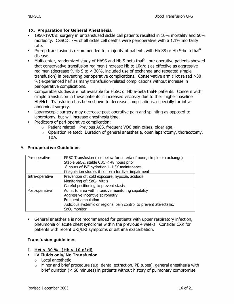
IX. Preparation for General Anesthesia 1950-1970's: surgery in untransfused sickle cell patients resulted in 10% mortality and 50%
morbidity. CSSCD: 7% of all sickle cell deaths were perioperative with a 1.1% mortality rate.
Pre-op transfusion is recommended for majority of patients with Hb SS or Hb S-beta thal0 disease.
Multicenter, randomized study of HbSS and Hb S-beta thal0 - pre-operative patients showed that conservative transfusion regimen (increase Hb to 10g/dl) as effective as aggressive regimen (decrease %Hb S to < 30%, included use of exchange and repeated simple transfusion) in preventing perioperative complications. Conservative arm (Hct raised >30 %) experienced half as many transfusion-related complications without increase in perioperative complications.
Comparable studies are not available for HbSC or Hb S-beta thal+ patients. Concern with simple transfusion in these patients is increased viscosity due to their higher baseline Hb/Hct. Transfusion has been shown to decrease complications, especially for intra-abdominal surgery.
Laparoscopic surgery may decrease post-operative pain and splinting as opposed to laporotomy, but will increase anesthesia time.
Predictors of peri-operative complication: o Patient related: Previous ACS, frequent VOC pain crises, older age. o Operation related: Duration of general anesthesia, open laparotomy, thoracotomy,
T&A.
A. Perioperative Guidelines Pre-operative PRBC Transfusion (see below for criteria of none, simple or exchange)
Stable SaO2, stable CBC < 48 hours prior 8 hours of IVF hydration 1-1.5X maintenance Coagulation studies if concern for liver impairment
Intra-operative Prevention of: cold exposure, hypoxia, acidosis. Monitoring of: Sa02, Vitals Careful positioning to prevent stasis
Post-operative Admit to area with intensive monitoring capability Aggressive incentive spirometry Frequent ambulation Judicious systemic or regional pain control to prevent atelectasis. SaO2 monitor
General anesthesia is not recommended for patients with upper respiratory infection,
pneumonia or acute chest syndrome within the previous 4 weeks. Consider CXR for patients with recent URI/LRI symptoms or asthma exacerbation.
Transfusion guidelines 1. Hct < 30 % (Hb < 10 g/dl) IV Fluids only/No Transfusion o Local anesthetic o Minor and brief procedure (e.g. dental extraction, PE tubes), general anesthesia with
brief duration (< 60 minutes) in patients without history of pulmonary compromise
Revised December 2003 16 of 21
NEPSCC Blood Transfusion CPG
Simple Transfusion: o For non-T&A, non-laparotomy, non-thoracotomy procedures o Advisable for those patients at higher risk for even minor procedures. Volume calculated to raise Hct to 30-35% should be transfused within 3 days of procedure.
Repeated simple transfusion (Elective Procedures): o Consider for elective procedures involving the airway (i.e. T&A), open laparotomy,
thoracotomy, or procedures > 90 minutes duration. o Will reduce the % Hb S to 30% without exchange transfusion. o Simple transfusion with volume calculated to increase Hct to 30% at 4 weeks and 2
weeks prior to procedure. o Admit night before procedure, If % Hb S not ~30% or if Hct <30% may give 'top-off’
transfusion. Exchange transfusion / Erythrocytapheresis (Urgent Procedures[kmr2]): o Consider for urgent procedures involving the airway, open laparotomy, thoracotomy, or
procedures > 90 minutes duration. o For elective procedures with the goal of reducing the % Hb S to 30% o Consider in those patients with prior severe ACS, chronic pulmonary disease or with
other significant risk factors 2. Hct >30 % (Hb > 10 g/dl):
o Patients with baseline Hct >30% (including patients with HbSC and Hb SβThal+, HU therapy) will be managed on a case by case basis in consultation with Pediatric Hematology, the Surgical service and Anesthesiology. These patients may not alwaysrequire t ansfusion.
r IV fluids/no transfusion o Local anesthetic o Minor and brief procedure (e.g. dental extraction, PE tubes, general anesthesia with
brief duration (< 60 minutes) in patients without pulmonary compromise Simple transfusion not advised Exchange transfusion / Erythrocytapheresis o Consider for elective procedures involving the airway (i.e. T&A), open laparotomy,
thoracotomy, or other procedures requiring extended general anesthesia. Especially advisable for those patients with prior severe ACS, chronic pulmonary disease or with other risk factors
Revised December 2003 17 of 21

NEPSCC Blood Transfusion CPG
References Adams et al. Erythrocytapheresis can reduce iron overload and prevent the need to chelation therapy in chronically transfused pediatric patients. Journal o Pediatric Hematology/Oncology. 1996; 18(1): 46-50. f
f t
f
f
American Academy of Pediatrics. Red Book, 2003. Ballas. Iron overload is a determinant of morbidity and mortality in adult patients with sickle cell disease. Seminars in Hematology. 2001; 38(1- S1): 30-36. Griffin TC, Buchanan GR Elective surgery in children with sickle cell disease without preoperative blood transfusion. J Pediatr Surg. 1993 May;28(5):681-5. Dzik. Non-infectious serious hazards of transfusion. Blood Bulletin 2002; 5(1). Files et al. Longitudinal changes in ferritin during chronic transfusion: A report from the stroke prevention trial in sickle cell anemia (STOP). Journal o Pedia ric Hematology/Oncology. 2002; 24(4): 284-290. Harmatz et al. Severity of iron overload in patients with sickle cell disease receiving chronic red blood cell transfusion therapy. Blood. 2000; 96(1): 76-79. Hillard at al. Erythrocytapheresis limits iron accumulation in chronically transfused sickle cell patients. AmericanJournal o Hematology. 1998; 59: 28-35. Infections Risks of Blood Transfusion. Blood Bulletin 2001; 4(2). Janes et al. Automated red cell exchange in sickle cell disease. Br J Haematology 1997; 92(2): 256-258. Koshy et al. Surgery and anesthesia in sickle cell disease. Blood 1995; 86(10): 3676-3684. Kushner, Porter and Olivieri. Secondary iron overload. Hematology. 2001; 47-61. Lane et al. Guidelines for comprehensive care, carepaths, and protocols for management of acute and chronic complications of sickle cell disease. Sickle cell disease care consortium 2001. Neumayr et al. Surgery in patients with Hemoglobin SC disease: Report of the preoperative transfusion in sickle cell disease study group. AJH 1998; 57: 101-108. NHLBI. The Management o Sickle Cell Disease. NIH guidelines, 4th edition (NIH publication 02-2117) Rev. July 2002. Accessed September 2003. O’Grady et al. Guidelines for the prevention of intravascular catheter-related infections. Pediatrics 2002; 110(5): e5. Ohne-Frempong et al. Indications for red blood cell transfusion in sickle cell disease. Sem Hematol 2001; 38(Suppl 1): 5 – 13. Olivieri. Progression of iron overload in sickle cell disease. Seminars in Hematology. 2001; 38(1-S1): 57-62. Rosse et al. Transfusion and alloimmunization in sickle cell disease. Blood 1990; 76(7): 1432-1437. Singer et al. Erthrocytapheresis for chronically transfused children with sickle cell disease: an effective method for maintaining a low hemoglobin S level and reducing iron overload. J Clin Apheresis 1994: 14: 122-125. Vichinsky et al. Alloimmunization in sickle cell anemia and transfusion of racially unmatched blood. NEJM 1990; 322(23): 1617-1621.
Revised December 2003 18 of 21

NEPSCC Blood Transfusion CPG
Vichinsky et al. A comparison of conservative and aggressive transfusion regimen in the perioperative management of sickle cell disease. NEJM 1995; 333(4): 206-213. Wales et al. Acute chest syndrome after abdominal surgery in children with sickle cell disease: Is a laproscopic approach better? J Pediatr Surg 2001; 36(5): 718-721. Wayne et al. Transfusion management of sickle cell disease. Blood 1993; 81(5): 1109 – 1123.
Revised December 2003 19 of 21

NEPSCC Blood Transfusion CPG
APPENDIX A: Boston Medical Center Protocol for Pediatric Manual Exchange Transfusion
• Equipment: o 20 - 60mL syringes o Empty blood banks (may be obtained from Blood Bank) o (2) 3-way stopcock o IV tubing o Extension tubing o Pall filter (if not pre-filtered) o CVR monitor o O2 saturation monitor o Thermometer, stethoscope, BP cuff o Blood warmer (if needed)
• Procedure o VS including HR, BP, RR, O2 saturation, and temperature before start of
procedure and q 15 minutes during procedure. o Attach primed extension tubing with three-way stopcock and large syringe to
end of lumen of line and attach to empty blood bag. Attach via a central stopcock to a second extension set. The second extension set will have stopcocks and large syringes for both the NS and PRBCs to be infused. Consider heparinizing the NS (1mg/mL).
o Withdraw patient whole blood into discard bag. Infuse NS and donor PRBCs administer exchange in aliquots prescribed.
Revised December 2003 20 of 21

NEPSCC Blood Transfusion CPG
Appendix B Ports and Related Supplies for Apheresis (Currently in use at Hasbro Children’s Hospital)
PORTS: All are made by Arrow International 4 different types/sizes of double lumen ports are made by Arrow but ONLY ONE is designed
for High Velocity use. # AP 06535 : High Velocity port. Very bulky to inset. Best to use in adolescents and adults
only. Would strongly recommend ruling out underlying hypercoaguability, and to start low dose coumadin shortly after placement.
# AP 06527/AP # 06528/ AP# 06530 ( Also known as AP# 01517/ AP # 01518/ AP#
01519) These ports in smaller kids with some degree of success. They tend to clot off or even break with high velocity use. There is a diaphragm in the center of these ports, which shifts side to side during the Apheresis procedure, and this can cause problems with the flow rates.
NEEDLES: Arrow Non-Coring Needles, 16G or 14G ¾” or 1” Depending on the child. Use the smallest
size that works on the patient. When using these needles in the ports other than the high velocity ports, the life of the port
is shortened considerably. Arrow literature states that the 14G needle has a puncture life of 100 ; a 16G Needle is 400. These numbers refer to a high velocity port ( AP# 06535) only. In a smaller port, port life is reduced by half.
EXTENSION SETS: Arrow: W21711 is an extension set with features designed to aid in Apheresis. It has a 9mm
stopcock attached. Normal stopcocks are 7mm. Tubing is designated for high pressure flows and is less likely to collapse and therefore
decrease flow rates during the procedure. Luer lock requires less manipulation close to child’s chest to tighten.
Contact information: Arrow International: 1-800-821-2737 www.arrowintl.com
Revised December 2003 21 of 21