block-it inducible pol ii mir rnai expression vector kit with emgfp · user guide for research use...
TRANSCRIPT

user guide
For Research Use Only. Not intended for any animal or human therapeutic or diagnostic use.
BLOCK-iT™ Inducible Pol II miR RNAi Expression Vector Kit with EmGFP Gateway®-adapted expression system for expression of microRNA (miRNA) in mammalian cells under control of a tetracycline-regulated Pol II promoter
Catalog number K4939-00
Revision date 16 April 2012 Publication Part number A10295
MAN0000685

ii

iii
Contents Expression Clone Generation for Experienced Users ...................................................................................... v Kit Contents and Storage ................................................................................................................................... vii Accessory Products ............................................................................................................................................. xii
Introduction ................................................................................................................................. 1 Overview ................................................................................................................................................................. 1 Using miRNA for RNAi Analysis ....................................................................................................................... 5 BLOCK-iT™ Inducible Pol II miR RNAi Expression Vector with EmGFP Kit ............................................... 9 The T-REx™ System .............................................................................................................................................. 13
Methods ..................................................................................................................................... 16 Designing the Single-Stranded DNA Oligos .................................................................................................... 16 Generating the Double-Stranded Oligo ............................................................................................................ 21 Performing the Ligation Reaction ...................................................................................................................... 26 Transforming One Shot® TOP10 Competent E. coli ........................................................................................ 28 Analyzing Transformants ................................................................................................................................... 29 Chaining pre-miRNAs ........................................................................................................................................ 32 Removing EmGFP Coding Sequence ................................................................................................................ 34 Using the pT-REx™-DEST30 Vector ................................................................................................................... 35 Transferring the Pre-miRNA Expression Cassette to Destination Vectors .................................................. 37 Performing the Rapid BP/LR Recombination Reaction ................................................................................. 38 General Considerations for Transfection and Regulated Expression ........................................................... 41 Using pcDNA™6/TR ........................................................................................................................................... 45 Generating a TetR-Expressing Host Cell Line ................................................................................................. 47 Transfecting Cells ................................................................................................................................................ 49 Detecting Fluorescence ....................................................................................................................................... 52 Generating a Stable Inducible miRNA Expressing Cell Line ........................................................................ 53 Troubleshooting ................................................................................................................................................... 55
Appendix .................................................................................................................................... 60 Blasticidin .............................................................................................................................................................. 60 Recipes ................................................................................................................................................................... 61 Map of pcDNA™6.2-GW/ EmGFP-miR ............................................................................................................ 62 Features of pcDNA™6.2-GW/EmGFP-miR ...................................................................................................... 64 Map of pcDNA™1.2/V5-GW/lacZ .................................................................................................................... 65 Map of pT-REx™-DEST30 .................................................................................................................................... 66 Features of the pT-REx™-DEST30 Vector .......................................................................................................... 67 Map of pT-REx/GW-30/lacZ ............................................................................................................................. 68 Map of pcDNA™6/TR Vector............................................................................................................................. 69 Features of pcDNA™6/TR Vector ...................................................................................................................... 70 Technical Support ................................................................................................................................................ 71 Purchaser Notification ........................................................................................................................................ 72 Gateway® Clone Distribution Policy ................................................................................................................. 73 References ............................................................................................................................................................. 74

iv

v
Expression Clone Generation for Experienced Users
Introduction This quick reference sheet is provided for experienced users of the BLOCK-iT™
Inducible Pol II miR RNAi Expression Vector Kit. If you are performing the annealing, cloning, or transformation procedures for the first time, follow the detailed protocols provided in the manual.
Step Action
Design single-stranded DNA oligos
Follow the guidelines on pages 16 to design single-stranded DNA oligos encoding the pre-miRNA of interest.
Anneal the single-stranded oligos to generate a ds oligo
1. Set up the following annealing reaction. 200 μM top strand oligo 5 μL 200 μM bottom strand oligo 5 μL 10X Oligo Annealing Buffer 2 μL DNase/RNase-free water 8 μL Total volume 20 μL
2. Heat the reaction mixture at 95°C for 4 minutes. 3. Remove the sample and set on the laboratory bench. Allow the reaction to cool
to room temperature for 5–10 minutes. 4. Spin down the sample in a microcentrifuge for 5 seconds. Mix gently. 5. Dilute the ds oligo mixture 5,000-fold by performing serial
100-fold and 50-fold dilutions: the first into DNase/RNase-free water and the second into 1X Oligo Annealing Buffer. Final concentration is 10 nM.
Clone the ds oligo into pcDNA™6.2-GW/EmGFP-miR
1. Set up the following ligation reaction. 5X Ligation Buffer 4 μL pcDNA™6.2-GW/EmGFP-miR (5 ng/μL), linearized 2 μL ds oligo (10 nM; 1:5,000 dilution) 4 μL DNase/RNase-Free water 9 μL T4 DNA Ligase (1 U/μL) 1 μL Total volume 20 μL
2. Mix reaction well and incubate for 5 minutes at room temperature. 3. Place reaction on ice and proceed to transform E. coli, below.
Transform One Shot® TOP10 Chemically Competent E. coli
1. Add 2 μL of the ligation reaction into a vial of One Shot® TOP10 chemically competent E. coli and mix gently.
2. Incubate on ice for 5 to 30 minutes. 3. Heat-shock the cells for 30 seconds at 42°C without shaking. Immediately
transfer the tube to ice. 4. Add 250 μL of room temperature S.O.C. Medium. 5. Incubate at 37°C for 1 hour with shaking. 6. Spread 20–100 μL of bacterial culture on a pre-warmed LB agar plate containing
50 μg/mL spectinomycin and incubate overnight at 37°C. 7. Isolate DNA using PureLink® HQ Mini Plasmid Purification Kit or equivalent
and verify positive clones by sequence analysis.
Continued on next page
vi
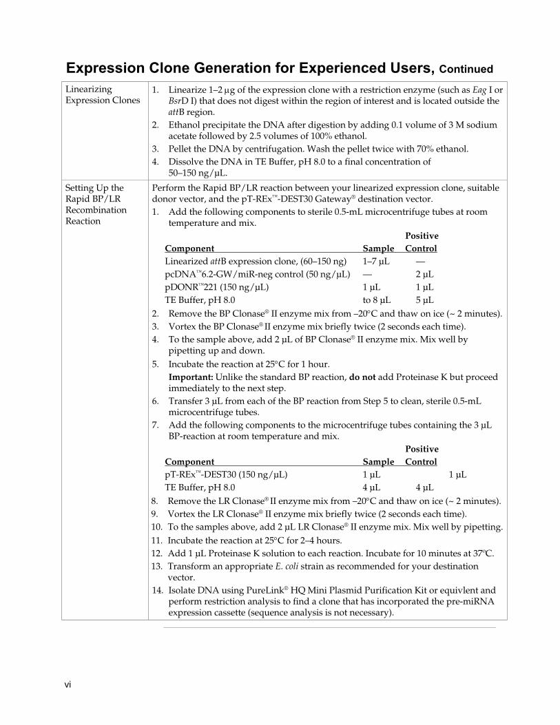
Expression Clone Generation for Experienced Users, Continued Linearizing Expression Clones
1. Linearize 1–2 μg of the expression clone with a restriction enzyme (such as Eag I or BsrD I) that does not digest within the region of interest and is located outside the attB region.
2. Ethanol precipitate the DNA after digestion by adding 0.1 volume of 3 M sodium acetate followed by 2.5 volumes of 100% ethanol.
3. Pellet the DNA by centrifugation. Wash the pellet twice with 70% ethanol. 4. Dissolve the DNA in TE Buffer, pH 8.0 to a final concentration of
50–150 ng/μL.
Setting Up the Rapid BP/LR Recombination Reaction
Perform the Rapid BP/LR reaction between your linearized expression clone, suitable donor vector, and the pT-REx™-DEST30 Gateway® destination vector. 1. Add the following components to sterile 0.5-mL microcentrifuge tubes at room
temperature and mix. Positive Component Sample Control Linearized attB expression clone, (60–150 ng) 1–7 μL — pcDNA™6.2-GW/miR-neg control (50 ng/μL) — 2 μL pDONR™221 (150 ng/μL) 1 μL 1 μL TE Buffer, pH 8.0 to 8 μL 5 μL
2. Remove the BP Clonase® II enzyme mix from –20°C and thaw on ice (~ 2 minutes).3. Vortex the BP Clonase® II enzyme mix briefly twice (2 seconds each time). 4. To the sample above, add 2 μL of BP Clonase® II enzyme mix. Mix well by
pipetting up and down. 5. Incubate the reaction at 25°C for 1 hour.
Important: Unlike the standard BP reaction, do not add Proteinase K but proceed immediately to the next step.
6. Transfer 3 μL from each of the BP reaction from Step 5 to clean, sterile 0.5-mL microcentrifuge tubes.
7. Add the following components to the microcentrifuge tubes containing the 3 μL BP-reaction at room temperature and mix.
Positive Component Sample Control pT-REx™-DEST30 (150 ng/μL) 1 μL 1 μL TE Buffer, pH 8.0 4 μL 4 μL
8. Remove the LR Clonase® II enzyme mix from –20°C and thaw on ice (~ 2 minutes).9. Vortex the LR Clonase® II enzyme mix briefly twice (2 seconds each time). 10. To the samples above, add 2 μL LR Clonase® II enzyme mix. Mix well by pipetting. 11. Incubate the reaction at 25°C for 2–4 hours. 12. Add 1 μL Proteinase K solution to each reaction. Incubate for 10 minutes at 37ºC. 13. Transform an appropriate E. coli strain as recommended for your destination
vector. 14. Isolate DNA using PureLink® HQ Mini Plasmid Purification Kit or equivlent and
perform restriction analysis to find a clone that has incorporated the pre-miRNA expression cassette (sequence analysis is not necessary).

vii
Kit Contents and Storage
Shipping/Storage The BLOCK-iT™ Inducible Pol II miR RNAi Expression Vector with EmGFP Kit is shipped as described below. Upon receipt, store each item as detailed below. For more detailed information about the reagents supplied in the BLOCK-iT™ Pol II miR RNAi Expression Vector Kits, refer to the BLOCK-iT™ Pol II miR RNAi Expression Vector Kit manual.
Box Component Shipping Storage
1 BLOCK-iT™ Pol II miR RNAi Expression Vector with EmGFP
Dry ice
–20°C
2 pT-REx™-DEST30 Gateway® Vector • pT-REx™-DEST30 Gateway® vector • pT-REx/GW-30/lacZ control vector
Dry ice –20°C –20°C
3 T-REx™ Support Kit • pcDNA™6/TR • Tetracycline, 10 mg/mL
Dry ice –20°C –20°C
4–6 One Shot® Stbl™3 Chemically Competent E. coli (3 kits) Dry ice –80°C
7 pDONR™221 Vector Room temperature
–20°C
8 Blasticidin Room temperature
–20°C
9 Gateway® BP Clonase® II • Gateway® BP Clonase® II Plus Enzyme Mix • Proteinase K solution • 30% PEG8000/30 mM MgCl2 Solution • pEXP7-tet Positive Control
Dry ice
–20°C
10 Gateway® LR Clonase® II Plus • Gateway® LR Clonase® II Plus Enzyme Mix • Proteinase K solution
Dry ice –20°C
Kit Contents
The BLOCK-iT™ Inducible Pol II miR RNAi Expression Vector with EmGFP Kit contains the following components listed in the table below. For a detailed description of the contents of the BLOCK-iT™ Inducible Pol II miR RNAi Expression Vector with EmGFP Kit, see next page..
Components Quantity
BLOCK-iT™ Pol II MiR RNAi Expression Vector with EmGFP 1 kit One Shot® TOP10 Chemically Competent E. coli 3 × 20 reactionsBP Clonase® II 20 reactions LR Clonase® II 20 reactions T-REx™ Regulatory Module 1 box pT-REx™-DEST30 Gateway® Vector 1 box pDONR™221 Gateway® Vector 6 μg Blasticidin 50 mg
Continued on next page

viii
Kit Contents and Storage, Continued
BLOCK-iT™ Inducible Pol II miR RNAi Expression Vector Reagents
The following reagents are included with the BLOCK-iT™ Inducible Pol II miR RNAi Expression Vector with EmGFP. Store the reagents at –20°C.
Reagent Composition Quantity
pcDNA™6.2-GW/EmGFP-miR, linearized 5 ng/μL in: 10 mM Tris-HCl, pH 8.0 1 mM EDTA, pH 8.0
4 × 10 μL
10X Oligo Annealing Buffer 100 mM Tris-HCl, pH 8.0 10 mM EDTA, pH 8.0 1 M NaCl
250 μL
DNase/RNase-Free Water — 3 × 1.5 mL
5X Ligation Buffer 250 mM Tris-HCl, pH 7.6 50 mM MgCl2 5 mM ATP 5 mM DTT 25% (w/v) polyethylene glycol-8000
80 μL
T4 DNA Ligase 1 (Weiss) U/μL in 10 mM Tris-HCl, pH 7.5 50 mM KCl 1 mM DTT 50% (v/v) glycerol
20 μL
EmGFP forward sequencing primer 100 ng/μL in TE Buffer, pH 8.0 20 μL
miRNA reverse sequencing primer 100 ng/μL in TE Buffer, pH 8.0 20 μL
miR-lacZ positive double-stranded (ds) control oligo
50 μM in 1X Oligo Annealing Buffer 4 μL
pcDNA™1.2/V5-GW/lacZ control plasmid 500 ng/μL in TE Buffer, pH 8.0 20 μL
pcDNA™6.2-GW/EmGFP-miR-neg control plasmid
500 ng/μL in TE Buffer, pH 8.0 20 μL
Unit Definition of T4 DNA Ligase
One (Weiss) unit of T4 DNA Ligase catalyzes the exchange of 1 nmol 32P-labeled pyrophosphate into [γ/β-32P]ATP in 20 minutes at 37°C (Weiss et al., 1968). One unit is equal to approximately 300 cohesive-end ligation units.
Product Use
For research use only. Not intended for any human or animal diagnostic or therapeutic uses.
Continued on next page

ix
Kit Contents and Storage, Continued
Primer Sequences The table below provides the sequence and the quantity supplied of the primers included in the kit.
Primer Sequence Quantity
EmGFP forward sequencing primer
5′-GGCATGGACGAGCTGTACAA-3′ 2 μg (323 pmol)
miRNA reverse sequencing primer
5′-CTCTAGATCAACCACTTTGT-3′ 2 μg (332 pmol)
LacZ Control Oligo Sequences
The sequences of the miR-lacZ positive ds control oligo are listed below. The miR-lacZ positive ds control oligo are annealed and are supplied in the kit as a 50 μM double-stranded oligo. The miR-lacZ positive ds control oligo needs to be reannealed and diluted 5000-fold to 10 nM before use in the ligation reaction.
lacZ DNA Oligo Sequence
Top strand 5’-TGCTGAAATCGCTGATTTGTGTAGTCGTTTTGGCCACTGACTGACGACTACACATCAGCGATTT-3’
Bottom strand 5’-CCTGAAATCGCTGATGTGTAGTCGTCAGTCAGTGGCCAAAACGACTACACAAATCAGCGATTTC-3’
One Shot® TOP10 Chemically Competent E. coli
The following reagents are included in the One Shot® TOP10 Chemically Competent E. coli kit. Transformation efficiency is ≥1 × 10⁹ cfu/μg plasmid DNA. Store at –80°C.
Reagent Composition Quantity
S.O.C. Medium (may be stored at +4°C or room temperature)
2% Tryptone 0.5% Yeast Extract 10 mM NaCl 2.5 mM KCl 10 mM MgCl2 10 mM MgSO4 20 mM glucose
3 × 6 mL
TOP10 cells — 3 × 21 × 50 μL
pUC19 Control DNA 10 pg/μL in 5 mM Tris-HCl, 0.5 mM EDTA, pH 8
3 × 50 μL
Genotype of TOP10 Cells
F- mcrA Δ(mrr-hsdRMS-mcrBC) φ80lacZΔM15 ΔlacX74 recA1 araD139 Δ(ara-leu)7697 galU galK rpsL (StrR) endA1 nupG
Continued on next page

x
Kit Contents and Storage, Continued
BP Clonase® II The following reagents are included with the BP Clonase® II. Store at –20°C for up to 6 months. For long-term storage, store at –80°C.
Reagent Composition Quantity
Gateway® BP Clonase® II Enzyme Mix Proprietary 40 μL
Proteinase K Solution 2 μg/μL in: 10 mM Tris-HCl, pH 7.5 20 mM CaCl2
50% Glycerol
40 μL
PEG Solution 30% PEG 8000 30 mM MgCl2
1mL
pEXP7-tet Positive Control 50 ng/μL in TE Buffer, pH 8.0
20 μL
LR Clonase® II The following reagents are included with the LR Clonase® II. Store at –20°C for
up to 6 months. For long-term storage, store at –80°C.
Reagent Composition Quantity
Gateway® LR Clonase® II Enzyme Mix Proprietary 40 μL
Proteinase K Solution 2 μg/μL in: 10 mM Tris-HCl, pH 7.5 20 mM CaCl2
50% Glycerol
40 μL
pENTR™-gus Positive Control 50 ng/μL in TE Buffer, pH 8.0
20 μL
Note: The pENTR™-gus control included with the LR Clonase® II Enzyme Mix may be used as a positive control for the LR recombination reaction only.
Inducing and Selection Agents
In addition to the vector provided, the T-REx™ Regulatory Module also includes the following inducing and selection agents. Store the tetracycline at –20°C protected from exposure to light. Store the blasticidin powder at +4°C.
Reagent Quantity and Concentration Comments
Tetracycline 1 mL (10mg/mL) Inducing agent
Blasticidin 50 mg, powder Selection agent for pcDNA™6/TR plasmid
Continued on next page
xi
Kit Contents and Storage, Continued
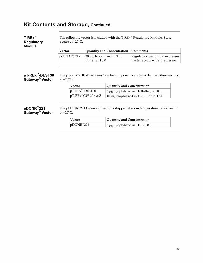
T-REx™ Regulatory Module
The following vector is included with the T-REx™ Regulatory Module. Store vector at –20°C.
Vector Quantity and Concentration Comments
pcDNA™6/TR© 20 μg, lyophilized in TE Buffer, pH 8.0
Regulatory vector that expresses the tetracycline (Tet) repressor
pT-REx™-DEST30 Gateway® Vector
The pT-REx™-DEST Gateway® vector components are listed below. Store vectors at –20°C.
Vector Quantity and Concentration
pT-REx™-DEST30 6 μg, lyophilized in TE Buffer, pH 8.0 pT-REx/GW-30/lacZ 10 μg, lyophilized in TE Buffer, pH 8.0
pDONR™221 Gateway® Vector
The pDONR™221 Gateway® vector is shipped at room temperature. Store vector at –20°C.
Vector Quantity and Concentration
pDONR™221 6 μg, lyophilized in TE, pH 8.0

xii
Accessory Products
Accessory Products
Some of the reagents supplied in the BLOCK-iT™ Inducible Pol II miR RNAi Expression Vector Kits as well as other products suitable for use with the kit are available separately from Life Technologies. Ordering information is provided below.
Item Quantity Catalog no.
BLOCK-iT™ Pol II miR RNAi Expression Vector Kit with EmGFP
20 reactions K4936-00
BLOCK-iT™ Lentiviral Pol II miR RNAi Expression System with EmGFP
20 reactions K4938-00
BLOCK-iT™ HiPerform™ Lentiviral Pol II miR RNAi Expression System with EmGFP
20 reactions K4934-00
BLOCK-iT™ Pol II miR-XXXX Validated miRNA DuoPak (XXXX=gene symbol)
10 μg V49300-01 through V49300-53
Gateway® pT-Rex™-DEST30 Vector 6 μg 12301-016
pcDNA™6/TR© 20 μg V1025-20
T4 DNA Ligase 100 units 500 units
15224-017 15224-025
One Shot® TOP10 Chemically Competent E. coli
10 reactions 20 reactions 40 reactions
C4040-10 C4040-03 C4040-06
Gateway® LR Clonase® II Enzyme Mix 20 reactions 100 reactions
11791-020 11791-100
Gateway® BP Clonase® II Enzyme Mix 20 reactions 100 reactions
11789-020 11789-100
pDONR™221 6 μg 12536-017
Blasticidin 50 mg R210-01
Geneticin® 1 g 5 g 20 mL (50 mg/mL) 100 mL (50 mg/mL)
11811-023 11811-031 10131-035 10131-027
Tetracycline 5 g Q100-19
PureLink® HQ Mini Plasmid Purification Kit 100 preps K2100-01
PureLink® HiPure Plasmid Midiprep Kit 25 preps K2100-04
Lipofectamine® 2000 Transfection Reagent 0.75 mL 1.5 mL
11668-027 11668-019
Lipofectamine® LTX Reagent 1.0 mL 15338-100
Continued on next page

xiii
Accessory Products, Continued
Accessory Products Continued
Item Quantity Catalog no.
Opti-MEM® I Reduced Serum Medium 100 mL 500 mL
31985-062 31985-070
Phosphate-Buffered Saline (PBS), pH 7.4 500 mL 10010-023
4% E-Gel® Starter Pak 9 gels and Base G6000-04
2% E-Gel® Starter Pak 9 gels and Base G6000-02
10 bp DNA Ladder 50 μg 10821-015
293FT Cell Line 3 × 10⁶ cells, frozen R700-07
PureLink® Quick Gel Extraction Kit 50 preps K2100-12
Spectinomycin For selection of pcDNA™6.2-GW/EmGFP-miR transformants in E. coli, you will
need to obtain spectinomycin. Spectinomycin dihydrochloride pentahydrate is available from Sigma (Catalog no. S4014). For a recipe to prepare spectinomycin for use, see Appendix, page 61.
RNAi Designer and RNAi Express
The BLOCK-iT™ RNAi Designer is an online tool to help you design and order microRNA sequences for any target gene of interest. The RNAi Designer incorporates the guidelines provided in this manual as well as other design rules into a proprietary algorithm to design microRNA sequences that are compatible for use in cloning into the BLOCK-iT™ Pol II miR RNAi Expression Vectors.
BLOCK-iT™ miR RNAi Select
Life Technologies has predesigned miR RNAi sequences, called BLOCK-iT™ miR RNAi Select, targeting >70% of the human, mouse and rat RefSeq genes.
BLOCK-iT™ miR RNAi Select provides up to four miR sequences per gene that are supplied as 8 tubes containing four top oligos and four bottom DNA oligos. Upon annealing and cloning into the BLOCK-iT™ Pol II miR RNAi Expression vector pcDNA™6.2-GW/EmGFP-miR, these oligos generate up to four different miR RNAi expression vectors directed against your gene of interest.
The resulting miR RNAi expression vectors can be transfected into cells to knock down the gene of interest, or the hairpins can be transferred into the T-REx™ inducible vector or other compatible DEST vectors (see Gateway® Destination Vectors, next page) to knock down the gene of interest in hard-to-transfect or primary cells. We guarantee that at least two out of the four miR RNAi expression vectors will result in >70% knockdown of the target gene (provided that the transfection efficiency in your experiment is at least 80%).
To order BLOCK-iT™ miR RNAi Select online using the BLOCK-iT™ RNAi Express search engine (www.lifetechnologies.com/rnaiexpress). Just enter the gene name, accession number, or keyword, and choose your desired BLOCK-iT™ miR RNAi Select.
Continued on next page
xiv
Accessory Products, Continued
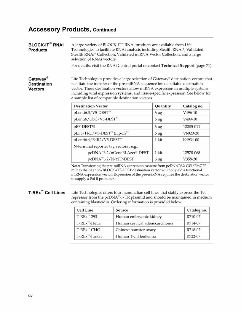
BLOCK-iT™ RNAi Products
A large variety of BLOCK-iT™ RNAi products are available from Life Technologies to facilitate RNAi analysis including Stealth RNAi®, Validated Stealth RNAi® Collection, Validated miRNA Vector Collection, and a large selection of RNAi vectors.
For details, visit the RNAi Central portal or contact Technical Support (page 71).
Gateway® Destination Vectors
Life Technologies provides a large selection of Gateway® destination vectors that facilitate the transfer of the pre-miRNA sequence into a suitable destination vector. These destination vectors allow miRNA expression in multiple systems, including viral expression systems, and tissue-specific expression. See below for a sample list of compatible destination vectors.
Destination Vector Quantity Catalog no.
pLenti6.3/V5-DEST™ 6 μg V496-10
pLenti6/UbC/V5-DEST™ 6 μg V499-10
pEF-DEST51 6 μg 12285-011
pEF5/FRT/V5-DEST™ (Flp-In™) 6 μg V6020-20
pLenti6.4/R4R2/V5-DEST™ 1 kit K4934-00
N-terminal reporter tag vectors , e.g.:
pcDNA™6.2/nGeneBLAzer®-DEST 1 kit 12578-068
pcDNA™6.2/N-YFP-DEST 6 μg V358-20
Note: Transferring the pre-miRNA expression cassette from pcDNA™6.2-GW/EmGFP-miR to the pLenti6/BLOCK-iT™-DEST destination vector will not yield a functional miRNA expression vector. Expression of the pre-miRNA requires the destination vector to supply a Pol II promoter.
T-REx™ Cell Lines Life Technologies offers four mammalian cell lines that stably express the Tet
repressor from the pcDNA™6/TR plasmid and should be maintained in medium containing blasticidin. Ordering information is provided below.
Cell Line Source Catalog no.
T-REx™-293 Human embryonic kidney R710-07
T-REx™-HeLa Human cervical adenocarcinoma R714-07
T-REx™-CHO Chinese hamster ovary R718-07
T-REx™-Jurkat Human T-c ll leukemia R722-07

1
Introduction
Overview
Introduction The BLOCK-iT™ Inducible Pol II miR RNAi Expression Vector Kit with EmGFP combines Life Technologies’ BLOCK-iT™ RNAi and T-REx™ technologies to facilitate tetracycline-regulated expression of a microRNA (miRNA) of interest from a Pol II/TO RNAi cassette for use in RNA interference (RNAi) analysis in mammalian cells. The Gateway®-adapted destination vector is designed to allow efficient, regulated expression (transient or stable) of miRNA in dividing mammalian cells. The pT-REx™-DEST30 vector is designed for use with the T-REx™ system and allows for high-level tetracycline-regulated expression of EmGFP and your miRNA of interest in mammalian cells expressing the Tet repressor. The T-REx™ System is a tetracycline-regulated mammalian expression system that uses regulatory elements from the E. coli Tn10-encoded tetracycline (Tet) resistance operon (Hillen and Berens, 1994; Hillen et al., 1983). Tetracycline regulation in the T-REx™ System is based on the binding of tetracycline to the Tet repressor and derepression of the promoter controlling expression of the gene of interest (Yao et al., 1998). For more information about the BLOCK-iT™ RNAi, T-REx™, and Gateway® technologies, see below.
Advantages of the BLOCK-iT™ Inducible Pol II miR RNAi Expression Vector Kit with EmGFP
Using the BLOCK-iT™ Inducible Pol II miR RNAi Expression Vector Kit with EmGFP for vector-based expression of miRNA provides the following advantages:
• Provides a rapid and efficient way to clone double-stranded oligonucleotide (ds oligo) duplexes encoding a desired miRNA target sequence into a vector containing a Pol II-driven expression cassette (i.e., CMV EmGFP-miR RNAi cassette) for use in RNAi analysis. The expression cassette can be easily transferred into pT-REx™-DEST30 using Gateway® recombination reaction.
• The expression construct containing the CMV/TO RNAi expression cassette may be directly transfected into mammalian cells expressing the Tet repressor to enable rapid, tetracycline-regulated screening of miRNA target sequences.
• The expression construct contains a neomycin resistance marker to allow generation of stable cell lines using Geneticin® that express the miRNA of interest upon tetracycline addition.
• Enables targeting of multiple genes or increasing knockdown of a single target gene with one construct (see Chaining of miRNAs on page 8).
• Permits visual or automated selection of cells expressing the pre-miRNA through co-cistronic expression of EmGFP
• Offers easy transfer of the pre-miRNA expression cassette into Gateway®-adapted viral expression systems or vectors driven by a variety of promoters, including tissue-specific and regulated promoters for in vivo experiments
• Permits design of predictable RNAi constructs with a high rate of success
• In conjunction with the pre-designed BLOCK-iT™ miR RNAi Select oligos, covers >70% of the human, mouse and rat RefSeq genes with a guaranteed rate of success
Continued on next page

2
Overview, Continued
Gateway® Technology
The Gateway® Technology is a universal cloning method that takes advantage of the site-specific recombination properties of bacteriophage lambda (Landy, 1989) to provide a rapid and highly efficient way to move your DNA sequence of interest (the miRNA sequence) into multiple vector systems.
To transfer your pre-miRNA expression cassette into the destination vector:
1. Generate an entry clone by performing a BP recombination reaction between the pcDNA™6.2-GW/EmGFP-miR expression clone and a suitable donor vector (pDONR™221).
2. Perform an LR recombination reaction between the resulting entry clone and a destination vector (pT-REx™-DEST30).
3. Co-transfect your expression clone and pcDNA™6/TR into the cell line of choice for tetracycline-regulated expression of EmGFP and your miRNA of interest.
For additional information about the Gateway® Technology, refer to the Gateway® Technology with Clonase® II manual which is available for downloading from www.lifetechnologies.com or by contacting Technical Support (see page 71).
Continued on next page
The BLOCK-iT™ Inducible Pol II miR RNAi Technology
The BLOCK-iT™ Pol II miR RNAi Technology is a next generation RNAi technology employing miRNA expression vectors that allow constitutive or regulated expression of knockdown cassettes driven by RNA Polymerase II (Pol II) promoters in mammalian cells. The BLOCK-iT™ Inducible Pol II miR RNAi Expression Vectors are specifically designed to allow expression of miRNA sequences and contain specific miR flanking sequences that allow proper processing of the miRNA. The expression vector design is based on the miRNA vector system developed in the laboratory of David Turner (U.S. Patent Publication No. 2004/0053876) and includes the use of endogenous murine miR-155 flanking sequences.
A variety of BLOCK-iT™ RNAi products are available from Life Technologies to facilitate RNAi analysis in mammalian and invertebrate systems. For more information about any of the BLOCK-iT™ RNAi products, see the RNAi Central application portal at www.lifetechnologies.com/rnai or contact Technical Support (see page 71).
Alternative Expression Systems
The pcDNA™6.2-GW/EmGFP-miR vector expresses EmGFP and the pre-miRNA in most mammalian cells at a high, constitutive level using the human cytomegalovirus (CMV) immediate early promoter. If different expression of the pre-miRNA is required, such as tissue-specific, regulated or lentiviral expression, the vector allows easy recombination with other suitable destination vectors using the Gateway® Technology.

3
Overview, Continued
Continued on next page
T-REx™ Technology
The T-REx™ Technology facilitates tetracycline-regulated expression of a gene and/or miRNA of interest in mammalian cells through the use of regulatory elements from the E. coli Tn10-encoded tetracycline (Tet) resistance operon (Hillen & Berens, 1994; Hillen et al., 1983). Tetracycline regulation in the T-REx™ System is based on the binding of tetracycline to the Tet repressor and derepression of the promoter controlling expression of the gene or miRNA of interest (Yao et al., 1998). In the T-REx™ System, the Tet repressor is expressed from the pcDNA™6/TR plasmid. The pT-REx™-DEST30 vector is designed for use with the T-REx™ System. This vector contains two tetracycline operator 2 (TetO2) sites within the human CMV promoter for tetracycline-regulated expression of EmGFP and your miRNA of interest (Yao et al., 1998). The TetO2 sequences serve as binding sites for 4 Tet repressor molecules (comprising two Tet repressor homodimers) and confer tetracycline-responsiveness to EmGFP and your miRNA of interest. In the absence of tetracycline, expression is repressed by the binding of Tet repressor homodimers to the TetO2 sequences. Addition of tetracycline to the cells derepresses the hybrid CMV/TetO2 promoter and allows expression of EMGFP and your miRNA of interest. The main components of the T-REx™ System include:
• An inducible expression construct to facilitate tetracycline-regulated expression of EmGFP and your miRNA of interest under the control of a hybrid promoter containing two tetracycline operator 2 (TetO2) sites.
• A regulatory expression construct that facilitates high-level, constitutive expression of the Tet repressor (TetR). In the T-REx™ System, expression of the TetR gene is controlled by the CMV promoter.
• Tetracycline for inducing expression.
For more details about the TetO2 sequences, see page 14. For more information about pcDNA™6/TR and the Tet repressor, refer to the T-REx™ System manual. This manual is available for downloading from www.lifetechnologies.com or by contacting Technical Support (see page 71).

4
Overview, Continued
Purpose of this Manual
This manual provides the following information:
• An overview of the pathway by which miRNA facilitates gene knockdown in mammalian cells.
• Guidelines to design the appropriate single-stranded oligonucleotides representing the target gene.
• Instructions to anneal the single-stranded oligonucleotides to generate a double-stranded oligonucleotide (ds oligo).
• Instructions to clone the ds oligo into the pcDNA™6.2-GW/EmGFP-miR vector, and transform the ligation reaction into competent E. coli.
• Instructions to perform a Rapid BP/LR recombination reaction with pT-REx™-DEST30 to generate a tetracycline-regulated expression clone.
• Instructions to generate stable, mammalian TetR-expressing cell lines.
• Instructions to transfect your EmGFP-miR expression clone into a mammalian TetR-expressing cell line to perform transient, tetracycline-regulated RNAi analysis.
• Guidelines to transfect your pT-REx™ expression construct into a mammalian TetR-expressing cell line and perform Geneticin® selection to generate a stable cell line for tetracycline-regulated RNAi analysis.
• Guidelines to perform the chaining reaction to enable co-cistronic expression of multiple pre-miRNAs from one construct.
• Guidelines for detection of the EmGFP expressed pT-REx expression constructs.
Important The BLOCK-iT™ Inducible Pol II miR RNAi Expression Vector Kit with EmGFP is
designed to help you generate a CMV promoter-based vector to express miRNA in mammalian cell lines for RNAi analysis. Although the kit has been designed to help you express miRNA representing a particular target sequence in the simplest, most direct fashion, use of the kit for RNAi analysis assumes that users are familiar with the principles of gene silencing, vector-based production of miRNA, transfection in mammalian systems, and cloning. We highly recommend that users possess a working knowledge of the RNAi pathway and lipid-mediated transfection.
For more information about miRNA and the RNAi pathways and expression of miRNA in mammalian cells, refer to published references (Ambros, 2004; Bartel, 2004; Boden et al., 2004; Cullen, 2004; Kim, 2005; McManus & Sharp, 2002; Zeng et al., 2002).
Refer to Molecular Biology handbooks, such as Current Protocols in Molecular Biology (Ausubel et al., 1994), if you are not familiar with the cloning steps involved.

5
Using miRNA for RNAi Analysis
Introduction RNA interference (RNAi) describes the phenomenon by which short, homologous RNA duplexes induce potent and specific inhibition of eukaryotic gene expression via the degradation of complementary messenger RNA (mRNA), and is functionally similar to the processes of post-transcriptional gene silencing (PTGS) or cosuppression in plants (Cogoni et al., 1994; Napoli et al., 1990; Smith et al., 1990; van der Krol et al., 1990) and quelling in fungi (Cogoni & Macino, 1997; Cogoni & Macino, 1999; Romano & Macino, 1992).
In plants, the PTGS response is thought to occur as a natural defense against viral infection or transposon insertion (Anandalakshmi et al., 1998; Jones et al., 1998; Li & Ding, 2001; Voinnet et al., 1999). In experimental settings, RNAi is widely used to silence genes through transfection of RNA duplexes or introduction of vector-expressed short hairpin RNA (shRNA).
The RNAi Pathway In eukaryotic organisms, dsRNA produced in vivo, introduced by pathogens, or
through research, is processed into 21–23 nucleotide double-stranded short interfering RNA duplexes (siRNA) by an enzyme called Dicer, a member of the RNase III family of double-stranded RNA-specific endonucleases (Bernstein et al., 2001; Ketting et al., 2001).
Each siRNA then incorporates into an RNA-induced silencing complex (RISC), an enzyme complex that serves to target cellular transcripts complementary to the siRNA for specific cleavage and degradation, or translational repression (Hammond et al., 2000; Nykanen et al., 2001). MicroRNAs (miRNAs) are endogenous RNAs that trigger gene silencing (Ambros, 2001; Carrington & Ambros, 2003).
miRNA Pathway MicroRNAs (miRNAs) are endogenously expressed small ssRNA sequences of
~22 nucleotides in length which naturally direct gene silencing through components shared with the RNAi pathway (Bartel, 2004). Unlike shRNAs, however, the miRNAs are found embedded, sometimes in clusters, in long primary transcripts (pri-miRNAs) of several kilobases in length containing a hairpin structure and driven by RNA Polymerase II (Lee et al., 2004), the polymerase also responsible for mRNA expression. Drosha, a nuclear RNase III, cleaves the stem-loop structure of the pri-miRNA to generate small hairpin precursor miRNAs (pre-miRNAs) which are ~70 nucleotides in length (Zeng et al., 2005). The pre-miRNAs are exported from the nucleus to the cytoplasm by exportin-5, a nuclear transport receptor(Bohnsack et al., 2004; Yi et al., 2003). Following the nuclear export, the pre-miRNAs are processed by Dicer into a ~22 nucleotides miRNA (mature miRNA) molecule, and incorporated into an miRNA-containing RNA-induced silencing complex (miRISC)(Cullen, 2004).
Continued on next page

6
Using miRNA for RNAi Analysis, Continued
Translational Repression versus Target Cleavage
The mature miRNAs regulate gene expression by mRNA cleavage (mRNA is nearly complementary to the miRNA) or translational repression (mRNA is not sufficiently complementary to the miRNA). Target cleavage can be induced artificially by altering the target or the miRNA sequence to obtain complete hybridization (Zeng et al., 2002).
In animals, most miRNAs imperfectly complement their targets and interfere with protein production without directly inducing mRNA degradation (Ambros, 2004). Nonetheless, these miRNAs are found associated with the RNAi nuclease AGO2 (Liu et al., 2004; Meister et al., 2004), and at least two miRNAs with close matches to their target sequences, particularly in their 5’ regions, have been shown to cleave cognate mRNAs (Yekta et al., 2004; Yu et al., 2005).
The engineered miRNAs produced by the BLOCK-iT™ Inducible Pol II miR RNAi Expression Vector Kit (see below) fully complement their target site and cleave the target mRNA. Sequence analysis showed that the primary cleavage site at the phosphodiester bond in the mRNA found opposite the tenth and eleventh bases of the engineered miRNA as predicted for RNAi-mediated cleavage (Elbashir et al., 2001) similar to siRNA mediated cleavage.
Using a Vector-Based System to Express Engineered miRNA
Use of siRNA (diced siRNA or synthetic siRNA) for RNAi analysis in mammalian cells is limited by their transient nature. To address this limitations, a number of groups have developed vector-based systems to facilitate expression of engineered short hairpin RNA (shRNA) sequences in mammalian cells using Pol III promoters (Brummelkamp et al., 2002; Paddison et al., 2002; Paul et al., 2002; Sui et al., 2002; Yu et al., 2002). However, the use of shRNA vectors for RNAi analysis requires the screening of large number of sequences to identify active sequences and the use of Pol III promoters limits applications such as tissue-specific expression.
To overcome limitations with siRNA and shRNA, we have developed Gateway®-adapted expression vectors that enable the expression of engineered miRNA sequences from Pol II promoters. The pcDNA™6.2-GW/EmGFP-miR expression vector facilitates the generation of an expression clone containing a ds oligo encoding a pre-miRNA sequence. The resulting expression construct may be introduced into mammalian cells for transient expression of the miRNA sequence, or stable transfectants can be generated. The BLOCK-iT™ Inducible Pol II miR RNAi Expression Vector Kit with EmGFP facilitates the easy and efficient transfer of the pre-miRNA sequences and EmGFP into the pT-REx™-DEST30 vector for tetracycline-regulated expression in the T-REx™ System. If desired, the EmGFP and pre-miRNA sequence may also be transferred into any other suitable destination vector by Gateway® recombination reactions (see page 37).
Continued on next page

7
Using miRNA for RNAi Analysis, Continued
miRNA Vector The BLOCK-iT™ Inducible Pol II miR RNAi Expression Vector Kit is supplied with pcDNA™6.2-GW/EmGFP-miR vector that allows the expression of your engineered pre-miRNA under the control of the strong, Pol II human CMV (cytomegalovirus) promoter and Herpes Simplex virus (HSV) thymidine kinase (TK) polyadenylation signal. The coding sequence of EmGFP (Emerald Green Fluorescent Protein) is incorporated into the vector such that the pre-miRNA insertion site is in the 3’ untranslated (3’UTR) region of the fluorescent protein mRNA. Addition of EmGFP allows tracking of the miRNA expression and provides strong correlation of EmGFP expression with the knockdown of the target gene by your miRNA.
Human CMV Promoter
The BLOCK-iT™ Inducible Pol II miR RNAi Expression Vector contain the human cytomegalovirus (CMV) immediate early promoter to allow high-level, constitutive miRNA expression in mammalian cells (Andersson et al., 1989; Boshart et al., 1985; Nelson et al., 1987).
We have chosen the human CMV promoter to control vector-based expression of miRNA molecules in mammalian cells for the following reasons:
• The promoter is recognized by RNA Polymerase II and controls high-level, constitutive expression of miRNA and co-cistronic reporter genes
• The promoter is active in most mammalian cell types
Note: Although highly active in most mammalian cell lines, activity of the viral CMV promoter can be down-regulated in some cell lines due to methylation (Curradi et al., 2002), histone deacetylation (Rietveld et al., 2002), or both.
Design of the Engineered Pre-miRNA
The engineered pre-miRNA sequence structure is based on the murine miR-155 sequence (Lagos-Quintana et al., 2002). The 5’ and 3’ flanking regions derived from the miR-155 transcript were inserted in the vector to preserve as much as possible of the miR-155 structure. We optimized the stem-loop structure and a 2 nucleotide internal loop results in higher knockdown rate than the 5 nucleotide /3 nucleotide internal loop found in native miR-155 molecule. An Msc I site was incorporated in the terminal loop to aid in sequence analysis. Below the changes are shown made to the native miR-155 to form an engineered pre-miRNAs directed against lacZ (targeting sequence in bold).
Continued on next page
UG| UU UUGGCC CUGAAAUCGCUGAU GUGUAGUCGUU \ |||||||||||||| ||||||||||: A GACUUUAGCGACUA--CACAUCAGCAG / AG^ UCAGUC
5’-UG| UGUGA UUGGCC CUGUUAAUGCUAAU UAGGGGUU \ |||||||||||||: ||||:||: U GACAAUUACGAUUG AUCCUCAG / 3’-G^ UCC-- UCAGUC
internal loop
terminal loop
MscI native miR-155 optimized miR-lacZ
internal loop
terminal loop

8
Using miRNA for RNAi Analysis, Continued
Structure of the Engineered Pre-miRNA
The pcDNA™6.2-GW/EmGFP-miR vectors are designed to accept engineered pre-miRNA sequences targeting your gene of interest. The engineered pre-miRNA sequence structure is based on the murine miR-155 sequence and the stem-loop structure was optimized to obtain a high knockdown rate as described on the previous page. For optimized knockdown results, we recommend that the ds oligo encoding the engineered pre-miRNA have the following structural features: • A 4 nucleotide, 5’ overhang (TGCT) complementary to the vector (required
for directional cloning) • A 5’G + short 21 nucleotide antisense sequence (mature miRNA) derived from
the target gene, followed by • A short spacer of 19 nucleotides to form the terminal loop and • A short sense target sequence with 2 nucleotides removed ( 2) to create an
internal loop • A 4 nucleotide, 5’ overhang (CAGG) complementary to the vector (required
for directional cloning) The structural features are depicted in the figure below.
For guidelines to design the oligonucleotides, refer to page 16. We recommend using Life Technologies’ RNAi Designer at www.lifetechnologies.com/rnai, an online tool to help you design and order pre-miRNA sequences for any target gene of interest.
Pre-miRNA Expression Cassette
The engineered pre-miRNA sequence is cloned into the cloning site of BLOCK-iT™
Inducible Pol II miR RNAi Expression Vector that is flanked on either side with sequences from murine miR-155 to allow proper processing of the engineered pre-miRNA sequence. The pre-miRNA sequence and adjacent miR-155 flanking regions are denoted as the pre-miRNA expression cassette and are shown below. This expression cassette is transferred between vectors during Gateway® recombination reactions.
Once the engineered pre-miRNA expression cassette is introduced into the mammalian cells for expression, the pre-miRNA forms an intramolecular stem-loop structure similar to the structure of endogenous pre-miRNA that is then processed by the endogenous Dicer enzyme into a 22 nucleotide mature miRNA. Note: The 21 nucleotides are derived from the target sequence while the 3’ most nucleotide is derived from the native miR-155 sequence.
Chaining of miRNAs
miRNAs are sometimes expressed in clusters in long primary transcripts driven by RNA Pol II (Lee et al., 2004). Our vectors support chaining of miRNAs to express them in one primary transcript, thus ensuring co-cistronic expression of multiple miRNAs. Note: Chaining should be done in the pcDNA™6/TR vector before transfer to pT-REx™-DEST30, as restriction sites present in the pT-REx™-DEST30 vector make it incompatible with the chaining protocol.
��������������� ������ ���������������
�������������������������� ������������������������
���������� ��������
!��"# ��������������� ������ ���������������
��������������������������
%��������� ��������

9
BLOCK-iT™ Inducible Pol II miR RNAi Expression Vector with EmGFP Kit
Description of the System
The BLOCK-iT™ Inducible Pol II miR RNAi Expression Vector Kit with EmGFPfacilitates the generation of an expression construct that permits tetracycline-regulated, high-level expression of a pre-miRNA in mammalian cells for RNAi analysis of a target gene. The kit contains the following major components:
• BLOCK-iT™ Pol II miR RNAi Expression Vector with EmGFP (pcDNA™6.2-GW/EmGFP-miR), pDONR™221 and pT-REx™-DEST30 Gateway® Vector for the production of a tetracycline-regulated expression clone containing a double-stranded oligonucleotide (ds oligo) encoding a pre-miRNA (oligos have to be ordered separately). The pcDNA™6.2-GW/EmGFP-miR vector is supplied linearized with 4-nucleotide 5′ overhangs on each strand to facilitate directional cloning of the ds oligo insert.
• pT-REx™-DEST30 Gateway® vector for high-leveltetracycline-regulated expression of your gene of interest in mammalian cells expressing the Tet repressor and that are designed for use with the T-REx™ System.
• Gateway® Clonase® II Enzyme Mixes for transfer of miRNA expression cassette into an inducible expression vector.
• T-REx™ Regulatory Module for high-level tetracycline-regulated expression of the miRNA of interest in mammalian cells expressing the Tet repressor.
• Tetracycline for induction of miRNA expression and blasticidin for selection of Tet repressor-expressing cells.
Controls The BLOCK-iT™ Inducible Pol II miR RNAi Expression Vector Kit with EmGFP
also includes a negative control plasmid, a ds positive control oligo , and T-REx™ cell tranfection and expression positive control. • The pcDNA™6.2-GW/EmGFP-miR-neg control plasmid contains an insert
that can form a hairpin structure that is processed into mature miRNA, but is predicted not to target any known vertebrate gene. Thus, this plasmid serves as a suitable negative control for pre-miRNA experiments with pcDNA™6.2-GW/EmGFP-miR expression vectors. The neg control sequence without 5’ overhangs is shown below (for map, see page 62):
5’-GAAATGTACTGCGCGTGGAGACGTTTTGGCCACTGACTGACGTCTCCACGCAGTACATTT-3’
• The miR-lacZ positive double-stranded (ds) control oligo serves as a positive control during the miRNA expression vector generation. Use this oligo to generate a pcDNA™6.2-GW/EmGFP-miR-lacZ expression clone.
• Co-transfecting the resulting pcDNA™6.2-GW/EmGFP-miR-lacZ expression clone targeting the lacZ gene and the pcDNA™1.2/V5-GW/lacZ reporter plasmid supplied with the kit into mammalian cells provide a means to assess the RNAi response in your cell line by assaying for knockdown of β-galactosidase.
• pT-REx™/GW-30/lacZ is provided as a positive control vector for mammalian cell transfection and expression (see page 68 for map) and may be used to optimize recombinant protein expression levels in your cell line. This vector allows expression of β-galactosidase which may be detected by Western blot or functional assay.
Continued on next page

10
BLOCK-iT™ Inducible Pol II miR RNAi Expression Vector with EmGFP Kit, Continued
Generating an miRNA Expression Vector Using the Kit
Using the reagents supplied in the BLOCK-iT™ Inducible Pol II miR RNAi Expression Vector Kits, you will perform the following steps to generate an expression clone in pcDNA™6.2-GW/EmGFP-miR:
1. Design and synthesize two complementary single-stranded DNA oligonucleotides, with one encoding the miRNA of interest. Alternatively, order BLOCK-iT™ miR RNAi Select oligos targeting your gene(s) (see page xiv).
2. Anneal the single-stranded oligonucleotides to generate a double-stranded oligo (ds oligo).
3. Clone the ds oligo into the linearized pcDNA™6.2-GW/EmGFP-miR vector.
4. Transform the ligation reaction into One Shot® TOP10 chemically competent E. coli and select for spectinomycin-resistant transformants.
5. Use the pcDNA™6.2-GW/EmGFP-miR expression construct for transient RNAi analysis in mammalian cells and perform a Gateway® recombination reaction with pT-REx™-DEST30 Gateway® destination vector and pDONR™221 to generate a tetracycline-regulated EmGFP-miRNA expression vector.
Features of the pcDNA™6.2-GW/EmGFP-miR Vector
The pcDNA™6.2-GW/EmGFP-miR vectors contain the following features:
• Human CMV promoter for high-level, constitutive expression of the miRNA from a RNA Polymerase II-dependent promoter
• 5’ and 3’ miR flanking regions for formation of an engineered pre-miRNA
• Cloning site containing 4-nucleotide 5′ overhangs on each DNA strand for directional cloning of the ds oligo encoding the pre-miRNA of interest
• Two recombination sites, attB1 and attB2 sites, flanking the pre-miRNA expression cassette for recombinational cloning of the pre-miRNA expression cassette into a Gateway® destination vector
• Herpes Simplex virus (HSV) thymidine kinase (TK) polyadenylation signal for termination and polyadenylation of the transcript
• Spectinomycin resistance gene for selection in E. coli
• pUC origin for high-copy maintenance of the plasmid in E. coli
• Blasticidin resistance gene for selection in E. coli and mammalian cells to generate cell lines stably expressing the miRNA
• pcDNA™6.2-GW/EmGFP-miR also contains an EmGFP coding sequence for co-cistronic expression with the pre-miRNA.
Continued on next page

11
BLOCK-iT™ Inducible Pol II miR RNAi Expression Vector with EmGFP, Continued
Green Fluorescent Protein (GFP)
The BLOCK-iT™ Inducible Pol II miR RNAi Expression Vector with EmGFP contains the Emerald Green Fluorescent Protein (EmGFP) derived from Aequorea victoria GFP within the pre-miRNA expression cassette. Green Fluorescent Protein (GFP) is a naturally occurring bioluminescent protein derived from the jellyfish Aequorea victoria (Shimomura et al., 1962). GFP emits fluorescence upon excitation, and the gene encoding GFP contains all of the necessary information for posttranslational synthesis of the luminescent protein. GFP is often used as a molecular beacon because it requires no species-specific cofactors for function, and the fluorescence is easily detected using fluorescence microscopy and standard filter sets. GFP can function as a reporter gene downstream of a promoter of interest and upstream of one or more pre-miRNAs. After transferring the pre-miRNA expression cassette into pT-REx™-DEST30, you may transfect tet repressor-expressing cells for tetracycline regulated expression of the EmGFP protein and miRNA, allowing you to visually track the cells in which knockdown is occurring.
GFP and Spectral Variants
Modifications have been made to the wild-type GFP to enhance its expression in mammalian systems. These modifications include amino acid substitutions that correspond to the codon preference for mammalian use, and mutations that increase the brightness of the fluorescence signal, resulting in “enhanced” GFP (Zhang et al., 1996). Mutations have also arisen or have been introduced into GFP that further enhance and shift the spectral properties of GFP such that these proteins will emit fluorescent color variations (reviewed in Tsien, 1998). The Emerald GFP (EmGFP) is such a variant of enhanced GFP.
Note We have observed reduced EmGFP expression from miRNA-containing vectors
due to processing of the transcripts. In most cases, EmGFP expression should remain detectable.
EmGFP The EmGFP variant has been described in a published review (Tsien, 1998) and is
summarized below. The amino acid mutations are represented by the single letter abbreviation for the amino acid in the consensus GFP sequence, followed by the codon number and the single letter amino acid abbreviation for the substituted amino acid.
Fluorescent Protein GFP Mutations*
EmGFP S65T, S72A, N149K, M153T, I167T
*Mutations listed are as described in the literature. When examining the actual sequence, the vector codon numbering starts at the first amino acid after the initiation methionine of the fluorescent protein, so that mutations appear to be increased by one position. For example, the S65T mutation actually occurs in codon 66 of EmGFP.
Continued on next page

12
BLOCK-iT™ Inducible Pol II miR RNAi Expression Vector with EmGFP, Continued
EmGFP Fluorescence
The EmGFP from the pcDNA™6.2-GW/EmGFP-miR expression vector has the following excitation and emission wavelengths, as published in the literature (Tsien, 1998):
Excitation (nm) Emission (nm)
487 509
Filter Sets for Detecting EmGFP Fluorescence
The EmGFP can be detected with standard FITC filter sets. However, for optimal detection of the fluorescence signal, you may use a filter set which is optimized for detection within the excitation and emission ranges for the fluorescent protein. The filter set for fluorescence microscopy and the manufacturer are listed below:
Filter Set Manufacturer
Omega XF100 Omega (www.omegafilters.com)

13
The T-REx™ System
Description of the T-REx™ System
In the T-REx™ System, expression of your gene of interest is repressed in the absence of tetracycline and induced in the presence of tetracycline (Yao et al., 1998). Unlike other tetracycline-regulated systems which use hybrid regulatory molecules and viral transactivation domains (Gossen and Bujard, 1992), the T-REx™ System uses only regulatory elements from the native Tet operon (Yao et al., 1998). Tetracycline-regulated gene expression in the T-REx™ System more closely resembles the regulation of the native bacterial tet operon (Hillen and Berens, 1994; Hillen et al., 1983) and avoids the potentially toxic effects of viral transactivation domains observed in some mammalian cell lines. The major component of the T-REx™ System that is provided in this kit, is the inducible pT-REx™-DEST30 expression plasmid. Expression of your miRNA of interest and EmGFP from pT-REx™-DEST30 is controlled by the strong CMV promoter (Andersson et al., 1989; Boshart et al., 1985; Nelson et al., 1987) into which 2 copies of the tet operator 2 (TetO2) sequence have been inserted in tandem. The TetO2 sequences consist of 2 copies of the 19 nucleotide sequence, 5´-TCCCTATCAGTGATAGAGA-3´ separated by a 2 base pair spacer (Hillen and Berens, 1994; Hillen et al., 1983). Each 19 nucleotide TetO2 sequence serves as the binding site for 2 molecules of the Tet repressor. For details on the Tet operator sequences and the specific features of each inducible expression vector, please refer to the manual for this vector. The second major component of the T-REx™ System is the pcDNA™6/TR regulatory vector which expresses high levels of the TetR gene (Postle et al., 1984) under the control of the human CMV promoter (see Appendix, pages 69 for more information about pcDNA™6/TR© and the TetR gene). Both T-REx™ vectors can be introduced into mammalian host cells by standard transfection methods.
Continued on next page
Features of the pT-REx™-DEST30 Vector
The pT-REx™-DEST vector contains the following elements. For a map of pT-REx™-DEST30, see page 66. • Hybrid promoter consisting of human cytomegalovirus immediate-early (CMV)
promoter/enhancer and tetracycline operator 2 (TetO2) sites for tetracycline-regulated expression in a wide range of mammalian cells
• Two recombination sites, attR1 and attR2, downstream of the CMV promoter for recombinational cloning of the gene of interest from an entry clone
• Chloramphenicol resistance gene located between the two attR sites for counterselection
• ccdB gene located between the two attR sites for negative selection • SV40 polyadenylation sequence for proper termination and processing of the
transcript • f1 intergenic region for production of single-strand DNA in F plasmid-
containing E. coli • SV40 early promoter and origin for expression of the neomycin resistance gene
and stable propagation of the plasmid in mammalian hosts expressing the SV40 large T antigen
• Neomycin resistance gene for selection of stable cell lines • The ampicillin (bla) resistance gene for selection in E. coli • The pUC origin for high copy replication and plasmid maintenance in E. coli

14
The T-REx™ System, Continued
Continued on next page
Tet Operator Sequences
The promoters of bacterial tet genes contain two types of operator sequences, O1
and O2, that serve as high affinity binding sites for the Tet repressor (Hillen and Berens, 1994; Hillen et al., 1983). Each O1 and O2 site binds to one Tet repressor homodimer. While Tet repressor homodimers bind to both tet operators with high affinity, studies have shown that the affinity of the Tet repressor homodimer for O2 is three- to five-fold higher than for O1 (Hillen and Berens, 1994).
Tet operators have been incorporated into heterologous eukaryotic promoters to allow tetracycline-regulated gene expression in mammalian cells (Gossen and Bujard, 1992; Yao et al., 1998). In the T-REx™ System, two copies of the O2 operator sequence (TetO2) are inserted into the strong CMV promoter of the pT-REx™-DEST vectors to allow regulated expression of your gene of interest by tetracycline. For more information about tet operators, refer to Hillen and Berens (1994).
Yao et al. (1998) have recently demonstrated that the location of tet operator sequences in relation to the TATA box of a heterologous promoter is critical to the function of the tet operator. Regulation by tetracycline is only conferred upon a heterologous promoter by proper spacing of the TetO2 sequences from the TATA box (Yao et al., 1998). For this reason, the first nucleotide of the TetO2 operator sequence has been placed 10 nucleotides after the last nucleotide of the TATA element in the CMV promoter for the pT-REx™-DEST vectors.
In other tetracycline-regulated systems, the TetO2 sequences are located upstream of the TATA element in the promoter of the inducible expression vector (Gossen and Bujard, 1992). These systems differ substantially from the T-REx™ System in that they use regulatory molecules composed of the Tet repressor fused to a viral transactivation domain. The presence of viral transactivation domains appears to overcome the requirement for specific positioning of the TetO2 sequences in relation to the TATA box of the heterologous promoter. However, the presence of viral transactivation domains has been found to have deleterious effects in some mammalian cell lines.
Mechanism of Repression
In the absence of tetracycline, the Tet repressor forms a homodimer that binds with extremely high affinity to each TetO2 sequence in the promoter of the inducible expression vector (Hillen and Berens, 1994). The 2 TetO2 sites in the promoter of the inducible expression vector serve as binding sites for 4 molecules (or 2 homodimers) of the Tet repressor. The affinity of the Tet repressor for the tet operator is KB = 2 × 10¹¹ M-1 (as measured under physiological conditions), where KB is the binding constant (Hillen and Berens, 1994). Binding of the Tet repressor homodimers to the TetO2 sequences represses transcription of your gene of interest. Upon addition, tetracycline binds with high affinity to each Tet repressor homodimer in a 1:1 stoichiometry and causes a conformational change in the repressor that renders it unable to bind to the Tet operator. The association constant, KA, of tetracycline for the Tet repressor is 3 × 10⁹ M-1 (Hillen and Berens, 1994). The Tet repressor:tetracycline complex then dissociates from the Tet operator and allows induction of transcription from the gene of interest.

15
The T-REx™ System, Continued The figure illustrates the components of the T-REx™ System.
�������� �������& ���� ����
���& ��������������
�������� ����
���&
���� ����
���& ��������������
���� ���& ���& ��������������
� ��� �����
�� ��������������� ����������������������������� �
�����������������
�� ��� ������������������������� ����������!���"������������������������������#�����$�������%�� ����������������%��������������
&� '��� �������$���� �(���������������������� �����������
)� *�����%�������������� ����������� �� �������� ���� ���� �%������� $����� ����������������� �����"�����$ ������������������ �����������������%���������������
!'��������(���������)
!'����������������)
����������������
���� �������� ����
��������
����
����
����#�*+
16
Methods
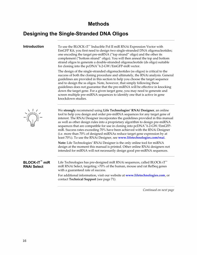
Designing the Single-Stranded DNA Oligos
Introduction To use the BLOCK-iT™ Inducible Pol II miR RNAi Expression Vector with EmGFP Kit, you first need to design two single-stranded DNA oligonucleotides; one encoding the target pre-miRNA (“top strand” oligo) and the other its complement (“bottom strand” oligo). You will then anneal the top and bottom strand oligos to generate a double-stranded oligonucleotide (ds oligo) suitable for cloning into the pcDNA™6.2-GW/EmGFP-miR vector.
The design of the single-stranded oligonucleotides (ss oligos) is critical to the success of both the cloning procedure and ultimately, the RNAi analysis. General guidelines are provided in this section to help you choose the target sequence and to design the ss oligos. Note, however, that simply following these guidelines does not guarantee that the pre-miRNA will be effective in knocking down the target gene. For a given target gene, you may need to generate and screen multiple pre-miRNA sequences to identify one that is active in gene knockdown studies.
We strongly recommend using Life Technologies’ RNAi Designer, an online tool to help you design and order pre-miRNA sequences for any target gene of interest. The RNAi Designer incorporates the guidelines provided in this manual as well as other design rules into a proprietary algorithm to design pre-miRNA sequences that are compatible for use in cloning into pcDNA™6.2-GW/EmGFP-miR. Success rates exceeding 70% have been achieved with the RNAi Designer (i.e. more than 70% of designed miRNAs reduce target gene expression by at least 70%). To use the RNAi Designer, see www.lifetechnologies.com/rnai.
Note: Life Technologies’ RNAi Designer is the only online tool for miRNA design at the moment this manual is printed. Other online RNAi designers not intended for miRNA will not necessarily design good pre-miRNA sequences.
BLOCK-iT™ miR RNAi Select
Life Technologies has pre-designed miR RNAi sequences, called BLOCK-iT™
miR RNAi Select, targeting >70% of the human, mouse and rat RefSeq genes with a guaranteed rate of success.
For additional information, visit our website at www.lifetechnologies.com, or contact Technical Support (see page 71).
Continued on next page
��
��
���
��

17
Designing the Single-Stranded DNA Oligos, Continued
Features of Pre-miRNA Insert
When designing the oligos encoding the pre-miRNA, consider that a pre-miRNA insert contains the following features (from 5’ to 3’ end):
• 5 nucleotides (TGCTG) derived from the endogenous miR-155, an endogenous murine miRNA that is the basis of the miRNA vector system developed in the laboratory of David Turner (Chung et al., 2006). This also provides a four nucleotide 5’ overhang, compatible with a 4 nucleotide overhang in the provided linearized pcDNA™6.2-GW/EmGFP-miR to clone the double-stranded oligo.
• Reverse complement of the 21-nucleotide target sequence (mature miRNA sequence). When transcribed, this is the core sequence that will target your gene of interest, and therefore needs to be antisense to the targeted messenger RNA.
• 19 nucleotides derived from miR-155 to form a terminal loop with an engineered Msc I site to aid in sequence analysis.
• Nucleotides 1–8 and 11–21 of the sense target sequence. Note that nucleotides 9 and 10 are removed to form a short internal loop in the mature miRNA, which results in more efficient knockdown.
• 4 nucleotides derived from endogenous miR-155. This also constitutes the four nucleotide 5’ overhang, compatible with a 4 nucleotide overhang in the provided linearized pcDNA™6.2-GW/EmGFP-miR to clone the double-stranded oligo.
Upon transcription, the mature miRNA sequence and its complement form a stem of the pre-miRNA with a short internal loop, separated by a larger terminal loop. The folded pre-miRNA structure of miR-lacZ is shown below (lacZ targeting sequence in bold)
Continued on next page
UG| UU UUGGCC CUGAAAUCGCUGAU GUGUAGUCGUU \ |||||||||||||| ||||||||||: A GACUUUAGCGACUA CACAUCAGCAG / AG^ -- UCAGUC

18
Designing the Single-Stranded DNA Oligos, Continued
Choosing the Target Sequence
When performing RNAi analysis on a particular gene, your choice of target sequence can significantly affect the degree of gene knockdown observed. We recommend following the guidelines below when choosing your target sequence. These are general recommendations only; exceptions may occur.
Length: The target sequence should be 21 nucleotides in length.
Complexity:
• Make sure that the target sequence does not contain runs of more than three of the same nucleotide.
• Choose a sequence with low to moderate GC content (~30–50% GC content is suggested).
• Do not choose a target sequence that is a known site for RNA-protein interaction.
• Avoid the following restriction sites, which may be used for optional, advanced features later.
Restriction site Sequence Advanced Feature Page
Msc I TGGCCA Alternate sequencing protocol 30
BamH I GGATCC miRNA chaining 32
Bgl II AGATCT miRNA chaining 32
Sal I GTCGAC miRNA chaining 32
Xho I CTCGAG miRNA chaining 32
Dra I TTTAAA Removal of EmGFP 34
Homology: Make sure that the target sequence does not contain significant homology to other genes as this can increase off-target RNAi effects.
Orientation: Choose a target sequence encoding the sense sequence of the target mRNA.
Generating the Top Oligo Sequence
To generate the top oligo sequence, combine these elements (from 5’ end to 3’ end):
1. 5’ TGCTG
2. Reverse complement of the 21-nucleotide sense target sequence. This is the Mature miRNA Sequence.
3. GTTTTGGCCACTGACTGAC (terminal loop).
4. Nucleotides 1–8 (5’-3’) of sense target sequence.
5. Nucleotides 11–21 (5’-3’) of sense target sequence.
Continued on next page

19
Designing the Single-Stranded DNA Oligos, Continued
Generating the Bottom Oligo Sequence
To generate the bottom oligo sequence, perform the following steps:
1. Remove 5’ TGCT from top oligo sequence (new sequence starts with G).
2. Take the reverse complement of the sequence from step 1.
3. Add CCTG to the 5’ end of the sequence from step 2.
Note • We recommend using Life Technologies’ RNAi Designer at
www.lifetechnologies.com/rnai, which automatically applies the design rules, and produces a high rate of knockdown success.
• It is not necessary to add 5′ phosphates to your single stranded oligos during synthesis. The phosphate groups necessary for ligation are present in the linearized pcDNA™6.2-GW/EmGFP-miR.
Example of ss Oligo Design
The diagram below illustrates the required features of the top strand and bottom strand single-stranded oligos as discussed in this section. This particular example lists the sequences of top and bottom strand oligos encoding an miRNA targeting the lacZ gene. These ss oligos were annealed to generate the miR-lacZ positive ds control oligo supplied in the kit.
We generally order unpurified, desalted single-stranded oligos using Life Technologies’ custom primer synthesis service (see www.lifetechnologies.com for more information) The ss oligos obtained anneal efficiently and provide optimal cloning results. Note however, that depending on which supplier you use, the purity and quality of the ss oligos may vary. If you obtain variable annealing and cloning results using unpurified, desalted oligos, you may want to order oligos that are HPLC or PAGE-purified.
Continued on next page
������������� ��
���������������������������������������������������������������������
���������������������������������������������������������������������
�����������������������������������������������
��������������������������������� ������� �� �
� ������������!����"���
��������)������
$����������)������������������#����"���
��������� ��������� ����������������������������#����"����
�
������
������������������������������������������������������������������������������������������������������������������������������������������
)�������
�������������� ��������������� ��
��
��
���
��

20
Designing the Single-Stranded DNA Oligos, Continued
Cloning Site and Recombination Region of pcDNA™6.2-GW/EmGFP-miR
Use the diagram below to help you design suitable DNA oligonucleotides to clone into pcDNA™6.2-GW/EmGFP-miR after annealing. Note the following features in the diagram below: • The pcDNA™6.2-GW/EmGFP-miR vector is supplied linearized between
nucleotides 1518 and 1519. The linearized vector contains 4 nucleotide overhangs derived from miR-155 sequences. Note that the annealed double-stranded (ds) oligo must contain specific 4 nucleotide 5′ overhangs on each strand as indicated.
• The light shaded region corresponds to DNA sequences that are transferred from the initial pre-miRNA expression vector into the Gateway® destination vector (e.g., pT-REx™-DEST30 Gateway® Vector) following recombination. The dark shaded region represents the EmGFP coding sequence. Note: Following recombination with a Gateway® destination vector, the resulting expression clone will contain a pre-miRNA expression cassette consisting of the EmGFP coding sequence, 5’ miR flanking region, miRNA sequence, and the 3’ miR flanking region.
Note: The complete sequence of pcDNA™6.2-GW/EmGFP-miR is available for downloading from our website (www.lifetechnologies.com) or by contacting Technical Support (see page 71). For a map of pcDNA™6.2-GW/EmGFP-miR, see page 62.
$��% %�%�&#������
$�'� ����
()*+���,��������������� � ����� � ��
���- ���
�.
+���� ���������� � � ���� ������
()*+
����������������������� � ����� � �� ����� �%/� �0����0��� ���� ��������-�
��
�..
�
��.
� #����� ���1��������� &#����� ���1���������
���
�.
�
�.
��
�2
�.
�&
�3
4�
5
6&&
63
�6
47
�������������
()*+���������������
������������������������������������������������������������������������������������������������������������������������
������������������������������������������������������������������������������������������������������������������������
������������������������������������������������������������������������������������������������������������������������
��������������������������������������������������������������������������������������������� �������������������������� ������������������
��������������������������������������������������������������������������������������������������������������������
������������������������������������������������������������������������������������������������������
������������������������������������������������������������������������������������������������������������������������
������������������������������������������������������������������������������������������������������������������������
21
Generating the Double-Stranded Oligo
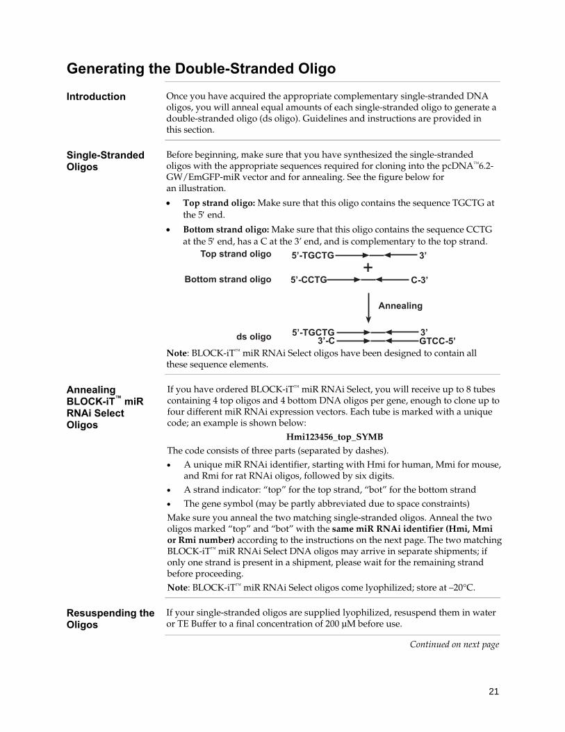
Introduction Once you have acquired the appropriate complementary single-stranded DNA oligos, you will anneal equal amounts of each single-stranded oligo to generate a double-stranded oligo (ds oligo). Guidelines and instructions are provided in this section.
Single-Stranded Oligos
Before beginning, make sure that you have synthesized the single-stranded oligos with the appropriate sequences required for cloning into the pcDNA™6.2-GW/EmGFP-miR vector and for annealing. See the figure below for an illustration.
• Top strand oligo: Make sure that this oligo contains the sequence TGCTG at the 5′ end.
• Bottom strand oligo: Make sure that this oligo contains the sequence CCTG at the 5′ end, has a C at the 3’ end, and is complementary to the top strand.
Note: BLOCK-iT™ miR RNAi Select oligos have been designed to contain all these sequence elements.
Annealing BLOCK-iT™ miR RNAi Select Oligos
If you have ordered BLOCK-iT™ miR RNAi Select, you will receive up to 8 tubes containing 4 top oligos and 4 bottom DNA oligos per gene, enough to clone up to four different miR RNAi expression vectors. Each tube is marked with a unique code; an example is shown below:
Hmi123456_top_SYMB The code consists of three parts (separated by dashes). • A unique miR RNAi identifier, starting with Hmi for human, Mmi for mouse,
and Rmi for rat RNAi oligos, followed by six digits. • A strand indicator: “top” for the top strand, “bot” for the bottom strand • The gene symbol (may be partly abbreviated due to space constraints) Make sure you anneal the two matching single-stranded oligos. Anneal the two oligos marked “top” and “bot” with the same miR RNAi identifier (Hmi, Mmi or Rmi number) according to the instructions on the next page. The two matching BLOCK-iT™ miR RNAi Select DNA oligos may arrive in separate shipments; if only one strand is present in a shipment, please wait for the remaining strand before proceeding. Note: BLOCK-iT™ miR RNAi Select oligos come lyophilized; store at –20°C.
Resuspending the Oligos
If your single-stranded oligos are supplied lyophilized, resuspend them in water or TE Buffer to a final concentration of 200 μM before use.
Continued on next page
8
������
��������)������
$����������)������
)�������
��,�����
��,����
��,���������,��
�,%�
%�,�
%�
%�

22
Generating the Double-Stranded Oligo, Continued
Amount of DNA Oligo to Anneal
You will anneal equal amounts of the top and bottom strand oligos to generate the ds oligos. We perform the annealing reaction at a final single-stranded oligo concentration of 50 μM. Annealing at concentrations below 5 μM significantly reduce the efficiency. Note that the annealing step is not 100% efficient.
Re-annealing LacZ2.1 Control Oligo
If you plan to use the miR-lacZ positive ds control oligo in the ligation reaction, make sure to re-anneal it along with the other oligos as described on the next page. Since the miR-lacZ positive ds control oligo already comes at a concentration of 50 μM in 1X Oligo Annealing Buffer, re-anneal the miR-lacZ positive ds control oligo without further dilution.
Materials Needed • Your “top strand” single-stranded oligo (200 μM in water or TE Buffer)
• Your “bottom strand” single-stranded oligo (200 μM in water or TE Buffer)
• 50 μM stock of miR-lacZ positive ds control oligo (thaw on ice)
• 10X Oligo Annealing Buffer (supplied with the kit)
• DNase/RNase-Free Water (supplied with the kit)
• 0.5-mL sterile microcentrifuge tubes
• 95°C water bath or heat block
Setting up the Annealing Reaction
Follow this procedure to set up the annealing reaction. Note that the final concentration of the oligo mixture is 50 μM.
1. In a 0.5-mL sterile microcentrifuge tube, set up the following annealing reaction at room temperature.
Reagent Quantity
Top strand DNA oligo (200 μM) 5 μL
Bottom strand DNA oligo (200 μM) 5 μL
10X Oligo Annealing Buffer 2 μL
DNase/RNase-Free Water 8 μL
Total volume 20 μL
2. If re-annealing the miR-lacZ positive ds control oligo, centrifuge its tube
briefly (~5 seconds), and transfer contents to a separate 0.5-mL sterile microcentrifuge tube.
Continued on next page

23
Generating the Double-Stranded Oligo, Continued
Annealing Procedure
Follow this procedure to anneal your single-stranded oligos to generate the ds oligo.
1. Incubate the tubes from the previous section (Setting up the Annealing Reaction) at 95°C for 4 minutes.
2. Remove the tube containing the annealing reaction from the water bath or the heat block and set on your laboratory bench.
3. Allow the reaction mixture to cool to room temperature for 5–10 minutes. The single-stranded oligos will anneal during this time.
4. Place the sample in a microcentrifuge and centrifuge briefly (~5 seconds). Mix gently.
5. Remove 1 μL of the annealing mixture and dilute the ds oligo as directed in Diluting the ds Oligo, below.
6. Store the remainder of the 50 μM ds oligo mixture at –20°C (stable for at least a year).
Diluting the ds Oligo
To clone your ds oligo or miR-lacZ positive ds control oligo into pcDNA™6.2-GW/EmGFP-miR, you must dilute the 50 μM stock to a final concentration of 10 nM (i.e., 5,000-fold dilution). We generally perform 100-fold and 50-fold serial dilutions, the first into DNase/RNase-free water and the second into the Oligo Annealing Buffer supplied with the kit. Follow the procedure below to dilute the ds oligo.
1. Dilute the 50 μM ds oligo mixture (from Annealing Procedure, Step 5, above) 100-fold into DNase/RNase-free water to obtain a final concentration of 500 nM. Vortex to mix thoroughly.
50 μM ds oligo 1 μL
DNase/RNase-free water 99 μL
Total volume 100 μL
2. Dilute the 500 nM ds oligo mixture (from Step 1) 50-fold into Oligo Annealing Buffer as follows to obtain a final concentration of 10 nM.
500 nM ds oligo 1 μL
10X Oligo Annealing Buffer 5 μL
DNase/RNase-free water 44 μL
Total volume 50 μL
3. Vortex to mix thoroughly. Store the remaining 500 nM ds oligo stock at –20°C.
4. Aliquot the 10 nM ds oligo stock and store at –20°C.
Continued on next page

24
Generating the Double-Stranded Oligo, Continued
Important The undiluted ds oligos are 5,000-fold more concentrated than the working concentration. When performing the dilutions, be careful not to cross-contaminate the different ds oligo stocks. Remember to wear gloves and change pipette tips after every manipulation.
Storing the ds Oligo
Once you have diluted your ds oligo, you should have three stocks of annealed ds oligo. Use each stock as follows:
• 50 μM ds oligo (undiluted): Use this stock for long-term storage, and to prepare new diluted ds oligo stocks if existing stocks become denatured or cross-contaminated.
• 500 nM ds oligo (100-fold dilution): Use this stock for gel analysis (see Checking the Integrity of the ds Oligo, below).
• 10 nM ds oligo (5,000-fold dilution): Use this stock for cloning (see Ligation Procedure, page 26). This stock is not suitable for long-term storage.
Store the three ds oligo stocks at –20°C.
Important When using the diluted ds oligo stock solutions (i.e., 100-fold or 5,000-fold
diluted stocks), thaw the solutions on ice. Do not heat or allow the ds oligo solutions to reach greater than room temperature as this causes the ds oligos to melt. The concentration of the oligos in the diluted solutions is not high enough to permit re-annealing and instead favors the formation of intramolecular hairpin structures. These intramolecular hairpin structures will not clone into pcDNA™6.2-GW/EmGFP-miR.
If your diluted ds oligo stock solution is heated, discard the ds oligo solution and prepare new diluted stocks using the procedure on the previous page.
Note: If the 50 μM ds oligo solution (undiluted stock) becomes heated, the oligos are sufficiently concentrated and may be re-annealed following the annealing procedure on page 23.
Checking the Integrity of the ds Oligo
You may verify the integrity of your annealed ds oligo using agarose gel electrophoresis, if desired. We suggest running an aliquot of the annealed ds oligo (5 μL of the 500 nM stock) and comparing it to an aliquot of each starting single-stranded oligo (dilute the 200 μM stock 400-fold to 500 nM; use 5 μL for gel analysis). Be sure to include an appropriate molecular weight standard. We generally use the following gel and molecular weight standard:
• Agarose gel: 4% E-Gel® (Life Technologies, Catalog no. G5000-04)
• Molecular weight standard: 10 bp DNA Ladder (Life Technologies, Catalog no. 10821-015)
Note: 4% E-Gel® resolves these fragments much better than regular 4% agarose gels.
Continued on next page

25
Generating the Double-Stranded Oligo, Continued
What You Should See
When analyzing an aliquot of the annealed ds oligo reaction by agarose gel electrophoresis, we generally see the following:
• A detectable higher molecular weight band representing annealed ds oligo running around 60–70 bp.
• A faintly detectable lower molecular weight band representing unannealed oligos that form hairpins, running at around 30–35 bp.
For an example of expected results obtained from agarose gel analysis, see the next page. If the band representing ds oligo is weak or if you do not see a band, see Troubleshooting, page 55 for tips to troubleshoot your annealing reaction.
Example of Expected Results
In this experiment, lacZ control oligos were annealed (50 μM final concentration) using the reagents supplied in the kit and following the procedure on page 23 to generate the lacZ ds control oligo. The annealing reaction was diluted 100-fold in water to a concentration of 500 nM. Aliquots of the diluted ds oligo (5 μL; 2.5 pmol) and the corresponding single-stranded oligos (mixed but not annealed; 2.5 pmol per oligo) were analyzed on a 4% E-Gel®.
Results: The lacZ oligo annealing reaction shows a clearly detectable, higher molecular weight band that differs in size from each component single-stranded oligo. Remaining unannealed ss oligos are also weakly detectable.
Note: The agarose gel is non-denaturing; therefore, the single-stranded oligos do not resolve at the expected size due to formation of secondary structure.
bp
100 -
60 - 50 - 40 - 30 - 20 -
1 2 Lane 1. single stranded lacZ oligos Lane 2. annealed lacZ oligos
ss oligo
ds oligo

26
Performing the Ligation Reaction
Introduction Once you have generated your ds oligo and have diluted it to the appropriate concentration, you will clone the ds oligo into the pcDNA™6.2-GW/EmGFP-miR vector and transform your ligation reaction into competent TOP10 E. coli. It is important to have everything you need set up and ready to use to ensure that you obtain the best results. We suggest that you read this section and Transforming One Shot® TOP10 Competent E. coli (page 28) before beginning.
Note: If you want to perform miRNA chaining, refer to page 32.
Important You will use T4 DNA Ligase and a 5X Ligation Buffer supplied with the kit to facilitate ligation of your ds oligo with the linearized pcDNA™6.2-GW/EmGFP-miR vector. When performing the ligation reaction, note the following:
• The T4 DNA Ligase and the 5X Ligation Buffer supplied with the kit have been optimized to permit ligation of the ds oligo into the pcDNA™6.2-GW/EmGFP-miR vector in 5 minutes at room temperature. T4 DNA Ligase preparations and reaction buffers available from other manufacturers may not be appropriate for use in this application.
Note: The T4 DNA Ligase and reaction buffer supplied in the BLOCK-iT™ Inducible Pol II miR RNAi Expression Vector Kits are available separately from Life Technologies (Catalog no. 15224-017).
• Traditional ligation reactions are performed at 16°C overnight. This is not recommended for this application. Follow the ligation procedure, next page.
Amount of ds Oligo to Use
For optimal results, use approximately a 15:1 molar ratio of ds oligo insert: vector for ligation.
Ligation Controls We recommend a ligation with DNase/RNase-Free Water instead of oligo as negative control for the ligation reaction.
We recommend including the miR-lacZ positive double-stranded (ds) control oligo supplied with the kit as a positive control in your ligation experiment. The miR-lacZ positive ds control oligo is supplied as a 50 μM stock in 1X Oligo Annealing Buffer, and needs to be re-annealed and diluted 5000-fold before use in a ligation reaction (see pagexx). See page xx for the sequence of each strand of the lacZ ds control oligo.
Note: Once you have cloned the lacZ ds control oligo into pcDNA™6.2-GW/EmGFP-miR, you may use the resulting expression clone as a positive control for the RNAi response in your mammalian cell line. Simply co-transfect the expression clone and the pcDNA™1.2/V5-GW/lacZ reporter plasmid supplied with the kit into your mammalian cell line and assay for knockdown of β-galactosidase expression.
Important Reminder: When using the 10 nM ds oligo stock solution for cloning, thaw the
solution on ice. Do not thaw the ds oligo by heating or the ds oligo duplexes may melt and form intramolecular hairpin structures. After use, return the tube to –20°C storage.
Continued on next page

27
Performing the Ligation Reaction, Continued
Materials Needed • Double-stranded oligo of interest (10 nM in 1X Oligo Annealing Buffer; thaw on ice before use)
• pcDNA™6.2-GW/EmGFP-miR, linearized (5 ng/μL, supplied with the kit; thaw on ice before use)
• 5X Ligation Buffer (supplied with the kit)
• DNase/RNase-Free Water (supplied with the kit)
• T4 DNA Ligase (1 U/μL, supplied with the kit)
Ligation Procedure
Follow the procedure below to perform the ligation reaction. If you wish to include a negative control, set up a separate ligation reaction but omit the ds oligo.
1. Set up a 20 μL ligation reaction at room temperature using the following reagents in the order shown.
Reagent Sample Positive
control Negative control
5X Ligation Buffer 4 μL 4 μL 4 μL
pcDNA™6.2-GW/EmGFP-miR, linearized (5 ng/μL) 2 μL 2 μL 2 μL
miR-ds oligo (10 nM; i.e., 1:5,000 dilution; page 23) 4 μL — —
miR-lacZ positive ds control oligo (10 nM; i.e., 1:5,000 dilution; page 23)
— 4 μL —
DNase/RNase-Free Water 9 μL 9 μL 13 μL
T4 DNA Ligase (1 U/μL) 1 μL 1 μL 1 μL
Total volume 20 μL 20 μL 20 μL
2. Mix reaction well by pipetting up and down.
Note: The presence of PEG and glycerol (supplied by the Ligation Buffer and the T4 DNA Ligase) will make the reaction mixture viscous. Be sure to mix the reaction thoroughly by pipetting up and down. Do not vortex.
3. Incubate for 5 minutes at room temperature.
Note: Extending the incubation time may result in a higher yield of colonies. Do not exceed 2 hours.
4. Place the reaction on ice and proceed to Transforming One Shot® TOP10 Competent E. coli, next page.
Note: You may store the ligation reaction at –20°C overnight.

28
Transforming One Shot® TOP10 Competent E. coli
Introduction Once you have performed the ligation reaction, transform your ligation mixture into competent E. coli. One Shot® TOP10 Chemically Competent E. coli are included with the kit to facilitate transformation. Follow the guidelines and instructions provided in this section.
Note: One Shot® TOP10 E. coli possess a transformation efficiency of 1 × 10⁹ cfu/μg DNA.
Materials Needed • Ligation reaction (from Step 4, previous page)
• One Shot® TOP10 Chemically Competent E. coli (supplied with the kit, one vial per transformation; thaw on ice immediately before use)
• S.O.C. Medium (supplied with the kit; warm to room temperature)
• pUC19 positive control (supplied with the kit, if desired)
• 42°C water bath
• LB plates containing 50 μg/mL spectinomycin (two for each transformation; warm at 37°C for 30 minutes before use). See Appendix, page 61 for recipe.
• LB plates containing 100 μg/mL ampicillin (if transforming pUC19 control)
• 37°C shaking and non-shaking incubator
Note: Low salt LB agar plates containing 100 μg/mL Blasticidin can also be used to select for transformants. Be sure to use low salt agar plates and check pH carefully for Blasticidin to work efficiently. For more information on Blasticidin and recipes, see page 60 and 61.
One Shot® TOP10 Transformation Procedure
Use this procedure to transform your ligation reaction into One Shot® TOP10 Chemically Competent E. coli. For a positive control, transform 10 pg (1 μL) of pUC19 plasmid into a vial of One Shot® TOP10 chemically competent E. coli.
1. Add 2 μL of the ligation reaction (from Step 3, previous page) into a vial of One Shot® TOP10 chemically competent E. coli and mix gently. Do not mix by pipetting up and down.
2. Incubate on ice for 5–30 minutes. Note: Longer incubations seem to have a minimal effect on transformation efficiency. The length of the incubation is at the user’s discretion.
3. Heat-shock the cells for 30 seconds at 42°C without shaking. 4. Immediately transfer the tubes to ice. 5. Add 250 μL of room temperature S.O.C. Medium. 6. Cap the tube tightly and shake horizontally (200 rpm) at 37°C for 1 hour. 7. Spread 50–200 μL from each transformation on a pre-warmed LB agar plate
containing 50 μg/mL spectinomycin and incubate overnight at 37°C. We recommend plating two different volumes to ensure that at least one plate will have well-spaced colonies. For the pUC19 control, plate 20–100 μL of the transformation reaction on LB plates containing 100 μg/mL ampicillin.
8. An efficient ligation reaction may produce several hundred colonies.
29
Analyzing Transformants
Analyzing Transformants
To analyze positive clones, we recommend that you:
1. Pick 5–10 spectinomycin-resistant colonies and culture them overnight in LB or SOB medium containing 50 μg/mL spectinomycin.
Note: Low salt LB containing 100 μg/mL Blasticidin can also be used to grow transformants. See page 61.
2. Isolate plasmid DNA using your method of choice. To obtain pure plasmid DNA for automated or manual sequencing, we recommend using the PureLink® HQ Mini Plasmid Purification Kit, or equivalent.
Sequence each pcDNA™6.2-GW/EmGFP-miR expression construct (see below) to confirm the following:
a. The presence and correct orientation of the ds oligo insert.
b. The sequence of the ds oligo insert.
Note: Because of the small size of the ds oligo insert, we do not recommend using restriction enzyme analysis to screen transformants.
Important We highly recommend sequencing positive transformants to confirm the
sequence of the ds oligo insert. When screening transformants, we find that up to 20% of the clones contain mutated inserts (generally 1 or 2 bp deletions within the ds oligo). The reason for this is not known, but may be due to triggering of repair mechanisms within E. coli as a result of the inverted repeat sequence within the ds oligo insert.
Note: Expression clones containing mutated ds oligo inserts generally elicit a poor RNAi response in mammalian cells. Identify expression clones with the correct ds oligo sequence and use these clones for your RNAi analysis.
Sequencing To facilitate sequencing of your pcDNA™6.2-GW/miR expression clones, use the
miRNA forward sequencing primer and miRNA reverse sequencing primer supplied with the kit. For pcDNA™6.2-GW/EmGFP-miR expression clones, use EmGFP forward sequencing primer and miRNA reverse sequencing primer. See the diagram on page 20 for the location of the priming sites.
Note If you download the sequence for pcDNA™6.2-GW/miR or pcDNA™6.2-
GW/EmGFP-miR from our website, note that the overhang sequences will be shown already hybridized to their complementary sequences (e.g., TGCT will be shown hybridized to ACGA and CAGG will be shown hybridized to GTCC).
Continued on next page

30
Analyzing Transformants, Continued
In some cases, you may have difficulty sequencing the ds oligo insert in your expression construct. This is because the hairpin sequence is an inverted repeat that can form secondary structure during sequencing, resulting in a drop in the sequencing signal when entering the hairpin. If you have difficulty sequencing your expression constructs, we suggest trying the following to improve your sequencing results:
• Use high-quality, purified plasmid DNA for sequencing. We recommend preparing DNA using Life Technologies’ PureLink® HQ Mini Plasmid Purification Kit.
• Add DMSO to the sequencing reaction to a final concentration of 5%.
• Increase the amount of template used in the reaction (up to twice the normal concentration).
• Standard sequencing kits typically use dITP in place of dGTP to reduce G:C compression. Other kits containing dGTP are available for sequencing G-rich and GT-rich templates. If you are using a standard commercial sequencing kit containing dITP, obtain a sequencing kit containing dGTP (e.g., dGTP BigDye® Terminator v3.0 Cycle Sequencing Ready Reaction Kit, Applied Biosystems, Catalog no. 4390229) and use a 7:1 molar ratio of dITP:dGTP in your sequencing reaction.
Alternative Sequencing Protocol
If sequencing problems persist, use an alternative sequencing protocol that employs Msc I-digestion:
1. Before sequencing, digest the vector with Msc I, which cuts once within the miRNA loop (and once elsewhere in the backbone).
2. Purify the digested product by cleanup on a miniprep column or extraction and precipitation (it is not necessary to gel purify).
3. Sequence the digested vector in independent runs with the forward and reverse primers. The reads will terminate in the middle of the Msc I site but should be very strong and clear to that point.
For an example of the sequence of the pre-miRNA insert of the positive control pcDNA™6.2-GW/ miR-lacZ after Msc I digestion see below.
BigDye® is a registered trademark of Applied Biosystems
Continued on next page
��
��
���
��

31
Analyzing Transformants, Continued
Long-Term Storage
Once you have identified the correct expression clone, be sure to purify the colony and make a glycerol stock for long-term storage. We recommend that you store a stock of plasmid DNA at –20°C.
1. Streak the original colony out for a single colony on an LB plate containing 50 μg/mL spectinomycin.
2. Isolate a single colony and inoculate into 1–2 mL of LB containing 50 μg/mL spectinomycin.
3. Grow until the culture reaches stationary phase.
4. Mix 0.85 mL of culture with 0.15 mL of sterile glycerol and transfer to a cryovial.
5. Store the glycerol stock at –80°C.
Note: Low salt LB containing 100 μg/mL Blasticidin can also be used to grow transformants.
What to Do Next Once you have obtained your pcDNA™6.2-GW/EmGFP-miR expression clone,
you have the following options:
• Transfect the expression clone directly into the mammalian cell line of interest to perform transient RNAi analysis (see Transfecting Cells, see page 49).
• Transfect the expression clone directly into the mammalian cell line of interest and isolate stable transfectants that knock-down the gene of interest constitutively (see Generating a Stable Cell Line, page 53).
• Perform miRNA chaining to express multiple pre-miRNAs from one single construct (see Chaining pre-miRNAs, page 32).
• Remove the EmGFP coding sequence from your pcDNA™6.2-GW/EmGFP-miR expression clone (see Removing EmGFP Coding Sequence, page 34).
• Perform an LR recombination reaction with your expression construct and a suitable Gateway® destination vector to generate an expression clone in an alternative backbone (see Performing the Rapid BP/LR Recombination Reaction, page 38).

32
Chaining pre-miRNAs
Introduction miRNAs are sometimes expressed in clusters in long primary transcripts driven by RNA Pol II (Lee et al., 2004). Our vectors support chaining of miRNAs to express them in one primary transcript, thus ensuring co-cistronic expression of multiple miRNAs. In the final construct, the original pattern of restriction sites is regenerated, making the construct amenable to multiples rounds of chaining. The figure below shows the principle of chaining two miRNAs, derived from two different miRNA vectors, into one miRNA expression vector. Note: Chaining together miRNAs targeting different genes usually results in slightly reduced knockdown of each gene. Chaining different miRNAs targeting the same gene or repeating one miRNA can enhance knock-down. Due to increased processing, EmGFP expression is attenuated by miRNA chaining.
Restriction Strategy
There are two possible strategies for restriction digestion:
1. A combination of BamH I and Xho I to excise the pre-miRNA insert, and Bgl II and Xho I to digest the pre-miRNA expression vector used as backbone
2. A combination of Sal I and Bgl II to excise the pre-miRNA insert, and Sal I and BamH I to digest the pre-miRNA expression vector used as backbone
Below the procedure for the first strategy is described. For the second strategy, change the restriction enzymes used in the procedure.
Before proceeding with the Procedure for Chaining, refer to the Note in Chaining of miRNAs (page 8)
Continued on next page
Dual pre-miRNA Expression Cassette 1 & 2
Bgl II
1
2 1
2
Sal IBamHI Bgl II
Xho I
BamHI Bgl II Xho I Sal I
Xho IBamHI
Sal I
pre-miRNA Expression Cassette 1
pre-miRNA Expression Cassette 2

33
Chaining pre-miRNAs, Continued
Procedure for Chaining
Below is a protocol for chaining of miRNAs.
1. Insert: Digest 2 μg pcDNA™6.2-GW/EmGFP-miR-1 with 10 units BamH I and 10 units Xho I for 2 hours at 37º C.
2. Backbone: Digest 1 μg pcDNA™6.2-GW/EmGFP-miR-2 with 10 units Bgl II and 10 units Xho I for 2 hours at 37º C.
3. Run fragments on 2% E-Gel® or other high percentage agarose gels.
4. Excise the backbone and insert fragments from the gel. Purify the fragments using the PureLink® Quick Gel Extraction Kit from Life Technologies or equivalent.
5. Ligate the purified backbone and insert fragment at a 1:4 molar ratio, using T4 DNA ligase from Life Technologies or equivalent.
6. Transform competent cells, such as E. coli. One Shot® TOP10 as described on page 28.
7. Analyze resulting clones as described on pages 29.
8. Test construct for both miRNAs by transfecting cells as described on page 49.
34
Removing EmGFP Coding Sequence
Introduction Depending on your experiment, you may not wish to express EmGFP from the pre-miRNA expression construct. You can remove the EmGFP coding sequence by Dra I digestion and self-ligation of the vector, forming a pcDNA™6.2-GW/ miR clone expressing the same pre-miRNA. This section describes a procedure for removing the EmGFP coding sequence from pcDNA™6.2-GW/EmGFP-miR.
Procedure for Removing EmGFP
Below is a protocol for removing the EmGFP Coding Sequence from pcDNA™6.2-GW/ EmGFP-miR.
1. Digest 1 μg pcDNA™6.2-GW/EmGFP-miR with 10 units Dra I for 2 hours at 37ºC.
2. Run fragments on 0.8% E-Gel® or other low percentage agarose gels.
3. Excise the vector fragment from the gel, purify the fragment. Purify the fragment using the PureLink® Quick Gel Extraction Kit from Life Technologies or equivalent.
4. Ligate the purified backbone and insert fragment at a 1:4 molar ratio, using T4 DNA ligase from Life Technologies or equivalent.
5. Transform competent cells, such as E. coli. One Shot® TOP10 as described on page 28.
6. Analyze resulting clones by restriction analysis with Dra I; no 750 bp fragment should be visible.

35
Using the pT-REx™-DEST30 Vector
Introduction Guidelines for using the pT-REx™-DEST30 vector are provided in this section.
Propagating the pT-REx™-DEST30 Vector
If you wish to propagate and maintain the pT-REx™-DEST30 vector, we recommend using Library Efficiency® DB3.1™ Competent Cells (Catalog no. 11782-018) from Life Technologies for transformation. The DB3.1™ E. coli strain is resistant to CcdB effects and can support the propagation of plasmids containing the ccdB gene.
Note: DO NOT use general E. coli cloning strains including TOP10 or DH5α™ for propagation and maintenance as these strains are sensitive to CcdB effects.
Resuspending the pT-REx™-DEST30 Vector
Before you perform the BP/LR Clonase® reaction, resuspend pT-REx™-DEST30 to 50–150 ng/μL in sterile water.
Recombination Reactions
Two recombination reactions constitute the basis of the Gateway® Technology:
BP Reaction
Facilitates recombination of an attB substrate (like a linearized attB expression clone) with an attP substrate (donor vector) to create an attL-containing entry clone. This reaction is catalyzed by BP Clonase® II enzyme mix.
LR Reaction
Facilitates recombination of an attL substrate (entry clone) with an attR substrate (destination vector) to create an attB-containing expression clone. This reaction is catalyzed by LR Clonase® II enzyme mix.
Continued on next page

36
Using the pT-REx™-DEST30 Vector, Continued
Generation of new miRNA expression clones
The two recombination reactions are both needed to transfer the pre-miRNA expression cassette from pcDNA™6.2-GW/EmGFP-miR to the pT-REx™-DEST30 destination vector.
BP Reaction
pcDNA™6.2-GW/EmGFP-miR is an expression clone that contains attB-sites and thus needs to be recombined with a attP substrate (such as pDONR™221) first to form an entry clone.
LR Reaction
The destination vectors mentioned in this section all contain attR substrates, therefore an entry clone formed by recombination of pcDNA™6.2-GW/EmGFP-miR and a donor vector like pDONR™221 can be recombined with the destination vector to form a new miRNA expression clone.
Confirming the Expression Clone
The ccdB gene mutates at a very low frequency, resulting in a very low number of false positives. True expression clones will be ampicillin-resistant and chloramphenicol-sensitive. Transformants containing a plasmid with a mutated ccdB gene will be both ampicillin- and chloramphenicol-resistant. To check your putative expression clone, test for growth on LB plates containing 30 μg/mL chloramphenicol. A true expression clone will not grow in the presence of chloramphenicol.

37
Transferring the Pre-miRNA Expression Cassette to Destination Vectors
Introduction Guidelines for transferring the pre-miRNA expression cassette to you destination
vector are provided in this section.
Compatible Destination Vectors
The various Gateway® vectors have widely different transcriptional and technical properties, which can be used to express the pre-miRNA. They offer custom promoter cloning, tissue-specific expression, regulated expression, and lentiviral transduction of the pre-miRNA. In addition, destination vectors providing N-terminal reporter genes can be used after removal of EmGFP. Below is a list of destination vectors that are compatible with the BLOCK-iT™ Pol II miR RNAi Expression Vector Kits. For more information or to order the destination vectors, refer to our website (www.lifetechnologies.com) or contact Technical Support (see page 71).
Destination Vector Quantity Catalog no.
pLenti6.3/V5-DEST™ 6 μg V496-10
pLenti6/UbC/V5-DEST™ 6 μg V499-10
pEF-DEST51 6 μg 12285-011
pEF5/FRT/V5-DEST™ (Flp-In™) 6 μg V6020-20
pLenti6.4/R4R2/V5-DEST™ 1 kit K4934-00
N-terminal reporter tag vectors , e.g.:
pcDNA™6.2/nGeneBLAzer®-DEST 1 kit 12578-068
pcDNA™6.2/N-YFP-DEST 6 μg V358-20
Note : The pLenti6.3/V5-DEST vector is also provided in the BLOCK-iT™ Lentiviral Pol II miR RNAi Expression Systems

38
Performing the Rapid BP/LR Recombination Reaction
Introduction The Rapid BP/LR protocol is used to transfer a gene from one expression clone into another destination vector in two consecutive steps—a BP reaction using a donor vector followed by an LR recombination reaction using a destination vectors without purification of the intermediate entry clone.
Using this protocol allows you to generate expression clones more rapidly than the standard BP and LR protocols. Fewer expression clones are obtained (at least 10–20% of the total number of expression clones) using the Rapid BP/LR protocol. If you wish to maximize the number of expression clones generated, do not use this protocol. Use the standard BP and LR as described in the Gateway® Technology with Clonase® II manual which is available from our website (www.lifetechnologies.com) or by contacting Technical Support (see page 71).
Important This protocol is for experienced Gateway® users. If you are unfamiliar with the
Gateway® system, refer to the Gateway® Technology with Clonase® II manual.
Positive Control We recommend using the pcDNA™6.2-GW/EmGFP miR-neg Control Plasmid
supplied with the BLOCK-iT™ Inducible Pol II miR RNAi Expression Kits as a positive control for the Rapid BP/LR protocol. Dilute the supplied negative control plasmid 1:10 in sterile water to obtain a final concentration of 50 ng/μL.
Do not use the pEXP7-tet supplied with the BP Clonase® II Enzyme Mix or pENTR™-gus supplied with the LR Clonase® II Enzyme Mix as a positive control for the rapid protocol.
Materials Needed • Expression clone
• pDONR™221 vector (resuspend to 150 ng/μL in sterile water).
• pT-REx™-DEST30 (150 ng/μL in TE Buffer, pH 8.0)
• pcDNA™6.2-GW/EmGFP miR-neg control, if desired (supplied with the kit)
• BP Clonase® II enzyme mix
• LR Clonase® II enzyme mix
• 2 μg/μL Proteinase K solution (supplied with Clonase® enzymes; thaw and keep on ice until use)
• TE Buffer, pH 8.0 (10 mM Tris-HCl, pH 8.0, 1 mM EDTA)
• Sterile 0.5-mL microcentrifuge tubes
Continued on next page
39
Performing the Rapid BP/LR Recombination Reaction, Continued
Linearizing Expression Clones
We recommend that you linearize the expression clone using Eag I or BsrD I.
1. Linearize 1–2 μg of the expression clone with a restriction enzyme (such as Eag I or BsrD I) that does not digest within the region of interest and is located outside the attB region.
2. Ethanol precipitate the DNA after digestion by adding 0.1 volume of 3 M sodium acetate followed by 2.5 volumes of 100% ethanol.
3. Pellet the DNA by centrifugation. Wash the pellet twice with 70% ethanol.
4. Dissolve the DNA in TE Buffer, pH 8.0 to a final concentration of 50–150 ng/μL.
Setting Up the Rapid BP/LR Recombination Reaction
Follow this procedure to perform the Rapid BP/LR reaction between your linearized expression clone, suitable donor vector, and regular Gateway® destination vector.
1. Add the following components to sterile 0.5-mL microcentrifuge tubes at room temperature and mix.
Component Sample Positive Control
Linearized attB expression clone, (60–150 ng) 1–7 μL —
pcDNA™6.2-GW/miR-neg control (50 ng/μL) — 2 μL
pDONR™221 vector (150 ng/μL) 1 μL 1 μL
TE Buffer, pH 8.0 to 8 μL 5 μL
2. Remove the BP Clonase® II enzyme mix from –20°C and thaw on ice
(~2 minutes). 3. Vortex the BP Clonase® II enzyme mix briefly twice (2 seconds each time). 4. To the sample above, add 2 μL of BP Clonase® II enzyme mix. Mix well by
pipetting up and down.
Reminder: Return BP Clonase® II enzyme mix to –20°C immediately after use.
5. Incubate the reaction at 25°C for 1 hour. Important: Unlike the standard BP reaction, do not add Proteinase K but proceed immediately to the next step.
6. Transfer 3 μL from each of the BP reaction from Step 5 to clean, sterile 0.5-mL microcentrifuge tubes.
Note: Save the remaining BP reaction mix at –20°C. You can transform the reaction mix into One Shot® TOP10 Chemically Competent E. coli as described on page 28 to check the efficiency of the BP reaction and will also allow you to isolate entry clones for future use.
7. Add the following components to the microcentrifuge tubes containing the 3 μL BP-reaction at room temperature and mix.
Component Sample Positive Control
pT-REx™ DEST30 Destination vector (150 ng/μL) 1 μL 1 μL
TE Buffer, pH 8.0 4 μL 4 μL
Continued on next page

40
Performing the Rapid BP/LR Recombination Reaction, Continued
Setting Up the Rapid BP/LR Recombination Reaction Continued
Protocol continued from the previous page.
8. Remove the LR Clonase® II enzyme mix from –20°C and thaw on ice (~2 minutes).
9. Vortex the LR Clonase® II enzyme mix briefly twice (2 seconds each time).
10. To the samples above, add 2 μl of LR Clonase® II enzyme mix. Mix well by pipetting up and down.
Reminder: Return LR Clonase® II enzyme mix to –20°C immediately after use.
11. Incubate the reaction at 25°C for 2–4 hours.
Note: The incubation time may be extended from 4 hours to overnight, if more colonies are required.
12. Add 1 μL of the Proteinase K solution to each reaction. Incubate for 10 minutes at 37ºC.
Note: You may store the reaction at –20°C for up to 1 week before transformation, if desired
13. Transform an appropriate E. coli strain as recommended for your destination vector.
Important: do not transform the LR recombination reaction into E. coli strains that contain the F′ episome (e.g., TOP10F′). These strains contain the ccdA gene and will prevent negative selection with the ccdB gene.
14. Isolate DNA using PureLink® HQ Mini Plasmid Purification Kit or equivlent and perform restriction analysis to find a clone that has incorporated the pre-miRNA expression cassette (sequence analysis is not necessary).
What to Do Next Once you have obtained your new expression clone, we recommend you test it by
transfecting an appropriate mammalian cell line to perform transient RNAi analysis if applicable (see Transfecting Cells, page 49). Proceed to General Considerations for Transfection and Regulated Expression (next page).

41
General Considerations for Transfection and Regulated Expression
Introduction Once you have generated your BLOCK-iT™ Inducible Pol II miR RNAi Expression
Vector entry construct, you are ready to express your miRNA of interest and to perform RNAi analysis of your target gene. This section provides general guidelines to help you design your transfection and RNAi experiment. We recommend that you read through this section before beginning.
Factors Affecting Gene Knockdown Levels
A number of factors can influence the degree to which expression of your gene of interest is reduced (i.e., gene knockdown) in an RNAi experiment including:
• Transfection efficiency (see page 49 for more information)
• Transcription rate of the target gene of interest
• Stability of the target protein
• Growth characteristics of your mammalian cell line
• Efficacy of the miRNA of interest
Take these factors into account when designing your RNAi experiments.
miRNA Expression Options
A number of options exist to express your miRNA of interest in the mammalian cell line of choice for RNAi analysis. Choose the option that best fits your needs.
Option Procedure Benefit
1 Obtain or generate a mammalian cell line that stably expresses the Tet repressor. Use this cell line as the host for the BLOCK-iT™ Inducible Pol II miR RNAi Expression Vector construct. Select for a double stable cell line, if desired.
Perform transient or stable, regulated miRNA expression experiments with multiple miRNA constructs using a cell line that consistently expresses the same amount of Tet repressor.
2 Transfer the CMV/TO RNAi cassette from BLOCK-iT™ Pol II miR RNAi Expression Vector with EmGFP into a suitable Gateway® destination vector (e.g., pLenti4/TO/V5-DEST) by BP/LR recombination to generate an expression clone.
Perform other RNAi applications (e.g., regulated miRNA expression in non-dividing mammalian cells using the pLenti4/TO/V5-DEST construct).
3 Transfect the BLOCK-iT™ Inducible Pol II miR RNAi Expression Vector construct into any non-TetR-expressing, dividing mammalian cell line. Select for a stable cell line, if desired.
Constitutively express the miRNA of interest.
Continued on next page

42
General Considerations for Transfection and Regulated Expression, Continued
Expression of Tet Repressor (TetR)
Because tetracycline-regulated miRNA expression in the BLOCK-iT™ Inducible RNAi System is based on a repression/derepression mechanism, the amount of Tet repressor that is expressed in the host cell line determines the level of transcriptional repression of the Tet operator sequences in your BLOCK-iT™ Inducible Pol II miR RNAi Expression Vector construct. Tet repressor levels need to be sufficiently high to suitably repress basal level transcription of the miRNA, thus suppressing target gene knockdown in uninduced cells. In addition, the most effective repression of basal miRNA expression is achieved when Tet repressor is present in mammalian cells prior to introduction of the BLOCK-iT™ Inducible Pol II miR RNAi Expression Vector construct. For these reasons, we recommend first generating a stable cell line expressing the Tet repressor, then using this cell line as the host for your BLOCK-iT™ Inducible Pol II miR RNAi Expression Vector expression construct (Option 2, previous page) or other suitable inducible expression construct (Option 3, previous page). This option is particularly recommended if you want to:
• Perform regulated RNAi knockdown experiments with several miRNA expression constructs in the same mammalian cell line.
• Obtain the lowest levels of basal miRNA expression (i.e., lowest levels of target gene knockdown in the absence of tetracycline)
To generate your own stable TetR-expressing cell line, see Generating a TetR-Expressing Host Cell Line, page 47. To obtain a TetR-expressing stable cell line from Life Technologies, see the Recommendation below.
Several T-REx™ cell lines that stably express the Tet repressor are available from Life Technologies (see page xx for ordering information). If you wish to assay for tetracycline-regulated expression of your gene of interest in 293, HeLa, CHO, or Jurkat cells, you may want to use one of the T-REx™ cell lines as the host for your BLOCK-iT™ Inducible Pol II miR RNAi Expression Vector entry construct.
Note: The T-REx™ cell lines stably express the Tet repressor from the pcDNA™6/TR expression plasmid. This plasmid is used to generate stable TetR-expressing cell lines in Life Technologies’ T-REx™ System. Both pLenti6/TR and pcDNA™6/TR contain the same TetR gene. For more information about the T-REx™ cell lines or pcDNA™6/TR, see our website (www.lifetechnologies.com) or contact Technical Support (see page 71).
Continued on next page
��
��
���
��

43
General Considerations for Transfection and Regulated Expression, Continued
Methods of Transfection
For established cell lines (e.g., COS, A549), consult original references or the supplier of your cell line for the optimal method of transfection. Pay particular attention to media requirements, when to pass the cells, and at what dilution to split the cells. Further information is provided in Current Protocols in Molecular Biology (Ausubel et al., 1994).
Methods for transfection include calcium phosphate (Chen & Okayama, 1987; Wigler et al., 1977), lipid-mediated (Felgner et al., 1989; Felgner & Ringold, 1989), and electroporation (Chu et al., 1987; Shigekawa & Dower, 1988). Choose the method and reagent that provides the highest efficiency transfection in your mammalian cell line. For a recommendation, see below.
For high-efficiency transfection in a broad range of mammalian cell lines, we recommend using the cationic lipid-based Lipofectamine® 2000 Reagent (Catalog no. 11668-027) available from Life Technologies (Ciccarone et al., 1999). Using Lipofectamine® 2000 to transfect plasmid DNA into eukaryotic cells offers the following advantages:
• Provides the highest transfection efficiency in many mammalian cell types.
• DNA-Lipofectamine® 2000 complexes can be added directly to cells in culture medium in the presence of serum Note: If you are using sensitive cell lines, such as HeLa, we recommend using Lipofectamine®LTX (page xii).
• Removal of complexes, medium change, or medium addition following transfection is not required, although complexes can be removed after 4–6 hours without loss of activity.
For more information on Lipofectamine® 2000 Reagent, refer to our website (www.lifetechnologies.com) or call Technical Support (see page 71).
Transient vs. Stable Expression of Your miRNA
When designing your RNAi experiment, you should consider how to assay for knockdown of the target gene. After you have transfected your BLOCK-iT™ Inducible Pol II miR RNAi Expression Vector construct into TetR-expressing mammalian cells, you may:
• Pool a heterogeneous population of cells and test for target gene knockdown after induction with tetracycline (i.e., transient knockdown). We recommend waiting for a minimum of 24–48 hours after induction before assaying for target gene knockdown to allow time for the miRNA to be expressed and processed.
• Select for stably transfected cells using Geneticin®. Cells will divide once or twice in the presence of lethal doses of Geneticin® Selective Antibiotic, so the effects of the drug take several days to become apparent. Complete selection can take from 2 to 3 weeks of growth in selective medium.
Continued on next page
��
��
���
��

44
General Considerations for Transfection and Regulated Expression, Continued
Tetracycline Tetracycline (MW = 444.4) is commonly used as a broad spectrum antibiotic and
acts to inhibit translation by blocking polypeptide chain elongation in bacteria. In the BLOCK-iT™ Inducible H1 RNAi System, tetracycline functions as an inducing agent to regulate transcription of the miRNA of interest from the H1/TO RNAi cassette. Tetracycline is supplied with the BLOCK-iT™ Inducible H1 RNAi Kits as a 10 mg/mL stock solution that is ready-to-use, but is also available separately from Life Technologies in powdered form (Catalog no. Q100-19).
Using Tetracycline To induce transcription of the miRNA of interest in mammalian cells, we generally
add tetracycline to a final concentration of 1 μg/mL in complete growth medium. If desired, you may vary the concentration of tetracycline used for induction from 0.001 to 1 μg/mL to modulate expression of the miRNA of interest.
Note: The concentrations of tetracycline used for induction in the BLOCK-iT™ Inducible H1 RNAi System are generally not high enough to be toxic to mammalian cells.
Follow the guidelines below when handling tetracycline.
• Tetracycline is light sensitive. Store the stock solution at –20°C, protected from light. Prepare medium containing tetracycline immediately before use.
• Tetracycline is toxic. Do not ingest solutions containing the drug. If handling the powdered form, do not inhale.
• Wear gloves, a laboratory coat, and safety glasses or goggles when handling tetracycline and tetracycline-containing solutions.
Tetracycline in Fetal Bovine Serum
When culturing cells in medium containing fetal bovine serum (FBS), note that many lots of FBS contain tetracycline as FBS is often isolated from cows that have been fed a diet containing tetracycline. If you culture your mammalian cells in medium containing FBS that is not reduced in tetracycline, you may observe some basal expression of your miRNA of interest in the absence of tetracycline. We generally culture our mammalian cells in medium containing FBS that may not be reduced in tetracycline, and have observed low basal expression of miRNA (as assayed by % target gene knockdown) in the absence of tetracycline. Depending on your application (e.g., if targeting a protein involved in cell viability), you may wish to culture your cells in tetracycline-tested FBS. You may obtain tetracycline-tested GIBCO® FBS from Life Technologies. Contact Technical Support (see page 71) for more information.

45
Using pcDNA™6/TR
Introduction The following section contains guidelines for maintaining and propagating the pcDNA™6/TR regulatory vector. You will use the pcDNA™6/TR vector to generate a TetR expressing host cell line.
E. coli Strain We recommend that you propagate the pcDNA6/TR© vector in the One Shot®
TOP10 Chemically Competent E. coli, included with the kit. However, you can use other E. coli strains that are recombination deficient (recA) and endonuclease A deficient (endA). Some compatible E.coliI strains are provided below:
Item Quantity Catalog no.One Shot® TOP10F´ (chemically competent cells) 21 × 50 μL C3030-03
Electrocomp™ TOP10F´ (electrocompetent cells) 5 × 80 μL C665-55
Max Efficiency® DH10B™ Chemically Competent Cells 5 × 0.2 mL 18297-010
Transformation Method
You may use any method of choice for transformation. Chemical transformation is the most convenient for many researchers. Electroporation is the most efficient and the method of choice for large plasmids.
Maintenance of Plasmids
The pcDNA™6/TR vector contains the ampicillin and blasticidin resistance genes to allow selection of the plasmid using ampicillin or blasticidin (see pages xx for more information about pcDNA™6/TR). For more information about preparation and handling of blasticidin, refer to the Appendix, page 60.
To propagate and maintain the pcDNA™6/TR plasmid, we recommend using the following procedure:
1. Resuspend the vector in 20 μL sterile water to prepare a 1 μg/μL stock solution. Store the stock solution at –20°C.
2. Use the stock solution to transform a recA, endA E. coli strain like TOP10F´, INV F´, or equivalent.
3. Select transformants on LB agar plates containing 50 to 100 μg/mL ampicillin or 50 μg/mL blasticidin in Low Salt LB (see the Appendix, page 61 for a recipe).
4. Prepare a glycerol stock of each plasmid for long-term storage (see the next page for a protocol).
Continued on next page
46
Using pcDNA6™/TR, Continued
Selection in E. coli To facilitate selection of blasticidin-resistant E. coli, the salt concentration of the medium must remain low (< 90 mM) and the pH must be 7.0. Prepare Low Salt LB broth and plates using the recipe in the Appendix, page 61.
Failure to lower the salt content of your LB medium will result in non-selection due to inhibition of the drug unless a higher concentration of blasticidin is used.
Preparing a Glycerol Stock
Once you have identified the correct clone, be sure to purify the colony and make a glycerol stock for long-term storage. It is also a good idea to keep a DNA stock of your plasmid at –20°C.
1. Streak the original colony out on an LB plate containing 50 μg/mL ampicillin or 100 μg/mL blasticidin in Low Salt LB. Incubate the plate at 37°C overnight.
2. Isolate a single colony and inoculate into 1–2 mL of LB containing 50 μg/mL ampicillin or 50 μg/mL blasticidin in Low Salt LB.
3. Grow the culture to mid-log phase (OD600 = 0.5–0.7).
4. Mix 0.85 mL of culture with 0.15 mL of sterile glycerol and transfer to a cryovial.
5. Store at –80°C.
Plasmid Preparation
Plasmid DNA for transfection into eukaryotic cells must be very clean and free from phenol and sodium chloride. Contaminants will kill the cells, and salt will interfere with lipids decreasing transfection efficiency. We recommend isolating DNA using the PureLink® HQ Mini Plasmid Purification Kit (Catalog no. K2100-01) or PureLink® HiPure Plasmid Midiprep Kit (Catalog no. K2100-04).
47
Generating a TetR-Expressing Host Cell Line
Introduction We recommend that you first create a stable cell line that expresses only the Tet repressor, then use that cell line to create a second cell line that expresses your miR RNAi from the inducible expression plasmid. To generate a TetR-expressing host cell line, transfect the pcDNA™6/TR plasmid into your mammalian line of interest. Use Blasticidin to select for a stable cell line.
T-REx™ cell lines that stably express the Tet repressor are available from Life Technologies (see page xiv for ordering information). If you wish to assay for tetracycline-inducible expression of your gene of interest in 293, HeLa, U2OS, and lurkat cells, you may want to use one of the T-REx™ cell lines as the host to establish your double stable cell line. For more information, visit www.lifetechnologies.com or contact Technical Support (see page 71).
Note Reminder: When generating a stable cell line expressing the Tet repressor, you will
want to select for clones that express the highest levels of Tet repressor to use as hosts for your inducible expression construct. Those clones that express the highest levels of Tet repressor should exhibit the most complete repression of basal transcription of your gene of interest.
Determining Antibiotic Sensitivity
To successfully generate a stable cell line expressing the Tet repressor and your miRNA, you need to determine the minimum concentration of each antibiotic (Blasticidin and Geneticin®) required to kill your untransfected host cell line. For each antibiotic, test a range of concentrations (see below) to ensure that you determine the minimum concentration necessary for your cell line. Use the protocol below to determine the minimal concentrations of Geneticin® and Blasticidin required to prevent growth of the parental cell line. Refer to the Appendix, page 60 for instructions on how to prepare and store blasticidin.
1. Plate or split a confluent plate so the cells will be approximately 25% confluent. For each antibiotic, prepare a set of 6–7 plates. Add the following concentrations of antibiotic to each plate in a set:
• For blasticidin selection, test 0, 1, 3, 5, 7.5, and 10 μg/mL blasticidin
• For Geneticin® selection, test 0, and 100–1000 μg/mL Geneticin® 2. Replenish the selective media every 3–4 days, and observe the percentage of
surviving cells. 3. Count the number of viable cells at regular intervals to determine the
appropriate concentration of antibiotic that prevents growth within 1–2 weeks after addition of the antibiotic.
Continued on next page
��
��
���
��

48
Generating a TetR-Expressing Host Cell Line, Continued
Possible Sites for Linearization of pcDNA™6/TR
To obtain stable transfectants, you may choose to linearize pcDNA™6/TR before transfection. While linearizing the vector may not improve the efficiency of transfection, it increases the chances that the vector does not integrate in a way that disrupts the Tet repressor gene or other elements necessary for expression in mammalian cells. The table below lists unique sites that may be used to linearize your construct prior to transfection. Other restriction sites are possible.
Enzyme Restriction Site (bp) Location Supplier
Bst1107 I 4470 Backbone AGS*, Fermentas, Takara
Sap I 4733 Backbone New England Biolabs
BspLU11 I 4849 Backbone Boehringer-Mannheim
Eam1105 I 5739 Ampicillin gene AGS*, Fermentas, Takara
Fsp I 5961 Ampicillin gene Many *Angewandte Gentechnologie Systeme
Selection of Stable Integrants
Once you have determined the appropriate antibiotic concentrations to use for selection, you can generate a stable cell line expressing pcDNA™6/TR and your inducible miR RNAi construct. 1. Transfect mammalian cells with pcDNA™6/TR using the desired protocol.
Remember to include a plate of untransfected cells as a negative control. 2. 24 hours after transfection, wash the cells and add fresh medium to the cells. 3. 48 hours after transfection, split the cells into fresh medium containing
blasticidin at the pre-determined concentration required for your cell line. Split the cells such that the cells are no more than 25% confluent. If the cells are too dense, the antibiotic will not kill the cells. Antibiotics work best on actively dividing cells.
4. Feed the cells with selective medium every 3–4 days until foci can be identified.
5. Pick at least 20 foci and expand them to test for tetracycline-inducible gene expression by transiently transfecting with the positive control plasmid expressing β-galactosidase. Screen for those clones that exhibit the lowest levels of basal transcription and the highest levels of β-galactosidase expression after addition of tetracycline (see below).
Maintaining TetR-Expressing Cell Lines
Once you have generated your stable TetR-expressing cell line and have verified that the cells express suitable levels of Tet repressor, we recommend the following:
• Maintain the cell line in medium containing Blasticidin
• Remember to freeze and store vials of early passage cells

49
Transfecting Cells
Introduction This section provides general guidelines to transfect your BLOCK-iT™ Inducible Pol II miR RNAi Expression Vector construct into a TetR-expressing mammalian cell line of interest to perform transient, regulated RNAi analysis. Performing transient RNAi analysis is useful to:
• Quickly test multiple miRNA sequences to a particular target gene
• Quickly screen for an RNAi response in your mammalian cell line
If you want to generate a stable cell line expressing the miRNA of interest, see the next section.
Factors Affecting Gene Knockdown Levels
A number of factors can influence the degree to which expression of your gene of interest is reduced (i.e., gene knockdown) in an RNAi experiment including:
• Transfection efficiency
• Transcription rate of the target gene of interest
• Stability of the target protein
• Growth characteristics of your mammalian cell line
• Efficacy of the miRNA of interest
• Activity of the promoter driving the miRNA expression cassette
Take these factors into account when designing your RNAi experiments.
Important Reminder: For optimal results, we recommend that you transfect your BLOCK-iT™
Inducible Pol II miR RNAi Expression Vector construct into a mammalian cell line that stably expresses high levels of the Tet repressor (i.e., use one of Life Technologies’ T-REx™ Cell Lines or a cell line that you have generated). If you have not generated a stable TetR-expressing cell line, you may co-transfect the BLOCK-iT™ Inducible Pol II miR RNAi Expression Vector plasmid with pcDNA™6/TR into your mammalian cell line. If you wish to use this method, we recommend using 6-fold more TetR expression plasmid DNA than BLOCK-iT™ Inducible Pol II miR RNAi Expression Vector plasmid DNA in the co-transfection. For example, use 600 ng of pcDNA™6/TR plasmid and 100 ng of BLOCK-iT™ Inducible Pol II miR RNAi Expression Vector entry construct DNA when transfecting cells plated in a 24-well format. Note that you may need to optimize repression and inducibility by varying the ratio of TetR expression plasmid:BLOCK-iT™ Inducible Pol II miR RNAi Expression Vector used for transfection.
Plasmid Preparation
Once you have obtained your expression clone, you must isolate plasmid DNA for transfection. Plasmid DNA for transfection into eukaryotic cells must be very clean and free from contamination with phenol or sodium chloride. Contaminants will kill the cells, and salt will interfere with lipid complexing, decreasing transfection efficiency. We recommend isolating plasmid DNA using the PureLink® Plasmid DNA Purification Kits.

50
Transfecting Cells, Continued
Positive and Negative Controls
If you have performed the positive control reaction and have cloned the miR-lacZpositive ds control oligo supplied with the kit into pcDNA™6.2-GW/EmGFP-miR, we recommend using the resulting pcDNA™6.2-GW/EmGFP-miR-lacZ expression construct as a positive control to assess the RNAi response in your cell line. Simply co-transfect the pcDNA™6.2-GW/EmGFP-miR-lacZ expression construct and the pcDNA™1.2/V5-GW/lacZ reporter plasmid supplied with the kit into your mammalian cells and assay for knockdown of β-galactosidase expression 24–48 hours post-transfection using western analysis or activity assay. For optimal results, we recommend using 6-fold more expression construct DNA than reporter plasmid DNA in the co-transfection. For example, use 600 ng of pcDNA™6.2-GW/miR-lacZ DNA and 100 ng of pcDNA™1.2/V5-GW/lacZ DNA when transfecting cells plated in a 24-well format. For more information about the pcDNA™1.2/V5-GW/lacZ reporter plasmid, recommendations for transfection, and methods to assay for β-galactosidase activity, see the next page.
As negative control, perform parallel transfections with the pcDNA™6.2-GW/EmGFP-miR-neg control plasmid.
pcDNA™1.2/V5-GW/lacZ Reporter Plasmid
The pcDNA™1.2/V5-GW/lacZ reporter plasmid is supplied with the kit for use as a positive control to assay for the RNAi response in your mammalian cell line. In this vector, β-galactosidase is expressed as a C-terminally tagged fusion protein under the control of the human cytomegalovirus (CMV) promoter (Andersson et al., 1989; Boshart et al., 1985; Nelson et al., 1987).
The pcDNA™1.2/V5-GW/lacZ vector is supplied as 500 ng/μL of plasmid DNA in TE Buffer, pH 8.0. Dilute the stock as necessary for use in transfection (see below). If you wish to propagate the plasmid, transform a recA, endA E. coli strain such as TOP10. Use 10 ng of plasmid for transformation and select on LB agar plates containing 100 μg/mL ampicillin.
Materials Needed • TetR-expressing mammalian cell line of interest (make sure that cells are
healthy and >90% viable before beginning)
Note: If your cell line expresses TetR from pcDNA™6/TR, maintain the cells in medium containing the appropriate concentration of Blasticidin.
• BLOCK-iT™ Inducible Pol II miR RNAi Expression Vector expression construct (pT-REx™-DEST30)
• pcDNA™1.2/V5-GW/lacZ plasmid (if performing the positive control transfection; supplied with the kit)
• pT-REx/GW-30/EmGFP-miR-lacZ plasmid (if you have performed the positive control ligation and BP/LR reactions and are performing the positive control transfection)
• Transfection reagent of choice (e.g., Lipofectamine® 2000)
• Tetracycline (supplied with the kit, 10 mg/mL stock solution)
• Appropriate tissue culture dishes and supplies
Continued on next page

51
Transfecting Cells, Continued
Guidelines for Transfection and Induction
Guidelines are provided below to transfect your BLOCK-iT™ Inducible Pol II miR RNAi Expression Vector into the TetR-expressing mammalian cell line of choice and to induce expression of the miRNA of interest with tetracycline.
1. One day before transfection, plate cells at a density recommended by the manufacturer of the transfection reagent you are using.
2. On the day of transfection (Day 1), transfect your BLOCK-iT™ Inducible Pol II miR RNAi Expression Vector construct into cells following the recommendations of the manufacturer of your transfection reagent. If you are co-transfecting the BLOCK-iT™ Inducible Pol II miR RNAi Expression Vector construct and a TetR expression plasmid or the pcDNA™1.2/V5-GW/lacZ and pT-REx/GW-30/EmGFP-miR-lacZ plasmids, use the appropriate amounts of each plasmid as recommended on page 49.
3. At an appropriate time (generally 3 to 24 hours) after transfection, remove medium and replace with fresh growth medium containing 1 μg/mL tetracycline to induce miRNA expression. Note the following:
• If you have transfected your cells using Lipofectamine® 2000, you may add tetracycline to induce expression of your miRNA as early as 3 hours following transfection.
• If you have included the lacZ positive control plasmids in your experiment, add tetracycline to cells 3 hours after transfection. This induces expression of the lacZ miRNA and prevents accumulation of β-galactosidase, enabling detectable measurement of lacZ knockdown that might otherwise be masked by the long half-life of β-galactosidase.
• If you have transfected your cells using another transfection reagent, you may need to replace the medium and allow cells to recover for 24 hours before induction.
4. Incubate cells in medium containing tetracycline for 24 to 96 hours, as appropriate before assaying for target gene knockdown.
Assaying for β-galactosidase Expression
If you perform RNAi analysis using the control expression clone containing the lacZ ds oligo (i.e., pT-REx/GW-30/EmGFP-miR-lacZ), you may assay for β-galactosidase expression and knockdown by western analysis or activity assay using cell-free lysates (Miller, 1972). Life Technologies offers the β-gal Antiserum (Catalog no. R901-25), the β-Gal Assay Kit (Catalog no. K1455-01), and the FluoReporter® lacZ/Galactosidase Quantitation Kit (Catalog no. F-2905) for detection of β-galactosidase expression. For an example of results obtained from a β-galacto-sidase knockdown experiment, see the next page.
Note: The β-galactosidase protein expressed from the pcDNA™1.2/V5-GW/lacZ control plasmid is fused to a V5 epitope and is approximately 119 kDa in size. If you are performing western blot analysis, you may also use the Anti V5 Antibodies available from Life Technologies (e.g., Anti-V5-HRP Antibody; Catalog no. R961-25 or Anti-V5-AP Antibody, Catalog no. R962-25) for detection. For more information, refer to our website (www.lifetechnologies.com) or call Technical Support (see page 71).
Continued on next page

52
Detecting Fluorescence
Introduction You can perform analysis of the EmGFP fluorescent protein from the expression clone in either transiently transfected cells or stable cell lines. Once you have transfected your expression clone into mammalian cells, you may detect EmGFP protein expression directly in cells by fluorescence microscopy or other methods that use light excitation and detection of emission. See below for recommended fluorescence microscopy filter sets.
Filters for Use with EmGFP
The EmGFP can be detected with standard FITC filter sets. However, for optimal detection of the fluorescence signal, you may use a filter set which is optimized for detection within the excitation and emission ranges for the fluorescent protein such as the Omega XF100 filter set for fluorescence microscopy.
The spectral characteristics of EmGFP are listed in the table below:
Fluorescent Protein Excitation (nm) Emission (nm)
EmGFP 487 509
For information on obtaining these filter sets, contact Omega Optical, Inc. (www.omegafilters.com) or Chroma Technology Corporation (www.chroma.com).
Fluorescence Microscope
You may view the fluorescence signal of EmGFP in cells using an inverted fluorescence microscope with FITC filter or Omega XF100 filter (available from www.omegafilters.com ) for viewing cells in culture or a flow cytometry system.
Color Camera If desired, you may use a color camera that is compatible with the microscope to
photograph the cells. We recommend using a digital camera or high sensitivity film, such as 400 ASA or greater.
Detecting Transfected Cells
After transfection, allow the cells to recover for 24 to 48 hours before assaying for fluorescence. Medium can be removed and replaced with PBS during viewing to avoid any fluorescence due to the medium. Be sure to replace PBS with fresh medium if you wish to continue growing the cells.
Note: Cells can be incubated further to optimize expression of EmGFP.
Note We have observed reduced EmGFP expression from miRNA-containing vectors
due to processing of the transcripts. In most cases, EmGFP expression should remain detectable.

53
Generating a Stable Inducible miRNA Expressing Cell Line
Introduction Once you have established that your miRNA can be inducibly expressed from pT-REx/GW-30/EmGFP-miR, you may wish to establish a stable cell line that constitutively expresses the Tet repressor and inducibly expresses your miRNA. As with transient transfection, we recommend using a cell line that stably expresses the Tet repressor as a host for your BLOCK-iT™ Inducible Pol II miR RNAi Expression Vector construct. Use a T-REx™ Cell Line available from Life Technologies or your own TetR-expressing cell line (see page 47 for guidelines to generate the cell line). The pT-REx™-DEST30 vector contains the neomycin resistance gene to allow selection of stable cell lines using Geneticin®. If you wish to create stable cell lines, transfect your construct into the mammalian cell line of choice and select for foci using Geneticin®. General guidelines are provided below.
To obtain stable transfectants, we recommend that you linearize your pT-REx/GW-30/EmGFP-miR construct before transfection. While linearizing the vector may not improve the efficiency of transfection, it increases the chances that the vector does not integrate in a way that disrupts elements necessary for expression in mammalian cells. To linearize your construct, cut at a unique site that is neither located within a critical element nor within your gene of interest.
Geneticin® Geneticin® blocks protein synthesis in mammalian cells by interfering with
ribosomal function. It is an aminoglycoside, similar in structure to neomycin, gentamycin, and kanamycin. Expression in mammalian cells of the bacterial aminoglycoside phosphotransferase gene (APH), derived from Tn5, results in detoxification of Geneticin® (Southern & Berg, 1982).
Geneticin® Selection Guidelines
Geneticin® is available from Life Technologies (see page xii). Use as follows:
1. Prepare Geneticin® in a buffered solution (e.g., 100 mM HEPES, pH 7.3). 2. Use 100 to 1000 μg/mL of Geneticin® in complete medium. 3. Calculate concentration based on the amount of active drug. 4. Test varying concentrations of Geneticin® on your cell line to determine the
concentration that kills your cells (kill curve). Cells differ in their susceptibility to Geneticin®.
Cells will divide once or twice in the presence of lethal doses of Geneticin® Selective Antibiotic, so the effects of the drug take several days to become apparent. Complete selection can take from 2–3 weeks of growth in selective medium.
Continued on next page
��
��
���
��

54
Generating a Stable Inducible miRNA Expressing Cell Line, Continued
Materials Needed • TetR-expressing mammalian cell line of interest (make sure that cells are
healthy and >90% viable before beginning)
Note: If your cell line expresses TetR from pcDNA™6/TR or pLenti6/TR, maintain the cells in medium containing the appropriate concentration of Blasticidin.
• BLOCK-iT™ Inducible Pol II miR RNAi Expression Vector (pT-REx™-DEST30)
• Transfection reagent of choice (e.g., Lipofectamine® 2000)
• Geneticin® in a buffered solution (e.g., 100 mM HEPES, pH 7.3)
• Blasticidin (to maintain the pcDNA™6/TR or pLenti6/TR construct) in the TetR-expressing cell line
• Tetracycline (supplied with the kit, 10 mg/mL stock solution)
• Appropriate tissue culture dishes and supplies
Guidelines for Transfection and Selection
Guidelines are provided below to transfect the pT-REx™-DEST30 vector into the TetR-expressing mammalian cell line of choice and to select for stable cell lines using Geneticin®.
1. One day before transfection, plate cells at a density recommended by the manufacturer of the transfection reagent you are using.
2. On the day of transfection (Day 1), transfect pT-REx™-DEST30 vector into cells following the recommendations of the manufacturer of your transfection reagent.
3. Four to six hours after transfection, remove the medium and replace with fresh growth medium. Incubate the cells overnight at 37°C.
4. The following day (Day 2), trypsinize and replate cells into a larger-sized tissue culture format in fresh complete medium containing the appropriate concentrations of Blasticidin and Geneticin® Note: Blasticidin is required to maintain the pcDNA™6/TR or pLenti6/TR construct in the TetR-expressing cells.
Example: If transfecting cells in a 6-well format, trypsinize and replate cells into a 10 cm tissue culture plate in medium containing Blasticidin and Geneticin®
5. Replace medium with fresh medium containing Blasticidin and Geneticin®
every 3–4 days until Blasticidin- and Geneticin®-resistant colonies can be identified (generally 10–14 days after selection).
6. Pick at least 10 Blasticidin- and Geneticin® -resistant colonies and expand each clone.
7. Induce expression of the miRNA of interest by adding tetracycline to a final concentration of 1 μg/mL. Assay for target gene knockdown 24–48 hours post-transfection. Compare to uninduced cells.

55
Troubleshooting
Introduction Use the information in this section to troubleshoot the annealing, cloning, transformation, and transfection procedures.
Annealing Reaction
The table below lists some potential problems and possible solutions that may help you troubleshoot the annealing reaction.
Problem Reason Solution
Weak band representing ds oligo observed on an agarose gel
Single-stranded oligos designed incorrectly
Verify that the sequence of the bottom strand oligo is complementary to the sequence of the top strand oligo. If not, re-synthesize the bottom strand oligo.
Allowed oligos to cool at +4°C instead of room temperature during annealing procedure
After heating to 95°C, anneal the oligos by setting the microcentrifuge tube at room temperature for 5–10 minutes (see the procedure on page 22).
Did not anneal equal amounts of top and bottom strand oligo
Anneal equal amounts of the top and bottom strand oligo using the procedure on page 22.
No band representing ds oligo observed on an agarose gel
Single-stranded oligos designed incorrectly
Verify that the sequence of the bottom strand oligo is complementary to the sequence of the top strand oligo. If not, re-synthesize the bottom strand oligo.
Used the wrong single-stranded oligos
Make sure that you mix single-stranded oligos with complementary sequence.
Did not anneal or annealed incorrect BLOCK-iT™ miR RNAi Select oligos
Anneal the two oligos marked “top” and “bot” with the same miR RNAi identifier (see page 21)
Ligation and Transformation Reactions
The table below lists some potential problems and possible solutions that may help you troubleshoot the ligation and transformation procedures.
Problem Reason Solution
Few spectinomycin-resistant colonies obtained on the selective plate
Single-stranded oligos designed incorrectly
Make sure that each single-stranded oligo contains the 4 nucleotides on the 5′ end required for cloning into pcDNA™6.2-GW/EmGFP-miR: Top strand oligo: include TGCT on the 5′ end. Bottom strand oligo: include CCTG on the 5′ end.
ds oligos were degraded Store the 10 nM ds oligo stock in 1X Oligo Annealing Buffer. Avoid repeated freeze/thaw cycles. Aliquot the 10 nM ds oligo stock and store at –20°C.
Continued on next page

56
Troubleshooting, Continued
Ligation and Transformation Reactions, Continued
Problem Reason Solution
Few spectinomycin-resistant colonies obtained on the selective plate, continued
ds oligos stored incorrectly Store the ds oligo stocks at –20°C.
500 nM ds oligo stock solution diluted into water instead of 1X Oligo Annealing Buffer
To dilute the 50 μM ds oligo reaction: Dilute the 50 μM stock 100-fold into DNase/RNase-free water to generate a 500 nM stock. Dilute the 500 nM stock 50-fold into 1X Oligo Annealing Buffer to generate a 10 nM stock. Use the 10 nM stock for cloning.
10 nM ds oligo stock solution heated above room temperature prior to use
Thaw ds oligo stock solution on ice or at +4°C prior to use. Important: Dilute ds oligos will melt and form intramolecular hairpins if heated above room temperature. These hairpins will not clone into pcDNA™6.2-GW/EmGFP-miR.
Incorrect vector: insert ratio used in ligation reaction Forgot to dilute annealed target ds oligo or miR-lacZ positive ds control oligo 1:5,000 before use Annealed ds oligo diluted incorrectly
Dilute the 50 μM ds oligo mixture as instructed on page 23 to generate a 10 nM stock. Use the 10 nM ds oligo stock for cloning.
Ligation reaction not adequately mixed or incorrectly mixed prior to incubation
Mix the ligation reaction well by pipetting up and down. Note: Flicking the tube is not adequate to mix the reagents. Do not vortex the ligation reaction.
Did not use the 5X Ligation Buffer supplied with the kit
Use the T4 DNA Ligase and 5X Ligation Buffer supplied with the kit for ligation as these reagents have been optimized to facilitate 5-minute ligation at room temperature. Important: Other T4 DNA Ligase and ligation buffers may not support 5-minute, room temperature ligation.
Ligation reaction not incubated for long enough
Extend the incubation time of the ligation reaction up to 2 hours at room temperature.
Ligation reaction incubated overnight at 16°C
The ligation conditions used to clone the ds oligo into pcDNA™6.2-GW/EmGFP-miR differ from traditional ligation conditions. Incubate the ligation reaction at room temperature for 5 minutes.
Continued on next page
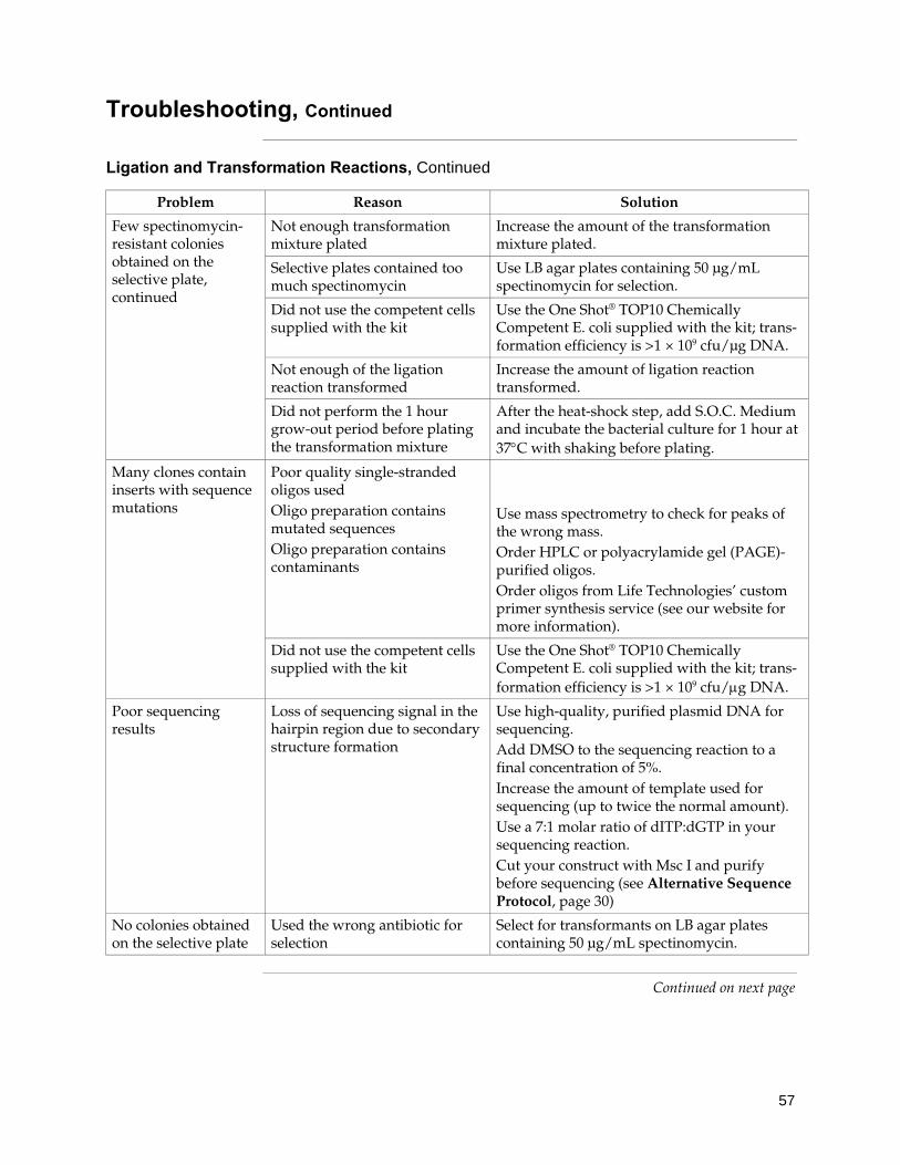
57
Troubleshooting, Continued
Ligation and Transformation Reactions, Continued
Problem Reason Solution
Few spectinomycin-resistant colonies obtained on the selective plate, continued
Not enough transformation mixture plated
Increase the amount of the transformation mixture plated.
Selective plates contained too much spectinomycin
Use LB agar plates containing 50 μg/mL spectinomycin for selection.
Did not use the competent cells supplied with the kit
Use the One Shot® TOP10 Chemically Competent E. coli supplied with the kit; trans- formation efficiency is >1 × 10⁹ cfu/μg DNA.
Not enough of the ligation reaction transformed
Increase the amount of ligation reaction transformed.
Did not perform the 1 hour grow-out period before plating the transformation mixture
After the heat-shock step, add S.O.C. Medium and incubate the bacterial culture for 1 hour at 37°C with shaking before plating.
Many clones contain inserts with sequence mutations
Poor quality single-stranded oligos used Oligo preparation contains mutated sequences Oligo preparation contains contaminants
Use mass spectrometry to check for peaks of the wrong mass. Order HPLC or polyacrylamide gel (PAGE)-purified oligos. Order oligos from Life Technologies’ custom primer synthesis service (see our website for more information).
Did not use the competent cells supplied with the kit
Use the One Shot® TOP10 Chemically Competent E. coli supplied with the kit; trans- formation efficiency is >1 × 10⁹ cfu/μg DNA.
Poor sequencing results
Loss of sequencing signal in the hairpin region due to secondary structure formation
Use high-quality, purified plasmid DNA for sequencing. Add DMSO to the sequencing reaction to a final concentration of 5%. Increase the amount of template used for sequencing (up to twice the normal amount). Use a 7:1 molar ratio of dITP:dGTP in your sequencing reaction. Cut your construct with Msc I and purify before sequencing (see Alternative Sequence Protocol, page 30)
No colonies obtained on the selective plate
Used the wrong antibiotic for selection
Select for transformants on LB agar plates containing 50 μg/mL spectinomycin.
Continued on next page

58
Troubleshooting, Continued
Transfection and RNAi Analysis
The table below lists some potential problems and possible solutions that may help you troubleshoot your transfection and knockdown experiment.
Problem Reason Solution
Low levels of gene knockdown observed due to low transfection efficiency
Antibiotics added to the media during transfection if using Lipofectamine® 2000 Reagent Cells too sparse at the time of transfection Not enough plasmid DNA transfected Not enough Lipofectamine® 2000 used
Do not add antibiotics to the media during transfection. Plate cells such that they will be 90–95% confluent at the time of transfection. Increase the amount of plasmid DNA transfected. Optimize the transfection conditions for your cell line by varying the amount of Lipofectamine® 2000 used.
Low levels of gene knockdown observed (other causes)
Didn’t wait long enough after transfection before assaying for gene knockdown
Repeat the transfection and wait for a longer period of time after transfection before assaying for gene knockdown. Perform a time course of expression to determine the point at which the highest degree of gene knockdown occurs.
ds oligo insert in your pcDNA™6.2-GW/EmGFP-miR construct contains mutations
When analyzing spectinomycin-resistant transformants, sequence the ds oligo insert to verify its sequence. Select constructs containing the correct ds oligo insert for use in RNAi analysis.
miRNA sequence not optimal due to selected target region.
Select a different target region. Order BLOCK-iT™ miR RNAi Select for your target gene (see page xiii), which allows you to generate four different miR RNAi expression vectors. We guarantee that at least two out of the four BLOCK-iT™ miR RNAi Select expression vectors will result in >70% knockdown of the target gene (provided that the transfection efficiency in your experiment is at least 80%).
Continued on next page
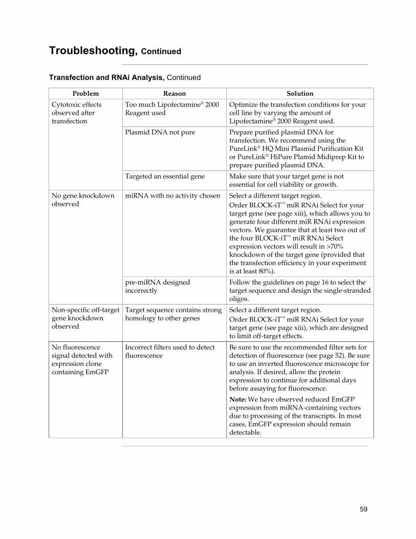
59
Troubleshooting, Continued
Transfection and RNAi Analysis, Continued
Problem Reason Solution
Cytotoxic effects observed after transfection
Too much Lipofectamine® 2000 Reagent used
Optimize the transfection conditions for your cell line by varying the amount of Lipofectamine® 2000 Reagent used.
Plasmid DNA not pure Prepare purified plasmid DNA for transfection. We recommend using the PureLink® HQ Mini Plasmid Purification Kit or PureLink® HiPure Plamid Midiprep Kit to prepare purified plasmid DNA.
Targeted an essential gene Make sure that your target gene is not essential for cell viability or growth.
No gene knockdown observed
miRNA with no activity chosen Select a different target region. Order BLOCK-iT™ miR RNAi Select for your target gene (see page xiii), which allows you to generate four different miR RNAi expression vectors. We guarantee that at least two out of the four BLOCK-iT™ miR RNAi Select expression vectors will result in >70% knockdown of the target gene (provided that the transfection efficiency in your experiment is at least 80%).
pre-miRNA designed incorrectly
Follow the guidelines on page 16 to select the target sequence and design the single-stranded oligos.
Non-specific off-target gene knockdown observed
Target sequence contains strong homology to other genes
Select a different target region. Order BLOCK-iT™ miR RNAi Select for your target gene (see page xiii), which are designed to limit off-target effects.
No fluorescence signal detected with expression clone containing EmGFP
Incorrect filters used to detect fluorescence
Be sure to use the recommended filter sets for detection of fluorescence (see page 52). Be sure to use an inverted fluorescence microscope for analysis. If desired, allow the protein expression to continue for additional days before assaying for fluorescence.
Note: We have observed reduced EmGFP expression from miRNA-containing vectors due to processing of the transcripts. In most cases, EmGFP expression should remain detectable.

60
Appendix Blasticidin
Blasticidin Blasticidin S HCl is a nucleoside antibiotic isolated from Streptomyces
griseochromogenes which inhibits protein synthesis in both prokaryotic and eukaryotic cells (Takeuchi et al., 1958; Yamaguchi et al., 1965). Resistance is conferred by expression of either one of two Blasticidin S deaminase genes: bsd from Aspergillus terreus (Kimura et al., 1994) or bsr from Bacillus cereus (Izumi et al., 1991). These deaminases convert Blasticidin S to a non-toxic deaminohydroxy derivative (Izumi et al., 1991).
Molecular Weight, Formula, and Structure
The formula for Blasticidin S is C17H26N8O5-HCl, and the molecular weight is 458.9. The diagram below shows the structure of Blasticidin.
Handling Blasticidin
Always wear gloves, mask, goggles, and protective clothing (e.g., a laboratory coat) when handling Blasticidin. Weigh out Blasticidin and prepare solutions in a hood.
Preparing and Storing Stock Solutions
Blasticidin may be obtained separately from Life Technologies (Catalog no. R210-01) in 50 mg aliquots. Blasticidin is soluble in water. Sterile water is generally used to prepare stock solutions of 5–10 mg/mL.
• Dissolve Blasticidin in sterile water and filter-sterilize the solution.
• Aliquot in small volumes suitable for one time use (see next to last point below) and freeze at –20°C for long-term storage or store at +4°C for short-term storage.
• Aqueous stock solutions are stable for 1–2 weeks at +4°C and 6–8 weeks at –20°C.
• pH of the aqueous solution should be 7.0 to prevent inactivation of Blasticidin.
• Do not subject stock solutions to freeze/thaw cycles (do not store in a frost-free freezer).
• Upon thawing, use what you need and store the thawed stock solution at +4°C for up to 2 weeks.
• Medium containing Blasticidin may be stored at +4°C for up to 2 weeks.
��
�
����
�
���
��
��
��
�
����

61
Recipes
Spectinomycin Use this procedure to prepare a 10 mg/mL stock solution of spectinomycin. Materials Needed • Spectinomycin Dihydrochloride (Sigma, Catalog no. S4014) • Sterile, deionized water Procedure 1. Weigh out 50 mg of spectinomycin and transfer to a sterile centrifuge tube. 2. Resuspend the spectinomycin in 5 mL of sterile, deionized water to produce
a 10 mg/mL stock solution. 3. Filter-sterilize. 4. Store the stock solution at +4°C for up to 2 weeks. For long-term storage,
store at –20°C.
LB (Luria-Bertani) Medium and Plates
Composition: 1.0% Tryptone 0.5% Yeast Extract 1.0% NaCl pH 7.0 1. For 1 liter, dissolve 10 g tryptone, 5 g yeast extract, and 10 g NaCl in 950 mL
deionized water. 2. Adjust the pH of the solution to 7.0 with NaOH and bring the volume up to
1 liter. 3. Autoclave on liquid cycle for 20 minutes at 15 psi. Allow solution to cool to
55°C and add antibiotic if needed. 4. Store at room temperature or at +4°C. 5. For LB agar plates: 6. Prepare LB medium as above, but add 15 g/L agar before autoclaving. 7. Autoclave on liquid cycle for 20 minutes at 15 psi. 8. After autoclaving, cool to ~55°C, add antibiotic if needed, and pour into
10 cm plates. 9. Let harden, then invert and store at +4°C.
Low Salt LB Plates with Blasticidin
Composition: 10 g Tryptone 5 g NaCl 5 g Yeast Extract 1. Combine the dry reagents above and add deionized, distilled water to
950 mL. Adjust pH to 7.0 with 1 N NaOH and bring the volume up to 1 liter. For plates, add 15 g/L agar before autoclaving.
2. Autoclave on liquid cycle at 15 psi and 121°C for 20 minutes. 3. Allow the medium to cool to at least 55°C before adding the Blasticidin to
100 g/mL final concentration. 4. Let harden, then invert and store at +4°C. Store plates at +4°C in the dark. Plates containing Blasticidin S HCl are stable for up to 2 weeks.

62
Map of pcDNA™6.2-GW/ EmGFP-miR
pcDNA™6.2-GW/EmGFP-miR
The figure below shows the features of the pcDNA™6.2-GW/EmGFP-miR vector. The vector is supplied linearized between nucleotides 1518 and 1519 with 4 base pair 5′ overhangs on each strand as indicated. The complete sequence of pcDNA™6.2-GW/ EmGFP-miR is available for downloading from our website (www.lifetechnologies.com) or by contacting Technical Support (see page 71).
Note: For an explanation of the features see page 64.
# �*+
�- ��
�+./���
�0 ���
$�������)�
!*1
�+./ ��
�2����
��(3��45 ,�67!��"#,���
�488�9�
����
��������:��
�������������� ��������!��"#���$0 ���$ %��������� ��
������
���
���
����
��
���
��
���
��
��
���
��
��������������(3��*�45 ,�67!��"#,����488�������)��
$�'� ����9�:����� �������- �����9�:�����4�7���576()*+9�:�����5 &� 6&�()*+���,��������������� ���������9�:����� 673� 6���#����� ���1���������9�:����� 63�� � ��#����"�����$�9�:����� � �� � ��#���"���9�:����� � 3� ���&#����� ���1���������9�:����� � 3� �4&���-��������$�9�:����� �3�� 4 4������������������������ �����������$�9�:����� 475� 4�4%/� �0����0������������9�:����� 46�� 3 4� ������9�:������7����6�4�'67�����0� ��������������9�:������6�&��53 (�5� ����9�:�������64��3 �-��������������������������9�:������3 &�&& �'67� �0����0������������9�:�����&643�&�33 ;$��������$�9�:�����&5&5�66 7� �����0���������������������$�9�:�����66�7��637� �����0���� ������$�9�:������63 ��4�6
�$��<�$ �������0�������

63
Map of pcDNA™6.2-GW/EmGFP-miR-neg Control Plasmid
pcDNA™6.2-GW/EmGFP-miR-Negative Control Plasmid
The figure below shows the features of the pcDNA™6.2-GW/EmGFP-miR-neg control plasmid. The vector contains an insert between bases 1519 and 1578 that can form a hairpin structure just as a regular pre-miRNA, but is predicted not to target any known vertebrate gene. The insert has been cloned according to the instructions in this manual (see page 16). Thus, this plasmid serves as a suitable negative control for pre-miRNA experiments with pcDNA™6.2-GW/EmGFP-miR expression vectors. The complete sequence of pcDNA™6.2-GW/EmGFP-miR-neg control plasmid is available for downloading from our website (www.lifetechnologies.com) or by contacting Technical Support (see page 71).
Note: For an explanation of the features see page 64.
# �*+
�- ��
�+./���
�0 ���
$�������)�
!*1
�+./ ��
�2����
��(3��45 ,�67!��"#,���,���������������)
�1�8�9�
��������:��
���������� ��������!��"#���$0 ���$ %��������� ��
���������,��������
���
���
����
��
���
��
���
��
��
���
��
��������������(3��*�45 ,�67!��"#,���,����������������)�1�8�������)��
$�'� ����9�:����� �������- �����9�:�����4�7�576()*+9�:�����5 &� 6&�()*+���,��������������� ���������9�:����� 673� 6���#����� ���1���������9�:����� 63�� � �������������9 �:����� � 3� �5�&#����� ���1���������9�:����� �53� 4�&���-��������$�9�:����� 4��� 454������������������������ �����������$�9�:����� 445� 4�4%/� �0����0������������9�:����� 57�� 354� ������9�:������7����� 4�'67�����0� ��������������9�:�������6&���� (�5� ����9�:������374��35�-��������������������������9�:������35&�&&5 �'67� �0����0������������9�:�����&��3�&4�3 ;$��������$�9�:�����&535�6657� �����0���������������������$�9�:�����6�67����7� �����0���� ������$�9�:�������� ��4�6
�$��<�$ �������0�������

64
Features of pcDNA™6.2-GW/EmGFP-miR
Explanation of Features
pcDNA™6.2-GW/EmGFP-miR vectors contain the following elements. All features have been functionally tested and the vectors have been fully sequenced.
Feature Benefit
CMV promoter Permits high-level, constitutive expression of the gene of interest (Andersson et al., 1989; Boshart et al., 1985; Nelson et al., 1987).
attB1 and attB2 sites Bacteriophage λ-derived recombination sequences that allow recombinational cloning of a gene of interest in the expression construct with a Gateway® destination vector (Landy, 1989).
EmGFP coding sequence Allows visual detection of transfected mammalian cells using fluorescence microscopy
EmGFP forward sequencing primer
Allows sequencing of the insert
5′ miR flanking region Allows formation of functional engineered pre-miRNA
5′ overhangs Allows ligase-mediated directional cloning of the double-stranded oligonucleotide of interest.
miR-neg control Allows formation of a pre-miRNA hairpin sequence predicted not to target any known vertebrate gene (only for pcDNA™6.2-GW/EmGFP-miR-neg control). Sequence without 5’ overhangs is shown below: 5’-GAAATGTACTGCGCGTGGAGACGTTTTGGCCACTGACTGACGTCTCCACGCAGTACATTT-3’
3′ miR flanking region Allows formation of functional engineered pre-miRNA
miRNA reverse sequencing primer
Allows sequencing of the insert
TK polyadenylation signal Allows transcription termination and polyadenylation of mRNA.
f1 origin Allows rescue of single-stranded DNA SV40 early promoter and origin Allows high-level expression of the selection marker and episomal
replication in cells expressing the SV40 large T antigen.
EM7 promoter Synthetic prokaryotic promoter for expression of the selection marker in E. coli.
Blasticidin (bsd) resistance gene Permits selection of stably transfected mammalian cell lines (Kimura et al., 1994).
SV40 polyadenylation signal Allows transcription termination and polyadenylation of mRNA.
pUC origin Permits high-copy replication and maintenance in E. coli.
Spectinomycin resistance gene (aadA1)
Allows selection of the plasmid in E. coli (Liebert et al., 1999).
Spectinomycin promoter Allows expression of the spectinomycin resistance gene in E. coli.

65
Map of pcDNA™1.2/V5-GW/lacZ
Description pcDNA™1.2/V5-GW/lacZ (6498 bp) is a control vector expressing a C-terminally-tagged β-galactosidase fusion protein under the control of the human cytomegalovirus (CMV) promoter (Andersson et al., 1989; Boshart et al., 1985; Nelson et al., 1987), and was generated using the MultiSite Gateway® Three-Fragment Vector Construction Kit available from Life Technologies (Catalog no. 12537-023). Briefly, a MultiSite Gateway® LR recombination reaction was performed with pDEST™R4-R3 and entry clones containing the CMV promoter, lacZ gene, and V5 epitope and TK polyadenylation signal (Cole & Stacy, 1985) to generate the pcDNA™1.2/V5-GW/lacZ vector. β-galactosidase is expressed as a C-terminal V5 fusion protein with a molecular weight of approximately 119 kDa. The complete sequence of pcDNA™1.2/V5-GW/lacZ is available for downloading from our website (www.lifetechnologies.com) or by contacting Technical Support (see page 71).
��������������(3�;05 7+�,�67 ���4.8<�������)��
���-69�:���������$�'� ����9�:����� &5�5�6���- 9�:�����4 6�4&5=��>������� �����9�:�����46&�&53�=��>�?�*9�:�����46&�&5 6���-�9�:�����&5 4�&5&3'��� �� �9�:�����&5&3�&5�7���>���,������ ����������9��67���3���>����������� ����������9� ��7� �&3��$�%/� �0����0������������9�:�����&�75�675����-&9�:�����6753�6733�� � ����9�:�����647&�657 � ��� � � �����������������������9�:�����657����4� ;$������9�:������575�4&�7
�$��<�� �������0�������
���� +���������
��(3�;05 7+�,�67���
4.8<�9�
�2� ���
# �*+
�- ��
���������
����

66
Map of pT-REx™-DEST30
Map The map below shows the elements of pT-REx™-DEST30. DNA from the entry clone replaces the region between bases 706 and 2389. The complete sequence of pT-REx™-DEST30 is available from our website (www.lifetechnologies.com) or by contacting Technical Support (see page 71).
��
�0 ���
�+./ ��
�+./���
���:�
������ �������������������������������
������������ ������������������������ ���� ����������������������������������������������� �!���"#������ �����������$���%������ ������������ �����&''���(�)�����)�����������������*����� �����'(����'����+�*����� ������'((���(����%������ ������������ �������$'��,�(�$��������� ������,&,��,�(�����������������-!.�,�������-�����������*����� ������'���(�,�/�����*���� �����(�$��(&�(.�,�����������������-����*���� �����($'��,�'�0����������������� %1�� �����,��$�,'��2����-�����������*����� ������������&(��������������!����������� %1�� ������,$,�&((,�3�����*���� �����&,$'�$���
����������������� !
���������
3���
:��
# �*
+ =
���&
�2����
����" �� ���# ����$

67
Features of the pT-REx™-DEST30 Vector
Features pT-REx™-DEST30 (7544 bp) contains the following elements. All features have been functionally tested.
Feature Benefit
Human cytomegalovirus (CMV) immediate-early promoter/enhancer
Allows efficient, high-level expression of your recombinant protein (Andersson et al., 1989; Boshart et al., 1985; Nelson et al., 1987)
Tetracycline operator (O2) sequences
Two tandem 19 nucleotide repeats which serve as binding sites for Tet repressor homodimers (Hillen and Berens, 1994)
attR1 and attR2 sites Allows recombinational cloning of the gene of interest from an entry clone
Chloramphenicol resistance gene Allows counterselection of expression clones
ccdB gene Allows negative selection of expression clones
T7 promoter (complementary strand)
Allows efficient in vitro transcription in the antisense orientation
SV40 polyadenylation signal Allows efficient transcription termination and polyadenylation of mRNA (Goodwin & Rottman, 1992)
f1 origin Allows rescue of single-stranded DNA
SV40 early promoter and origin Allows high-level expression of the neomycin resistance gene and episomal replication in cells expressing the SV40 large T antigen
Neomycin resistance gene Allows selection of stable transfectants in mammalian cells (Southern and Berg, 1982)
Polyadenylation signal Allows efficient transcription termination and polyadenylation of mRNA
Ampicillin resistance gene (β-lactamase)
Allows selection of transformants in E. coli
pUC origin Allows high-copy number replication and growth in E. coli

68
Map of pT-REx™/GW-30/lacZ
Description pT-REx™/GW-30/lacZ is an 8999 bp control vector containing the gene for β-galactosidase. pT-REx™/GW-30/lacZ was constructed using the Gateway® LR recombination reaction between an entry clone containing the lacZ gene and pT-REx™-DEST30. The molecular weight of β-galactosidase is approximately 116 kDa.
Map of pT-REx/GW-30/lacZ
The map below shows the elements of pT-REx/GW-30/lacZ. The complete sequence of pT-REx/GW-30/lacZ is available from our website (www.lifetechnologies.com) or by contacting Technical Support (see page 71).
��
�0 ���
�+./ ��
�+./���
���:�
������%&'���%���()��� !
���������
3���
:��
# �*
+ =
���&
�2����
������ ���������%&'���%���(*)))�����������
������������ ������������������������ ���� ����������������������������������������������� �!���"#������ �����������$���+������ ������������ �����&''�$�(���4� %1�� �����$,(�($''���+������ ������������ �����(�(,�(����$��������� �����('�'�('(������������������-!.�,�������-�����������*����� �����,($��,���/�����*���� �����,&(������.�,�����������������-����*���� �������,�����(0����������������� %1�� ������&���&,�&2����-�����������*����� �����&,$��&�����������������!����������� %1�� �����&'�'�$$�'�3�����*���� �����$'(,��&�$
���(���#" ���#$

69
Map of pcDNA™6/TR Vector
Map of pcDNA™6/TR
pcDNA™6/TR is a 6662 bp vector that expresses the Tet repressor under the control of the human CMV promoter. The figure below summarizes the features of the pcDNA™6/TR vector. The complete sequence for pcDNA™6/TR is available for downloading from our website (www.lifetechnologies.com) or by contacting Technical Support (see page 71).
TetR Gene The TetR gene used in pcDNA™6/TR was originally isolated from the Tn10transposon which confers resistance to tetracycline in E. coli and other enteric bacteria (Postle et al., 1984). The TetR gene from Tn10 encodes a class B Tet repressor and is often referred to as TetR(B) in the literature (Hillen and Berens, 1994).
The TetR gene encodes a repressor protein of 207 amino acids with a calculated molecular weight of 23 kDa. For more information about the Tet repressor and its interaction with the Tet operator, please refer to the review by Hillen and Berens (1994).
Continued on next page
��(3�47��>
444 �9�
�2� ��� �+./ ��$��
��
���)�
�+�����
!*,1
�0 ���
���������
���� �+./ ��
������ ������,-.%��...$������������
������������� ����������� �������������������55��� ��������������������� ����������������� �������� ���� ����� !�"��� ����������##$����������� ������#����%������������ ������������� ������%���#%6��#���������� ����#�%��#��&� ��"��������� �"������ ����#������������ �������� ���� ����� !�"��� �������������'�����7����������� �����%��%%��������������� ����%������%�(�)*���"������(���)����� �"������ ���%�����%���(�)
# �*+
���9
�?+�

70
Features of pcDNA™6/TR Vector
Features of pcDNA™6/TR
The table below describes the relevant features of pcDNA™6/TR. The vector includes the rabbit β-globin intron II to enhance expression of the TetR gene. For a more detailed description of the TetR gene and the Tet repressor, please refer to the previous page. All features have been functionally tested.
Feature Benefit
Human cytomegalovirus (CMV) immediate early promoter
Permits high-level expression of the TetR gene (Andersson et al., 1989; Boshart et al., 1985; Nelson et al., 1987)
Rabbit β-globin intron II (IVS) Enhances expression of the TetR gene (van Ooyen et al., 1979) in cultured cells
TetR gene Encodes the Tet repressor that binds to tet operator sequences to repress transcription of the gene of interest in the absence of tetracycline (Postle et al., 1984; Yao et al., 1998)
SV40 early polyadenylation signal Permits efficient transcription termination and polyadenylation of mRNA
f1 origin Allows rescue of single-stranded DNA
SV40 early promoter and origin Allows efficient, high-level expression of the blasticidin resistance gene in mammalian cells and episomal replication in cells expressing SV40 large T antigen
EM-7 promoter Synthetic prokaryotic promoter for expression of the blasticidin resistance gene in E. coli
Blasticidin (bsd) resistance gene Allows selection of stable transfectants in mammalian cells (Kimura et al., 1994)
SV40 early polyadenylation signal Allows efficient transcription termination and polyadenylation of mRNA
pUC origin Permits high-copy number replication and growth in E. coli
bla promoter Allows expression of the ampicillin (bla) resistance gene
Ampicillin (bla) resistance gene (β-lactamase)
Allows selection of transformants in E. coli

71
Technical Support
Obtaining Support For the latest services and support information for all locations, go to www.lifetechnologies.com.
At the website, you can:
• Access worldwide telephone and fax numbers to contact Technical Support and Sales facilities
• Search through frequently asked questions (FAQs)
• Submit a question directly to Technical Support ([email protected])
• Search for user documents, SDSs, vector maps and sequences, application notes, formulations, handbooks, certificates of analysis, citations, and other product support documents
• Obtain information about customer training
• Download software updates and patches
Safety Data Sheets (SDS)
Safety Data Sheets (SDSs) are available at www.lifetechnologies.com/sds.
Certificate of Analysis
The Certificate of Analysis provides detailed quality control and product qualification information for each product. Certificates of Analysis are available on our website. Go to www.lifetechnologies.com/support and search for the Certificate of Analysis by product lot number, which is printed on the box.
Limited Product Warranty
Life Technologies and/or its affiliate(s) warrant their products as set forth in the Life Technologies General Terms and Conditions of Sale found on the Life Technologies website at www.lifetechnologies.com/termsandconditions. If you have any questions, please contact Life Technologies at www.lifetechnologies.com/support.
72
Purchaser Notification
Introduction Use of the BLOCK-iT™ Inducible Pol II miR RNAi Expression Vector Kits are covered under the licenses detailed below.
Limited Use Label License No. 358: Research Use Only
The purchase of this product conveys to the purchaser the limited, non-transferable right to use the purchased amount of the product only to perform internal research for the sole benefit of the purchaser. No right to resell this product or any of its components is conveyed expressly, by implication, or by estoppel. This product is for internal research purposes only and is not for use in commercial applications of any kind, including, without limitation, quality control and commercial services such as reporting the results of purchaser’s activities for a fee or other form of consideration. For information on obtaining additional rights, please contact [email protected] or Out Licensing, Life Technologies, 5791 Van Allen Way, Carlsbad, California 92008.

73
Gateway® Clone Distribution Policy
Introduction The information supplied in this section is intended to provide clarity concerning Life Technologies’ policy for the use and distribution of cloned nucleic acid fragments, including open reading frames, created using Life Technologies’ commercially available Gateway® Technology.
Gateway® Entry Clones
Life Technologies understands that Gateway® entry clones, containing attL1 and attL2 sites, may be generated by academic and government researchers for the purpose of scientific research. Life Technologies agrees that such clones may be distributed for scientific research by non-profit organizations and by for-profit organizations without royalty payment to Life Technologies.
Gateway® Expression Clones
Life Technologies also understands that Gateway® expression clones, containing attB1 and attB2 sites, may be generated by academic and government researchers for the purpose of scientific research. Life Technologies agrees that such clones may be distributed for scientific research by academic and government organizations without royalty payment to Life Technologies. Organizations other than academia and government may also distribute such Gateway® expression clones for a nominal fee ($10 per clone) payable to Life Technologies.
Additional Terms and Conditions
We would ask that such distributors of Gateway® entry and expression clones indicate that such clones may be used only for research purposes, that such clones incorporate the Gateway® Technology, and that the purchase of Gateway® Clonase® from Life Technologies is required for carrying out the Gateway® recombinational cloning reaction. This should allow researchers to readily identify Gateway® containing clones and facilitate their use of this powerful technology in their research. Use of Life Technologies’ Gateway® Technology, including Gateway® clones, for purposes other than scientific research may require a license, and questions concerning such commercial use should be directed to Life Technologies’ licensing department at 760-603-7200.

74
References Ambros, V. (2001) MicroRNAs: Tiny Regulators with Great Potential. Cell 107, 823-826 Ambros, V. (2004) The functions of animal microRNAs. Nature 431, 350-355 Anandalakshmi, R., Pruss, G. J., Ge, X., Marathe, R., Mallory, A. C., Smith, T. H., and Vance, V. B. (1998)
A Viral Suppressor of Gene Silencing in Plants. Proc. Natl. Acad. Sci. USA 95, 13079-13084 Andersson, S., Davis, D. L., Dahlbäck, H., Jörnvall, H., and Russell, D. W. (1989) Cloning, Structure, and
Expression of the Mitochondrial Cytochrome P-450 Sterol 26-Hydroxylase, a Bile Acid Biosynthetic Enzyme. J. Biol. Chem. 264, 8222-8229
Ausubel, F. M., Brent, R., Kingston, R. E., Moore, D. D., Seidman, J. G., Smith, J. A., and Struhl, K. (1994) Current Protocols in Molecular Biology, Greene Publishing Associates and Wiley-Interscience, New York
Bartel, D. P. (2004) MicroRNAs: genomics, biogenesis, mechanism, and function. Cell 116, 281-297 Bernstein, E., Caudy, A. A., Hammond, S. M., and Hannon, G. J. (2001) Role for a Bidentate Ribonuclease
in the Initiation Step of RNA Interference. Nature 409, 363-366 Boden, D., Pusch, O., Silbermann, R., Lee, F., Tucker, L., and Ramratnam, B. (2004) Enhanced gene
silencing of HIV-1 specific siRNA using microRNA designed hairpins. Nucleic Acids Res 32, 1154-1158
Bohnsack, M. T., Czaplinski, K., and Gorlich, D. (2004) Exportin 5 is a RanGTP-dependent dsRNA-binding protein that mediates nuclear export of pre-miRNAs. RNA 10, 185-191.
Boshart, M., Weber, F., Jahn, G., Dorsch-Häsler, K., Fleckenstein, B., and Schaffner, W. (1985) A Very Strong Enhancer is Located Upstream of an Immediate Early Gene of Human Cytomegalovirus. Cell 41, 521-530
Brummelkamp, T. R., Bernards, R., and Agami, R. (2002) A System for Stable Expression of Short Interfering RNAs in Mammalian Cells. Science 296, 550-553
Carrington, J. C., and Ambros, V. (2003) Role of MicroRNAs in Plant and Animal Development. Science 301, 336-338
Chen, C., and Okayama, H. (1987) High-Efficiency Transformation of Mammalian Cells by Plasmid DNA. Mol. Cell. Biol. 7, 2745-2752
Chu, G., Hayakawa, H., and Berg, P. (1987) Electroporation for the Efficient Transfection of Mammalian Cells with DNA. Nucleic Acids Res. 15, 1311-1326
Chung, K. H., Hart, C. C., Al-Bassam, S., Avery, A., Taylor, J., Patel, P. D., Vojtek, A. B., and Turner, D. L. (2006) Polycistronic RNA polymerase II expression vectors for RNA interference based on BIC/miR-155. Nucleic Acids Res 34, e53.
Ciccarone, V., Chu, Y., Schifferli, K., Pichet, J.-P., Hawley-Nelson, P., Evans, K., Roy, L., and Bennett, S. (1999) Lipofectamine® 2000 Reagent for Rapid, Efficient Transfection of Eukaryotic Cells. Focus 21, 54-55
Cogoni, C., and Macino, G. (1997) Isolation of Quelling-Defective (qde) Mutants Impaired in Posttranscriptional Transgene-Induced Gene Silencing in Neurospora crassa. Proc. Natl. Acad. Sci. USA 94, 10233-10238
Cogoni, C., and Macino, G. (1999) Gene Silencing in Neurospora crassa Requires a Protein Homologous to RNA-Dependent RNA Polymerase. Nature 399, 166-169
Cogoni, C., Romano, N., and Macino, G. (1994) Suppression of Gene Expression by Homologous Transgenes. Antonie Van Leeuwenhoek 65, 205-209
Continued on next page

75
References, Continued Cole, C. N., and Stacy, T. P. (1985) Identification of Sequences in the Herpes Simplex Virus Thymidine
Kinase Gene Required for Efficient Processing and Polyadenylation. Mol. Cell. Biol. 5, 2104-2113 Cullen, B. R. (2004) Derivation and function of small interfering RNAs and microRNAs. Virus Res 102, 3-9. Cullen, B. R. (2004) Transcription and processing of human microRNA precursors. Mol Cell 16, 861-865 Curradi, M., Izzo, A., Badaracco, G., and Landsberger, N. (2002) Molecular Mechanisms of Gene Silencing
Mediated by DNA Methylation. Mol. Cell. Biol. 22, 3157-3173 Elbashir, S. M., Harborth, J., Lendeckel, W., Yalcin, A., Weber, K., and Tuschl, T. (2001) Duplexes of 21-
Nucleotide RNAs Mediate RNA Interference in Cultured Mammalian Cells. Nature 411, 494-498 Felgner, P. L., Holm, M., and Chan, H. (1989) Cationic Liposome Mediated Transfection. Proc. West.
Pharmacol. Soc. 32, 115-121 Felgner, P. L. a., and Ringold, G. M. (1989) Cationic Liposome-Mediated Transfection. Nature 337, 387-388 Goodwin, E. C., and Rottman, F. M. (1992) The 3´-Flanking Sequence of the Bovine Growth Hormone
Gene Contains Novel Elements Required for Efficient and Accurate Polyadenylation. J. Biol. Chem. 267, 16330-16334
Hammond, S. M., Bernstein, E., Beach, D., and Hannon, G. J. (2000) An RNA-Directed Nuclease Mediates Genetic Interference in Caenorhabditis elegans. Nature 404, 293-296
Hillen, W., and Berens, C. (1994) Mechanisms Underlying Expression of Tn10 Encoded Tetracycline Resistance. Annu. Rev. Microbiol. 48, 345-369
Hillen, W., Gatz, C., Altschmied, L., Schollmeier, K., and Meier, I. (1983) Control of Expression of the Tn10-encoded Tetracycline Resistance Genes: Equilibrium and Kinetic Investigations of the Regulatory Reactions. J. Mol. Biol. 169, 707-721
Izumi, M., Miyazawa, H., Kamakura, T., Yamaguchi, I., Endo, T., and Hanaoka, F. (1991) Blasticidin S-Resistance Gene (bsr): A Novel Selectable Marker for Mammalian Cells. Exp. Cell Res. 197, 229-233
Jones, A. L., Thomas, C. L., and Maule, A. J. (1998) De novo Methylation and Co-Suppression Induced by a Cytoplasmically Replicating Plant RNA Virus. EMBO J. 17, 6385-6393
Ketting, R. F., Fischer, S. E., Bernstein, E., Sijen, T., Hannon, G. J., and Plasterk, R. H. (2001) Dicer Functions in RNA Interference and in Synthesis of Small RNA Involved in Developmental Timing in C. elegans. Genes Dev. 15, 2654-2659
Kim, V. N. (2005) MicroRNA biogenesis: coordinated cropping and dicing. Nat Rev Mol Cell Biol 6, 376-385.
Kimura, M., Takatsuki, A., and Yamaguchi, I. (1994) Blasticidin S Deaminase Gene from Aspergillus terreus (BSD): A New Drug Resistance Gene for Transfection of Mammalian Cells. Biochim. Biophys. ACTA 1219, 653-659
Lagos-Quintana, M., Rauhut, R., Yalcin, A., Meyer, J., Lendeckel, W., and Tuschl, T. (2002) Identification of tissue-specific microRNAs from mouse. Curr Biol 12, 735-739.
Landy, A. (1989) Dynamic, Structural, and Regulatory Aspects of Lambda Site-specific Recombination. Ann. Rev. Biochem. 58, 913-949
Lee, Y., Kim, M., Han, J., Yeom, K. H., Lee, S., Baek, S. H., and Kim, V. N. (2004) MicroRNA genes are transcribed by RNA polymerase II. Embo J 23, 4051-4060
Li, W. X., and Ding, S. W. (2001) Viral Suppressors of RNA Silencing. Curr. Opin. Biotechnol. 12, 150-154
Continued on next page

76
References, Continued Liebert, C. A., Watson, A. L., and Summers, A. O. (1999) Transposon Tn21, Flagship of the Floating
Genome. Microbiol. Mol. Biol. Rev. 63, 507-522 Liu, J., Carmell, M. A., Rivas, F. V., Marsden, C. G., Thomson, J. M., Song, J. J., Hammond, S. M., Joshua-
Tor, L., and Hannon, G. J. (2004) Argonaute2 is the catalytic engine of mammalian RNAi. Science 305, 1437-1441
McManus, M. T., and Sharp, P. A. (2002) Gene Silencing in Mammals by Small Interfering RNAs. Nature Rev. Genet. 3, 737-747
Meister, G., Landthaler, M., Patkaniowska, A., Dorsett, Y., Teng, G., and Tuschl, T. (2004) Human Argonaute2 mediates RNA cleavage targeted by miRNAs and siRNAs. Mol Cell 15, 185-197
Miller, J. H. (1972) Experiments in Molecular Genetics, Cold Spring Harbor Laboratory, Cold Spring Harbor, New York
Napoli, C., Lemieux, C., and Jorgensen, R. (1990) Introduction of a Chalcone Synthase Gene into Petunia Results in Reversible Co-Suppression of Homologous Genes in trans. Plant Cell 2, 279-289
Nelson, J. A., Reynolds-Kohler, C., and Smith, B. A. (1987) Negative and Positive Regulation by a Short Segment in the 5´-Flanking Region of the Human Cytomegalovirus Major Immediate-Early Gene. Molec. Cell. Biol. 7, 4125-4129
Nykanen, A., Haley, B., and Zamore, P. D. (2001) ATP Requirements and Small Interfering RNA Structure in the RNA Interference Pathway. Cell 107, 309-321
Paddison, P. J., Caudy, A. A., Bernstein, E., Hannon, G. J., and Conklin, D. S. (2002) Short Hairpin RNAs (shRNAs) Induce Sequence-Specific Silencing in Mammalian Cells. Genes Dev. 16, 948-958
Paul, C. P., Good, P. D., Winer, I., and Engelke, D. R. (2002) Effective Expression of Small Interfering RNA in Human Cells. Nat. Biotechnol. 20, 505-508
Rietveld, L. E., Caldenhoven, E., and Stunnenberg, H. G. (2002) In vivo Repression of an Erythroid-Specific Gene by Distinct Corepressor Complexes. EMBO J. 21, 1389-1397
Romano, N., and Macino, G. (1992) Quelling: Transient Inactivation of Gene Expression in Neurospora crassa by Transformation with Homologous Sequences. Mol. Microbiol. 6, 3343-3353
Shigekawa, K., and Dower, W. J. (1988) Electroporation of Eukaryotes and Prokaryotes: A General Approach to the Introduction of Macromolecules into Cells. BioTechniques 6, 742-751
Shimomura, O., Johnson, F. H., and Saiga, Y. (1962) Extraction, Purification and Properties of Aequorin, a Bioluminescent Protein from the Luminous hHydromedusan, Aequorea. Journal of Cellular and Comparative Physiology 59, 223-239
Smith, C. J., Watson, C. F., Bird, C. R., Ray, J., Schuch, W., and Grierson, D. (1990) Expression of a Truncated Tomato Polygalacturonase Gene Inhibits Expression of the Endogenous Gene in Transgenic Plants. Mol. Gen. Genet. 224, 477-481
Southern, P. J., and Berg, P. (1982) Transformation of Mammalian Cells to Antibiotic Resistance with a Bacterial Gene Under Control of the SV40 Early Region Promoter. J. Molec. Appl. Gen. 1, 327-339
Sui, G., Soohoo, C., Affar, E. B., Gay, F., Shi, Y., Forrester, W. C., and Shi, Y. (2002) A DNA Vector-Based RNAi Technology to Suppress Gene Expression in Mammalian Cells. Proc. Natl. Acad. Sci. USA 99, 5515-5520
Takeuchi, S., Hirayama, K., Ueda, K., Sakai, H., and Yonehara, H. (1958) Blasticidin S, A New Antibiotic. The Journal of Antibiotics, Series A 11, 1-5
Tsien, R. Y. (1998) The Green Fluorescent Protein. Annu. Rev. Biochem. 67, 509-544
Continued on next page

77
References, Continued van der Krol, A. R., Mur, L. A., Beld, M., Mol, J. N., and Stuitje, A. R. (1990) Flavonoid Genes in Petunia:
Addition of a Limited Number of Gene Copies May Lead to a Suppression of Gene Expression. Plant Cell 2, 291-299
Voinnet, O., Pinto, Y. M., and Baulcombe, D. C. (1999) Suppression of Gene Silencing: A General Strategy Used by Diverse DNA and RNA Viruses of Plants. Proc. Natl. Acad. Sci. USA 96, 14147-14152
Weiss, B., Jacquemin-Sablon, A., Live, T. R., Fareed, G. C., and Richardson, C. C. (1968) Enzymatic Breakage and Joining of Deoxyribonucleic Acid. VI. Further Purification and Properties of Polynucleotide Ligase from Escherichia coli Infected with Bacteriophage T4. J. Biol. Chem. 243, 4543-4555
Wigler, M., Silverstein, S., Lee, L.-S., Pellicer, A., Cheng, Y.-C., and Axel, R. (1977) Transfer of Purified Herpes Virus Thymidine Kinase Gene to Cultured Mouse Cells. Cell 11, 223-232
Yamaguchi, H., Yamamoto, C., and Tanaka, N. (1965) Inhibition of Protein Synthesis by Blasticidin S. I. Studies with Cell-free Systems from Bacterial and Mammalian Cells. J. Biochem (Tokyo) 57, 667-677
Yao, F., Svensjo, T., Winkler, T., Lu, M., Eriksson, C., and Eriksson, E. (1998) Tetracycline Repressor, tetR, Rather than the tetR-Mammalian Cell Transcription Factor Fusion Derivatives, Regulates Inducible Gene Expression in Mammalian Cells. Hum. Gene Ther. 9, 1939-1950
Yekta, S., Shih, I. H., and Bartel, D. P. (2004) MicroRNA-directed cleavage of HOXB8 mRNA. Science 304, 594-596
Yi, R., Qin, Y., Macara, I. G., and Cullen, B. R. (2003) Exportin-5 mediates the nuclear export of pre-microRNAs and short hairpin RNAs. Genes Dev 17, 3011-3016
Yu, J. Y., DeRuiter, S. L., and Turner, D. L. (2002) RNA Interference by Expression of Short-interfering RNAs and Hairpin RNAs in Mammalian Cells. Proc. Natl. Acad. Sci. USA 99, 6047-6052
Yu, Z., Raabe, T., and Hecht, N. B. (2005) MicroRNA122a Reduces Expression of the Post-Transcriptionally Regulated Germ Cell Transition Protein 2 (Tnp2) Messenger RNA (mRNA) by mRNA Cleavage. Biol Reprod 18
Zeng, Y., Wagner, E. J., and Cullen, B. R. (2002) Both Natural and Designed MicroRNAs Can Inhibit the Expression of Cognate mRNAs When Expressed in Human Cells. Mol Cell 9, 1327-1333
Zeng, Y., Yi, R., and Cullen, B. R. (2005) Recognition and cleavage of primary microRNA precursors by the nuclear processing enzyme Drosha. Embo J 24, 138-148
Zhang, G., Gurtu, V., and Kain, S. (1996) An Enhanced Green Fluorescent Protein Allows Sensitive Detection of Gene Transfer in Mammalian Cells. Biochem. Biophys. Res. Comm. 227, 707-711
©2012 Life Technologies Corporation. All rights reserved. The trademarks mentioned herein are the property of Life Technologies Corporation or their respective owners. LIFE TECHNOLOGIES CORPORATION AND/OR ITS AFFILIATE(S) DISCLAIM ALL WARRANTIES WITH RESPECT TO THIS DOCUMENT, EXPRESSED OR IMPLIED, INCLUDING BUT NOT LIMITED TO THOSE OF MERCHANTABILITY, FITNESS FOR A PARTICULAR PURPOSE, OR NON-INFRINGEMENT. TO THE EXTENT ALLOWED BY LAW, IN NO EVENT SHALL LIFE TECHNOLOGIES AND/OR ITS AFFILIATE(S) BE LIABLE, WHETHER IN CONTRACT, TORT, WARRANTY, OR UNDER ANY STATUTE OR ON ANY OTHER BASIS FOR SPECIAL, INCIDENTAL, INDIRECT, PUNITIVE, MULTIPLE OR CONSEQUENTIAL DAMAGES IN CONNECTION WITH OR ARISING FROM THIS DOCUMENT, INCLUDING BUT NOT LIMITED TO THE USE THEREOF.

78
Notes


Headquarters5791 Van Allen Way | Carlsbad, CA 92008 USA | Phone +1 760 603 7200 | Toll Free in USA 800 955 6288For support visit www.invitrogen.com/support or email [email protected]
www.lifetechnologies.com