blepharospasm round up - novel: home · -corneal trauma 1 -entropion 1 (b) ocular symptoms prior to...
TRANSCRIPT

BLEPHAROSPASM ROUND UP Shirley H. Wray, M.D., Ph.D.

Blepharospasm is a disabling focal dystonia confined to the eyelids
Manifested by repetitive involuntary
sustained contractions of the palpebral portion of the orbicularis oculi muscle.

The cause of blepharospasm is unknown.
One precipitating trigger may be the high incidence of local ocular symptoms and signs prior to or at the onset.
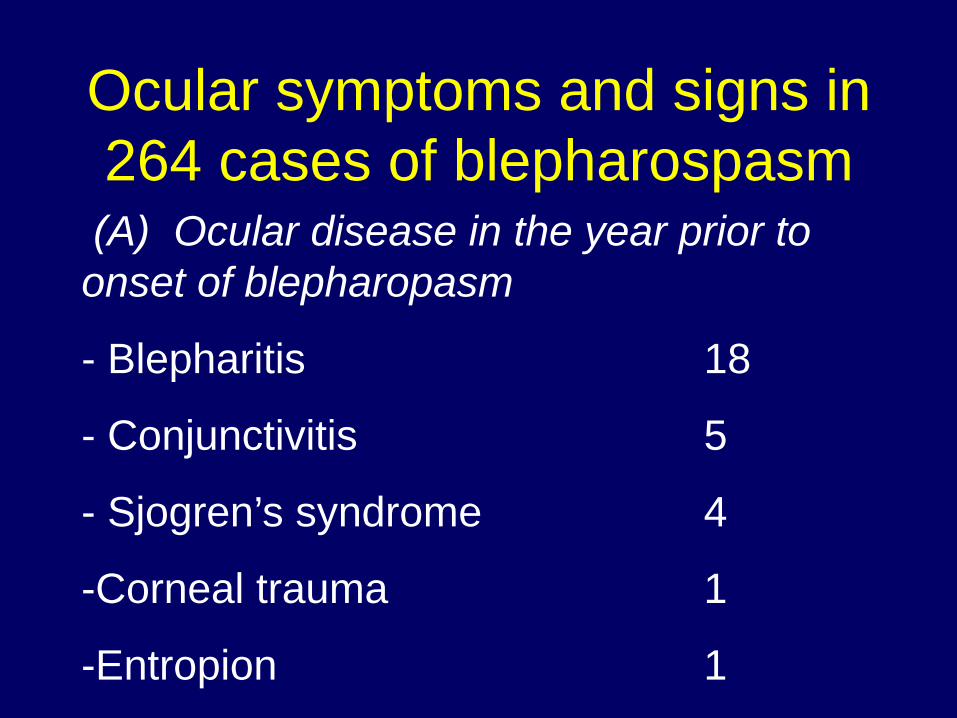
Ocular symptoms and signs in 264 cases of blepharospasm
(A) Ocular disease in the year prior to onset of blepharopasm
- Blepharitis 18
- Conjunctivitis 5
- Sjogren’s syndrome 4
-Corneal trauma 1
-Entropion 1
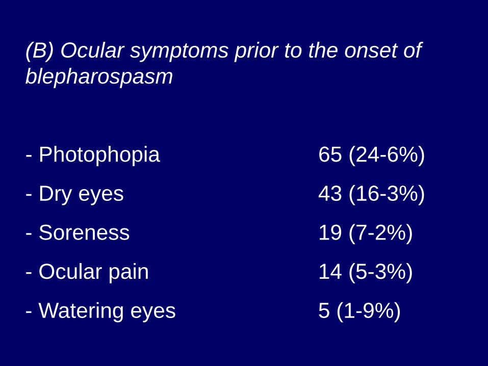
(B) Ocular symptoms prior to the onset of blepharospasm
- Photophopia 65 (24-6%)
- Dry eyes 43 (16-3%)
- Soreness 19 (7-2%)
- Ocular pain 14 (5-3%)
- Watering eyes 5 (1-9%)

A family history of blepharospasm or dystonia elsewhere suggests a genetic predisposition.
Dystonia involving other muscles occurs in approximately 78% of patients within 6 years as an orderly temporal progression of dystonia in the cranial-cervical area.

Distribution of dystonia in 264 cases of blepharospasm
Orbicuarlis oculi 264 (100%)
Oro-mandibular 188 (71-2%)
Neck 60 (22-7%)
Laryngeal 46 (17-4%)
Respiratory 39 (14-8%)
Arm/Hand 26 (9-8%)
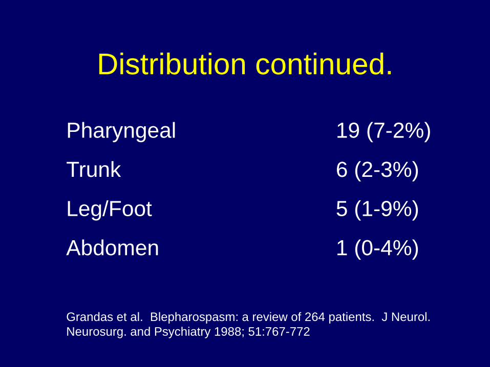
Distribution continued.
Pharyngeal 19 (7-2%)
Trunk 6 (2-3%)
Leg/Foot 5 (1-9%)
Abdomen 1 (0-4%) Grandas et al. Blepharospasm: a review of 264 patients. J Neurol. Neurosurg. and Psychiatry 1988; 51:767-772

Blepharospasm is associated with progressive
neurodegenerative diseases:
Parkinson’s Disease – a dopamine deficiency
Progressive Supranuclear Palsy – a tauopathy Multiple System Atrophy
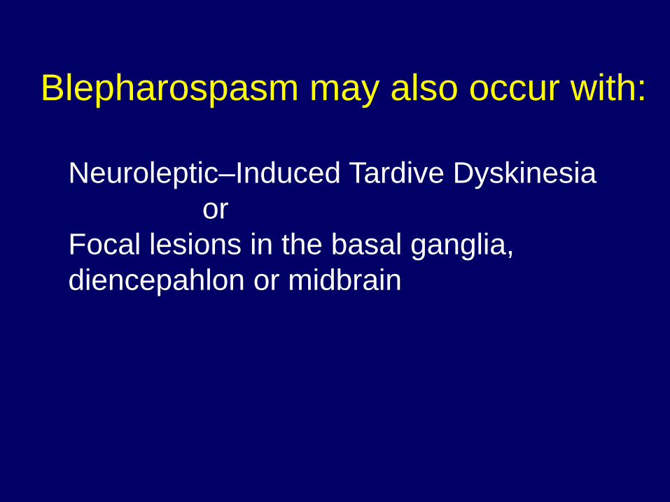
Blepharospasm may also occur with:
Neuroleptic–Induced Tardive Dyskinesia or
Focal lesions in the basal ganglia, diencepahlon or midbrain


Eye examination
Observe the eyes and face when taking
the history Assess lid position in different gaze
directions Look for blepharoclonus on gentle eye
closure Count the blink rate

Eye examination continued.
Check for suppression of blepharospasm by visual attention (OKN drum)
Look for a positive Glabella tap – An inability to inhibit a blink when the forehead is tapped and
Pay attention to the latency and speed of voluntary vertical and horizontal eye movements on command.
Question
What happened to interrupt the
dynamics of normal blinking? What is the basis for the repetitive and
sustained contractions of the orbicularis oculi – for the dystonia?

Pathophysiology
A key player in the pathophysiological
process is the Levator Palpebrae Superioris muscle which elevates the lid.
This muscle contains only singly-
innervated fibres of the types suitable for fatigue-resistant tonic activity.

The motor neurons that activate the levator
are located in a single midline Central Caudal Nucleus of the 3rd nerve complex in the midbrain.
The muscle is innervated by the superior branch of the 3rd nerve.

The levator acting alone controls:
Tonic lid elevation to keep the eyes open
and
Voluntary eye closure and eye opening.

Two further muscles, innervated by the facial nerve, act on the eyelid.
The frontalis muscle which helps to retract the lid in extreme upward gaze
The orbicularis oculi muscle which controls periodic and reflex blinking and firm eye closure in protective and expressive acts like sneezing. Movement Disorders in Clinical Practice 2001; Sawle, G.

In all kinds of blinks the levator is abruptly inhibited to allow the eyes to close and then it resumes its prior level of activity once the contraction of the palpebral portion of the orbicularis oculi, closing the eyelids momentarily, is over.
Conversely, the orbicularis oculi activity precedes and outlasts the levator inhibition in firm eye closure.
The Brain’s Control
The cerebral cortex (R>L) controls the tonic activity of the levator and voluntary eye opening and eye closure.
The dynamics of normal blinks, spontaneous and voluntary, and frequency of periodic blinks depend on the affective, attentional and cognitive state of the patient.
During sleep and when the eyes are gently closed, activity of the levator ceases completely.
The Brain’s Control
The extrapyramidal dopaminergic circuit influences the execution of blinks and blink frequency.
The basal ganglia play a role in the
inhibition of the levator during blinks and eye closure.

ROUND UP

The late age of onset of blepharospasm and the company that blepharospasm keeps with the progressive neurodegenerative diseases strongly suggests that this focal dystonic disorder –
is a central disturbance of one or more neurotransmitters and/or synaptic transmission in genetically predisposed patients.
Differential Diagnosis
Stress related excess blinking Cranial dystonia or Meige’s Syndrome Hemifacial spasm Apraxia of eyelid opening


Cranial Dystonia or Meige’s Syndrome
Movement Disorders; Riley, D.E., Lang, A.E.; Neurology in Clinical Practice 1996; Bradley, W.G.; Daroff, R.B.; FenichelG.M.; Marsden, C.D.

Cranial Dystonia or Meige’s Syndrome
Movement Disorders; Riley, D.E., Lang, A.E.; Neurology in Clinical Practice 1996; Bradley, W.G.; Daroff, R.B.; FenichelG.M.; Marsden, C.D.


Hemifacial Spasm is characterized by causing
- paroxysmal, involuntary clonic and tonic
- synchronous contraction of the muscles
- innervated by the facial nerve on one side.
The spasms are due to brief bursts of normal motor units firing at high frequency.
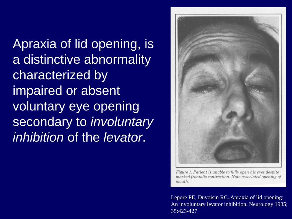
Apraxia of lid opening, is a distinctive abnormality characterized by impaired or absent voluntary eye opening secondary to involuntary inhibition of the levator.
Lepore PE, Duvoisin RC. Apraxia of lid opening: An involuntary levator inhibition. Neurology 1985; 35:423-427

My thanks to Nancy Lombardo, Systems Librarian and her team at the Spencer S. Eccles Health Sciences Library. University of Utah.
Acknowledgements
