bird respiratory system - · pdf fileopen in browser pro version are you a developer? try out...
TRANSCRIPT
pdfcrowd.comopen in browser PRO version Are you a developer? Try out the HTML to PDF API
BIO 554/754 Ornithology
Avian Respiration
This page has been translated into Belorussian by Paul Bukhovko andis available at www.movavi.com/opensource/birdrespiration-be
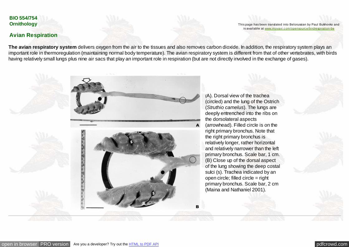
The avian respiratory system delivers oxygen from the air to the tissues and also removes carbon dioxide. In addition, the respiratory system plays animportant role in thermoregulation (maintaining normal body temperature). The avian respiratory system is different from that of other vertebrates, with birdshaving relatively small lungs plus nine air sacs that play an important role in respiration (but are not directly involved in the exchange of gases).
(A). Dorsal view of the trachea(circled) and the lung of the Ostrich(Struthio camelus). The lungs aredeeply entrenched into the ribs onthe dorsolateral aspects(arrowhead). Filled circle is on theright primary bronchus. Note thatthe right primary bronchus isrelatively longer, rather horizontaland relatively narrower than the leftprimary bronchus. Scale bar, 1 cm.(B) Close up of the dorsal aspectof the lung showing the deep costalsulci (s). Trachea indicated by anopen circle; filled circle = rightprimary bronchus. Scale bar, 2 cm(Maina and Nathaniel 2001).

pdfcrowd.comopen in browser PRO version Are you a developer? Try out the HTML to PDF API
Avian respiratory system (hd = humeral diverticulum of the clavicular air sac; adapted from Sereno et al. 2008)
The air sacs permit a unidirectional flow of air through the lungs. Unidirectional flow means that air moving through bird lungs is largely 'fresh' air & has ahigher oxygen content. In contrast, air flow is 'bidirectional' in mammals, moving back and forth into and out of the lungs. As a result, air coming into amammal's lungs is mixed with 'old' air (air that has been in the lungs for a while) & this 'mixed air' has less oxygen. So, in bird lungs, more oxygen isavailable to diffuse into the blood (avian respiratory system).

pdfcrowd.comopen in browser PRO version Are you a developer? Try out the HTML to PDF API
Pulmonary air-sac system of a Common Teal (Anas crecca). a. Latex injection (blue) highlighting the location of air sacs.b, Main components of the avian flow-through system. Abd, abdominal aire sac; Cdth, caudal thoracic air sac; Cl, clavicularair sac; Crth, cranial thoracic air sac; Cv, cervical air sac; Fu, furcula; Hu, humerus; Lu, lung; Lvd, lateral vertebral diverticula;
Pv, pelvis; and Tr, trachea (From: O'Connor and Claessens 2005).

pdfcrowd.comopen in browser PRO version Are you a developer? Try out the HTML to PDF API
The alveolar lungs of mammals (Rhesus monkey; A) and parabronchial lungs of birds (pigeon; B) are subdivided into largenumbers of extremely small alveoli (A, inset) or air capillaries (radiating from the parabronchi; B, inset). The mammalian respiratory
system is partitioned homogeneously, so the functions of ventilation and gas exchange are shared by alveoli and much of the lung volume.The avian respiratory system is partitioned heterogeneously, so the functions of ventilation and gas exchange are separate in the air sacs
(shaded in gray) and the parabronchial lung, respectively. Air sacs act as bellows to ventilate the tube-like parabronchi (Powell and Hopkins 2004).

pdfcrowd.comopen in browser PRO version Are you a developer? Try out the HTML to PDF API
Comparison of the avian 'unidirectional' respiratory system (a) where gases are exchanged between the lungs and the blood in the parabronchi, and thebidirectional respiratory system of mammals (b) where gas exchange occurs in small dead-end sacs called alveoli (From: West et al. 2007).

pdfcrowd.comopen in browser PRO version Are you a developer? Try out the HTML to PDF API
Animated gif created by Eleanor Lutz (Eleanor's website: http://tabletopwhale.com/2014/10/24/3-different-ways-to-breathe.html)

pdfcrowd.comopen in browser PRO version Are you a developer? Try out the HTML to PDF API
Credit: Zina Deretsky, National Science Foundation
Bird-like respiratory systems in dinosaurs -- A recent analysis showing the presence of a very bird-like pulmonary, or lung, system inpredatory dinosaurs provides more evidence of an evolutionary link between dinosaurs and birds. First proposed in the late 19th century,
theories about the animals' relatedness enjoyed brief support but soon fell out of favor. Evidence gathered over the past 30 years hasbreathed new life into the hypothesis. O'Connor and Claessens (2005) make clear the unique pulmonary system of birds, which has fixed
lungs and air sacs that penetrate the skeleton, has an older history than previously realized. It also dispels the theory that predatorydinosaurs had lungs similar to living reptiles, like crocodiles.
The avian pulmonary system uses "flow-through ventilation," relying on a set of nine flexible air sacs that act like bellows to move air throughthe almost completely rigid lungs. Air sacs do not take part in the actual oxygen exchange, but do greatly enhance its efficiency and allow forthe high metabolic rates found in birds. This system also keeps the volume of air in the lung nearly constant. O'Connor says the presence of
an extensive pulmonary air sac system with flow-through ventilation of the lung suggests this group of dinosaurs could have maintained astable and high metabolism, putting them much closer to a warm-blooded existence. "More and more characteristics that once defined
birds--feathers, for example--are now known to have been present in dinosaurs, so, many avian features may really be dinosaurian," saidO'Connor. A portion of the air sac actually integrates with the skeleton, forming air pockets in otherwise dense bone. The exact function ofthis skeletal modification is not completely understood, but one explanation theorizes the skeletal air pockets evolved to lighten the bone
structure, allowing dinosaurs to walk upright and birds to fly.

pdfcrowd.comopen in browser PRO version Are you a developer? Try out the HTML to PDF API
Some hollow bones are providing solid new evidence of how birds evolved from dinosaurs.

pdfcrowd.comopen in browser PRO version Are you a developer? Try out the HTML to PDF API
Most birds have 9 air sacs:
one interclavicular sactwo cervical sacstwo anterior thoracicsacstwo posterior thoracicsacstwo abdominal sacs
Functionally, these 9 air sacscan be divided into anteriorsacs (interclavicular,cervicals, & anteriorthoracics) & posterior sacs(posterior thoracics &abdominals). Air sacs havevery thin walls with few bloodvessels. So, they do not playa direct role in gas exchange.Rather, they act as a 'bellows'to ventilate the lungs (Powell2000).
Source: http://numbat.murdoch.edu.au/Anatomy/avian/fig3.2.GIF

pdfcrowd.comopen in browser PRO version Are you a developer? Try out the HTML to PDF API
Air sacs and axial pneumatization in an extant avian. The body of bird in left lateral view, showing the cervical (C), interclavicular (I), anterior thoracic (AT),posterior thoracic (PT), and abdominal (AB) air sacs. The hatched area shows the volume change during exhalation. The cervical and anterior thoracic
vertebrae are pneumatized by diverticula of the cervical air sacs. The posterior thoracic vertebrae and synsacrum are pneumatized by the abdominal airsacs in most taxa. Diverticula of the abdominal air sacs usually invade the vertebral column at several points. Diverticula often unite when they come into
contact, producing a system of continuous vertebral airways extending from the third cervical vertebra to the end of the synsacrum. Modified from Duncker1971 (Wedel 2003).
Computerized axial tomogram of an awake, spontaneously breathing

pdfcrowd.comopen in browser PRO version Are you a developer? Try out the HTML to PDF API
Computerized axial tomogram of an awake, spontaneously breathinggoose; air is darkest. A large percentage of the bird's body is filledwith the several air sacs. Upper left: At the level of the shoulder joints(hh, humeral head) is the intraclavicular air sac (ICAS), which extendsfrom the heart cranially to the clavicles (i.e., furcula or wishbone). S,sternum; FM, large flight muscles with enclosed air sac diverticula,arrowheads; t, trachea. Upper right: At the level of the caudal heart(H) is the paired cranial thoracic air sacs (TAS). Arrowhead points tothe medial wall of the air sac (contrast enhanced with aerosolizedtantalum powder). The dorsal body cavity is filled with the lungs, whichare tightly attached to the dorsal and lateral body wall. V, thoracicvertebrae. Lower left: At the level of the knees (K) is the pairedcaudal thoracic air sacs (PTAS) and paired abdominal air sacs, withthe abdominal viscera (AV) filling the ventral body cavity. Themembrane separating the abdominal air sacs from one another(arrowhead) and from the caudal thoracic air sacs (arrows) can beseen. Lower right: At the level of the caudal pelvis, the abdominal airsacs, which extend to the bird's tail, can be seen. Arrow, membraneseparating abdominal air sacs (Brown et al. 1997).
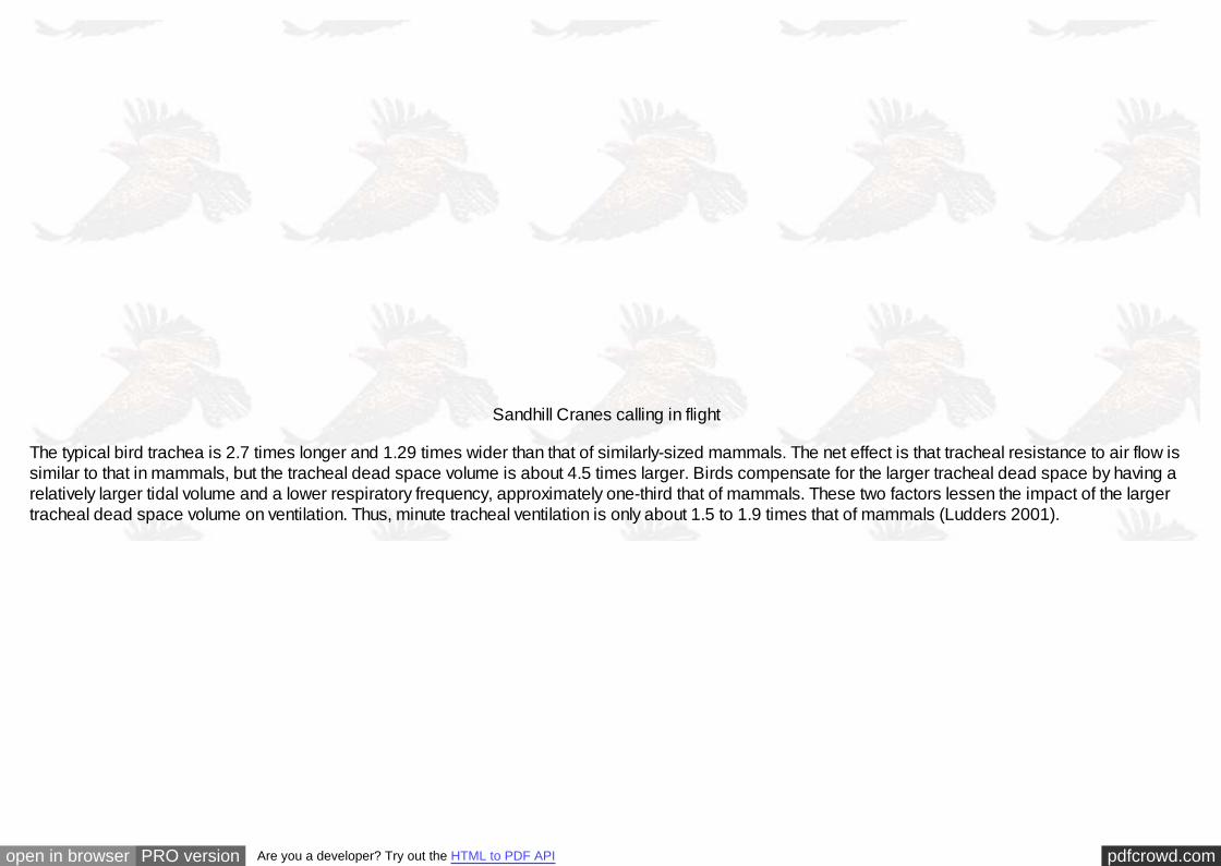
Birds can breathe through the mouth or the nostrils (nares). Air entering these openings (during inspiration) passes through the pharynx & then into thetrachea (or windpipe). The trachea is generally as long as the neck. However, some birds, such as cranes, have an exceptionally long (up to 1.5 m) tracheathat is coiled within the hollowed keel of the breastbone (shown below). This arrangement may give additional resonance to their loud calls (check this shortvideo of calling Sandhill Cranes).
pdfcrowd.comopen in browser PRO version Are you a developer? Try out the HTML to PDF API
Sandhill Cranes calling in flight
The typical bird trachea is 2.7 times longer and 1.29 times wider than that of similarly-sized mammals. The net effect is that tracheal resistance to air flow issimilar to that in mammals, but the tracheal dead space volume is about 4.5 times larger. Birds compensate for the larger tracheal dead space by having arelatively larger tidal volume and a lower respiratory frequency, approximately one-third that of mammals. These two factors lessen the impact of the largertracheal dead space volume on ventilation. Thus, minute tracheal ventilation is only about 1.5 to 1.9 times that of mammals (Ludders 2001).

pdfcrowd.comopen in browser PRO version Are you a developer? Try out the HTML to PDF API
Examples of tracheal loops found in Black Swans (Cygnus atratus), Whooper Swans (Cygnus cygnus), White Spoonbills (Platalea leucorodia), Helmeted Curassow (Crax pauxi),
and Whooping Cranes (Grus americana). Source: http://www.ivis.org/advances/Anesthesia_Gleed/ludders2/chapter_frm.asp
The trachea bifurcates (or splits) into two primary bronchi at the syrinx. The syrinx is unique to birds & is their 'voicebox' (in mammals, sounds are producedin the larynx). The primary bronchi enter the lungs & are then called mesobronchi. Branching off from the mesobronchi are smaller tubes calleddorsobronchi. The dorsobronchi, in turn, lead into the still smaller parabronchi. Parabronchi can be several millimeters long and 0.5 - 2.0 mm in diameter(depending on the size of the bird) (Maina 1989) and their walls contain hundreds of tiny, branching, & anastomosing 'air capillaries' surrounded by aprofuse network of blood capillaries (Welty and Baptista 1988). It is within these 'air capillaries' that the exchange of gases (oxygen and carbon dioxide)between the lungs and the blood occurs. After passing through the parabronchi, air moves into the ventrobronchi.

pdfcrowd.comopen in browser PRO version Are you a developer? Try out the HTML to PDF API
Semi-schematic drawing of the lung-air sac system in situ. The cranial half of the dorsobronchi (4) and the parabronchi (6) has been removed. 1 = trachea, 2= primary bronchus, 3 = ventrobronchi with the connections into (A) cervical, (B) interclavicular and (C) cranial thoracic air sacs, 5 = laterobronchi and the
caudal primary bronchus open into the (D) posterior thoracic and (E) abdominal air sacs (From: Duncker 2004).

pdfcrowd.comopen in browser PRO version Are you a developer? Try out the HTML to PDF API
Avian respiratory system showing the bronchi located inside the lungs. Dorsobronchi and ventrobronchi branch off of the primary bronchus; parabronchi
extend from the dorsobronchi to the ventrobronchi. Light blue arrows indicate the direction of air flow through the parabronchi. The primary bronchuscontinues through the lung and opens into the abdominal air sac. (Source: http://www.iv is.org/adv ances/Anesthesia_Gleed/ludders2/chapter_f rm.asp)
Birds exhibit some variation in lung structure and, specifically, in the arrangement of parabronchi. Most birds have two sets of parabronchi, thepaleopulmonic (‘ancient lung’) and neopulmonic (‘new lung’) parabronchi. However, the neopulmonic region is absent in some birds (e.g., penguins) andpoorly developed in others (e.g., storks [Ciconiidae] and ducks [Anatidae]). In songbirds (Passeriformes), pigeons (Columbiformes), and gallinaceous birds(Galliformes), the neopulmonic region of the lung is well-developed (Maina 2008). In these latter groups, the neopulmonic parabronchi contain about 15 to20% of the gas exchange surface of the lungs (Fedde 1998). Whereas airflow through the paleopulmonic parabronchi is unidirectional, airflow through theneopulmonic parabronchi is bidirectional. Parabronchi can be several millimeters long and 0.5 - 2.0 mm in diameter (depending on the size of the bird)(Maina 1989) and their walls contain hundreds of tiny, branching, and anastomosing air capillaries surrounded by a profuse network of blood capillaries.

pdfcrowd.comopen in browser PRO version Are you a developer? Try out the HTML to PDF API

pdfcrowd.comopen in browser PRO version Are you a developer? Try out the HTML to PDF API
Differences among different birds in the development of the neopulmonic region of the lung. (a) Penguin lungs are entirely paleopulmonic. (b) Some birds, such as ducks, have a relatively small neopulmonic region. (c) Songbirds have a well-developed neopulmonic region.
1, trachea, 2, primary bronchus, 3, ventrobronchus, 4, dorsobronchus, 5, lateral bronchus, 6, paleopulmonic parabronchi, 7, neopulmonic parabronchi; A, cervical air sac, B, interclavicular air sac, C, cranial thoracic air sac, D, caudal thoracic air sac,
E, abdominal air sac. The white arrows indicate changes in volume of the air sacs during the respiratory cycle (From: McLelland 1989).

pdfcrowd.comopen in browser PRO version Are you a developer? Try out the HTML to PDF API
So, how does air flow through the avian lungs & air sacs during respiration?
Air flow through the avian respiratory system during inspiration (a) and expiration (b).1 - interclavicular air sac, 2 - cranial thoracic air sac, 3 - caudal thoracic air sac, 4 - abdominal air sac
(From: Reese et al. 2006).
A schematic of the avian respiratory system, illustrating the major air sacs and their connections to the lung. (A) The lateral and dorsal direction of motion ofthe rib cage during exhalation is indicated by arrows. (B) The direction of airflow during inspiration. (C) The direction of flow during expiration (From:
Plummer and Goller 2008).

pdfcrowd.comopen in browser PRO version Are you a developer? Try out the HTML to PDF API
Avian respiratory cycle
This Flash diagram shows the paths that air takes through the respiratory system when a bird breathes.
Use the toolbar to step through the five pages of the diagram.Depending on your browser - you may need to click the toolbar one time or two times to fully activate it.The toolbar will respond to the IMB/PC keyboard keys: Up, Down, Left, Right, Home, End, Page Up, and Page Down.Some pages have notes that contain anatomical terms that may not be familiar to you. Put your cursor over the labels button (furthestright on the toolbar) or click on it to see what they refer to.

pdfcrowd.comopen in browser PRO version Are you a developer? Try out the HTML to PDF API
During inhalation, air moves into the posterior air sacs and, simultaneously,
into the lungs and through the parabronchi and into the anterior air sacs.
During exhalation, air moves out of the posterior air sacs into and through the parabronchi and, simultaneously,
out of the anterior air sacs and out of the body via the trachea.

pdfcrowd.comopen in browser PRO version Are you a developer? Try out the HTML to PDF API
During inhalation, all air sacs expand as inhaled air enters the posterior air sacs and lungs and, simultaneously, air moves out of the lungs
and into the anterior air sacs. During exhalation, the air sacs diminish in volume as air moves (1) from the posterior air sacs through the lungs and (2) from the anterior air sacs and out of the body via the trachea.
The above Shockwave Flash and Adobe Flash animations were created by John McAuley (Thanks John!).(To install Adobe Shockwave Player, go to http://get.adobe.com/shockwave/.
To install Adobe Flash: http://get.adobe.com/flashplayer/ and, for 64 bit,

pdfcrowd.comopen in browser PRO version Are you a developer? Try out the HTML to PDF API
http://labs.adobe.com/downloads/flashplayer11.html).
Respiratory airflow in avian lungs. Filled and open arrows denote direction of air flow during inspiration (filled arrows) and expiration (open arrows),respectively. Relative thickness of the arrows indicates the proportion of air streaming through the different areas of the respiratory system during therespiratory cycle. Dotted arrows indicate the volume changes of air sacs. In bird lungs (A), most air directly enters the caudal air sacs during inspiration(thick black arrow), whereas a lesser part flows through the parabronchi/air capillaries into cranial air sacs (thin black arrows). During expiration the majorpart of inspired air streams from the reservoirs (caudal air sacs, thick open arrows) through the parabronchi/air capillaries into major distal airways, where itmixes with the deoxygenated respiratory gas stored in cranial air sacs during the inspiratory phase. Consequently, respiratory gas flow through theparabronchi, atria, and the gas-exchanging air capillaries is unidirectional and continuous during both inspiration and expiration. This principle is achievedby cranio-caudal pressure gradients in the respiratory system changing between inspiration and expiration and the consecutive opening and closing of valvesystems between mesobronchi/air sacs and the parabronchi (not indicated in the figure). Hence, airflow is constant and high in the parabronchi, atria, andthe gas-exchanging air capillaries (From: Bernhard et al. 2004).
Surfactant SP-B (in the figure above) is mixture of phospholipids and specific proteins that functions to maintain airflow through the 'tubes' of the avianrespiratory system. Surfactant SP-A has only been detected in the mesobronchi of birds. SP-A plays an important role in innate host defense and regulationof inflammatory processes and may be important in the mesobronchi because air flow is slower and small particles could tend to accumulate there (seefigure below). Surfactant SP-C is not found in the avian respiratory system (or, if so, in very small quantities), but is found in the alveoli of mammals along withSP-A and SP-B. Because the mammalian respiratory system (below) includes structures that are collapsible (alveoli) and areas with low airflow, all threesurfactants are important for reducing surface tension and innate host defense (Bernhard et al. 2004).
pdfcrowd.comopen in browser PRO version Are you a developer? Try out the HTML to PDF API
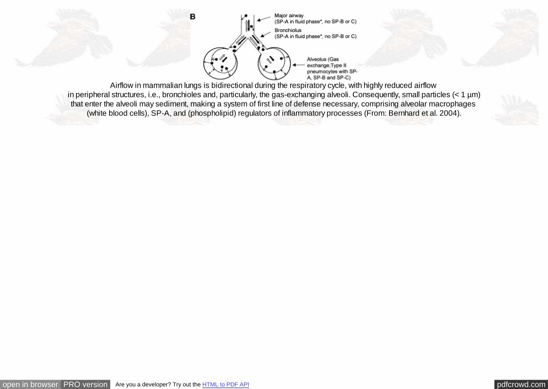
Airflow in mammalian lungs is bidirectional during the respiratory cycle, with highly reduced airflow in peripheral structures, i.e., bronchioles and, particularly, the gas-exchanging alveoli. Consequently, small particles (< 1 µm)that enter the alveoli may sediment, making a system of first line of defense necessary, comprising alveolar macrophages
(white blood cells), SP-A, and (phospholipid) regulators of inflammatory processes (From: Bernhard et al. 2004).

pdfcrowd.comopen in browser PRO version Are you a developer? Try out the HTML to PDF API
A: A high-power view of a foreign particle (p) being engulfed by an epithelial cell (e) in an avian lung. Arrows, elongated microvilli. B: Surface of an atrium of the lung of the domestic fowl showing red blood
cells with one of them (r) being engulfed by the underlying epithelial cell (arrow): e, epithelial surface; m, a free (surface) macrophage. Scale bars: A = 0.5 µm; B = 10 µm (From: Nganpiep and Maina 2002).
Air flow is driven by changes in pressure within the respiratory system:
During inspiration:
pdfcrowd.comopen in browser PRO version Are you a developer? Try out the HTML to PDF API
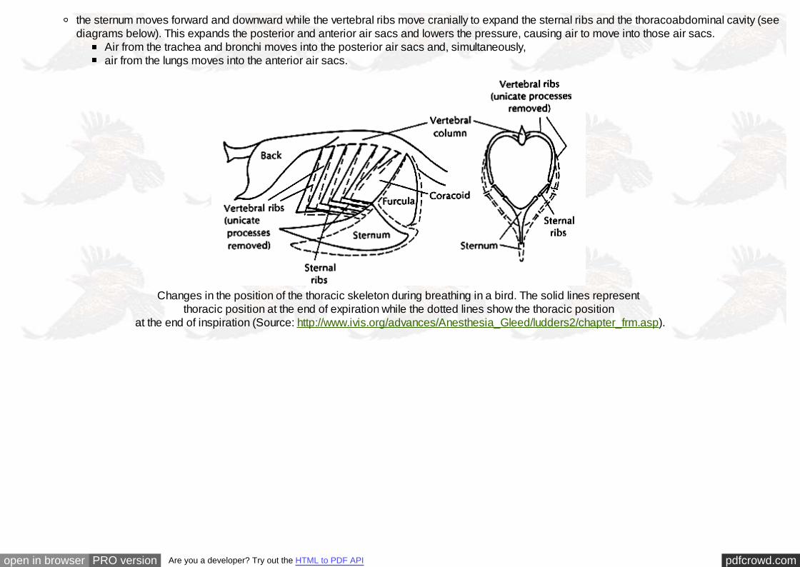
the sternum moves forward and downward while the vertebral ribs move cranially to expand the sternal ribs and the thoracoabdominal cavity (seediagrams below). This expands the posterior and anterior air sacs and lowers the pressure, causing air to move into those air sacs.
Air from the trachea and bronchi moves into the posterior air sacs and, simultaneously,air from the lungs moves into the anterior air sacs.
Changes in the position of the thoracic skeleton during breathing in a bird. The solid lines represent
thoracic position at the end of expiration while the dotted lines show the thoracic position at the end of inspiration (Source: http://www.ivis.org/advances/Anesthesia_Gleed/ludders2/chapter_frm.asp).

pdfcrowd.comopen in browser PRO version Are you a developer? Try out the HTML to PDF API
Drawing of a bird coelom in transverse section during expiration (gray bones) and inspiration (white bones). Dashed lines illustrate the horizontal septum that separates the pleural cavity (PC) where the lungs are located from the subpulmonary cavity (SP) where most
of the air sacs are located (except the abdominals that are in the peritoneal cavity), and the oblique septum that separates the air sacs from the abdominal cavity (AC) and digestive viscera. Both septa insert on the ventral keel of vertebrae. The volume of the pleural cavity changes very little with respiratory rib movements, but the volume of the subpulmonary cavity (and the air sacs) is greatly increased when the oblique
septum is stretched during inspiration (Adapted from: Klein and Owerkowicz 2006). The increase in volume lowers air pressure and draws air into the air sacs.

pdfcrowd.comopen in browser PRO version Are you a developer? Try out the HTML to PDF API
Schematic representation of the lungs and air sacs of a bird and the pathway of gas flow through the pulmonary system during inspiration and expiration. For purposes of clarity, the neopulmonic lung
is not shown. The intrapulmonary bronchus is also known as the mesobronchus. A - Inspiration. B - Expiration Source: http://www.ivis.org/advances/Anesthesia_Gleed/ludders2/chapter_frm.asp
During expiration:the sternum moves backward and upward & the vertebral ribs move caudally to retract the sternal ribs and reduce the volume of thethoracoabdominal cavity. The reduces the volume of the anterior & posterior air sacs, causing air to move out of those sacs.

pdfcrowd.comopen in browser PRO version Are you a developer? Try out the HTML to PDF API
Air from the posterior sacs moves into the lungs &, simultaneously,air from the anterior sacs moves into the trachea & out of the body.
So, air always moves unidirectionally through the lungs and, as a result, is higher in oxygen content than, for example, air in the alveoli of humans and othermammals.
Role of uncinate processes and associatedmuscles in avian respiration -- Codd et al.(2005) examined the activity of three musclesassociated with the uncinate processes, (1)external intercostal, (2) appendicocostalis and(3) external oblique (labeled in drawing to the left)examined using electrodes during sitting,standing and moderate speed treadmill runningin a Giant Canada Goose. The externalintercostal muscles demonstrated no respiratoryactivity, being active only during running,suggesting they play some role in trunkstabilization. The appendicocostalis and externaloblique muscles are respiratory muscles, beingactive during inspiration and expiration,respectively. The activity of theappendicocostalis muscle increased whensternal movements were restricted, suggestingthat activity of these muscles may be particularlyimportant during prolonged sitting such as duringegg incubation. Codd et al. (2005) suggestedthat the uncinate processes in birds facilitatemovements of the ribs and sternum duringbreathing and therefore are integral to thebreathing mechanics of birds.

pdfcrowd.comopen in browser PRO version Are you a developer? Try out the HTML to PDF API
Variation in length of uncinate processes -- Birds with different forms of locomotion exhibit morphologicaldifferences in their rib cages: (A) terrestrial (walking) species, Cassowary (Casuaris casuaris); (B) a typical flyingbird, Eagle Owl (Bubo bubo); and (C) an aquatic, diving species, Razorbill (Alca torda). Uncinate processes are
shorter in walking species, of intermediate length in typical birds, and relatively long in diving species (scale bar, 5cm). Muscles attached to uncinate processes (appendicocostales muscles) help rotate the ribs forwards, pushing
the sternum down and inflating the air sacs during inspiration. Another muscle (external oblique) attached touncinate processes pulls the ribs backward, moving the sternum upward during expiration. The longer uncinate
processes of diving birds are probably related to the greater length of the sternum and the lower angle of the ribs tothe backbone and sternum. The insertion of the appendicocostales muscles near the end of the uncinate
processes may provide a mechanical advantage for moving the elongated ribs during breathing (Tickle et al.2007).
Ultra-Low Oxygen Could Have Spurred Bird Breathing System -- Recent evidence suggests that oxygen levels weresuppressed worldwide 175 - 275 million years ago, low enough to make breathing the air at sea level feel like respiration at highaltitude. Peter Ward, a University of Washington paleontologist, theorizes that low oxygen and repeated short but substantialtemperature increases because of greenhouse warming sparked two major mass-extinction events.In addition, he believes the conditions spurred the development of an unusual breathing system inSaurischian dinosaurs. Rather than having a diaphragm to force air in and out of lungs, theSaurischians had lungs attached to a series of thin-walled air sacs that appear to have functionedsomething like bellows to move air through the body. This breathing system, still found in today'sbirds, made the Saurischian dinosaurs better equipped than mammals to survive the harshconditions in which oxygen content of air at the Earth's surface was only about half of today's 21%."The literature always said that the reason birds had sacs was so they could breathe when they fly.But I don't know of any brontosaurus that could fly," Ward said. "However, when we considered that birds fly at altitudes whereoxygen is significantly lower, we finally put it all together with the fact that the oxygen level at the surface was only 10 - 11% at thetime the dinosaurs evolved. That's the same as trying to breathe at 14,000 feet. If you've ever been at 14,000 feet, you know it's noteasy to breathe," he said.
Ward presented his ideas at the 2003 annual meeting of the American Geological Society in Seattle. See: http://www.nature.com/nsu/031103/031103-7.html

pdfcrowd.comopen in browser PRO version Are you a developer? Try out the HTML to PDF API
Exchange of gases:
In the avian lung, oxygen diffuses (by simple diffusion) from the air capillaries into the blood & carbon dioxide from the blood into the air capillaries (shown inthis figure and in figures below ). This exchange is very efficient in birds for a number of reasons. First, the complex arrangement of blood and air capillariesin the avian lung creates a substantial surface area through which gases can diffuse. The surface area available for exchange (SAE) varies with bird size.For example, the ASE is about 0.17 m 2 for House Sparrows (about 30 gms; Passer domesticus), 0.9 m 2 for Rock Pigeons (about 350 gms; Columbalivia), 3.0 m 2 for a Mallard (about 1150 gms; Anas platyrhynchos), and 8.9 m 2 for a male Graylag Goose (about 3.7 kg; Anser anser) (Maina 2008).However, smaller birds have a greater SAE per unit mass than do larger birds. For example, the SAE is about 90 cm 2/gm for Violet-eared Hummingbirds(Colibri coruscans; Dubach 1981), about 26 cm 2/gm for Mallards, and about 5.4 cm 2/gm for Emus (Dromaius novaehollandiae; Maina and King 1989).Among mammals, there is also a negative relationship between SAE and body size, with smaller mammals like shrews having a greater SAE per unit massthan larger mammals. However, for birds and mammals of similar size, the SAE of birds is generally about 15% greater (Maina et al. 1989).
A second reason why gas exchange in avian lungs is so efficient is that the blood-gas barrier through which gases diffuse is extremely thin. This is importantbecause the amount of gas diffusing across this barrier is inversely proportional to its thickness. Among terrestrial vertebrates, the blood-gas barrier isthinnest in birds. Natural selection has favored thinner blood-gas barriers in birds and mammals because endotherms use oxygen at higher rates thanectotherms like amphibians and reptiles. Among birds, the thickness of the blood-gas barrier varies, with smaller birds generally having thinner blood-gasbarriers than larger birds. For example, the blood-gas barrier is 0.099 μm thick in Violet-eared Hummingbirds and 0.56 μm thick in Ostriches (West 2009).
Comparison of the mean thickness of the blood-gas barrier of 34 species of birds, 37 species of mammals, 16 species of reptiles, and 10 species of amphibians revealed that birds had significantly thinner blood-gas
barriers than the other taxa (West 2009).
Also contributing to the efficiency of gas exchange in avian lungs is a process called cross-current exchange. Air passing through air capillaries and bloodmoving through blood capillaries generally travel at right angles to each other in what is called cross-current flow (Figure below; Makanya and Djonov 2009).As a result, oxygen diffuses from the air capillaries into the blood at many points along the length of the parabronchi, resulting in a greater concentration ofoxygen (i.e., higher partial pressures) in the blood leaving the lungs than is possible in the alveolar lungs of mammals (Figures below).

pdfcrowd.comopen in browser PRO version Are you a developer? Try out the HTML to PDF API
Diagram of parabronchial anatomy, gas-exchange region of the bird's lung-air-sacrespiratory system. The few hundred tothousand parabronchi, one of which is fullyshown here, are packed tightly into ahexagonal array. The central parabronchiallumen, through which gas flows unidirectionallyduring both inspiration and expiration issurrounded by gas-exchange tissue composedof an intertwined network of blood and aircapillaries. On the left side of this diagram, thelumen of the parabronchus leads into multiplechambers called atria (A) that, in turn, lead intosmaller chambers called infundibulae (I).Branching from the infundibulae are numerousair capillaries. On the right side of this diagramare the blood vessels. Arteries (a) lead into thecapillaries that are closely associated with theair capillaries. It is here (air and bloodcapillaries) where oxygen and carbon dioxideare exchanged. After flowing through thecapillaries, blood then moves into the veins (v)that will take the blood out of the lungs (From:Duncker 1971 as reprinted in Powell 2000).

pdfcrowd.comopen in browser PRO version Are you a developer? Try out the HTML to PDF API
Three-dimensional reconstruction of the gas-exchangeregion.
AC = air capillaries. Several air capillaries coalesce intoan infundibulum (INF) (Brown et al. 1997).
In this cross-section, note the intertwined network of blood capillaries, labeled with the presence of
erythrocytes (*), and air capillaries (AC) that make up the parabronchi'smantle of gas-exchange tissue (Brown et al. 1997).
pdfcrowd.comopen in browser PRO version Are you a developer? Try out the HTML to PDF API
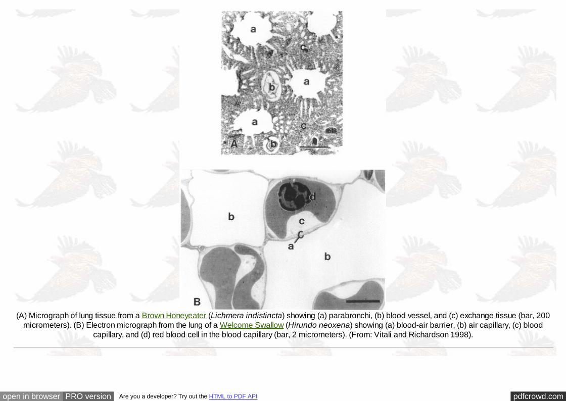
(A) Micrograph of lung tissue from a Brown Honeyeater (Lichmera indistincta) showing (a) parabronchi, (b) blood vessel, and (c) exchange tissue (bar, 200micrometers). (B) Electron micrograph from the lung of a Welcome Swallow (Hirundo neoxena) showing (a) blood-air barrier, (b) air capillary, (c) blood
capillary, and (d) red blood cell in the blood capillary (bar, 2 micrometers). (From: Vitali and Richardson 1998).

pdfcrowd.comopen in browser PRO version Are you a developer? Try out the HTML to PDF API
A) Medial view of the lung of a domestic chicken (Gallus gallus domesticus). p, primary bronchus; v, ventrobronchus; d, dorsobronchus; r, parabronchi.Scale bar, 1 cm. (B) An intraparabronchial artery (i) giving rise to blood capillaries (c) in the lung of an Emu (Dromiceus novaehollandiae). a, air capillaries.Scale bar, 15 μm. (C) Air capillaries closely associated with blood capillaries (arrows) in a chicken lung. Scale bar, 10 μm. (D) Blood capillaries (c) closely
associated with air capillaries (spaces) in a chicken lung. Scale bar, 12 μm. (From: Maina 2002).
pdfcrowd.comopen in browser PRO version Are you a developer? Try out the HTML to PDF API
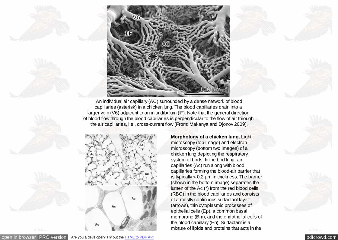
An individual air capillary (AC) surrounded by a dense network of bloodcapillaries (asterisk) in a chicken lung. The blood capillaries drain into a
larger vein (V6) adjacent to an infundibulum (IF). Note that the general direction of blood flow through the blood capillaries is perpendicular to the flow of air through
the air capillaries, i.e., cross-current flow (From: Makanya and Djonov 2009).
Morphology of a chicken lung. Lightmicroscopy (top image) and electronmicroscopy (bottom two images) of achicken lung depicting the respiratorysystem of birds. In the bird lung, aircapillaries (Ac) run along with bloodcapillaries forming the blood-air barrier thatis typically < 0.2 µm in thickness. The barrier(shown in the bottom image) separates thelumen of the Ac (*) from the red blood cells(RBC) in the blood capillaries and consistsof a mostly continuous surfactant layer(arrows), thin cytoplasmic processes ofepithelial cells (Ep), a common basalmembrane (Bm), and the endothelial cells ofthe blood capillary (En). Surfactant is amixture of lipids and proteins that acts in the

pdfcrowd.comopen in browser PRO version Are you a developer? Try out the HTML to PDF API
air capillaries of avian lungs both as an"antiglue" (preventing the adhesion ofrespiratory surfaces that may occur when thelungs collapse, e.g., during diving,swallowing of prey or on expiration) and toprevent liquid influx into the lungs (Daniels etal. 1998). Magnifications: top image - ×270;middle image - ×1,600; bottom image -×88,000 (Image from Bernhard et al. 2001).
In birds, the thickness of the blood-gas barrier in the 7.3-g Violet-eared Hummingbird (Colibri coruscans) is 0.099 µm, whereas that of an immature 40-kgOstrich (Struthio camelus) is 0.56 µm (Maina and West 2005).
Relationship between the harmonic mean thickness of the blood-gas barrier (the thickness of the barrier that affects the diffusion of oxygen from aircapillaries into blood capillaries) against body mass in the lungs of bats, birds, and non-flying mammals. Birds have particularly thinner barriers than bats
and non-flying mammals (Maina 2000).

pdfcrowd.comopen in browser PRO version Are you a developer? Try out the HTML to PDF API
Light micrographs of a portion of the lung of a chicken (A) and rabbit (B). Note the small diameter of the air capillaries in the chicken lung vs. that of the rabbit alveoli (same magnification).(A) In the chicken lung, pulmonary capillaries are supported by 'struts' of epithelium (arrows). (B) In the rabbit lung,
pulmonary capillaries are suspended in the large spaces between alveoli (Watson et al. 2007).
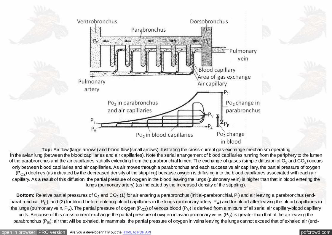
Cross-current exchange:
pdfcrowd.comopen in browser PRO version Are you a developer? Try out the HTML to PDF API
Top: Air flow (large arrows) and blood flow (small arrows) illustrating the cross-current gas-exchange mechanism operatingin the avian lung (between the blood capillaries and air capillaries). Note the serial arrangement of blood capillaries running from the periphery to the lumenof the parabronchus and the air capillaries radially extending from the parabronchial lumen. The exchange of gases (simple diffusion of O2 and CO2) occurs
only between blood capillaries and air capillaries. As air moves through a parabronchus and each successive air capillary, the partial pressure of oxygen(PO2) declines (as indicated by the decreased density of the stippling) because oxygen is diffusing into the blood capillaries associated with each air
capillary. As a result of this diffusion, the partial pressure of oxygen in the blood leaving the lungs (pulmonary vein) is higher than that in blood entering thelungs (pulmonary artery) (as indicated by the increased density of the stippling).
Bottom: Relative partial pressures of O2 and CO2 (1) for air entering a parabronchus (initial-parabronchial, PI) and air leaving a parabronchus (end-parabronchial, PE), and (2) for blood before entering blood capillaries in the lungs (pulmonary artery, PA) and for blood after leaving the blood capillaries inthe lungs (pulmonary vein, PV). The partial pressure of oxygen (PO2) of venous blood (PV) is derived from a mixture of all serial air capillary-blood capillary
units. Because of this cross-current exchange the partial pressure of oxygen in avian pulmonary veins (PV) is greater than that of the air leaving theparabronchus (PE); air that will be exhaled. In mammals, the partial pressure of oxygen in veins leaving the lungs cannot exceed that of exhaled air (end-

pdfcrowd.comopen in browser PRO version Are you a developer? Try out the HTML to PDF API
expiratory gas, or PE) (Figure adapted from Scheid and Piiper 1987). Importantly, the partial pressure of oxygen in blood leaving the avian lung is the resultof 'mixing'; blood from a series of capillaries associated with successive air capillaries along the length of a parabronchus is mixed as the blood leaves thecapillaries and enters small veins. As a result, the direction of air flow through a parabronchus does not effect the efficiency of the cross-current exchange(because gases are only exchanged between blood capillaries and air capillaries, not between the parabronchus and the blood). So, in above diagram,
reversing the direction of air flow would obviously mean that the air capillary on the far left would have the highest partial pressure of oxygen rather than theair capillary on the far right (so the stippling pattern that indicates the amount of oxygen in each air capillary would be reversed). However, because of the
'mixing' of blood just mentioned, this reversal would have little effect on the PV, the partial pressure of oxygen in blood leaving via pulmonary veins (the PO2would likely be a bit lower because some oxygen would have been lost the first time air passed through the neopulmonic parabronchi). This is importantbecause most birds have neopulmonic parabronchi as well as paleopulmonic parabronchi and, although air flow through paleopulmonic parabronchi is
unidirectional, air flow through neopulmonic parabronchi is bidirectional.
Diagram showing the flow of air from the parabronchial lumen (PL) into the air capillaries (not shown) and arterial blood from the periphery of the

pdfcrowd.comopen in browser PRO version Are you a developer? Try out the HTML to PDF API
parabronchus into the area of gas exchange (exchange tissue, ET). The orientation between the flow of air along the parabronchus and that of blood into the exchange tissue (ET) from the periphery is perpendicular or cross-current (dashed arrows). The exchange tissue is supplied with arterial blood
by interparabronchial arteries (IPA) that give rise to arterioles (stars) that terminate in blood capillaries. After passing through the capillaries, blood flows into the intraparabronchial venules (asterisks) that drain into interparabronchial veins (IPV). These in turn empty into the pulmonary vein which returns the
blood to the heart. (From: Maina and Woodward 2009).
Control of Ventilation:
Ventilation and respiratory rate are regulated to meet the demands imposed by changes in metabolic activity (e.g., rest and flight) as well as other sensoryinputs (e.g., heat and cold). There is likely a central respiratory control center in the avian brain, but this has not been unequivocally demonstrated. As inmammals, the central control area appears to be located in the pons and medulla oblongata with facilitation and inhibition coming from higher regions of thebrain. It also appears that the chemical drive on respiratory frequency and inspiratory and expiratory duration depend on feedback from receptors in the lungas well as on extrapulmonary chemoreceptors, mechanoreceptors, and thermoreceptors (Ludders 2001).
Central chemoreceptors affect ventilation in response to changes in arterial PCO2 and hydrogen ion concentration. Peripheral extrapulmonarychemoreceptors, specifically the carotid bodies (located in the carotid arteries), are influenced by PO2 and increase their discharge rate as PO2 decreases,thus increasing ventilation; they decrease their rate of discharge as PO2 increases or PCO2 decreases. These responses are the same as those observed inmammals. Unlike mammals, birds have a unique group of peripheral receptors located in the lung called intrapulmonary chemoreceptors (IPC) that areacutely sensitive to carbon dioxide and insensitive to hypoxia. The IPC affect rate and volume of breathing on a breath-to-breath basis by acting as theafferent limb of an inspiratory-inhibitory reflex that is sensitive to the timing, rate, and extent of CO2 washout from the lung during inspiration (Ludders 2001).
Respiration by Avian Embryos
During avian development there are three sequential stages of respiration(Tazawa 1987): prenatal (embryonic), paranatal (hatching), and postnatal(posthatching). During the prenatal stage respiratory gas exchange occurs viadiffusion between the external environment and the initial gas exchanger (i.e., thearea vasculosa, or the region of blood island formation and forerunner of thechorioallantoic membrane) in early embryonic life and later the vascular bed of thechorioallantois. The paranatal stage starts when the beak penetrates into the airpocket (air cell) between the inner and outer shell membranes (both internal toshell; i.e., internal pipping) this occurs during the last 2-3 days of incubation.During this stage, the lungs begin to replace the chorioallantois as the gasexchanger, yet diffusion remains the major mechanism moving gas across theshell. The postnatal stage begins when the beak penetrates the shell (i.e., externalpipping) (Brown et al. 1997).
Source: www.ece.utexas.edu/~bevans/courses/. . .

pdfcrowd.comopen in browser PRO version Are you a developer? Try out the HTML to PDF API
Chicken embryo
Literature Cited:
Bernhard, W., A. Gebert, G. Vieten, G. A. Rau1, J. M. Hohlfeld, A. D. Postle, and J. Freihorst. 2001. Pulmonary surfactant in birds: coping with surfacetension in a tubular lung. American Journal of Physiology - Regulatory Integrative and Comparative Physiology 281: R327-R337.
Bernhard, W., P. L. Haslam, and J. Floros. 2004. From birds to humans: new concepts on airways relative to alveolar surfactant. American Journal ofRespiratory Cell and Molecular Biology 30: 6-11.
Brown, R.E., J. D. Brain, and N. Wang. 1997. The avian respiratory system: a unique model for studies of respiratory toxicosis and for monitoring air quality.Environ Health Perspectives 105:188-200.
Codd, J. R., D. F. Boggs, S. F. Perry, and D. R. Carrier. 2005. Activity of three muscles associated with the uncinate processes of the giant Canada GooseBranta canadensis maximus. Journal of Experimental Biology 208:849-857.
Daniels, C.B., O. V. Lopatko, and S. Orgeig. 1998. Evolution of surface activity related functions of vertebrate pulmonary surfactant. Clin Exp PharmacolPhysiol. 25:716-721.
Dubach, M. 1981. Quantitative analysis of the respiratory system of the House Sparrow, Budgerigar, and Violet-eared Hummingbird. Respiration Physiology46: 43-60.

pdfcrowd.comopen in browser PRO version Are you a developer? Try out the HTML to PDF API
Duncker, H.-R. 1971. The lung air sac system of birds. Advances in Anatomy, Embryology, and Cell Biology 45: 1–171.
Duncker, H.-R. 2004. Vertebrate lungs: structure, topography and mechanics: A comparative perspective of the progressive integration of respiratorysystem, locomotor apparatus and ontogenetic development. Respiratory Physiology & Neurobiology 144: 111-124.
Klein, W., and T. Owerkowicz. 2006. Function of intracoelomic septa in lung ventilation of amniotes: lessons from lizards. Physiological and BiochemicalZoology 79: 1019-1032.
Ludders, J.W. 2001. Inhaled anesthesia for birds. In: Recent advances in veterinary anesthesia and analgesia: companion animals (R. D. Gleed and J. W.Ludders, eds.). International Veterinary Information Service, Ithaca, NY. (www.ivis.org/advances/Anesthesia_Gleed/ludders2/chapter_frm.asp)
Maina, J.N. 1989. The morphometry of the avian lung. Pp. 307-368 in Form and function in birds (A.S. King and J. McLelland, eds.). Academic Press,London.
Maina, J. N. 2000. Comparative respiratory morphology: Themes and principles in the design and construction of the gas exchangers. Anatomical Record 261: 25-44.
Maina, J. N. 2002. Structure, function and evolution of the gas exchangers: comparative perspectives. Journal of Anatomy 201: 281-304.
Maina, J. N. 2008. Functional morphology of the avian respiratory system, the lung-air system: efficiency built on complexity. Ostrich 79: 117-132.
Maina, J. N., and A. S. King. 1989. The lung of the Emu, Dromaius novaehollandiae: a microscopic and morphometric study. Journal of Anatomy 163: 67-74.
Maina, J. N., A. S. King, and G. Settle. 1989. An allometric study of the pulmonary morphometric parameters in birds, with mammalian comparison.Philosophical Transactions of the Royal Society of London B 326: 1-57.
Maina, J. N., and J. B. West. 2005. Thin and strong! The bioengineering dilemma in the structural and functional design of the blood-gas barrier. Physiol. Rev. 85: 811-844.
Maina, J. N., and C. Nathaniel. 2001. A qualitative and quantitative study of the lung of an Ostrich, Struthio camelus. Journal of Experimental Biology 204:2313-2330.
Maina, J. N., and J. D. Woodward. 2009. Three-dimensional serial section computer reconstruction of the arrangement of the structural components of theparabronchus of the Ostrich, Struthio camelus lung. Anatomical Record 292: 1685-1698.
Makanya, A. N., and V. Djonov. 2009. Parabronchial angioarchitecture in developing and adult chickens. Journal of Applied Physiology 106: 1959-1969,2009.
McLelland, J. 1989. Anatomy of the lungs and air sacs. In: Form and function in birds, vol. 4 (A. S. King and J. McLelland, eds.), pp. 221-279. AcademicPress, San Diego, CA.
Nganpiep, L. N. and J. N. Maina. 2002. Composite cellular defence stratagem in the avian respiratory system: functional morphology of the free (surface)macrophages and specialized pulmonary epithelia. Journal of Anatomy 200: 499-516.

pdfcrowd.comopen in browser PRO version Are you a developer? Try out the HTML to PDF API
O'Connor, P. M. and L. P. Claessens. 2005. Basic avian pulmonary design and flow-through ventilation in non-avian theropod dinosaurs. Nature 436:253-256.
Plummer, E. M., and F. Goller. 2008. Singing with reduced air sac volume causes uniform decrease in airflow and sound amplitude in the Zebra Finch. Journal of ExperimentalBiology 211: 66-78.
Powell, F.L. 2000. Respiration. Pp. 233-264 in Avian physiology, fifth edition (G. Causey Whittow, ed.). Academic Press, New York, NY.
Powell, F. L. and S. R. Hopkins. 2004. Comparative physiology of lung complexity: implications for gas exchange. News in Physiological Science 19:55-60.
Reese, S., G. Dalamani, and B. Kaspers. 2006. The avian lung-associated immune system: a review. Vet. Res. 37: 311-324.
Scheid, P., and J. Piiper. 1987. Gas exchange and transport. In: Bird respiration, volume 1 (T. J. Seller, ed.), pp. 97-129. CRC Press, Inc., Boca Raton, FL.
Sereno, P. C., R. N. Martinez, J. A. Wilson, D. J. Varricchio, O. A. Alcober, and H. C. E. Larsson. 2008. Evidence for avian intrathoracic air sacs in a newpredatory dinosaur from Argentina. PLoS ONE 3(9): e3303.
Tazawa, H. 1987. Embryonic respiration. Pp. 3 - 24 in Bird respiration, vol. 2 (T. J. Seller, ed.). CRC Press, Boca Raton, FL.
Tickle, P. G., A. R. Ennos, L. E. Lennox, S. F. Perry, and J. R. Codd. 2007. Functional significance of the uncinate processes in birds. Journal ofExperimental Biology 210: 3955-3961.
Vitali, S. D., and K. C. Richardson. 1998. Evaluation of pulmonary volumetric morphometry at the light and electron microscopy level in several species ofpasserine birds. Journal of Anatomy 193: 573-580.
Watson, R. R., Z. Fu, and J. B. West. 2007. Morphometry of the extremely thin pulmonary blood-gas barrier in the chicken lung. American Journal ofPhysiology. Lung Cellular and Molecular Physiology 36: L769-L777.
Wedel, M.J. 2003. Vertebral pneumaticity, air sacs, and the physiology of sauropod dinosaurs. Paleobiology 29: 243–255.
Welty, J.C. and L. Baptista. 1988. The life of birds, fourth edition. Saunders College Publishing, New York, NY.
West, J. B. 2009. Comparative physiology of the pulmonary blood-gas barrier: the unique avian solution. American Journal of Physiology - Regulatory,Integrative and Comparative Physiology 297: R1625-R1634.
West, J. B., R. R. Watson, and Z. Fu. 2007. The human lung: did evolution get it wrong? European Respiratory Journal 29: 11-17.
Useful links:
How Animals Work: Avian Respiratory Dynamics Animation

pdfcrowd.comopen in browser PRO version Are you a developer? Try out the HTML to PDF API
More lecture notes:
Energy Balance & Thermoregulation
Back to BIO 554/754 Syllabus
Back to Avian Biology