biphasic growth of orbital volume in chinese children · biphasic growth of orbital volume in...
TRANSCRIPT

Biphasic growth of orbital volume in ChinesechildrenNan Wei,1 Hua Bi,2 Bin Zhang,2 Xue Li,1 Fengyuan Sun,1 Xuehan Qian1
1Department ofOphthalmology, TianjinMedical University EyeHospital, Tianjin, China2College of Optometry, NovaSoutheastern University, Davie,Florida, USA
Correspondence toDr Xuehan Qian, Departmentof Ophthalmology, TianjinMedical University EyeHospital, 251 Fukang Road,Nankai District, Tianjin300384, China;[email protected]
Received 28 October 2016Revised 31 January 2017Accepted 6 February 2017
To cite: Wei N, Bi H,Zhang B, et al. Br JOphthalmol PublishedOnline First: [please includeDay Month Year]doi:10.1136/bjophthalmol-2016-309848
ABSTRACTObjective The aim of this study was to map out thedevelopmental curve of the orbital volume of Chinesechildren aged 1–15 years.Methods CT scanning was performed on 109 childrenand the orbital volume, interlateral orbital rim distance(IORD), and extent of exophthalmos were measured onthe CT images and plotted against age.Results The development of the orbit structurefollowed a biphasic pattern. The first growth phase wasbefore 3 years and the second growth phase wasbetween 7 years and 12 years of age. The growth speedin the first phase was about 3 times that of the secondone (first vs second phase: 2.28 cm3/year vs 0.67 cm3/year for orbital volume, 5.01 mm/year vs 1.57 mm/yearfor IORD, 1.29 mm/year vs 0.42 mm/year for theexophthalmos). During development, there was nosignificant difference between the left and right orbits.There was no significant difference between boys andgirls before 12 years of age. However, after 12 years ofage, boys had significantly larger orbital volumes (22.16±2.28 cm3/year vs 18.57±1.16 cm3/year, p<0.001) anda greater IORD (96.29±3.18 mm/year vs 91.00±4.54mm/year, p<0.001) than girls.Conclusions In Chinese children, the development oforbital volume follows a biphasic pattern and a sexdifference becomes significant after the age of 12 years.
INTRODUCTIONThe dynamic development of orbital volumeduring childhood creates several challenges forpaediatric surgeons in clinical work.1 2 Thesurgeon needs to refer to the normal value at a spe-cific age to provide a sufficient ocular implant afterremoving orbital content. By doing so, the orbitalbones can be adequately stimulated and this canpreserve partial or even full development, whichwill reduce the risk of craniofacial deformation inadulthood.3–5 In addition, it is important to selectthe optimal timing of the surgery.6–9 If the condi-tion starts during a period of fast development, it ispreferable to perform the surgery as early as pos-sible. However, if it happens during a period ofslow development, it might be worth waiting forthe child to become physically stronger beforeundergoing the risks of surgery. It is also importantto know at which age separate standards should beused for boys and girls because of sex differ-ences.1 2 10 11 Therefore, mapping out the normaldevelopmental timing curve of the orbits duringchildhood is of great clinical significance.Although several studies have been done in this
field, some important controversies still exist. It isnot clear if the orbital volume development followsa linear pattern with an even speed at any age, or if
the developmental process has a certain phase withfast development and a certain phase with a slowerspeed. Although boys tend to have a larger orbitalvolume than girls, at which age the sex differencebecomes significant is still debateable.1 2 10 11 Dueto factors such as race, ethnicity and geographicallocations, the normal developmental values col-lected from previous studies show large variations.There is relatively a lack of relevant data on thenormal development of orbital volumes in Chinesechildren.10 12
Therefore, the aim of this study was to estimatethe orbital volumes in Chinese children up to15 years of age using a spiral CT scan that has highaccuracy and good repeatbility. The collected datacould help with answer to address the controversiesin this field.
METHODSSubjectsThe subjects, all of Han ethnicity, were childrenfrom many different parts of China who visited theDepartment of Ophthalmology at Tianjin MedicalUniversity for various ophthalmic diseases. CTscans were performed on each child. Children whohad conditions that may disrupt the normaldevelopment of orbital structures, such as orbitallesions, craniofacial deformity or orbital fractureswere excluded from the study. A total of 110 chil-dren were recruited, including 76 boys (69.1%)and 34 girls. The detailed breakdown of age andsex information is shown in table 1. All subjectsand their guardians have been informed the detailsof the study and written consent forms were signedby their legal guardians.
CT scanThe CT scans were performed on a double spiralCT (Prospeed, General Electronics, Chicago, USA).The voltage was set at 120 kV and the current wasset at 150 mA. Each slice was 2 mm thick with apitch distance of 1.5 mm to form a matrix of512×512 pixels with a 200 mm field of view.Scanning was done in a horizontal orientation withthe line connecting the external wall of the orbitand the external auditory canal was the baseline.13
MeasurementsOrbital volume: For each slice of scanning, aviewing window was set at 2000×200 HounsfieldUnits (HU) (width×height), and the graphic pro-cessing software that came with the CT machinewas used to perform manual tracing of the orbitalboundaries. The interface between the bone wallsand soft tissue contents of the orbits was outlined.With the chosen setting, bone appeared as a high
Wei N, et al. Br J Ophthalmol 2017;0:1–6. doi:10.1136/bjophthalmol-2016-309848 1
Clinical science BJO Online First, published on March 3, 2017 as 10.1136/bjophthalmol-2016-309848
Copyright Article author (or their employer) 2017. Produced by BMJ Publishing Group Ltd under licence.
on June 1, 2020 by guest. Protected by copyright.
http://bjo.bmj.com
/B
r J Ophthalm
ol: first published as 10.1136/bjophthalmol-2016-309848 on 3 M
arch 2017. Dow
nloaded from
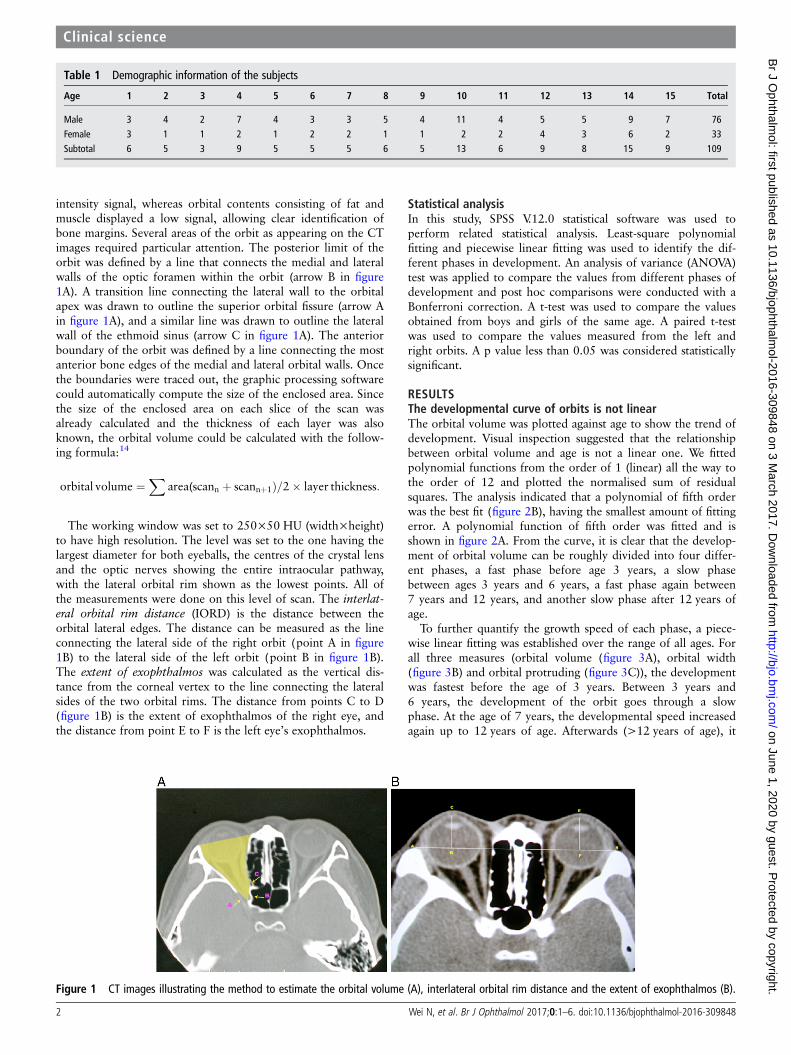
intensity signal, whereas orbital contents consisting of fat andmuscle displayed a low signal, allowing clear identification ofbone margins. Several areas of the orbit as appearing on the CTimages required particular attention. The posterior limit of theorbit was defined by a line that connects the medial and lateralwalls of the optic foramen within the orbit (arrow B in figure1A). A transition line connecting the lateral wall to the orbitalapex was drawn to outline the superior orbital fissure (arrow Ain figure 1A), and a similar line was drawn to outline the lateralwall of the ethmoid sinus (arrow C in figure 1A). The anteriorboundary of the orbit was defined by a line connecting the mostanterior bone edges of the medial and lateral orbital walls. Oncethe boundaries were traced out, the graphic processing softwarecould automatically compute the size of the enclosed area. Sincethe size of the enclosed area on each slice of the scan wasalready calculated and the thickness of each layer was alsoknown, the orbital volume could be calculated with the follow-ing formula:14
orbital volume ¼X
area(scann þ scannþ1Þ=2� layer thickness:
The working window was set to 250×50 HU (width×height)to have high resolution. The level was set to the one having thelargest diameter for both eyeballs, the centres of the crystal lensand the optic nerves showing the entire intraocular pathway,with the lateral orbital rim shown as the lowest points. All ofthe measurements were done on this level of scan. The interlat-eral orbital rim distance (IORD) is the distance between theorbital lateral edges. The distance can be measured as the lineconnecting the lateral side of the right orbit (point A in figure1B) to the lateral side of the left orbit (point B in figure 1B).The extent of exophthalmos was calculated as the vertical dis-tance from the corneal vertex to the line connecting the lateralsides of the two orbital rims. The distance from points C to D(figure 1B) is the extent of exophthalmos of the right eye, andthe distance from point E to F is the left eye’s exophthalmos.
Statistical analysisIn this study, SPSS V.12.0 statistical software was used toperform related statistical analysis. Least-square polynomialfitting and piecewise linear fitting was used to identify the dif-ferent phases in development. An analysis of variance (ANOVA)test was applied to compare the values from different phases ofdevelopment and post hoc comparisons were conducted with aBonferroni correction. A t-test was used to compare the valuesobtained from boys and girls of the same age. A paired t-testwas used to compare the values measured from the left andright orbits. A p value less than 0.05 was considered statisticallysignificant.
RESULTSThe developmental curve of orbits is not linearThe orbital volume was plotted against age to show the trend ofdevelopment. Visual inspection suggested that the relationshipbetween orbital volume and age is not a linear one. We fittedpolynomial functions from the order of 1 (linear) all the way tothe order of 12 and plotted the normalised sum of residualsquares. The analysis indicated that a polynomial of fifth orderwas the best fit (figure 2B), having the smallest amount of fittingerror. A polynomial function of fifth order was fitted and isshown in figure 2A. From the curve, it is clear that the develop-ment of orbital volume can be roughly divided into four differ-ent phases, a fast phase before age 3 years, a slow phasebetween ages 3 years and 6 years, a fast phase again between7 years and 12 years, and another slow phase after 12 years ofage.
To further quantify the growth speed of each phase, a piece-wise linear fitting was established over the range of all ages. Forall three measures (orbital volume (figure 3A), orbital width(figure 3B) and orbital protruding (figure 3C)), the developmentwas fastest before the age of 3 years. Between 3 years and6 years, the development of the orbit goes through a slowphase. At the age of 7 years, the developmental speed increasedagain up to 12 years of age. Afterwards (>12 years of age), it
Table 1 Demographic information of the subjects
Age 1 2 3 4 5 6 7 8 9 10 11 12 13 14 15 Total
Male 3 4 2 7 4 3 3 5 4 11 4 5 5 9 7 76Female 3 1 1 2 1 2 2 1 1 2 2 4 3 6 2 33Subtotal 6 5 3 9 5 5 5 6 5 13 6 9 8 15 9 109
Figure 1 CT images illustrating the method to estimate the orbital volume (A), interlateral orbital rim distance and the extent of exophthalmos (B).
2 Wei N, et al. Br J Ophthalmol 2017;0:1–6. doi:10.1136/bjophthalmol-2016-309848
Clinical science on June 1, 2020 by guest. P
rotected by copyright.http://bjo.bm
j.com/
Br J O
phthalmol: first published as 10.1136/bjophthalm
ol-2016-309848 on 3 March 2017. D
ownloaded from

slowed down. Fittings to all three developmental measures weresignificant as indicated by the F values and p values in eachpanel of figure 3.
Over the first 15 years of age, the orbit grew a lot, with a sig-nificant increase in orbital volume, IORD and the extent ofexophthalmos (one-way ANOVA, p<0.001 in all three tests).The development of the orbital volume was fastest in the firstphase (<3 years of age) with a slope of about 2.28. In thesecond phase (3–6 years of age), there was not much develop-ment at all with the slope close to zero. During the third phase(7–12 years of age), the development accelerated again with theslope increased to 0.67. In the last stage (>12 years of age), thedevelopment slowed down again with the slope reduced to 0.11(table 2A and B). Similar patterns could be seen in the develop-ment of the IORD and exophthalmos, with the fastest develop-ment (steepest slopes) appearing before 3 years of age and thesecond fastest development appearing between 7 years and12 years of age. The development in the other two phases wasrather slow (table 2).
The left eye and right eye have no differences duringdevelopmentThe values from the right and left orbits were highly correlatedfor both the mean orbital volume (r=0.98) and the extent ofexophthalmos (r=0.96). In each developmental phase, a pairedt-test revealed no statistically significant difference between thetwo sides (table 3).
The sex difference in orbital developmentThe overall developmental trends were very similar in boys andgirls. Both groups demonstrated a fast phase before 3 years of
age, slow phases between 3 years and 6 years, another fastphase between 7 years and 12 years, and a final slow phase after12 years of age. This developmental pattern was clear fororbital volume, IORD and the extent of exophthalmos.
However, there were some subtle differences between boysand girls during the course of development. In the first two fastphases (up to 6 years of age), there was no difference in orbitalvolume (p=0.227 and p=0.62, respectively). In the second fastphase (7–12 years of age), the difference between boys and girlsstarted to increase, with boys showing a larger orbital volume.The difference was not quite significant (p=0.075, table 4). Inthe final slow phase (>13 years of age), the orbital volumes inthe boys were significantly larger than those of the girls (figure4A, p<0.001). A similar pattern could be found in the develop-ment of the interlateral orbital rim distance, with the differencebetween boys and girls becoming larger and reaching a statistic-ally significant level after 12 years of age (figure 4B and table 4,p<0.001). For the development of the extent of exophthalmos,the difference between boys and girls never reached the signifi-cance level (figure 4C and table 4).
The correlation between orbital volume, IORD andexophthalmosFor the subjects studied in this project, their orbital volumes,interlateral orbital rim distances and the extent of exophthalmoswere significantly correlated with each other linearly (figure 5).The correlation between the orbital volume and IORD was thestrongest (R=0.83, p<0.01). In contrast, the correlationsbetween the extent of exophthalmos and IORD and orbitalvolume were relatively weaker (R=0.69, p<001; R=0.49,p<0.01, respectively).
Figure 2 Developmental trend oforbital volume. (A) Developmentaldata fitted into a polynomial functionof the fifth order. (B) Fitting errorsversus the order of polynomialfunctions.
Figure 3 Piecewise linear fitting showing the developmental trend of orbital volumes (A), orbital width (B) and orbital protruding (C). In eachpanel, each circle represents a subject. Black lines represent the developmental trend and blue diamonds represent the mean value±SD of subjectsin each of the developmental phases, outlined by the grey vertical lines.
Wei N, et al. Br J Ophthalmol 2017;0:1–6. doi:10.1136/bjophthalmol-2016-309848 3
Clinical science on June 1, 2020 by guest. P
rotected by copyright.http://bjo.bm
j.com/
Br J O
phthalmol: first published as 10.1136/bjophthalm
ol-2016-309848 on 3 March 2017. D
ownloaded from

DISCUSSIONPotential explanations for the biphasic developmentConsidering the development of structures close to orbits, thebiphasic development of the orbital volume could potentially beexplained in the following way. The first fast phase, before3 years age, is mainly determined by the fast growth of facialbones, the eyeball and orbital bones. At birth, the cranium toface ratio is 8:1. By 5 years of age, it is reduced to 4:1, and sta-bilises to 2:1 in adulthood.15 The fastest development of theeyeball occurs within the first year, when it reaches an axiallength of 22.5–23 mm. Between 3 years and 14 years of age,the eyeball only increases by 1 mm in axial length. For orbitalbone growth, the first year after birth is often considered to bethe critical period.2 10 The orbital bone develops most quicklyduring the first 3 years.3 16 During the second fast phase orbitaldevelopment is closely related to the process of sinus
pneumatisation.17 The ethmoid sinuses grow gradually to adultsize by the age of 12 years. The maxillary sinuses are the first todevelop. The growth of these paired sinuses is biphasic, occur-ring between 0–3 years and 7–12 years of age. This matchedour biphasic development of orbital volume extremely well. Thefrontal sinus pneumatisation begins around 7 years of age,which also matches the start of the second phase.
Clinical implicationsIn the management of the nasolacrimal duct obstruction, oneoften has to choose between conservative medical therapy,probing and silicone intubation, and dacryocystorhinostomy(DCR).18 The reasons that some paediatricians may hesitate toperform DCR on children include the poorly defined and chan-ging anatomy, rapidly growing facial bone, and a vigorous repairprocess producing hyperplastic scar tissue.19 Here, we reported
Table 2 Statistics of orbital volume, interlateral orbital rim distance (IORD) and exophthalmos in the four developmental stages
A
Age (years) N
Volume (cm3) IORD (mm) Exophthalmos (mm)
Mean±SD Mean±SD Mean±SD
<3 11 14.72±1.76 2.28 78.73±4.96 5.01 11.80±1.33 1.293–6 22 16.79±1.15 −0.08 84.54±2.91 0.44 12.77±1.42 −0.077–12 44 18.82±2.27 0.67 90.41±4.35 1.57 13.95±1.76 0.42>12 32 20.92±2.61 0.11 94.47±4.445 −0.16 15.31±1.75 0.02
B
Age groups (years) p Value Age groups (years) p Value Age groups (years) p Value
<3 vs 3–6 <0.001 <3 vs 3–6 <0.001 <3 vs 3–6 0.07<3 vs 7–12 <0.001 <3 vs 7–12 <0.001 <3 vs 7–12 <0.001<3 vs >12 <0.001 <3 vs >12 <0.001 <3 vs >12 <0.0013–6 vs 7–12 <0.001 3–6 vs 7–12 <0.001 3–6 vs 7–12 0.00823–6 vs >12 <0.001 3–6 vs >12 <0.001 3–6 vs >12 <0.0017–12 vs >12 <0.001 7–12 vs >12 <0.001 7–12 vs >12 0.0014
(A) Values are presented in the format of mean±SD. (B) The p value indicates the results from the possible combinations of comparisons.
Table 3 Statistical comparison of orbits on the right side and left side
Age (years) N
Volume (cm3) Width (mm)
OD OS OD-OS p Value OD OS OD-OS p Value
<3 11 14.76±1.77 14.67±1.77 0.09±0.41 0.905 11.68±1.31 11.91±1.375 −0.22±0.41 0.6963–6 22 16.91±1.19 16.67±1.19 0.23±0.61 0.522 12.77±1.34 12.77±1.602 0±0.82 1.0007–12 44 18.80±2.30 18.85±2.27 −0.04±0.52 0.923 14.01±1.77 13.90±1.80 0.11±0.58 0.767>12 32 21.02±2.61 20.83±2.64 0.18±0.62 0.778 15.34±1.83 15.28±1.69 0.06±0.35 0.887
Values are presented in the format of mean±SD. The p value indicates the comparison between the two sides (paired t-test).OD, right eye, OS, left eye.
Table 4 Statistics of orbital volume, interlateral orbital rim distance (IORD) and exophthalmos in the four developmental stages
Age (years)
N Volume (cm3) IORD (mm) Exophthalmos (mm)
G B Girl Boy p Value Girl Boy p Value Girl Boy p Value
<3 4 7 13.84±1.49 15.22±1.80 0.227 77.50±4.65 79.43±5.35 0.563 11.75±0.96 11.82±1.57 0.9363–6 6 16 17.00±0.88 16.71±1.26 0.62 84.33±3.08 84.63±2.94 0.839 12.42±1 0.07 12.91±1.54 0.4857–12 12 32 17.83±2.20 19.20±2.22 0.075 89.42±5.62 90.78±3.82 0.361 13.92±2.01 13.97±1.70 0.932>12 11 21 18.57±1.16 22.16±2.28 <0.001 91.00±4.54 96.29±3.18 <0.001 15.00±1.28 15.48±1.96 0.474
Values are presented in the format of mean±SD. The p value indicates the comparison between the boys and girls (t-test).
4 Wei N, et al. Br J Ophthalmol 2017;0:1–6. doi:10.1136/bjophthalmol-2016-309848
Clinical science on June 1, 2020 by guest. P
rotected by copyright.http://bjo.bm
j.com/
Br J O
phthalmol: first published as 10.1136/bjophthalm
ol-2016-309848 on 3 March 2017. D
ownloaded from

that between 3 years and 6 years of age, the orbital growth israther slow. That would provide a nice time window for thesurgeon to perform DCR with less concern on the rapidly chan-ging anatomy. Indeed, when performed by experienced lacrimalsurgeons, DCR can achieve a high cure rate and low rate ofcomplication in treating primary nasolacrimal ductobstruction.20
Exenterated orbit in childhood often experiences growthretardation as a result of reduced volume stimulus.21 Postsurgeryradiation also contributes to the retardation by damaging the pro-liferative cells inside the growth plates.22 Our data showed thatthe orbit develops rapidly during the fast phase, particularlywithin the first 3 years of life. This agrees with the clinical obser-vation that younger patients at the time of enucleation haveexhibited greater deformities.23 Implants can largely reduce thegrowth retardation and ameliorate the orbital asymmetry.Therefore, during the rapid growth phase, it is critical to measurethe orbital volume periodically and increases in implant size toreflect the increase in volume that naturally occurs with age.
Although Graves’ disease is rare in children younger than4 years old, it can seriously interfere with growth and develop-ment if not recognised and treated.24 Common symptoms areupper eyelid retraction, conjunctival injection, and proptosisand periorbital oedema. However, due to the fast growth inchildhood, these symptoms may not be easily noticed.Therefore, early detection and high suspicion of Graves’ diseasebecome crucial. The younger the patients are, the closer is the
monitoring required considering the rapid development inorbital structures during the first 3 years.
In our study, the strong correlation between the orbitalvolume and IORD may be attributed to the fact that both struc-tures are primarily bony structures. In contrast, the correlationsbetween exophthalmos and orbital volume were rather weak,although significant. The extent of exophthalmos is determinedby the combination of several factors including orbital volume,soft content and the size of the globe. For exophthalmos seen inGraves’ disease, it is the mismatch between increased orbitalcontents and normal bony orbital container. In non-syndromicexorbitism, it is the relatively small orbital capacity versus thenormal volume of the orbital contents. High myopia is an aggra-vating factor due to the relatively larger size of the globe.25
Therefore it would be inappropriate to plan an orbital expan-sion or decompression procedure based on the extent of exoph-thalmos. Only a thorough analysis of the relationship betweenthe orbital structure, the orbital content and the globe itself willhelp to create a rational treatment plan.
ConclusionOur data revealed a biphasic development model of the orbitalvolumes, with most of the development happening within thefirst fast phase. Clinically, when considering the timing for anysurgery involving craniofacial/orbital structures or orbital con-tents, it is important to reference the developmental phase.Since development of the orbit depends on the stimulation from
Figure 4 Line plots showing the developmental trend of the orbital volume (A), interlateral orbital rim distance (B) and extent of exophthalmos (C).In each panel, each blue circle represents a boy and each grey triangle represents a girl. Blue and black lines represent the developmental trend ofboys and girls, respectively. Blue and black diamonds represent the mean value±SD of boys and girls in each of the developmental phases, outlinedby the grey vertical lines.
Figure 5 Scatter plots showing the correlations among orbital volume, interlateral orbital rim distance and exophthalmos. Each dot represents asubject and the straight line represents the best-fit regression line.
Wei N, et al. Br J Ophthalmol 2017;0:1–6. doi:10.1136/bjophthalmol-2016-309848 5
Clinical science on June 1, 2020 by guest. P
rotected by copyright.http://bjo.bm
j.com/
Br J O
phthalmol: first published as 10.1136/bjophthalm
ol-2016-309848 on 3 March 2017. D
ownloaded from
the soft tissues located inside the orbit, in the fast phase, thesurgery should be performed as early as possible to provide asufficient implant. Hopefully the orbital bones can beadequately stimulated in order to preserve partial or even fulldevelopment, with less risk of craniofacial deformation as anadult.
Contributors XQ had full access to all of the data in the study and takesresponsibility for the integrity of the data and the accuracy of the data analysis.Study concept and design: XQ, NW and BZ. Acquisition, analysis or interpretation ofdata: all authors. Drafting of the manuscript: BZ and NW. Critical revision of themanuscript for important intellectual content: all authors. Statistical analysis: BZ andNW.
Competing interests None declared.
Patient consent Guardian consent obtained..
Ethics approval This study protocol was approved by the Institutional ReviewBoard of the Tianjin Medical University.
Provenance and peer review Not commissioned; externally peer reviewed.
Open Access This is an Open Access article distributed in accordance with theCreative Commons Attribution Non Commercial (CC BY-NC 4.0) license, whichpermits others to distribute, remix, adapt, build upon this work non-commercially,and license their derivative works on different terms, provided the original work isproperly cited and the use is non-commercial. See: http://creativecommons.org/licenses/by-nc/4.0/
REFERENCES1 Furuta M. Measurement of orbital volume by computed Tomography: especially on
the Growth of the orbit. Jpn J Ophthalmol 2001;45:600–6.2 Bentley RP, Sgouros S, Natarajan K, et al. Normal changes in orbital volume during
childhood. J Neurosurg 2002;96:742–6.3 Apt L, Isenberg S. Changes in orbital dimensions following enucleation. Arch
Ophthalmol 1973;90:393–5.4 Lin HY, Liao SL. Orbital development in survivors of retinoblastoma treated by
enucleation with hydroxyapatite implant. Br J Ophthalmol 2011;95:630–3.5 Hou ZJ, Xian JF, Chang QL, et al. Digital evaluation of orbital development after
self-inflating hydrogel expansion in Chinese children with congenitalmicrophthalmia. J Plast Reconstr Aesthet Surg 2016;69:706–14.
6 Yago K, Furuta M. Orbital growth after unilateral enucleation in infancy without anorbital implant. Jpn J Ophthal 2001;45:648–52.
7 Bentley RP, Sgouros S, Natarajan K, et al. Changes in orbital volume duringchildhood in cases of craniosynostosis. J Neurosurg 2002;96:747–54.
8 Caruso PA, Harris GJ, Padwa BL. CT imaging of craniofacial malformations.Neuroimaging Clin N Am 2003;13:541–72.
9 Morrow BT, Albright WB, Neves RI, et al. Orbital expansion for congenitalanophthalmia may be achievable in infancy but not in childhood. J Craniofac Surg2016;27:1642–6.
10 Chau A, Fung K, Yip L, et al. Orbital development in Hong Kong Chinese subjects.Ophthalmic Physiol Opt 2004;24:436–9.
11 Yang G, Zhang Y, Chang Q, et al. Measurement of orbital development in childrenfrom birth to 6 years of age. Zhonghua Yan Ke Za Zhi 2015;51:576–80.
12 Yang H, Li DM, Wang ZC, et al. Multi-slice spiral CT evaluation of the orbitaldevelopment in children. Ophthalmol China 2009;18:321–3.
13 Deveci M, Ozturk S, Sengezer M, et al. Measurement of orbital volume by a3-dimensional software program: an experimental study. J Oral Maxillofac Surg2000;58:645–8.
14 Fan XQ, Zhang DS, Long G, et al. The establishment of measurement method bycomputed 3D on normal orbital. J Clin Ophthalmol 2000;8:4–7.
15 Oppenheimer AJ, Monson LA, Buchman SR. Pediatric orbital fractures.Craniomaxillofac Trauma Reconstr 2013;6:9–20.
16 Li DM. Give attention to standardized management of orbital development inChinese with microphthalmos or anophthalmos. Zhonghua Yan Ke Za Zhi2013;49:676–8.
17 Waitzman AA, Posnick JC, Armstrong DC, et al. Craniofacial skeletal measurementsbased on computed tomography: part II, Normal values and growth trends. CleftPalate Craniofac J 1992;29:118–28.
18 Freitag SK, Woog JJ. Congenital nasolacrimal obstruction. Ophthalmol Clin NorthAm 2000;13:705–18.
19 Nowinski TS, Flanagan JC, Mauriello J. Pediatric dacryocystorhinostomy. ArchOphthalmol 1985;103:1226–8.
20 Barnes EA, Abou-Rayyah Y, Rose GE. Pediatric Dacryocystorhinostomy forNasolacrimal Duct Obstruction. Ophthalmol 2001;108:1562–4.
21 Fountain TR, Goldberger S, Murphree AL. Orbital development after enucleation inearly childhood. Ophthal Plast Reconstr Surg 1999;15:32–6.
22 Peylan-Ramu N, Bin-Nun A, Skleir-Levy M, et al. Orbital growth retardationin retinoblastoma survivors: work in progress. Med Pediatr Oncol2001;37:465–70.
23 Kaste SC, Chen G, Fontanesi J, et al. Orbital development in long-term survivors ofretinoblastoma. J Clin Oncol 1997;15:1183–9.
24 Ho YH, Chung EC, Park SA. A 3-year-old girl with Graves’ disease with literaturereview. Ann Pediatr Endocrinol Metab 2014;19:154–8.
25 Baujat B, Krastinova D, Bach CA, et al. Orbital morphology in exophthalmos andexorbitism. Plast Reconstr Surg 2006;117:542–50.
6 Wei N, et al. Br J Ophthalmol 2017;0:1–6. doi:10.1136/bjophthalmol-2016-309848
Clinical science on June 1, 2020 by guest. P
rotected by copyright.http://bjo.bm
j.com/
Br J O
phthalmol: first published as 10.1136/bjophthalm
ol-2016-309848 on 3 March 2017. D
ownloaded from