biphasic aire expression in early embryos and in medullary thymic
TRANSCRIPT

The Rockefeller University Press $30.00J. Exp. Med. Vol. 207 No. 5 963-971www.jem.org/cgi/doi/10.1084/jem.20092144
963
Br ief Definit ive Repor t
The mechanisms underlying the autoimmune pathology caused by autoimmune regulator (Aire) deficiency are a focus of intense research that could help to answer the fundamental ques-tion of how the immune system discriminates between self and non-self within the thymic microenvironment (Kyewski and Klein, 2006). The discovery of Aire-dependent transcrip-tional control of many tissue-restricted antigen (TRA) genes from medullary thymic epithelial cells (mTECs), where Aire is most strongly ex-pressed (Anderson et al., 2002), raises the ques-tion of how the single Aire gene can influence the transcription of such a large number of TRA genes within mTECs (Gillard and Farr, 2005; Cheng et al., 2007; Matsumoto, 2007; Peterson et al., 2008; Mathis and Benoist, 2009). One important step toward solving this issue is to elucidate the exact timing of Aire ex-pression during the course of mTEC differen-tiation. Because Aire+CD80high or Aire+MHC
class IIhigh mTECs develop from AireCD80low (Gäbler et al., 2007; Rossi et al., 2007) or AireMHC class IIlow (Gray et al., 2007) im-mature mTECs, respectively, and Aire+ mTECs are postmitotic (Gray et al., 2007), it is now clear that Aire is expressed in mature mTECs. Consistent with this notion, Aire+ mTECs are negative for p63 expression, a regulator of the proximal stages of epithelial cell differentia-tion (Senoo et al., 2007; Dooley et al., 2008; Yano et al., 2008). However, it is not yet clear whether Aire+CD80high mTECs maintain this cellular signature until they die or whether they undergo further differentiation accom-panied by phenotypic change before their cell death event. In other words, it has not yet been
CORRESPONDENCE Mitsuru Matsumoto: [email protected]
Abbreviations used: Ab, anti-body; Aire, autoimmune regulator; APECED, autoim-mune polyendocrinopathy-candidiasis-ectodermal dystrophy; BAC, bacterial artificial chromosome; cTEC, cortical thymic epithelial cell; EGFP, enhanced GFP; EpCAM, epithelial cell adhesion molecule 1; ES, embryonic stem; FSC, forward scatter; iPS, induced pluripotent stem; MEF, mouse embryonic fibroblast; mTEC, medullary thymic epithelial cell; SSC, side scatter; Tg, transgenic; TRA, tissue-restricted antigen; UEA-1, Ulex europaeus agglutinin 1.
Biphasic Aire expression in early embryos and in medullary thymic epithelial cells before end-stage terminal differentiation
Yumiko Nishikawa,1 Fumiko Hirota,1 Masashi Yano,1 Hiroyuki Kitajima,2 Jun-ichi Miyazaki,3 Hiroshi Kawamoto,4 Yasuhiro Mouri,1 and Mitsuru Matsumoto1
1Division of Molecular Immunology, Institute for Enzyme Research, University of Tokushima, Tokushima 770-8503, Japan2Division of Human Stem Cell Technology, RIKEN Center for Developmental Biology, Kobe 650-0047, Japan3Division of Stem Cell Regulation Research (G6), Osaka University Graduate School of Medicine, Osaka 565-0871, Japan4Laboratory for Lymphocyte Development, RIKEN Research Center for Allergy and Immunology, Kanagawa 230-0045, Japan
The roles of autoimmune regulator (Aire)–expressing medullary thymic epithelial cells (mTECs) in the organization of the thymic microenvironment for establishing self-tolerance are enig-matic. We sought to monitor the production and maintenance of Aire-expressing mTECs by a fate-mapping strategy in which bacterial artificial chromosome transgenic (Tg) mice expressing Cre recombinase under the control of the Aire regulatory element were crossed with a GFP reporter strain. We found that, in addition to its well recognized expression within mature mTECs, Aire was expressed in the early embryo before emergence of the three germ cell layers. This observation may help to explain the development of ectodermal dystrophy often seen in patients with AIRE deficiency. With the use of one Tg line in which Cre recombinase expression was confined to mTECs, we found that Aire+CD80high mTECs further progressed to an AireCD80intermediate stage, suggesting that Aire expression is not constitutive from after its induction until cell death but instead is down-regulated at the beginning of terminal differen-tiation. We also demonstrated that many mTECs of Aire-expressing lineage are in close contact with thymic dendritic cells. This close proximity may contribute to transfer of tissue-restricted self-antigens expressed by mTECs to professional antigen-presenting cells.
© 2010 Nishikawa et al. This article is distributed under the terms of an At-tribution–Noncommercial–Share Alike–No Mirror Sites license for the first six months after the publication date (see http://www.rupress.org/terms). After six months it is available under a Creative Commons License (Attribution–Noncom-mercial–Share Alike 3.0 Unported license, as described at http://creativecommons .org/licenses/by-nc-sa/3.0/).
The
Journ
al o
f Exp
erim
enta
l M
edic
ine
on January 24, 2019jem.rupress.org Downloaded from http://doi.org/10.1084/jem.20092144Published Online: 19 April, 2010 | Supp Info:

964 Fate mapping of Aire-expressing cells | Nishikawa et al.
with anti–epithelial cell adhesion molecule 1 [EpCAM] mAb) but also from the cortex. More surprisingly, thymo-cytes also expressed GFP on flow cytometric analysis (as ex-emplified by mouse #13, line 410–4; Fig. S1 A, first column). Furthermore, T and B cells in the spleen and CD45+ BM cells also expressed GFP (Fig. S1 A). The results prompted us to examine GFP expression in other organs beyond the thymus, and we found that GFP was expressed in all the tissues examined in these four double Tg lines (i.e., brain, ectodermal origin; kidney, mesodermal origin; intestine, endodermal origin; skin, ectodermal and mesodermal origin; Fig. S2, A [mouse #5, line 415–2] and B [mouse #4, line 461–1]; these mice will hereafter be referred to as ubiqui-tous type). It would be important to mention that this is not simply a result of the Tg overexpression of Cre recom-binase gene in mice because the same results were obtained when we generated Aire/Cre knockin mice by homologous recombination in embryonic stem (ES) cells and crossed them with the reporter strain for GFP expression (Fig. S3). A noteworthy finding was that the neomycin resistance (neor) gene cassette harboring loxP sites at both ends in the targeted allele was deleted spontaneously during the establishment of Aire/Cre knockin mice (Fig. S3 C), most likely because of expression of the Aire gene during early embryogenesis (demonstrated in this study) and/or spermatogenesis (Schaller et al., 2008; see next paragraph).
The unexpected results obtained from the fate-mapping study described in the previous paragraph suggest that Aire, beyond its well recognized expression in mTECs, must be expressed at a time in early development before the emer-gence of the three germ cell layers (i.e., before gastrulation). We therefore directly investigated Aire expression in WT mouse embryos. RNA was extracted from whole embryos starting from embryonic day (E) 6.5 until E8.0 and were subjected to real-time PCR. Significant levels of Aire gene expression were detected at E6.5, just around the timing of gastrulation in mice, and expression subsequently declined to near background levels (Fig. 2 A). Semiquantitative RT-PCR also demonstrated that embryos at the two-cell stage, blasto-cysts, and ES cells also expressed Aire (Fig. 2 B). By sequenc-ing, we confirmed that the RT-PCR products were indeed the Aire gene product (unpublished data). Aire expression from ES cells was also confirmed by the flow cytometric analysis, with ES cells used for the establishment of Aire+/gfp mice (Yano et al., 2008; Fig. 2 C). Interestingly, Aire/GFP expression was observed only from a half population of the Aire+/gfp ES cells, suggesting a possible stochastic and/or os-cillating nature of Aire gene expression in ES cells. Whole-mount fluorescence microscopic analysis also revealed GFP expression from epiblast of E6.5 embryos, in which thymic organogenesis has not yet begun, of double Tg mice of ubiq-uitous type (line 415–2; Fig. 2 D). Consistent with these re-sults, both ES cells and induced pluripotent stem (iPS) cells of human origin also expressed AIRE (Fig. 2 E). RT-PCR products were confirmed to be the AIRE gene product by sequencing (unpublished data).
determined whether Aire is expressed at the end stage of terminal differentiation (the former possibility, model 1) or at the beginning of terminal differentiation (the latter pos-sibility, model 2). These models are difficult to test in the situation where we do not know whether Aire expression is constitutive or transient after its induction. It is possible that further differentiation of mTECs with changes in cell signa-ture might be associated with loss of Aire expression.
To overcome the difficulties described in the previous paragraph, and to better understand the roles of Aire within mTECs and of Aire-expressing mTECs in organizing the thy-mic microenvironment, we have used a fate-mapping strategy in which we can permanently mark cells expressing a gene of interest even after extinction of its transcription (Rodewald, 2008). Unexpectedly, fate mapping of Aire-expressing cells, together with subsequent analysis of Aire gene expression during early embryogenesis, demonstrated that Aire is ex-pressed before emergence of the three germ cell layers, be-fore its thymic expression. One possible manifestation of Aire gene expression before gastrulation is the development of ectodermal dystrophy, a characteristic of the human disease autoimmune polyendocrinopathy-candidiasis-ectodermal dystrophy (APECED) which is caused by AIRE deficiency (Peterson et al., 2008; Mathis and Benoist, 2009). With the use of a transgenic (Tg) mouse line in which cell marking with GFP subsequent to Aire expression was confined to the mTEC differentiation program, we were also able to track the phenotype of Aire-expressing lineage from Aire+CD80high to AireCD80int, supporting model 2 (described in the previ-ous paragraph). With this particular Tg line, we were also able to demonstrate that many mTECs of Aire-expressing lineages are in close contact with thymic DCs irrespective of differen-tiation stage, suggesting an efficient cross-presentation of TRA genes from mTECs of Aire-expressing lineages. Thus, our stud-ies on thymic and extra-thymic Aire gene expression by the fate-mapping strategy have revealed many fundamental and previously unknown characteristics of Aire-expressing cells.
RESULTS AND DISCUSSIONFate-mapping study of Aire-expressing cells reveals Aire gene expression before gastrulationWe generated bacterial artificial chromosome (BAC) Tg mice expressing Cre recombinase under the control of the Aire regulatory element (Aire/Cre BAC-Tg). Six indepen-dent Aire/Cre BAC-Tg lines were generated, and each line was individually crossed with a reporter Tg strain express-ing enhanced GFP (EGFP) upon Cre-mediated recombina-tion (CAG-CAT-EGFP, line 39; Kawamoto et al., 2000). First, we used immunohistochemistry with anti-GFP antibody (Ab) to monitor GFP expression in the thymus from double Tg mice. In four of the six double Tg lines (derived from Aire/Cre BAC-Tg lines 410–4, 413–3, 415–2, and 461–1; Table S1), we unexpectedly observed GFP expression from the entire region of the thymus (as exemplified by mouse #2, line 461–1; Fig. 1 A, first column). The stromal component expressed GFP not only from the medulla (densely stained

JEM VOL. 207, May 10, 2010 965
Br ief Definit ive Repor t
easy access for autoimmune attack, it is conceivable that the abnormalities of tissues of ectodermal origin in this disease are unconnected with any autoimmune pathology caused by AIRE deficiency but rather with a deficiency of the early embryonic AIRE gene expression that we have observed.
Fate mapping of Aire-expressing cells in the thymusIn two other double Tg lines (derived from Aire/Cre BAC-Tg lines 410–3 and 410–5; Table S1), GFP expression from the thymus was characteristic in that it was confined to particular components and/or cells (Fig. 1 A). Remarkably, individual offspring, even among the same littermates, exhibited dif-ferent staining patterns in line 410–5. Offspring of this line showed two distinct GFP expression patterns, as demonstrated
Beyond mTECs, Aire is expressed in secondary lymphoid tissues (Gardner et al., 2008) and testis (Schaller et al., 2008). The former has been suggested to be involved in deletional tolerance by a similar mechanism used in the thymus (Lee et al., 2007; Gardner et al., 2008), whereas the latter has been implicated in the early waves of apoptosis in spermatogenesis (Schaller et al., 2008). One possible implication of Aire gene expression before gastrulation could be its association with the development of ectodermal dystrophy (e.g., dystrophies of dental enamel and nails) in patients with APECED, al-though the corresponding phenotypes are not discernible in Aire-deficient mice (Anderson et al., 2002; Ramsey et al., 2002; Kuroda et al., 2005; Hubert et al., 2009). Because dental enamel and nails are sites where immune cells have no
Figure 1. GFP expression in the thymus of Aire/Cre BAC-Tg mice crossed with a GFP reporter strain. (A) GFP expression in the thymus was examined by immunohistochemistry with anti-GFP Ab (green). The medullary region was identified by staining with anti-EpCAM mAb (red). Mouse #2 from line 461–1 showed GFP expression from the entire region of the thymus, whereas offspring from line 410–5 (mice #2 and #5) showed distinct GFP expression patterns. Bar, 100 µm. (B) Concomitant expression of GFP (green) and endogenous mouse Aire (red) in the thymus from an mTEC-type mouse (mouse #2, line 410–5) assessed by immuno-histochemistry. Cells positive for both GFP and Aire are marked with arrows. Bar, 50 µm. One representative experiment from a total of five repeats is shown.

966 Fate mapping of Aire-expressing cells | Nishikawa et al.
[Fig. S1 B, second column] from mouse #24), we speculate that in this line Cre/GFP expression was a faithful marker of endogenous Aire expression in the thymus and did not respond to Aire expression at early embryonic stages, prob-ably because of rather weak expression of the Aire/Cre-BAC transgene. These mice will hereafter be referred to as mTEC type. This observation also suggests that, on a per-cell basis, the promoter activity of Aire in Aire-expressing cells in the early embryo is weaker than that in mTECs. The results from real-time PCR in Fig. 2 A are consistent with this notion (Aire gene expression from WT total thymus was deter-mined to be 1).
Because spleen from mTEC type (e.g., mouse #2, line 410–5) showed no cells of stromal component marked with GFP (Fig. S2 C, second column), it is reasonable to speculate that Aire-expressing cells in the peripheral lymphoid organs
in Fig. 1 A. Approximately half of the offspring (e.g., mice #2, 24, and 29; Table S1) from independent deliveries pos-sessed GFP-expressing mTECs (costained with anti-EpCAM Ab) with dendritic to fibroblastic morphology, which is remi-niscent of authentic Aire+ mTECs (Fig. 1 A, second column). Indeed, when the thymus was costained with anti-Aire mAb, we found that approximately one-third of GFP+ cells con-tained endogenous Aire protein (Fig. 1 B); out of 351 GFP+ cells counted, 111 cells showed concomitant Aire expression and 240 cells did not. Significant coexpression of GFP and Aire within mTECs validates the specificity of the Aire/Cre BAC-Tg line used in this study. Because these mice did not show any obvious GFP expression from other organs beyond the thymus (Fig. S2 A, brain and kidney from mouse #2) and beyond mTECs (i.e., thymocytes, spleen cells, BM cells [Fig. S1 A, second column] and thymic DCs/macrophages
Figure 2. Aire expression at early embry-onic stages. (A) RNA was extracted from WT or Aire-deficient (Aire KO) embryos at the indi-cated time points of gestation. Quantitative RT-PCR for Aire was performed with Hprt expression level as an internal control for the assay. Aire gene expression relative to WT total thymus (determined as 1) was calculated for individual embryos. Each circle corresponds to the relative expression value, and lines repre-sent mean values. Aire gene expression from WT embryos later than E7.0 was near to the background levels found in Aire KO embryos and Aire KO total thymus. One representative result from a total of three repeats is shown. (B) RNA was extracted from a total of 108 WT two-cell eggs, from a total of 48 WT blastocysts, and from ES cells (EB3) maintained in regular media containing LIF but without feeder layers. cDNA prepared from total thymus of WT mice served as a positive control, and cDNA from total thymus of Aire-KO served as a negative control. Gapdh was used to verify equal amounts of RNA in each sample, except for that of two-cell eggs. Note that expression of Aire together with Gapdh from two-cell eggs is apparently low because of the limited amount of cDNA available for the assay. One represen-tative result from a total of three repeats is shown. (C) GFP expression from Aire+/gfp ES cells with flow cytometric analysis. Control ES cells showed no GFP expression. One representative experiment from a total of two repeats is shown. (D) GFP expression from epiblast of E6.5 embryos of double Tg mice of ubiquitous type (line 415–2) by direct observation under the fluorescence microscope. The photo is presented as a merge with light-field picture (right). One representative experiment from a total of three
repeats is shown. Bar, 0.5 mm. (E) RNA was extracted from human ES cells, human iPS cells, HeLa cells, and mouse embryonic fibroblasts (MEFs). Genomic DNA prepared from mice Tg for human AIRE cDNA (not depicted) and WT mice served as positive and negative controls, respectively. HeLa cells and MEF (used as the feeder cells for ES cells and iPS cells) showed no AIRE expression. GAPDH was used to verify the presence of equal amounts of RNA in each sample, although it was absent from MEF because of the human GAPDH-specific primers. One representative result from a total of four repeats is shown.

JEM VOL. 207, May 10, 2010 967
Br ief Definit ive Repor t
this patchy distribution of GFP+ mesh-like structures in the cortex is a result of the creation of somatic mosaics in Cre/GFP-expressing cells. Because mosaicism of GFP labeling was also observed in other tissues in these animals, includ-ing brain, kidney, intestine, and skin (Fig. S2, A [mouse #3, third column] and B, [mouse #7 middle column]), we also speculate that Cre/GFP expression was turned on before gastrulation, similar to the ubiquitous type. These mice will hereafter be referred to as mosaic type. Thus, subtle variation in Aire/Cre BAC-Tg expression levels over the threshold necessary for efficient Cre-mediated recombination within line 410–5 resulted in either thymus-specific GFP expres-sion confined to Aire+ mTEC lineages (i.e., mTEC type) or GFP expression in multiple germ cell layers with mosaicism (i.e., mosaic type). Stromal component of the spleen was also labeled with GFP in mosaic type (Fig. S2 C, third column). Offspring from another line 410–3 also showed mosaic type GFP expression (Table S1).
are not migrating Aire+ mTECs originating in the thymus. Rather, Aire+ cells in peripheral lymphoid organs are likely to develop independently from thymic organogenesis.
In addition to the mTEC type, line 410–5 produced off-spring exhibiting completely different GFP expression pat-terns in the thymus, as exemplified by mouse #5 (Fig. 1 A). In some sections (Fig. 1 A, third column), we observed GFP expression predominantly from the medullary region. GFP was expressed both from stromal cells with dentritic to fi-broblastic cell shape (some of which contained Aire protein, as observed for mTEC type; not depicted) and from thymo-cytes (as exemplified by mouse #25; Fig. S1 A, third column). In the cortex, we observed scattered GFP-expressing stromal cells with dentritic to fibroblastic morphology (Fig. 1 A, third column). In other sections from the same animal, we also observed GFP+ mesh-like structures scattered in the cortex (Fig. 1 A, fourth column). Those cells were positive for kera-tin 8 (K8) expression (unpublished data). We speculate that
Figure 3. Flow cytometric analysis of GFP-expressing stromal cells in the thymus. (A) Thymic stromal cells were isolated enzymatically and CD45 cells were stained with markers for cTECs (anti-Ly51) and mTECs (UEA-1; top). GFP expression in mTECs (Ly51UEA-1+; middle) and cTECs (Ly51+UEA-1; bottom) was determined for mTEC type (mouse #29, line 410–5), Aire/GFP-KI (Aire+/gfp mice), ubiquitous type (mouse #17, line 410–4), and negative control (CAG-CAT-EGFP). Ubiquitous type contained GFP+ cells in both mTECs and cTECs, whereas mTEC type and Aire/GFP-KI possessed GFP+ cells only in the mTEC fractions. Percentages of the cells in the indicated regions are included. (B and C) Expression of CD80 and MHC class II from GFP+ mTECs (gated for CD45UEA-1+ fractions). (B) mTEC type contained GFP+ cells in CD80high and CD80int fractions at approximately a two to one ratio (top left), whereas GFP+ cells were predominant in the CD80high fraction in Aire/GFP-KI (bottom left). Percentages of cells in the outlined areas are indicated. Boundaries between CD80high and CD80int, and between CD80int and CD80low, were determined as described in the text. (C) Similarly, mTEC type contained GFP+ cells in MHC class IIhigh and MHC class IIlow fractions at approximately a two to one ratio (top left), whereas GFP+ cells were predominant in the MHC class IIhigh fraction in Aire/GFP-KI (bottom left). (D) FSC/SSC profiles of CD80high (top left) and CD80int (top right) fractions, and FSC/SSC profiles of MHC class IIhigh (bottom left) and MHC class IIlow (bottom right) fractions from the mTEC-type mouse shown in B and C, respectively. One representative experiment from a total of three repeats is shown.
968 Fate mapping of Aire-expressing cells | Nishikawa et al.
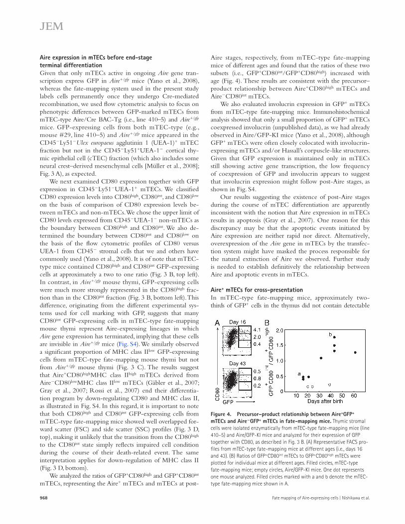
Aire stages, respectively, from mTEC-type fate-mapping mice of different ages and found that the ratios of these two subsets (i.e., GFP+CD80int/GFP+CD80high) increased with age (Fig. 4). These results are consistent with the precursor–product relationship between Aire+CD80high mTECs and AireCD80int mTECs.
We also evaluated involucrin expression in GFP+ mTECs from mTEC-type fate-mapping mice. Immunohistochemical analysis showed that only a small proportion of GFP+ mTECs coexpressed involucrin (unpublished data), as we had already observed in Aire/GFP-KI mice (Yano et al., 2008), although GFP+ mTECs were often closely colocated with involucrin-expressing mTECs and/or Hassall’s corpuscle-like structures. Given that GFP expression is maintained only in mTECs still showing active gene transcription, the low frequency of coexpression of GFP and involucrin appears to suggest that involucrin expression might follow post-Aire stages, as shown in Fig. S4.
Our results suggesting the existence of post-Aire stages during the course of mTEC differentiation are apparently inconsistent with the notion that Aire expression in mTECs results in apoptosis (Gray et al., 2007). One reason for this discrepancy may be that the apoptotic events initiated by Aire expression are neither rapid nor direct. Alternatively, overexpression of the Aire gene in mTECs by the transfec-tion system might have masked the process responsible for the natural extinction of Aire we observed. Further study is needed to establish definitively the relationship between Aire and apoptotic events in mTECs.
Aire+ mTECs for cross-presentationIn mTEC-type fate-mapping mice, approximately two-thirds of GFP+ cells in the thymus did not contain detectable
Aire expression in mTECs before end-stage terminal differentiationGiven that only mTECs active in ongoing Aire gene tran-scription express GFP in Aire+/gfp mice (Yano et al., 2008), whereas the fate-mapping system used in the present study labels cells permanently once they undergo Cre-mediated recombination, we used flow cytometric analysis to focus on phenotypic differences between GFP-marked mTECs from mTEC-type Aire/Cre BAC-Tg (i.e., line 410–5) and Aire+/gfp mice. GFP-expressing cells from both mTEC-type (e.g., mouse #29, line 410–5) and Aire+/gfp mice appeared in the CD45Ly51Ulex europaeus agglutinin 1 (UEA-1)+ mTEC fraction but not in the CD45Ly51+UEA-1 cortical thy-mic epithelial cell (cTEC) fraction (which also includes some neural crest–derived mesenchymal cells [Müller et al., 2008]; Fig. 3 A), as expected.
We next examined CD80 expression together with GFP expression in CD45Ly51UEA-1+ mTECs. We classified CD80 expression levels into CD80high, CD80int, and CD80low on the basis of comparison of CD80 expression levels be-tween mTECs and non-mTECs. We chose the upper limit of CD80 levels expressed from CD45UEA-1 non-mTECs as the boundary between CD80high and CD80int. We also de-termined the boundary between CD80int and CD80low on the basis of the flow cytometric profiles of CD80 versus UEA-1 from CD45 stromal cells that we and others have commonly used (Yano et al., 2008). It is of note that mTEC-type mice contained CD80high and CD80int GFP-expressing cells at approximately a two to one ratio (Fig. 3 B, top left). In contrast, in Aire+/gfp mouse thymi, GFP-expressing cells were much more strongly represented in the CD80high frac-tion than in the CD80int fraction (Fig. 3 B, bottom left). This difference, originating from the different experimental sys-tems used for cell marking with GFP, suggests that many CD80int GFP-expressing cells in mTEC-type fate-mapping mouse thymi represent Aire-expressing lineages in which Aire gene expression has terminated, implying that these cells are invisible in Aire+/gfp mice (Fig. S4). We similarly observed a significant proportion of MHC class IIlow GFP-expressing cells from mTEC-type fate-mapping mouse thymi but not from Aire+/gfp mouse thymi (Fig. 3 C). The results suggest that Aire+CD80highMHC class IIhigh mTECs derived from AireCD80lowMHC class IIlow mTECs (Gäbler et al., 2007; Gray et al., 2007; Rossi et al., 2007) end their differentia-tion program by down-regulating CD80 and MHC class II, as illustrated in Fig. S4. In this regard, it is important to note that both CD80high and CD80int GFP-expressing cells from mTEC-type fate-mapping mice showed well overlapped for-ward scatter (FSC) and side scatter (SSC) profiles (Fig. 3 D, top), making it unlikely that the transition from the CD80high to the CD80int state simply reflects impaired cell condition during the course of their death-related event. The same interpretation applies for down-regulation of MHC class II (Fig. 3 D, bottom).
We analyzed the ratios of GFP+CD80high and GFP+CD80int mTECs, representing the Aire+ mTECs and mTECs at post-
Figure 4. Precursor–product relationship between Aire+GFP+ mTECs and AireGFP+ mTECs in fate-mapping mice. Thymic stromal cells were isolated enzymatically from mTEC-type fate-mapping mice (line 410–5) and Aire/GFP-KI mice and analyzed for their expression of GFP together with CD80, as described in Fig. 3 B. (A) Representative FACS pro-files from mTEC-type fate-mapping mice at different ages (i.e., days 16 and 43). (B) Ratios of GFP+CD80int mTECs to GFP+CD80high mTECs were plotted for individual mice at different ages. Filled circles, mTEC-type fate-mapping mice; empty circles, Aire/GFP-KI mice. One dot represents one mouse analyzed. Filled circles marked with a and b denote the mTEC-type fate-mapping mice shown in A.
JEM VOL. 207, May 10, 2010 969
Br ief Definit ive Repor t
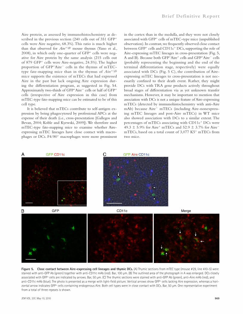
in the cortex than in the medulla, and they were not closely associated with GFP+ cells of mTEC-type mice (unpublished observation). In contrast, we frequently observed close contact between GFP+ cells and CD11c+ DCs, supporting the role of Aire-expressing mTEC lineages in cross-presentation (Fig. 5, A and B). Because both GFP+Aire+ cells and GFP+Aire cells (probably representing the beginning and the end of the terminal differentiation stage, respectively) were equally associated with DCs (Fig. 5 C), the contribution of Aire- expressing mTEC lineages to cross-presentation is not nec-essarily confined to their death event. Rather, they might provide DCs with TRA gene products actively throughout broad stages of differentiation via as yet unknown transfer mechanisms. However, it may be important to mention that association with DCs is not a unique feature of Aire-expressing mTECs (detected by immunohistochemistry with anti-Aire mAb) because Aire mTECs (including Aire-nonexpress-ing mTEC lineages and post-Aire mTECs) in WT mice also showed association with DCs to a similar extent. The percentages of mTECs associating with CD11c+ DCs were 49.3 ± 5.9% for Aire+ mTECs and 52.9 ± 3.7% for Aire mTECs, based on a total count of 3,077 K5+ mTECs from two mice.
Aire protein, as assessed by immunohistochemistry as de-scribed in the previous section (240 cells out of 351 GFP+ cells were Aire negative, 68.3%). This ratio is much higher than that observed for Aire+/gfp mouse thymus (Yano et al., 2008), in which only one-quarter of GFP+ cells were neg-ative for Aire protein by the same analysis (215 cells out of 879 GFP+ cells were Aire-negative, 24.5%). The higher proportion of GFP+Aire cells in the thymus of mTEC-type fate-mapping mice than in the thymus of Aire+/gfp mice supports the existence of mTECs that had expressed Aire in the past but lack ongoing Aire expression dur-ing the differentiation program, as suggested in Fig. S4. Approximately two-thirds of GFP+Aire cells or half of GFP+ cells (irrespective of Aire expression in this case) from mTEC-type fate-mapping mice can be estimated to be of this cell type.
It is believed that mTECs contribute to self-antigen ex-pression by being phagocytosed by professional APCs at the expense of their death (i.e., cross-presentation [Gallegos and Bevan, 2004; Koble and Kyewski, 2009]). We therefore used mTEC-type fate-mapping mice to examine whether Aire-expressing mTEC lineages have close contact with macro-phages or DCs. F4/80+ macrophages were more prominent
Figure 5. Close contact between Aire-expressing cell lineages and thymic DCs. (A) Thymic sections from mTEC type (mouse #29, line 410–5) were stained with anti-GFP Ab (green) together with anti-CD11c mAb (red). Bar, 100 µm. (B) The outlined area of the photograph in A was enlarged. DCs closely associated with GFP+ cells are indicated by arrows. Bar, 50 µm. (C) The thymic sections were stained with anti-GFP Ab (green), anti-Aire mAb (red), and anti-CD11c mAb (blue). The photo is presented as a merge with light-field picture. Vertical arrows show GFP+ cells lacking Aire expression, whereas a hori-zontal arrow indicates GFP+ cells containing endogenous Aire. Both cell types were in close contact with DCs. Bar, 50 µm. One representative experiment from a total of three repeats is shown.

970 Fate mapping of Aire-expressing cells | Nishikawa et al.
Real-time PCR and semiquantitative RT-PCR. RNA was extracted from TECs with RNeasy Mini kits (QIAGEN) and made into cDNA with SuperScript III RT kits (Invitrogen) according to the manufacturer’s instruc-tions. Real-time PCR (Yano et al., 2008) and semiquantitative RT-PCR (Kajiura et al., 2004) for the Aire gene were performed as described previously.
Semiquantitative RT-PCR for AIRE from human ES and iPS cells. Human ES cells (KhES-1) and human iPS cells (clone 253G1) were provided by N. Nakatsuji and S. Yamanaka (Kyoto University, Kyoto, Japan), respec-tively, and maintained on mitomycin C–treated MEF as feeder cells (Suemori et al., 2006), in conformity with The Guidelines for Derivation and Utiliza-tion of Human Embryonic Stem Cells of the Ministry of Education, Culture, Sports, Science and Technology, Japan, after obtaining approval from the Institutional Review Board. RNA was extracted with a QuickGene RNA cultured cell kit S (Fuji Film) and converted to cDNA with ReverTra Ace (Toyobo) in accordance with the manufacturer’s instructions. The primers for the human AIRE and GAPDH genes were the following: AIRE primers, 5-CAGGCAACAGTCCAGGAG-3 and 5-GCACCAGGAGCCAG-GTT-3; and GAPDH primers, 5-CCACCCATGGCAAATTCCATG-GCA-3 and 5-TCTAGACGGCAGGTCAGGTCCACC-3.
Online supplemental material. Fig. S1 shows flow cytometric analysis of GFP-expressing hemopoietic cells in the thymus, spleen, and BM from Aire/Cre BAC-Tg mice crossed with a GFP reporter strain. Fig. S2 shows extra-thymic expression of Aire/Cre BAC-Tg with immunohistochemical analysis. Fig. S3 shows generation and characterization of Aire/Cre knockin mice for fate-mapping study. Fig. S4 shows that Aire is expressed before end-stage terminal differentiation in mTECs. Table S1 contains a list of mice presented in the fate-mapping study. Online supplemental material is avail-able at http://www.jem.org/cgi/content/full/jem.20092144/DC1.
We thank Dr. K. Kakugawa for the mouse transfer and Drs. N. Nakatsuji and S. Yamanaka for the cell lines. We also thank Drs. H. Niwa, A. Hijikata, T.A. Endo, and H. Koseki for helpful discussion.
This work was supported in part by Grants-in-Aid for Scientific Research from the Japan Society for the Promotion of Science and from the Ministry of Education, Culture, Sports, Science and Technology of Japan (M. Matsumoto).
The authors have no conflicting financial interests.
Submitted: 5 October 2009Accepted: 3 March 2010
REFERENCESAnderson, M.S., E.S. Venanzi, L. Klein, Z. Chen, S.P. Berzins, S.J. Turley,
H. von Boehmer, R. Bronson, A. Dierich, C. Benoist, and D. Mathis. 2002. Projection of an immunological self shadow within the thy-mus by the aire protein. Science. 298:1395–1401. doi:10.1126/science .1075958
Brennan, C.A., and K.V. Anderson. 2004. Drosophila: the genetics of innate immune recognition and response. Annu. Rev. Immunol. 22:457–483. doi:10.1146/annurev.immunol.22.012703.104626
Cheng, M.H., A.K. Shum, and M.S. Anderson. 2007. What’s new in the Aire? Trends Immunol. 28:321–327. doi:10.1016/j.it.2007.05.004
Dooley, J., M. Erickson, and A.G. Farr. 2008. Alterations of the medul-lary epithelial compartment in the Aire-deficient thymus: implica-tions for programs of thymic epithelial differentiation. J. Immunol. 181: 5225–5232.
Gäbler, J., J. Arnold, and B. Kyewski. 2007. Promiscuous gene expression and the developmental dynamics of medullary thymic epithelial cells. Eur. J. Immunol. 37:3363–3372. doi:10.1002/eji.200737131
Gallegos, A.M., and M.J. Bevan. 2004. Central tolerance to tissue-specific antigens mediated by direct and indirect antigen presentation. J. Exp. Med. 200:1039–1049. doi:10.1084/jem.20041457
Gardner, J.M., J.J. Devoss, R.S. Friedman, D.J. Wong, Y.X. Tan, X. Zhou, K.P. Johannes, M.A. Su, H.Y. Chang, M.F. Krummel, and M.S. Anderson. 2008. Deletional tolerance mediated by extrathymic Aire-expressing cells. Science. 321:843–847. doi:10.1126/science.1159407
ConclusionA fate-mapping study of Aire-expressing cells has unex-pectedly revealed Aire gene expression during early em-bryogenesis. Because offspring of mice homozygous for Aire deficiency were born in the numbers expected from the heterozygous crossing (Anderson et al., 2002; Ramsey et al., 2002; Kuroda et al., 2005; Hubert et al., 2009), the role of Aire in early embryonic development, other than its possible association with ectodermal dystrophy, remains unknown. Nevertheless, it is intriguing to speculate that the evolution of the immune system has co-opted the Aire gene from an initial role in determining body plan. A precedent for such a process might be the Toll-like re-ceptors. In this regard, it is interesting that the Toll gene in Drosophila melanogaster plays an essential role in the es-tablishment of dorsal-ventral polarity, whereas deletion of genes involved in Toll-like receptor signaling in mice re-sults in no discernible effect on their embryonic develop-ment (Takeda et al., 2003; Brennan and Anderson, 2004). Consequently, we might expect ablation of the Aire gene in lower vertebrates (Saltis et al., 2008) to result in obvious phenotypes related to embryonic development. Integration of the roles of Aire expressed at early embryonic stages with current perspectives on the involvement of Aire in organizing the thymic microenvironment may provide a novel viewpoint on the fundamental features of self versus non-self discrimination.
MATERIALS AND METHODSMice. A BAC Tg construct containing 97.1 kb of the 5 region and 69 kb of the 3 region flanking the Aire gene was generated from BAC clone RP23-461E7, in which the Aire start codon was replaced with an open reading frame encoding a Cre recombinase followed by a poly-A signal from the rabbit globin gene using the Red/ET Counter Selection BAC Modification kit (Gene Bridges), which does not leave any drug selection genes in the BAC Tg construct (Muyrers et al., 2000). Aire/Cre BAC-Tg mice were generated by injecting linearized BAC Tg construct into pro-nuclei of fertilized C57BL/6 oocytes, and Tg founders were chosen by Southern blot analysis. A reporter Tg strain expressing EGFP upon Cre-mediated recombination (CAG-CAT-EGFP, line 39; Kawamoto et al., 2000) and Aire/GFP knockin mice (Yano et al., 2008) were generated as described previously. The mice were maintained under pathogen-free conditions. The protocols used in this study were in accordance with the Guidelines for Animal Experimentation of Tokushima University School of Medicine.
Immunohistochemistry. Immunohistochemical analysis of the thymus with goat polyclonal anti-GFP Ab (Novus Biologicals), rabbit polyclonal anti-GFP Ab (Invitrogen), rat anti-EpCAM mAb (BD), and rabbit poly-clonal anti-K5 Ab (Covance) was performed as described previously (Yano et al., 2008). Rabbit polyclonal anti-Aire Ab and rat anti-Aire mAb (clone RF33-1; IgG1) were produced in our laboratory by immunization with peptides corresponding to the C-terminal portion of mouse Aire. Anti-CD11c mAb (clone HL3) was obtained from BD.
TEC preparation and flow cytometric analysis. Preparation of TECs and flow cytometric analysis with a FACSCalibur (BD) was performed as described previously (Yano et al., 2008). The mAbs used were anti-CD4, anti-CD8, anti-CD11c (clone N418), anti-CD45, anti-CD80, anti-B220, anti-I-A/I-E, anti-Ly51, and anti-F4/80 (eBioscience). UEA-1 was pur-chased from Vector Laboratories.
JEM VOL. 207, May 10, 2010 971
Br ief Definit ive Repor t
Gillard, G.O., and A.G. Farr. 2005. Contrasting models of promiscu-ous gene expression by thymic epithelium. J. Exp. Med. 202:15–19. doi:10.1084/jem.20050976
Gray, D., J. Abramson, C. Benoist, and D. Mathis. 2007. Proliferative arrest and rapid turnover of thymic epithelial cells expressing Aire. J. Exp. Med. 204:2521–2528. doi:10.1084/jem.20070795
Hubert, F.X., S.A. Kinkel, P.E. Crewther, P.Z. Cannon, K.E. Webster, M. Link, R. Uibo, M.K. O’Bryan, A. Meager, S.P. Forehan, et al. 2009. Aire-deficient C57BL/6 mice mimicking the common human 13-base pair deletion mutation present with only a mild autoimmune pheno-type. J. Immunol. 182:3902–3918. doi:10.4049/jimmunol.0802124
Kajiura, F., S. Sun, T. Nomura, K. Izumi, T. Ueno, Y. Bando, N. Kuroda, H. Han, Y. Li, A. Matsushima, et al. 2004. NF-B-inducing kinase estab-lishes self-tolerance in a thymic stroma-dependent manner. J. Immunol. 172:2067–2075.
Kawamoto, S., H. Niwa, F. Tashiro, S. Sano, G. Kondoh, J. Takeda, K. Tabayashi, and J. Miyazaki. 2000. A novel reporter mouse strain that ex-presses enhanced green fluorescent protein upon Cre-mediated recombi-nation. FEBS Lett. 470:263–268. doi:10.1016/S0014-5793(00)01338-7
Koble, C., and B. Kyewski. 2009. The thymic medulla: a unique microen-vironment for intercellular self-antigen transfer. J. Exp. Med. 206:1505–1513. doi:10.1084/jem.20082449
Kuroda, N., T. Mitani, N. Takeda, N. Ishimaru, R. Arakaki, Y. Hayashi, Y. Bando, K. Izumi, T. Takahashi, T. Nomura, et al. 2005. Development of autoimmunity against transcriptionally unrepressed target antigen in the thymus of Aire-deficient mice. J. Immunol. 174:1862–1870.
Kyewski, B., and L. Klein. 2006. A central role for central tolerance. Annu. Rev. Immunol. 24:571–606. doi:10.1146/annurev.immunol.23.021704 .115601
Lee, J.W., M. Epardaud, J. Sun, J.E. Becker, A.C. Cheng, A.R. Yonekura, J.K. Heath, and S.J. Turley. 2007. Peripheral antigen display by lymph node stroma promotes T cell tolerance to intestinal self. Nat. Immunol. 8:181–190. doi:10.1038/ni1427
Mathis, D., and C. Benoist. 2009. Aire. Annu. Rev. Immunol. 27:287–312. doi:10.1146/annurev.immunol.25.022106.141532
Matsumoto, M. 2007. Transcriptional regulation in thymic epithelial cells for the establishment of self tolerance. Arch. Immunol. Ther. Exp. (Warsz.). 55:27–34. doi:10.1007/s00005-007-0007-9
Müller, S.M., C.C. Stolt, G. Terszowski, C. Blum, T. Amagai, N. Kessaris, P. Iannarelli, W.D. Richardson, M. Wegner, and H.R. Rodewald. 2008. Neural crest origin of perivascular mesenchyme in the adult thy-mus. J. Immunol. 180:5344–5351.
Muyrers, J.P., Y. Zhang, V. Benes, G. Testa, W. Ansorge, and A.F. Stewart. 2000. Point mutation of bacterial artificial chromosomes by ET re-combination. EMBO Rep. 1:239–243. doi:10.1093/embo-reports/ kvd049
Peterson, P., T. Org, and A. Rebane. 2008. Transcriptional regulation by AIRE: molecular mechanisms of central tolerance. Nat. Rev. Immunol. 8:948–957. doi:10.1038/nri2450
Ramsey, C., O. Winqvist, L. Puhakka, M. Halonen, A. Moro, O. Kämpe, P. Eskelin, M. Pelto-Huikko, and L. Peltonen. 2002. Aire deficient mice develop multiple features of APECED phenotype and show al-tered immune response. Hum. Mol. Genet. 11:397–409. doi:10.1093/ hmg/11.4.397
Rodewald, H.R. 2008. Thymus organogenesis. Annu. Rev. Immunol. 26:355–388. doi:10.1146/annurev.immunol.26.021607.090408
Rossi, S.W., M.Y. Kim, A. Leibbrandt, S.M. Parnell, W.E. Jenkinson, S.H. Glanville, F.M. McConnell, H.S. Scott, J.M. Penninger, E.J. Jenkinson, et al. 2007. RANK signals from CD4+3 inducer cells regulate develop-ment of Aire-expressing epithelial cells in the thymic medulla. J. Exp. Med. 204:1267–1272. doi:10.1084/jem.20062497
Saltis, M., M.F. Criscitiello, Y. Ohta, M. Keefe, N.S. Trede, R. Goitsuka, and M.F. Flajnik. 2008. Evolutionarily conserved and divergent re-gions of the autoimmune regulator (Aire) gene: a comparative analysis. Immunogenetics. 60:105–114. doi:10.1007/s00251-007-0268-9
Schaller, C.E., C.L. Wang, G. Beck-Engeser, L. Goss, H.S. Scott, M.S. Anderson, and M. Wabl. 2008. Expression of Aire and the early wave of apoptosis in spermatogenesis. J. Immunol. 180:1338–1343.
Senoo, M., F. Pinto, C.P. Crum, and F. McKeon. 2007. p63 Is essential for the proliferative potential of stem cells in stratified epithelia. Cell. 129:523–536. doi:10.1016/j.cell.2007.02.045
Suemori, H., K. Yasuchika, K. Hasegawa, T. Fujioka, N. Tsuneyoshi, and N. Nakatsuji. 2006. Efficient establishment of human embryonic stem cell lines and long-term maintenance with stable karyotype by enzymatic bulk passage. Biochem. Biophys. Res. Commun. 345:926–932. doi:10.1016/ j.bbrc.2006.04.135
Takeda, K., T. Kaisho, and S. Akira. 2003. Toll-like receptors. Annu. Rev. Immunol. 21:335–376. doi:10.1146/annurev.immunol.21.120601 .141126
Yano, M., N. Kuroda, H. Han, M. Meguro-Horike, Y. Nishikawa, H. Kiyonari, K. Maemura, Y. Yanagawa, K. Obata, S. Takahashi, et al. 2008. Aire controls the differentiation program of thymic epithelial cells in the medulla for the establishment of self-tolerance. J. Exp. Med. 205:2827–2838. doi:10.1084/jem.20080046