biotechnology & biomedical engineering - jscimed … · 2014-07-01 · bioassay-guided...
TRANSCRIPT
JSMBiotechnology & Biomedical Engineering
Special Issue on
Industrial Biotechnology-Made in Germany: The path from policies to sustainable energy, commodity and specialty productsEdited by:Dr. Thomas BrückProfessor of Industrial Biocatalysis, Dept. of Chemistry, Technische Universität München (TUM), Germany
Central

Cite this article: Mundt S, Bui HT, Preisitsch M, Kreitlow S, Bui HTN, et al. (2014) Microalgae - A Promising Source of Novel Therapeutics. JSM Biotechnol Bioeng 2(1): 1032.
*Corresponding authorPD.Dr. Sabine Mundt, Institute of Pharmacy, Ernst-Moritz-Arndt-University, Jahnstr.17, 17487 Greifswald, Germany, Tel: +493834864869; Fax: 493834864885; Email:
Submitted: 15 April 2013
Accepted: 12 May 2014
Published: 14 May 2014
ISSN: 2333-7117
Copyright© 2014 Mundt et al.
OPEN ACCESS
KeywordsMicroalgae; Antimicrobial screening; Fatty acids; Lyngbyazothrins; Carbamidocyclophanes; Microparticles
Research Article
Microalgae - A Promising Source of Novel TherapeuticsMundt, S.1*, Bui, H.T.1, Preisitsch, M.1, Kreitlow, S.1, Bui, H.T.1, Pham, H.T.1, Zainuddin, E.1, Le, T.T.1, Lukowski, G.2 and Jülich, W.D. 1
1Institute of Pharmacy, Ernst-Moritz-Arndt-University, Germany2Institute of Marine Biotechnology, Rathenaustr, Germany
Abstract
Due to the growing resistance of pathogenic bacteria and fungi against commercially available therapeutics, the search for new antimicrobial substances is of increasing importance. Based on the hypothesis that microorganisms living in an aquatic environment produce secondary metabolites as chemical weapons to survive in their daily fight against cohabitants of the biotope, a screening of 133 microalgae (121 cyanobacteria, 12 eukaryotic microalgae) was started. Biomass extracts and cultivation media were tested for activity against the Gram-positive bacteria Bacillus subtilis and Staphylococcus aureus, the Gram-negative bacteria Escherichia coli and Pseudomonas aeruginosa as well as the yeast Candida maltosa. Our data indicates that 56 cyanobacterial strains and 5 eukaryotic algae exhibited antimicrobial activity. Interestingly, 19 of the screened cyanobacteria inhibited the growth of MRSA. Further, screening experiments revealed activity against Helicobacter pylori as well as fish pathogenic bacteria and plant pathogenic fungi. Strains exhibiting significant antimicrobial activity were cultivated at 40L scale in order to conduct a bioassay-guided isolation and structure elucidation of the bioactive components. This procedure allowed identification of bioactive secondary metabolites encompassing the hydroxylated fatty acids coriolic and dimorphecolic acid, lyngbyazothrins, cyclic depsispeptides and carbamidocyclophanes, belonging to the class of polyketides, which are responsible for the observed antimicrobial activity.
In addition to tests of purified bioactive compounds, whole biomass of selected microalgae was used to prepare microparticles by high pressure homogenization. Subsequent, in-vitro tests have shown that microparticles from biomass of the microalgal strain Bio 33, named Maresome®, inhibit dermal colonization of different MRSA strains. Since preliminary, clinical tests confirm the in-vitro data, the anti-pathogenic potential of microalgae might be utilized in form of a prophylactic skin care product to prevent nosocomial infections.
ABBREVIATIONSAA: Arachidonic Acid; ADC: Antibody Drug Conjugate; ALCL:
Anaplastic Large Cell Lymphoma; Aound: 3-amino-2,5,7,8-tetrahydroxy-10-methylundecanoic acid; DCM: Dichloromethane; DHA: Docosahexaenoic Acid; EPA: Eicosapentaenoic Acid; EtOAc: Ethyl Acetate; GCMS: Gas Chromatography Mass Spectrometry; GLA: Gamma Linolenic Acid; MMA: Monomethylauristatin; MRSA: Methicillin-Resistant Staphylococcus aureus; MSSA: Methicillin-Sensitive Staphylococcus aureus; NES: North German MRSA Epidemic Strain; PSP: Paralytic Shellfish Poisoning; PUFA: Polyunsaturated Fatty Acid; VRSA: Vancomycin Resistant Staphylococcus aureus
INTRODUCTIONMicroalgae are photoautotrophic microorganisms containing
chlorophyll a and other accessory pigments to convert sunlight, carbon dioxide and water to carbohydrates, proteins and lipids in the process of photosynthesis. These organisms are found in both aquatic and terrestrial environments. The term microalgae does not describe a distinct taxonomic group, because it includes eukaryotes such as diatoms, dinoflagellates, green, red, brown and golden algae as well as prokaryotic cyanobacteria (blue-green algae). While identified eukaryotic microalgae number between
50,000 and 60,000 species, cyanobacterial species amount to only approximately 2000 species. Nonetheless, far more bioactive compounds acting in micromolar to nanomolar range have been isolated from cyanobacteria. Examples of bioactive compounds derived from cyanobacteria include hepato- and neurotoxins isolated from genera such as Microcystis, Anabaena, Nodularia, Aphanizomenon, Cylindrospermopsis and Oscillatoria. In addition to these toxins a variety of bioactive secondary metabolites with cytotoxic/antitumoral, antibiotic, antiviral, anti-inflammatory and antiparasitic activity have been published [1-10].
An array of pharmaceutically relevant substances constitutes linear or cyclic peptides containing unusual amino acids, which are often connected to aliphatic residues, forming lipopeptides. Additional pharmacologically relevant peptides comprise depsipeptides. The majority of bioactive peptides are derived from polyketide synthase/nonribosomal peptide synthetase activity. By contrast, ribosomally synthesized and posttranslational modified peptides with pharmaceutical activity have rarely been described [1-5,11,12]. In addition to microalgae derived bioactive peptides, pharmaceutically relevant natural products belonging to the class of alkaloids, carbohydrates and terpenes have also been described [1-12].

Central
Mundt et al. (2014)Email:
JSM Biotechnol Bioeng 2(1): 1032 (2014) 3/12
Cyanobacteria belonging to the orders Nostocales and Oscillatoriales with the genera Nostoc, Anabaena and Lyngbya are important producers of bioactive secondary metabolites. The filamentous cyanobacterium Lyngbya majuscula has been identified as a “high-natural product production strain” because in excess of 25% of all cyanobacterial natural products have been isolated from this species [1,4,13]. Prominent example of these natural products are the dolastatins, which display antineoplastic activities [1-4,12-14]. Monomethyl auristatin E (MMAE), a synthetic dolastatin 10 derivative was coupled to a monoclonal antibody that can be targeted to specific surface antigen such as CD30, Nectin-4 or glycoprotein NMB, which are expressed by different types of cancers. The Antibody-Drug-Conjugate (ADC) is internalized and cleaved only in cancer cells expressing the typical surface antigen,where the cytotoxic dolastatin analog is released. The ADC targets CD 30 has been approved by the FDA in 2011 [15] and in 2012 in Europe as Brentuximab vedotin (Adcetris®) [16] for treatment of patients with Hodgkins lymphoma and patients with systemic anaplastic large cell lymphoma (ALCL), respectively [17,18]. Brentuximab vedotin is the first approved drug derived from a cyanobacterial secondary metabolite. Further, CDX-011, an ADC with MMA E targets glycoprotein NMB is in phase II of clinical trials [19].
Eukaryotic microalgae containing considerable amounts of polyunsaturated fatty acids (PUFA) such as DHA, EPA, AA and GLA, polysaccharides, pigments and essential amino acids are used commercially as foodstuff and animal feed. PUFAs may reduce the risk of coronary heart disease and prevent development of atherosclerosis [7]. DHA derived from the microalgae Crypthecodinium, Schizochytrium and Ulkenia sp. is used as supplement for baby food to promote development of the infant’s eyes and brain [6]. Recently, DHA, EPA and higher unsaturated fatty acids were shown to have antimicrobial activities against Propionibacterium acnes and Staphylococcus aureus [20,21]. The first antibacterial compound isolated from microalgae was Chlorellin from Chlorella spp., which was identified as a mixture of fatty acids [7,22]. Furthermore, a glycoprotein isolated from Chlorella vulgaris culture supernatant has shown protective activity against tumor metastasis and chemotherapy-induced immune suppression in mice [23]. Another prominent example is the polysaccharide β-1,3-glucan can, which is accumulated in several microalgae such as Chlorella, Skeletonema, Nannochloropsis, Euglena and linked with antiviral, antitumor and immune stimulating effects. More recently, β-1,3-glucans were linked to regulation of blood glucose and insulin response in humans [6,24]. Sulfated polysaccharides of the red microalgae Porphyridium cruentum displayed antiviral activity against Herpex simplex and Varicella zoster viruses, and may prevent colon cancer [6]. Carotenoids with antioxidant effects such as α- and β-caroten, lutein, zeaxanthin, astaxanthin seem to be responsible for prevention of neurodegenerative and cardiovascular diseases, diabetes, osteoporoses and cancer [25].
In contrast to cyanobacteria, which produce numerous bioactive natural products with activities in the low micromolar or nanomolar range, such compounds are rarely isolated from eukaryotic algae. An exception are dinoflagellates which produce highly active sodium channel blockers such as saxitoxin and neosaxitoxin. Polyether compounds such as brevetoxin A from
Karenia brevis and maitotoxin from Gambierdiscus toxicus are activators of sodium and calcium channels respectively, resulting in strong neurotoxicity. The diatoms Nitzschia and Pseudonitzschia are producers of the excitatory amino acid domoic acid, acting at the cerebral glutamate receptor [3].
In this paper we present an antimicrobial natural product screen of 121 cyanobacteria and 12 eukaryotic algae belonging to the genera chlorophyta, heterokontophyta, charophyta, haptophyta and rhodophyta. Our screening procedure aims to identify compounds displaying activity against bacteria and yeast/fungi, which are pathogenic in humans, fish and plants. Subsequently, strains showing target activity were chosen for bioassay-guided isolation and structure elucidation of the active compounds. In this context we focus on three classes of structures, which we could identify structurally. Finally, we tested microparticles prepared from algae biomass by Maresome® technology for its ability to inhibit dermal colonization of MRSA.
MATERIALS AND METHODSCultivation of microalgae
121 cyanobacterial strains and 12 strains of microalgae deposited in the Culture collection of the Institute of Pharmacy, Department of Pharmaceutical Biology, Ernst-Moritz-Arndt-University Greifswald were used for screening. The microalgae were cultivated batchwise in 2 L Fehrnbach flasks with 1000 mL BG 11 medium [26] at 25°C under continuous illumination with 20 µmol m-2 s-1 with cool white fluorescent lamps over 35 d. Large scale cultivation was carried out in a 50 L cylindrical air-lift bioreactor [27]. Biomass and cultivation medium were harvested after 35 d by centrifugation. The biomass was washed with distilled water, lyophilized and kept at -20°C until use. The cultivation medium was filtrated and frozen at -20°C.
Extract preparation
1 g dried biomass was extracted with 3x150 mL n-hexane, methanol and water successively. The supernatants of each extraction were combined; the organic solvents were removed in vacuum at 40°C and the water by lyophilization. A volume of 3000 mL culture medium was reduced to 300 mL in vacuum at 40°C. 300 mL ethyl acetate (EtOAc) was added and the mixture was shaken for 24 h. The EtOAc layer was separated and the aqueous layer was shaken again with 300 mL EtOAc. The EtOAc layers were combined, dried over sodium sulphate and the EtOAc was removed in vacuum. For isolation of the pure compounds, extractions were carried out according to [28-30].
In vitro screening test system
The antibacterial and antifungal activity of the n-hexane, methanol, water and EtOAc extracts were evaluated in agar diffusion assay according to the methods of the European Pharmacopoeia [31]. Extracts (2 mg/6 mm paper disc) were tested against the ATCC strains Bacillus subtilis 6051, Staphylococcus aureus 6538, Escherichia coli 11229, Pseudomonas aeruginosa 27853. The yeast Candida maltosa SBUG 700 was provided by the Institute of Microbiology of the Ernst-Moritz-Arndt-University, Greifswald. The following antibiotics were tested as reference: Ampicillin 10µg B. subtilis (inhibition zone 33 mm) and S. aureus (inhibition zone 35 mm), 50µg E. coli (inhibition zone 26 mm);

Central
Mundt et al. (2014)Email:
JSM Biotechnol Bioeng 2(1): 1032 (2014) 4/12
Antimicrobial activity Strains tested Active strains Extracts tested Active extracts
Cyanobacteria 121 56 410 97
n-hexane 102 16 (4)
activity* ≤10mm >10 mm to ≤ 16mm >16mm to ≥ 31 mm
12 3 1
methanol 121 48 (17)
activity* ≤10mm >10 mm to ≤ 16mm >16 mm to ≥ 31 mm
16 28 4
water 121 8 (3)
activity* ≤10mm >10 mm to ≤ 16mm >16 mm to ≥ 31 mm
4 3 1
EtOAc (medium) 66 25 (6)
activity* ≤10mm >10 mm to ≤ 16mm >16 mm to ≥ 31 mm
10 9 6
Eukaryotic algae 12 5 96 18
n-hexane 23 7
activity* ≤10mm >10 mm to ≤ 16mm >16 mm to ≥ 31 mm
5 2 0
dichloromethane 20 1
activity* ≤10mm >10 mm to ≤ 16mm >16 mm to ≥ 31 mm
1 0 0
methanol 23 2 (1)
activity* ≤10mm >10 mm to ≤ 16mm >16 mm to ≥ 31 mm
2 0 0
water 23 2
activity* ≤10mm >10 mm to ≤ 16mm >16 mm to ≥ 31 mm
2 0 0
EtOAc (medium) 7 6
activity* ≤10mm >10 mm to ≤ 16mm >16 mm to ≥ 31 mm
2 4 0
MRSA activity Strains tested Active strains Extracts tested Active extracts
Cyanobacteria 47 19 144 20
n-hexane 26 0
methanol 50 18
activity* ≤10mm >10 mm to ≤ 16mm >16 mm to ≥ 31 mm
4 9 2
water 50 0
dichloromethane 6 0
EtOAc (medium) 12 2
activity* ≤10mm >10 mm to ≤ 16mm >16 mm to ≥ 31 mm
0 1 1
Eukaryotic algae 11 0 60 0
n-hexane 14 0
dichloromethane 14 0
methanol 14 0
water 14 0
EtOAc) medium 4 0
Table 1: Antimicrobial activity of cyanobacterial and microalgal extracts obtained from different solvents.

Central
Mundt et al. (2014)Email:
JSM Biotechnol Bioeng 2(1): 1032 (2014) 5/12
Activity to fishpathogenic bacteria
Strains tested Active strains Extracts tested Active extracts
Cyanobacteria 42 11 106 21
n-hexane 22 9
activity* ≤10mm >10 mm to ≤ 16mm >16 mm to ≥ 31 mm
0 9 0
methanol 42 11
activity* ≤10mm >10 mm to ≤ 16mm >16 mm to ≥ 31 mm
3 5 3
water 42 1
activity* ≤10mm >10 mm to ≤ 16mm >16 mm to ≥ 31 mm
0 0 1
Eukaryotic algae 7 3 19 5
n-hexane 5 2
activity* ≤10mm >10 mm to ≤ 16mm >16 mm to ≥ 31 mm
0 2 0
methanol 7 3
activity* ≤10mm >10 mm to ≤ 16mm >16 mm to ≥ 31 mm
1 2 0
water 7 0
Activity to Helicobacter pylori Strains tested Active strains Extracts tested Active extracts
Cyanobacteria 15 6 56 17
n-hexane 14 5
activity* ≤10mm >10 mm to ≤ 16mm >16 mm to ≥ 31 mm
2 3 0
dichloromethane 5 3
activity* ≤10mm >10 mm to ≤ 16mm >16 mm to ≥ 31 mm
2 0 1
methanol 15 6
activity* ≤10mm >10 mm to ≤ 16mm >16 mm to ≥ 31 mm
4 1 1
water 11 1
activity* ≤10mm >10 mm to ≤ 16mm >16 mm to ≥ 31 mm
1 0 0
EtOAc (medium) 11 2
activity* ≤10mm >10 mm to ≤ 16mm >16 mm to ≥ 31 mm
2 0 0
Eukaryotic algae n.t. n.t. n.t. n.t.
Abbreviations: n.t. not tested; ( ) extracts with antifungal activity; n=3 with two parallels in agar diffusion assay, 2 mg extract/6mm paper disk;*inhibition zone including diameter of paper disc; inhibition zone ≤ 10mm – low activity; inhibition zone >10 mm to ≤ 16mm – moderate activity; inhibition zone >16mm to ≥ 31 mm – good activity [32].
Gentamicin 20µg P. aeruginosa (inhibition zone 26 mm); Nystatin 10µg C. maltosa (inhibition zone 22 mm). Tests for antibacterial effects against multiresistant strains of human pathogenic bacteria were carried out in cooperation with the Institute of hygiene, Greifswald. The following multiresistant strains were used: North German MRSA epidemic strain (NES), MRSA 34289 (Friedrich Loeffler Institute of Medical Microbiology, Ernst-Moritz-Arndt-University, Greifswald); Staphylococcus epidermidis 847, Staphylococcus haemolyticus 535, Pseudomonas
aeruginosa 595 (Institute of hygiene Greifwald). Selected extracts were tested for activity against Helicobacter pylori in cooperation with Institute of hygiene Greifwald.
To investigate activity against fish pathogenic bacteria, the strains Pseudomonas anguilliseptica DSMZ 12111, Vibrio anguillarum DSMZ 11323, Aeromononas salmonicida ssp.salmonicida ATCC 51413, Aeromonas hydrophila ssp. hydrophila DSMZ 6173, Flexibacter maritimus DSMZ 17995 and Yersinia

Central
Mundt et al. (2014)Email:
JSM Biotechnol Bioeng 2(1): 1032 (2014) 6/12
Activity phytopathogenic fungi Strains tested Active strains Extracts tested Active extracts
Cyanobacteria 34 3 152 3
n-hexane 13 0
methanol 68 3
water 68 0
EtOAc (medium) 3 0
Eukaryotic algae n.t. n.t. n.t. n.t.
Table 2: Activity of cyanobacteria against phytopathogenic fungi.
Abbreviations: n.t. not tested n=3 with two parallels, agar diffusion assay, 2 mg extract/6mm paper disk; inhibition zone including diameter of paper disc.
ruckeri ATCC 29493 were investigated. As reference, 10µg of oxytetracylcline was used. Depending on the bacterial strain inhibition zones between 27 and 55 mm were measured. The phytopathogenic fungi Alternaria sp., Botrytis cinerea, Drechslera sp. and Microdochium nivale were obtained from the DSMZ Braunschweig.
Diameters of the inhibition zones were measured over the whole zone including the paper disc (6 mm). The evaluation of the antibacterial and antifungal activity, respectively, was carried out according to the following schema: 1. Inhibition zone ≤ 10mm – low activity; 2. inhibition zone >10 mm to ≤ 16mm – moderate activity; 3. inhibition zone >16mm to ≤ 31 mm – good activity; 4. inhibition zone >31mm – excellent activity [32].
Preparation of Maresome®
The microparticles were produced using a special microencapsulation technique [33]. Biomass and n-hexane were mixed (1:10 v/v) and the organic solvent was evaporated in a rotatory evaporator. The remaining dried biomass was dispersed in a surfactant-water mixture and homogenized by a high pressure homogenizer. The zeta potential was estimated with a zetasizer 4 (Malvern Instruments, UK). The particle mixture was incorporated into an ointment (Heitland & Petre International GmbH, Celle, Germany). In the same manner ointments based on biomasses of Chlorella, Arthrospira (formerly Spirulina) and Spirogyra were produced.
Test of Maresome®
The Maresome® were tested in the animal models mouse ear (direct contamination) and cow udder teat (skin to skin transmission) [34]. Mouse ears were prepared with Maresome® ointment and Maresome® free ointment as control in the direct contamination test. The prepared mouse ears were contaminated with MRSA strain NES. Thereafter the mouse ears were streaked on Mueller-Hinton II-agar plates and the agar plates were incubated for 48 h at 30oC, checked for bacterial growth and the colonies were enumerated. For skin to skin transmission test the “donor”- cow udder teat skin was contaminated also with MRSA strain NES. The “acceptor”- cow udder teats were treated with Maresome® ointment. Maresome® - free ointment was used as control. The “acceptor skin” was brought into contact to the “donor skin” for 10 s. Subsequently the cow udder teats were streaked on the agar plates. The agar plates were incubated for 48 h at 30°C and the MRSA colony number was determined.
GC/MS
N-hexane and EtOAc extracts were analyzed after hydrolysis (5 mg biomass + 0.5 mL NaOH in 50% aqueous MeOH, 45 min 80°C), extraction of the fatty acids with ether and derivatization with diazomethane by a GC 80007 MD 800 (Fisons instruments). A DB5 MS column (J &W Scientific] was used and the temperature was increased from 80°C to 280°C (8°C per min).
RESULTS AND DISCUSSIONGeneral antimicrobial screening of algae extracts
In a basic screening for antimicrobial activity 506 extracts of different polarity prepared from biomass and cultivation medium of 121 cyanobacteria and 12 strains of eukaryotic microalgae have been tested in agar diffusion test. The activity screen encompassed two Gram-positive bacteria (Bacillus subtilis, Staphylococcus aureus), two Gram-negative bacteria (Escherichia coli, Pseudomonas aeruginosa) and the yeast Candida maltosa (Table 1).
Of the evaluated extracts derived from cyanobacteria, 56 strains exhibited antimicrobial activities and a total of 97 extracts inhibited at least one of the test organisms. Methanol extracts showed the highest bioactivity score with 40% of the tested extracts showing activity. The lowest bioactivity score with 7% was observed with water extracts. Tests with the lipophilic n-hexane extracts derived from cyanobacterial biomass indicated that overall 16% showed antimicrobial activity. By contrast 38% of the EtOAc extracts derived from the cultivation medium inhibited the growth of the test organisms. Yields for EtOAc and hexane extracts were only 0.2 and 1.5 %, respectively, therefore limiting further tests. GCMS analyses of the n-hexane and EtOAc extracts revealed the presence of saturated and unsaturated fatty acids may be responsible for the antimicrobial activity [21,35,36]. The tested extracts inhibited the growth of Gram-positive bacteria. Additionally, methanol extracts of the biomass of the freshwater Lyngbya sp. SAG 36.91 as well as of two Vietnamese cyanobacteria Westiellopsis sp. and Calothrixelenkinii showed low to moderate activity with inhibition zones of 8 and 12 mm against the Gram-negative bacteria Pseudomonas aeruginosa and Escherichia coli. Of the assayed extracts listed in table 1, 30 extracts displayed antifungal activity. From these extracts 3 methanol extracts derived from the biomass of two Aphanizomenon spp. and one Anabaena sp. specifically inhibited the growth of the yeast Candida maltosa, while not showing any antibacterial activity.

Central
Mundt et al. (2014)Email:
JSM Biotechnol Bioeng 2(1): 1032 (2014) 7/12
HN
NH
OO
OHNH
O
OHN
NO
HN
HO O
N
O
HN
O
R1
OH OH
O OHR2
OO
NH
HN NH2
O
N O
Arom-9
DeH-Thr-7
L-Thr-8
L-Ser-6
D-aIlo-Ile-5
L-Pro-4
L-Pro-3
Gly-2
D-Gln-1
L-Pro-10
OO
NOH
R2=C12H14NO3
Lynbyazothrins A - D
BA
CD
R1 R2
OCH3
HOCH3
H
HH
C12H14NO3
C12H14NO3
COOH
CH3
HO9-HODE dimorphecolic acid
COOH
CH3
13-HODE coriolic acidOH
R3 HO OH
R4
HO OH
R1
R2
R6
R5
Carbamidocyclophans A - E
R1 R2 R3 R4 R5
ABCDE
ClClClClH
ClClClHH
ClClHHH
ClHHHH
OCONH2
OCONH2
OCONH2
OCONH2
OCONH2
F Cl Cl Cl Cl OHG Cl Cl Cl Cl OCOCH3
R6
OCONH2
OCONH2
OCONH2
OCONH2
OCONH2
OCONH2
OCONH2
Figure 1 Antimicrobial active compounds from cyanobacteria.
Screening of 96 extracts of 12 eukaryotic microalgae belonging to the chlorophyta, haptophyta, charophyta, heterokontophyta and rhodophyta resulted in identification of 5 active strains. Of the 43 tested lipophilic (n-hexane and DCM) biomass derived extracts 8 displayed activity to Gram-positive bacteria. Additionally, two aqueous extracts caused inhibition activity against E. coli and Pseudomonas aeruginosa; and 1 methanol extract inhibited the growth of the yeast with inhibition zones ≤ 10mm. Although 6 of 7 medium extracts exhibited antibacterial activity, inhibition zones between 8 and 15 mm indicated low to moderate activity and mixtures of fatty acids seem to be responsible for the effect.
Targeting screening of algae extracts against MRSA and Helicobacter pylori
Today worldwide nosocomial infections are caused by multiresistant bacteria such as MRSA but also VRSA and multi drug resistant Streptococcus and Enterococcus strains, Mycobacterium tuberculosis, Pseudomonas aeruginosa, E. coli and Klebsiella sp. Generally, multidrug resistance of various bacterial strains represents an increasing clinical problem. Recent systematic
reviews have estimated the prevalence of nosocomial infections in high-income countries at 7.6 % and in low and middle-income countries at 10.1 % [37]. Commonly, infections with multidrug resistant strains result in high mortality rates. Based on this background, the search for novel reserve antibiotics is an ongoing research challenge. Therefore we have extended our screening to four multiresistent Staphylococcus strains and Pseudomonas aeruginosa. Initially we tested 144 extracts from 47 cyanobacteria (Table 1) using the agar-diffusion assay. Interestingly, 19 cyanobacterialstrains and 20 extracts, demonstrated inhibitory activity against MRSA. No activity was observed with n-hexane, dichloromethane and water extracts. Also, no growth inhibition of Pseudomonas aeruginosa was observed. Extracts of the tested eukaryotic microalgae displayed no activity to multiresistant test organisms.
Further screening of cyanobacteria for activity towards Helicobacter pylori, a Gram-negative, microaerophilic bacterium known as cause for developing chronic gastritis, ulcer and gastric cancer, disclosed that 6 of 15 tested strains (Table 1) exhibited activity.

Central
Mundt et al. (2014)Email:
JSM Biotechnol Bioeng 2(1): 1032 (2014) 8/12
Screening of algae extracts against fish pathogens and plant pathogenic fungi
Although bacterial diseases of fish such as saddleback disease, erythrodermatitis, red spot disease, fin rot, furunculosis and vibriosis are caused by Gram-negative bacteria, a screening of 106 extracts from 42 cyanobacteria resulted in 11 active strains and 21 active extracts (Table 1). The main activity was found in methanol extracts, 11 of 42 extracts displayed activity. From the tested eukaryotic microalgae 3 of 7 strains and 5 of 19 extracts caused growth inhibition of the pathogens with inhibition zones between 7 and 15 mm in agar-diffusion assay.
Furthermore, selected cyanobacterial strains were tested for activity to plant pathogenic fungi (Table 2). From 34 strains and 152 extracts, the biomass derived methanol extracts of Cylindrospermum majus, Calothrix gracilis and Oscillatoria sp. revealed activity to Microdochium nivale, the pathogen causes snow mould, a cereal diseaseof great economic importance. The described activity of Scenedesmus extracts against Alternaria sp.
[38] showed that eukaryotic algae also produce active secondary metabolites inhibiting proliferation of plant pathogens.
Based on the results of the screening the three extracts were selected for bioassay-guided isolation and structural elucidation of the active compounds: 1. the n-hexane biomass extract of the freshwater cyanobacterium Limnothrix redekei HUB 051 with inhibition zones between 18 and 20 mm against Gram-positive bacteria the most active n-hexane extract; 2. the biomass derived methanol extract of the freshwater Lyngbya sp. with bioactivity against Gram-positive and Gram-negative bacteria and 3. the methanol extract prepared from the biomass of the Vietnamese cyanobacterium Nostoc sp. inhibiting growth of MRSA. The structures of the compounds discussed are shown in (Figure 1).
Fatty acids from Limnothrix redekei HUB 051
From the n-hexane biomass extract of Limnothrix redekei HUB 051, (formerly Oscillatoria redekei ) α-linolenic acid and two hydroxylated derivatives of α-linoleic acid, α-dimorphecolic acid (19-HODE) and coriolic acid (13-HODE) (Figure 1) were isolated.
0
200
400
600
800
1000
1200
control Arthrospira Spirogyra Chlorella Bio 33
colo
ny fo
rmin
g un
its
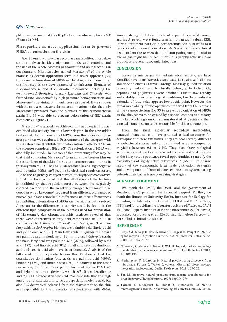
Figure 2 Influence of Maresome®prepared from biomass of Bio 33 (n=50) Arthrospira, Spirogyra and Chlorella (n = 6) on the colonization of MRSA strain NES in the model “Mouse ear”(direct contamination).
0
200
400
600
800
1000
1200
1400
1600
1800
2000
donator(NES) acceptor(NES) control(NES) donator(ATCC) acceptor(ATCC) control(ATCC)
colo
ny fo
rmin
g un
its
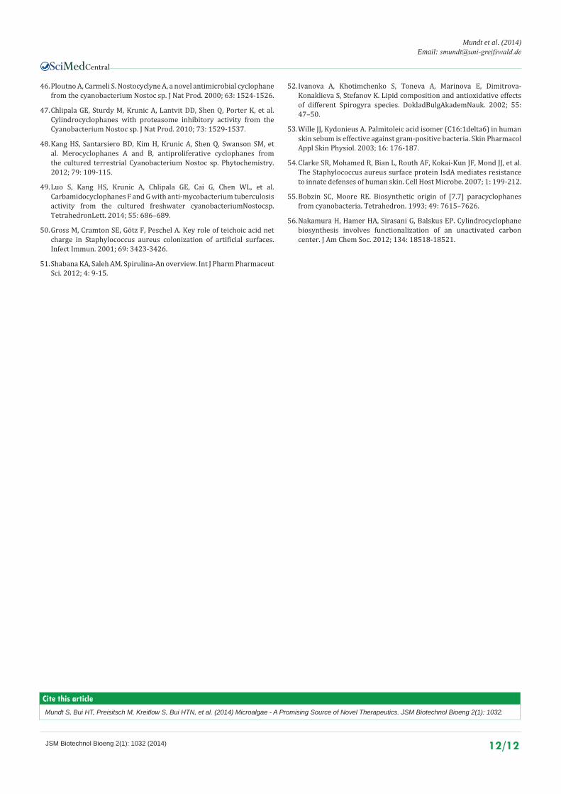
Figure 3 Influence of Bio 33 Maresome® (n = 18) on the colonization of MRSA Strain NES and MSSA strain ATCC 6835 in the model “Cow udder teat” (skin to skin transmission).
Central
Mundt et al. (2014)Email:
JSM Biotechnol Bioeng 2(1): 1032 (2014) 9/12
In agar diffusion tests, these acids and also the non-hydroxylated α-linoleic acid exhibited activity towards Staphylococcus aureus strains. Yet only α-linoleic and α-linolenic acid were able to limit growth of MRSA [27,28]. Fatty acids and their derivates are known as antimicrobial active substances. Already the first antimicrobial substance Chlorellin, isolated from microalgae of the genus Chlorella [22] was identified as a mixture of C18 fatty acids. Apart from antibacterial effects, the release of fatty acids into the cultivation medium resulted in allelopathic effects, a mechanism to improve the survival of the producers in their environment [39]. Mixtures of polyunsaturated and saturated fatty acids, such as eicosapentaenoic acid and α-linolenic acid produced by microalgae are identified as antimicrobial agents especially towards Gram-positive bacteria or even MRSA [20]. Recently, long-chain unsaturated fatty acids and hydroxylated derivatives such as 15-hydroxyeicosapentaenoic acid and 15-hydroxyeicosatrienoic acid with inhibitor activity to Staphylococcus aureus and Propionobacterium acnes were discussed as potential agents in treatment of skin infections [21]. Even though the exact mechanism of action for the antimicrobial activity of fatty acids is not fully clarified, the most probable target seems to be the cell membrane. Damage to the membrane will lead to cell leakage and eventually lysis of bacterial cells. A peroxidative mechanism involving hydrogen peroxide is proposed by Dubois [40].
Cyclic depsipeptides from Lyngbya sp. SAG 36.91
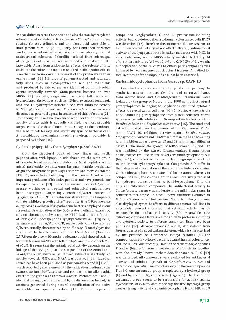
From the structural point of view, linear and cyclic peptides often with lipophilic side chains are the main group of cyanobacterial secondary metabolites. Most peptides are of mixed polyketide synthase/nonribosomal peptide synthetase origin and biosynthetic pathways are more and more elucidated [11]. Cyanobacteria belonging to the genus Lyngbya are important producers of bioactive peptides and have potential for therapeutically use [13]. Especially marine strains of Lyngbya, present worldwide in tropical and subtropical regions, have been investigated. Surprisingly, methanol/water extracts of Lyngbya sp. SAG 36.91, a freshwater strain from the temperate climate, inhibited growth of Bacillus subtilis, E. coli, Pseudomonas aeruginosa as well as all fish pathogenic bacteria employed in our screening. Fractionation of the 50% water methanol extract by column chromatography including HPLC lead to identification of four cyclic undecapeptides, lyngbyazothrins A-D (Figure 1) as binary mixtures A/B and C/D, respectively. Only the mixture C/D, structurally characterized by an N-acetyl-N-methyltyrosine residue at the free hydroxyl group at C5 of Aound (3-amino-2,5,7,8-tetrahydroxy-10-methylundecanoic acid) showed activity towards Bacillus subtilis with MIC of 16µM and to E. coli with MIC of 65µM. It seems that the antimicrobial activity depends on the linkage of the acyl group at the C-5 position of the Aound unit, as only the binary mixture C/D showed antibacterial activity. No activity towards MSSA and MRSA was observed [29]. Identical structures have been published as portoamides A and B [41,42], which reportedly are released into the cultivation medium by the cyanobacterium Oscillatoria sp. and responsible for allelopathic effects to the green alga Chlorella vulgaris. Portoamides C and D, identical to lyngbyazothrins A and B, are discussed as hydrolysis artefacts generated during natural detoxification of the active metabolites in aqueous medium [41]. For the separated
compounds lyngbyzothrin C and D proteasome-inhibiting activity, but no cytotoxic effects to human colon cancer cells HT29 was described [42].Therefore, the antimicrobial activity seems to be not associated with cytotoxic effects. Overall, antimicrobial activity of the lyngbyazothrins is rather moderate with MICs in micromolar range and no MRSA activity was detected. The yield of the binary mixtures A/B was 0.1% and C/D 0.2% of dry weight but separation of the mixtures to obtain pure compounds was hindered by rearrangement of structural isomers. A method for total synthesis of the compounds has not been described.
Carbamidocyclophanes from Nostoc sp. CAVN 10
Cyanobacteria also employ the polyketide pathway to synthesize natural products. Cylindro- and nostocyclophanes from Nostoc linkia and Cylindrospermum licheniforme were isolated by the group of Moore in the 1990 as the first natural paracyclophanes belonging to polyketides exhibited cytotoxic effects to several tumor cell lines [43-45]. Nostocyclyne, a triple bond containing paracyclophane from a field-collected Nostoc sp. caused growth inhibition of Gram-positive bacteria such as Bacillus subtilis and Staphylococcus aureus [46]. The methanol extract prepared from the biomass of the Vietnamese Nostoc strain CAVN 10, exhibited activity against Bacillus subtilis, Staphylococcus aureus and Candida maltosa in the basic screening with inhibition zones between 13 and 18 mm in agar diffusion assay. Furthermore, the growth of MRSA strains 535 and 847 was inhibited by the extract. Bioassay-guided fragmentation of the extract resulted in five novel carbamidocyclophanes A-E (Figure 1), characterized by two carbamidogroups in contrast to the known cylindrocyclophanes. Compounds A-D differ in their degree of chlorination at the end of the butyl side chains. Carbamidocyclophane A contains 4 chlorine atoms whereas in compounds B-E, the chlorine groups are successively replaced by hydrogen atoms so that carbamidocyclophane E is the only non-chlorinated compound. The antibacterial activity to Staphylococcus aureus was moderate in the milli molar range. In contrast to that, ampicillin, a well-known antibiotic, exhibited an MIC of 2.2 µmol in our test system. The carbamidocyclophanes also displayed cytotoxic effects to different tumor cell lines in micromolar concentrations, so that cytotoxic effects may be responsible for antibacterial activity [30]. Meanwhile, new cylindrocyclophanes from a Nostoc sp. with protease inhibiting and cytotoxic activity to different tumor cell lines have been published [47]. Merocyclophanes A and B, also isolated from Nostoc, consist of a novel carbon skeleton, which is characterized by the presence of α-branched methyl residues [48].The compounds display cytotoxic activity against human colon cancer cell line HT-29. Most recently, isolation of carbamidocyclophanes F and G (Figure 1) from a freshwater Nostoc strain together with the already known carbamidocyclophanes A, B, C [49] was described. All compounds were evaluated for antibacterial activity and inhibited growth of Staphylococcus aureus and Enterococcus faecalis in micromolar range. In the new compounds F and G, one carbamido group is replaced by a hydroxyl group (F) and by acetate (G), respectively (Figure 1). The loss of one carbamido group seems to be responsible for activity against Mycobacterium tuberculosis, especially the free hydroxyl group causes strong activity of carbamidocyclophane F with MIC of 0.8
Central
Mundt et al. (2014)Email:
JSM Biotechnol Bioeng 2(1): 1032 (2014) 10/12
µM in comparison to MICs >10 µM of carbamidocyclophanes A-C (Figure 1) [49].
Microparticle as novel application form to prevent MRSA colonization on the skin
Apart from low molecular secondary metabolites, microalgae contain polysaccharides, pigments, lipids and proteins and the use of the whole biomass as foodstuff and animal feed is in development. Microparticles named Maresome® of the whole biomass as dermal application form is a novel approach [33] to prevent colonization of MRSA on the skin, which constitutes the first step in the development of an infection. Biomass of 3 cyanobacteria and 3 eukaryotic microalgae, including the well-known Arthrospira, formely Spirulina and Chlorella, was formed into Maresome® by high-pressure homogenization and Maresome® containing ointments were prepared. It was shown with the mouse ear assay, a direct contamination model, that only Maresome® prepared from the biomass of the cyanobacterial strain Bio 33 was able to prevent colonization of NES strain completely (Figure 2).
Maresome® prepared from Chlorella and Arthrospira biomass exhibited also activity but to a lower degree. In the cow udder teat model, the transmission of MRSA from the donor skin to an acceptor skin was evaluated. Pretreatment of the acceptor with Bio 33 Maresome® inhibited the colonization of attached NES on the acceptor completely (Figure 3). The colonization of MSSA was also fully inhibited. The reason of this protecting effect may be that lipid containing Maresome® form an anti-adhesion film on the outer layer of the skin, the stratum corneum, and interact in this way with MRSA. The Bio 33 Maresome® have a high negative zeta potential (-38.8 mV) leading to electrical repulsion forces. Due to the negatively charged surface of Staphylococcus aureus, [50] it can be speculated that the attachment of the bacteria is inhibited by that repulsion forces between the negatively charged bacteria and the negatively charged Maresome®. The question why Maresome® prepared from different biomasses of microalgae show remarkable differences in their effectiveness in inhibiting colonization of MRSA on the skin is not resolved. A reason for the differences in activity could be found in the different lipid composition of the biomass used for preparation of Maresome®. Gas chromatographic analyses revealed that there were differences in fatty acid composition of Bio 33 in comparison to Arthrospira, Chlorella and Spirogyra. The main fatty acids in Arthrospira biomass are palmitic acid, linoleic acid and γ-linolenic acid [51]. Main fatty acids in Spirogyra biomass are palmitic and linolenic acid [52]. In the used Chlorella strain the main fatty acid was palmitic acid (27%), followed by oleic acid (17%) and linoleic acid (8%); small amounts of palmitoleic acid and stearic acid also have been detected. Analysis of the fatty acids of the cyanobacterium Bio 33 showed that the quantitative dominating fatty acids are palmitic acid (49%), linolenic (32%) and linoleic acid (8%). In contrast to the other microalgae, Bio 33 contains palmitoleic acid isomer C16:1 Δ7 and higher unsaturated derivatives such as 7,10 hexadecadienoic and 7,10,13 hexadecatrienoic acid. We conclude that the high amount of unsaturated fatty acids, especially linolenic acid, but also C16 derivatives released from the Maresome® on the skin are responsible for the prevention of colonization with MRSA.
Similar strong inhibition effects of a palmitoleic acid isomer against S. aureus were found also in human skin sebum [53]. Dermal treatment with cis-6-hexadecenoic acid also leads to a reduction of S. aureus colonization [54]. Since preliminary clinical tests confirm the in-vitro data, the anti-pathogenic potential of microalgae might be utilized in form of a prophylactic skin care product to prevent nosocomial infections.
CONCLUSIONScreening microalgae for antimicrobial activity, we have
identified several prokaryotic cyanobacterial strains with distinct and specific effects in-vitro. Through bioassay guided isolation secondary metabolites, structurally belonging to fatty acids, peptides and polyketides were obtained. Due to low activity and stability under physiological conditions, the therapeutically potential of fatty acids appears low at this point. However, the remarkable ability of microparticles prepared from the biomass of the cyanobacterium Bio 33 to prevent colonization of MRSA on the skin seems to be caused by a special composition of fatty acids. Especially high amounts of unsaturated fatty acids and their unusual isomers seem to be responsible for this phenomenon.
From the small molecular secondary metabolites, paracyclophanes seem to have potential as lead structures for development of new antibiotics. They are produced by different cyanobacterial strains and can be isolated as pure compounds in yields between 0.1 to 0.2%. They also show biological activities against multidrug resistant bacteria and first insights in the biosynthetic pathways reveal opportunities to modify the biosynthesis of highly active substances [48,55,56]. To ensure supply of the compounds, large scale cultivation strategies and development of heterologous expressions systems using heterotrophic bacteria are promising strategies.
ACKNOWLEDGEMENTWe thank the BMBF, the DAAD and the government of
Mecklenburg-Vorpommern for financial support. Further, we thank the Humboldt-University-Berlin, Institute for Ecology for providing the laboratory culture of HUB 051 and Dr. N. V. Tran, IBT Hanoi for providing the laboratory culture of Nostoc sp. CAVN 10. Beate Cuypers, Institute of Marine Biotechnology, Greifswald, is thanked for isolating strain Bio 33 and Hannelore Bartrow for her skillful technical assistance.
REFERENCES1. Burja AM, Banaigs B, Abou-Mansour E, Burgess JG, Wright PC. Marine
cyanobacteria – a prolific source of natural products. Tetrahedron. 2001; 57: 9347–9377
2. Nunnery JK, Mevers E, Gerwick WH. Biologically active secondary metabolites from marine cyanobacteria. Curr Opin Biotechnol. 2010; 21: 787-793.
3. Niedermeyer T, Brönstrup M. Natural product drug discovery from microalgae. Posten C, Walter C, editors. Microalgal biotechnology: integration and economy. Berlin: De Gruyter. 2012; 169-202.
4. Tan LT. Bioactive natural products from marine cyanobacteria for drug discovery. Phytochemistry. 2007; 68: 954-979.
5. Tarman K, Lindequist U, Mundt S. Metabolites of Marine microorganisms and their pharmacological activities. Kim SK, editor.

Central
Mundt et al. (2014)Email:
JSM Biotechnol Bioeng 2(1): 1032 (2014) 11/12
Marine Microbiology: bioactive compounds and biotechnological applications. Weinheim: Wiley-VCH; 2013; 393-416.
6. Rosello Sastre R. Products from microalgae: an overview. Posten C, Walter C, editors. Microalgal biotechnology: integration and economy. Berlin: De Gruyter; 2012; 13–50.
7. Mostafa SSM. Microalgal biotechnology. Prospects and applications. Dhal NK, Sahu SC, editors. Plant Science. In Tech; 2012; 275–314.
8. Blunt JW, Copp BR, Keyzers RA, Munro MHG, Prinsep M. Marine natural products. Nat Prod Rep 2012; 29: 144–222.
9. Blunt JW, Copp BR, Keyzers RA, Munro MH, Prinsep MR. Marine natural products. Nat Prod Rep. 2013; 30: 237-323.
10. Blunt JW, Copp BR, Keyzers RA, Munro MH, Prinsep MR. Marine natural products. Nat Prod Rep. 2014; 31: 160-258.
11. Sivonen K, Börner T. Bioactive compounds produced by cyanobacteria. Herrero A, Flores E, editors. The cyanobacteria: molecular biology, genomics and evolution. Norfolk: Caister Academic Press. 2008; 159-198.
12. Singh RK, Tiwari SP, Rai AK, Mohapatra TM. Cyanobacteria: an emerging source for drug discovery. J Antibiot (Tokyo). 2011; 64: 401-412.
13. Liu L, Rein KS. New peptides isolated from Lyngbya species: a review. Mar Drugs. 2010; 8: 1817-1837.
14. Tan LT. Filamentous tropical marine cyanobacteria: A rich source of natural products for anticancer drug discovery. J Appl Phycol. 2010; 22: 659–676.
15. U.S. Food and Drug Administration
16. European Medicines Agency
17. Younes A, Yasothan U, Kirkpatrick P. Brentuximab vedotin. Nat Rev Drug Discov. 2012; 11: 19-20.
18. Katz J, Janik JE, Younes A. Brentuximab Vedotin (SGN-35). Clin Cancer Res. 2011; 17: 6428-6436.
19. Marine Pharmaceuticals: The Clinical Pipeline
20. Amaro HM, Guedes AC, Malcata FX. Antimicrobial activities of microalgae: an invited review. 3rd edn. Mendez-Villas A, editor. Science against microbial pathogens: communicating current research and technological advances. Badajoz: Formatex; 2011; 12721–12280.
21. Desbois AP, Lawlor KC. Antibacterial activity of long-chain polyunsaturated fatty acids against Propionibacterium acnes and Staphylococcus aureus. Mar Drugs. 2013; 11: 4544-4557.
22. Pratt R, Daniels TC, Eiler JJ, Gunnison JB, Kumler WD, Oneto JF, Strait LA. CHLORELLIN, AN ANTIBACTERIAL SUBSTANCE FROM CHLORELLA. Science. 1944; 99: 351-352.
23. Barsanti L, Gualtieri P. editors. Algae. Anatomy, biochemistry and biotechnology. Boca Raton: CRC press. 2006.
24. Santek B, Felski M, Fries K, Lotz M, Flaschel E. Production of paramylon, a ß- 1,3-glucan, by heterotrophic cultivation of Euglena gracilis. Eng Life Sci. 2009; 9: 23–28.
25. Zhang J, Sun Z, Sun P, Chen T, Chen F. Microalgal carotenoids: beneficial effects and potential in human health. Food Funct. 2014; 5: 413-425.
26. Rippka R, Herdman H. editors. Pasteur Culture Collection of cyanobacteria: Catalogue and taxonomic handbook. I. Catalogue of strains. Paris: Institut Pasteur. 1992.
27. Mundt S, Kreitlow S, Nowotny A, Effmert U. Biochemical and pharmacological investigations of selected cyanobacteria. Int J Hyg
Environ Health. 2001; 203: 327-334.
28. Mundt S, Kreitlow S, Jansen R. Fatty acids with antibacterial activity from the cyanobacterium Oscillatoria redekei HUB 051. J Appl Phycol. 2003; 15: 263–267.
29. Zainuddin EN, Jansen R, Nimtz M, Wray V, Preisitsch M, Lalk M, et al. Lyngbyazothrins A-D, antimicrobial cyclic undecapeptides from the cultured Cyanobacterium lyngbya sp. J Nat Prod. 2009; 72: 1373-1378.
30. Bui HT, Jansen R, Pham HT, Mundt S. Carbamidocyclophanes A-E, chlorinated paracyclophanes with cytotoxic and antibiotic activity from the Vietnamese cyanobacterium Nostoc sp. J Nat Prod. 2007; 70: 499-503.
31. European Pharmacopoeia. Microbial assay of antibiotics, 5th edn. Council of Europe (COE), European Directorate for the Quality of Medicines (EDQM). 2005; 5.3: 188.
32. Bansemir A, Phytochemische und biologische Untersuchungen ausgewählter Makroalgen unter besonderer Berücksichtigung ihrer Wirksamkeit gegen die Erreger bakterieller Fischkrankheiten [dissertation]. Greifswald: Ernst-Moritz-Arndt-University, 2004.
33. Lukowski G, Lindequist U, Jülich WD, Mundt S. Pharmazeutisch oder kosmetisch wirksame Mittel aus lipidhaltigen marinen Organismen. EP 2 255 821 A1. 2010.
34. Lukowski G, Lindequist U, Mundt S, Kramer A, Julich WD. Inhibition of dermal MRSA colonization by microalgal micro- and nanoparticles. Skin Pharmacol Physiol. 2008; 21: 98-105.
35. Wu JT, Chiang YR, Huang WY, Jane WN. Cytotoxic effects of free fatty acids on phytoplankton algae and cyanobacteria. Aquat Toxicol. 2006; 80: 338-345.
36. Pham HTL. Bioassay-guided isolation of cytotoxic and antifungal compounds from cyanobacteria [dissertation]. Greifswald: Ernst-Moritz-Arndt-University. 2007.
37. Report on the burden of endemic health care associated infection word wide. 2011.
38. Ördög VO, Stirk WA, Lenobel R, Bancirova M, Strnad M, van Staden J, et al. Screening microalgae for some potentially useful agricultural and pharmaceutical secondary metabolites. J Appl Phycol. 2004; 16: 309–314.
39. DellaGreca M, Zarrelli A, Fergola P, Cerasuolo M, Pollio A, Pinto G. Fatty acids released by Chlorella vulgaris and their role in interference with Pseudokirchneriella subcapitata: experiments and modelling. J Chem Ecol. 2010; 36: 339-349.
40. Desbois AP, Mearns-Spragg A, Smith VJ. A fatty acid from the diatom Phaeodactylum tricornutum is antibacterial against diverse bacteria including multi-resistant Staphylococcus aureus (MRSA). Mar Biotechnol (NY). 2009; 11: 45-52.
41. Leão PN, Pereira AR, Liu WT, Ng J, Pevzner PA, Dorrestein PC, et al. Synergistic allelochemicals from a freshwater cyanobacterium. Proc Natl Acad Sci U S A. 2010; 107: 11183-11188.
42. Zi J, Lantvit DD, Swanson SM, Orjala J. Lyngbyaureidamides A and B, two anabaenopeptins from the cultured freshwater cyanobacterium Lyngbya sp. (SAG 36.91). Phytochemistry. 2012; 74: 173-177.
43. Moore BS, Chen JL, Patterson GML, Moore RE, Brinen L, Kazo Y, et al. [7.7] paracyclophanes from blue-green algae. J Am ChemSoc. 1990; 112: 4016–4063.
44. Chen JL, Moore RE, Patterson GML. Structures of nostocyclophanes A–D. J Org Chem. 1991; 56: 4360–4364.
45. Moore BS, Chen JL, Patterson GML, Moore RE. Structures of cylindrocyclophanes A–F. Tetrahedron. 1992; 48: 3001–3006.
Central
Mundt et al. (2014)Email:
JSM Biotechnol Bioeng 2(1): 1032 (2014) 12/12
Mundt S, Bui HT, Preisitsch M, Kreitlow S, Bui HTN, et al. (2014) Microalgae - A Promising Source of Novel Therapeutics. JSM Biotechnol Bioeng 2(1): 1032.
Cite this article
46. Ploutno A, Carmeli S. Nostocyclyne A, a novel antimicrobial cyclophane from the cyanobacterium Nostoc sp. J Nat Prod. 2000; 63: 1524-1526.
47. Chlipala GE, Sturdy M, Krunic A, Lantvit DD, Shen Q, Porter K, et al. Cylindrocyclophanes with proteasome inhibitory activity from the Cyanobacterium Nostoc sp. J Nat Prod. 2010; 73: 1529-1537.
48. Kang HS, Santarsiero BD, Kim H, Krunic A, Shen Q, Swanson SM, et al. Merocyclophanes A and B, antiproliferative cyclophanes from the cultured terrestrial Cyanobacterium Nostoc sp. Phytochemistry. 2012; 79: 109-115.
49. Luo S, Kang HS, Krunic A, Chlipala GE, Cai G, Chen WL, et al. Carbamidocyclophanes F and G with anti-mycobacterium tuberculosis activity from the cultured freshwater cyanobacteriumNostocsp. TetrahedronLett. 2014; 55: 686–689.
50. Gross M, Cramton SE, Götz F, Peschel A. Key role of teichoic acid net charge in Staphylococcus aureus colonization of artificial surfaces. Infect Immun. 2001; 69: 3423-3426.
51. Shabana KA, Saleh AM. Spirulina-An overview. Int J Pharm Pharmaceut Sci. 2012; 4: 9-15.
52. Ivanova A, Khotimchenko S, Toneva A, Marinova E, Dimitrova-Konaklieva S, Stefanov K. Lipid composition and antioxidative effects of different Spirogyra species. DokladBulgAkademNauk. 2002; 55: 47–50.
53. Wille JJ, Kydonieus A. Palmitoleic acid isomer (C16:1delta6) in human skin sebum is effective against gram-positive bacteria. Skin Pharmacol Appl Skin Physiol. 2003; 16: 176-187.
54. Clarke SR, Mohamed R, Bian L, Routh AF, Kokai-Kun JF, Mond JJ, et al. The Staphylococcus aureus surface protein IsdA mediates resistance to innate defenses of human skin. Cell Host Microbe. 2007; 1: 199-212.
55. Bobzin SC, Moore RE. Biosynthetic origin of [7.7] paracyclophanes from cyanobacteria. Tetrahedron. 1993; 49: 7615–7626.
56. Nakamura H, Hamer HA, Sirasani G, Balskus EP. Cylindrocyclophane biosynthesis involves functionalization of an unactivated carbon center. J Am Chem Soc. 2012; 134: 18518-18521.