biosynthesis of silver nanoparticles using an endophytic...
TRANSCRIPT

Journal of Nanoscience and Nanoengineering
Vol. 1, No. 4, 2015, pp. 241-247
http://www.aiscience.org/journal/jnn
* Corresponding author
E-mail address: [email protected] (S. Muthukrishnan)
Biosynthesis of Silver Nanoparticles Using an Endophytic Fungus, Curvularialunata and Its Antimicrobial Potential
Parthasarathy Ramalingmam, Sathiyabama Muthukrishnan*, Prabha Thangaraj
Department of Plant Science, School of Life Sciences, Bharathidasan University, Tiruchirappalli, TamilNadu, India
Abstract
The development of reliable green process for the synthesis of silver nanoparticles is an important branch of Nanobiotechnology.
In the present investigation the use of the endophytic fungus Curvularialunata for the extracellular biosynthesis of silver
nanoparticles (AgNPs) from silver nitrate solution is reported. It was observed that the aqueous silver (Ag+) ions, when exposed
to a filtrate of C. lunata, were reduced in solution, resulting in the formation of stable AgNPs. These AgNPs were characterized
by means of several techniques. The nanoparticles show maximum absorbance at 422 nm on ultraviolet-visible spectra. The
presence of protein was identified by Fourier Transform infrared spectroscopy. The reduction of Ag+ ion to elemental silver was
characterized by Energy - dispersive X-ray (EDX) Spectroscopy. Scanningelectron micrograph revealed the formation of
polydispersed nanoparticles of 10-50 nm. The nanoparticles were also evaluated for their enhanced antimicrobial activities with
various antibiotics against gram positive and gram negative bacteria. The antibacterial activities of Ampicillin, Rifampicin,
Chloramphenicol, Erythromycin, and Kanamycin were increased in the presence of AgNPs against test strain. The highest fold
increase of area was found for Erythromycin and Carbenicilin against E. coli, Ampicilin against S. paratyphi, Erythromycin
against B. subtillis. The results showed that the combination of antibiotics with AgNPshas better antimicrobial effects.
Keywords
Endophytic Fungi, Silver Nanoparticles, SEM, FTIR, Antibacterial Activity
Received: August 19, 2015 / Accepted: September 24, 2015 / Published online: December 6, 2015
@ 2015 The Authors. Published by American Institute of Science. This Open Access article is under the CC BY-NC license.
http://creativecommons.org/licenses/by-nc/4.0/
1. Introduction
Antibiotic resistance by pathogenic bacteria and fungi has
been continuously increasing over the past decade and there is
a need for the development of new antimicrobial agents. In the
present scenario, AgNPs have appeared as a promising
antibacterial candidate in the medical field (SindhuPriya et al.,
2013 [1]). Currently, nanobiotechnology represents an
alternative for chemical and physical methods for the
synthesis of NPs (Ahmed et al., 2003 [2], Anusuya and
Sathiyabama 2014 [3]). Recognizing the importance of
developing an eco-friendly synthesis of nanoparticles, number
of researches have turned to microorganisms that have shown
an ability to reduce metal ions. Synthesis of NPs from
microbes is a boon for advance research in nanotechnology.
The use of fungus for the synthesis of NPs could be more
advantageous, because of the tolerance, bioaccumulation
(Slawson et al., 1992 [4]) and extracellular production.
Further advantages includes its economic viability and ease in
handling biomass, large scale production of enzymes. NPs
such as Ag, Au, Pt and Pd are widely exploited due to its
special physicochemical properties in biochemical
applications. Several studies suggested that biomolecules like
proteins, phenols and flavonoids play a vital role in the
reduction of Ag+ ions (Ahmad et al., 2011 [5]) which are
242 Parthasarathy Ramalingmam et al.: Biosynthesis of Silver Nanoparticles Using an Endophytic
Fungus, Curvularialunata and Its Antimicrobial Potential
present in the fungal cultures. Therefore the exploitation of the
fungal culture filtrate in the synthesis of nanoparticles is
fascinating. Colloidal silver has been known for a long time to
possess antimicrobial properties and also it is non-toxic and
environmentally safe (Sathishkumar et al., 2012 [6]). Silver
ions have more advantage against conventional antibiotics;
(Antony et al., 2011 [7]).
Endophytes are microorganisms that live to complete at least
one stage of its life without causing apparent diseases are
obviously a rich and reliable source of bioactive metabolites
with large medical, agricultural and industrial potentials
(Strobel and Daisy, 2013 [8]; Parthasarathy and Sathiyabama
2014,2015 [9-10]). However, very few endophytic fungi such
as Aspergillusclavatus, Epicoccumnigrum have been reported
to biosynthesize of AgNPs (Verma et al., 2010 [11]; Qian et al.,
2013 [12]). Clearly, endophytes are relatively unexplored as a
potential resource for the synthesis of AgNPs.
The present study involves with the extracellular synthesis and
characterization of AgNPs using an endophytic fungus
C.lunata and evaluating the antibacterial effect of synthesized
silver nanoparticles against some human pathogens.
2. Materials and Methods
2.1. Microorganism and Production of
Biomass
The fungus C.lunata (ITCC No.8428.11), isolated from leaves
of Catharanthusroeus,was grown in 250 ml Erlenmeyer flasks
containing 100 ml Potato dextrose broth (PDB) at 27°C for 72
hrs. After incubation, mycelialbiomass were separated by
filtration, washed with sterile distilled water to remove the
traces of media components.
2.2. Synthesis of Silver Nanoparticles
Typically 15g of biomass (wet weight) was re-suspended in
100ml sterile double distilled water for 48 h at 27°C in an
Erlenmeyer flask and incubated. After incubation, the cell
filtrate was filtered by Whatman filter Paper No. 1. To the cell
filtrate silver nitrate was added (1mM), and incubated at room
temperature under dark condition for 24 h.
2.3. Characterization of Silver Nanoparticles
After 2 hours of incubation of the above mixture, the
preliminary detection of AgNPs was carried out by visual
observation of color change of the cell filtrate. The samples
were later subjected to optical measurements, which were
carried out by using a UV-visible spectrophotometer
(Shimadzu, Japan) and scanning the spectra between 200 to
700 nm at the resolution of 1nm.The pellet was re-suspended
in Milli-Q water and freeze dried. FTIR spectroscopic
studies were carried out to find possible bio-reducing agent
present in the culture filtrate of C. lunata. The FTIR
spectrum of the dried sample was recorded on a PerkinElmer
1600 instrument in the range of 4000 cm-1
and 400cm-1
. To
perform SEM analysis, thin films of the sample were
prepared on a carbon coated copper grid. The morphology,
size were measured at 100 Kev using Hitachi S-4500 SEM.
The size of the distributed silver nanoparticleswas also
measured by Dynamic Light Scattering (DLS) technique
using Malvern Zeta Sizer Nano series compact scattering
spectrometer. The stability of the nanoparticle in suspension
was determined by measuring the zeta potential (Zeta sizer
Nano ZS, Malvern). EDX spectroscopy analysis were carried
out for the confirmation of elemental silver in the
sample.XRD patterns of silver nanoparticles were obtained
by D max -2200, Rigaku, Japan.
2.4. Screening of Antimicrobial Activity
The antimicrobial effect of synthesized AgNPswas evaluated
against some human pathogens such as gram negative (E. coli,
Pseudomonas aeruginosa, Salmonella paratyphi) and gram
positive (Bacillus subtilis, Staphylococcus aureus, Bacillus
cereus) bacteria by disc diffusion method. Cultures were
maintained at -80°C on glycerol stock. They were
sub-cultured in Nutrient Broth for 24hrs at 30°C. Each strain
was swabbed uniformly into the individual Muller-Hinton
agar plates using sterile cotton swabs. Using sterile
micropipette, 30 µL of crude culture filtrate, AgNPs were
loaded on to sterile paper disc and it was allowed to dry. The
sample loaded discs along with standard antibiotic discwere
impregnated in the Muller-Hinton agar medium. The doses
were selected based on the preliminary data obtained from a
laboratory. After 24 hrs incubation at 37°C, the levels of zone
of inhibition were measured.
3. Results
3.1. Synthesis of AgNPs
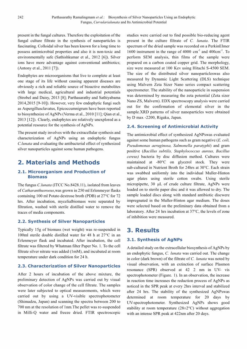
A detailed study on the extracellular biosynthesis of AgNPs by
an endophytic fungus, C. lunata was carried out. The change
in color (dark brown) of the filtrate of C. lunata was noted by
visual observation, with an extinction of surface Plasmon
resonance (SPR) observed at 42 2 nm in UV- vis
spectrophotometer (Figure. 1). In an observation, the increase
in reaction time increases the reduction process of AgNPs as
noticed in the SPR peak at every 2hrs interval and stabilized
after 24 hrs. The stability of the synthesized AgNPswas
determined at room temperature for 20 days by
UV-spectrophotometer. Synthesized AgNPs shows good
stability at room temperature (28±2°C) without aggregation
with an intense SPR peak at 422nm after 20 days.
Journal of Nanoscience and Nanoengineering Vol. 1, No. 4, 2015, pp. 241-247 243
Figure 1. UV-vis spectra at different time intervals.
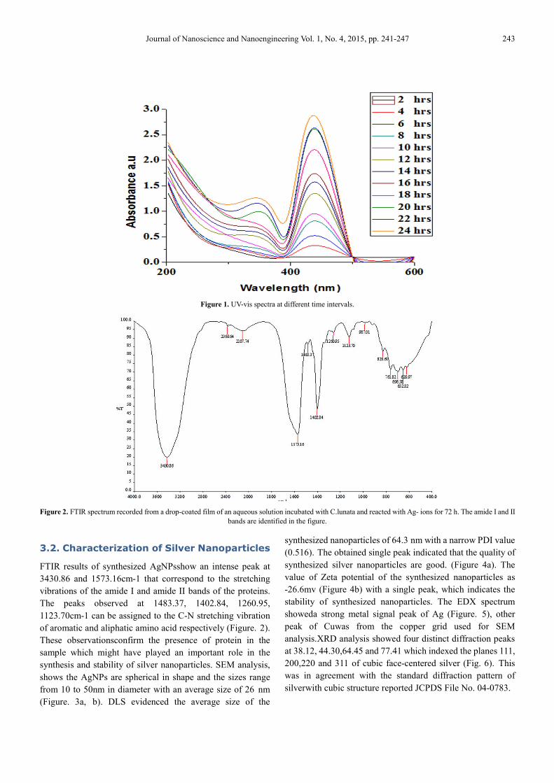
Figure 2. FTIR spectrum recorded from a drop-coated film of an aqueous solution incubated with C.lunata and reacted with Ag- ions for 72 h. The amide I and II
bands are identified in the figure.
3.2. Characterization of Silver Nanoparticles
FTIR results of synthesized AgNPsshow an intense peak at
3430.86 and 1573.16cm-1 that correspond to the stretching
vibrations of the amide I and amide II bands of the proteins.
The peaks observed at 1483.37, 1402.84, 1260.95,
1123.70cm-1 can be assigned to the C-N stretching vibration
of aromatic and aliphatic amino acid respectively (Figure. 2).
These observationsconfirm the presence of protein in the
sample which might have played an important role in the
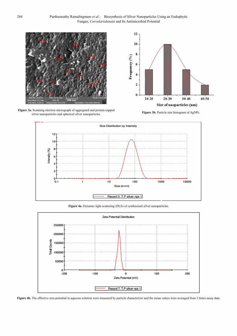
synthesis and stability of silver nanoparticles. SEM analysis,
shows the AgNPs are spherical in shape and the sizes range
from 10 to 50nm in diameter with an average size of 26 nm
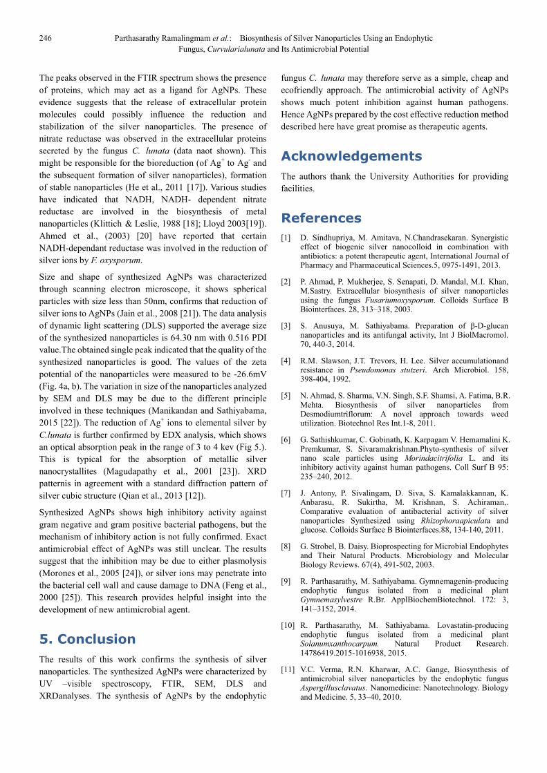
(Figure. 3a, b). DLS evidenced the average size of the
synthesized nanoparticles of 64.3 nm with a narrow PDI value
(0.516). The obtained single peak indicated that the quality of
synthesized silver nanoparticles are good. (Figure 4a). The
value of Zeta potential of the synthesized nanoparticles as
-26.6mv (Figure 4b) with a single peak, which indicates the
stability of synthesized nanoparticles. The EDX spectrum
showeda strong metal signal peak of Ag (Figure. 5), other
peak of Cuwas from the copper grid used for SEM
analysis.XRD analysis showed four distinct diffraction peaks
at 38.12, 44.30,64.45 and 77.41 which indexed the planes 111,
200,220 and 311 of cubic face-centered silver (Fig. 6). This
was in agreement with the standard diffraction pattern of
silverwith cubic structure reported JCPDS File No. 04-0783.
244 Parthasarathy Ramalingmam et al.: Biosynthesis of Silver Nanoparticles Using an Endophytic
Fungus, Curvularialunata and Its Antimicrobial Potential
Figure 3a. Scanning electron micrograph of aggregated and protein-capped
silver nanoparticles and spherical silver nanoparticles.
Figure 3b. Particle size histogram of AgNPs.
Figure 4a. Dynamic light scattering (DLS) of synthesized silver nanoparticles.
Figure 4b. The effective zeta-potential in aqueous solution were measured by particle characterizer and the mean values were averaged from 3 times assay data.

Journal of Nanoscience and Nanoengineering Vol. 1, No. 4, 2015, pp. 241-247 245
Figure 5. Energy-dispersive spectroscopy spectrum of silver nanoparticles.
Various x-ray emission peaks are labeled.
Figure 6. XRD Pattern of synthesized silver nanoparticles.
3.3. Antimicrobial Activity of AgNPs
Antimicrobial effects of synthesized AgNPswas tested against
human pathogens like E. coli, Pseudomonas aeruginosa,
Salmonella paratyphi, Bacillus subtillis, Staphylococcus
aureus and Bacillus cereus (Table. 1). Synthesized AgNps
showed inhibitory activity against gram negative and gram
positive bacterial pathogens.
Table 1. Zone of inhibition (mm) of different antibiotics against test strains
(in absence of and in presence of AgNPs at concentration of 10µg/disk gram
positive and gram negative bacteria).
Antibiotic
E. coli
Antibiotic (a) Antibiotic with
AgNPs (b)
Increase fold in
area
(b2-a2/a2)
Penicillin 8 10.6 0.75
Ampicilin 5 6.66 0.77
Rifampicin 10.33 14.33 0.92
Erythromycin 10 14.33 1.05
Kanamycin 10.66 13 0.48
Chloramphenicol 11.33 14.66 0.67
Carbenicilin 15 22 1.15
Antibiotic
Salmonella paratyphi
Antibiotic (a) Antibiotic with
AgNPs (b)
Increase fold in
area(b2-a2/a2)
Penicillin 12.33 15 0.48
Ampicilin 8 11.33 1.00
Rifampicin 10 12 0.44
Erythromycin 10.33 13 0.58
Kanamycin 11 13.66 0.54
Chloramphenicol 14.33 17 0.40
Carbenicilin 11.33 15 0.75
Antibiotic
Bacillus subtillis
Antibiotic (a) Antibiotic with
AgNPs (b)
Increase fold in
area(b2-a2/a2)
Penicillin 6.33 8.33 0.73
Ampicilin 8 9.66 0.45
Rifampicin 10.66 12.66 0.41
Erythromycin 8.33 12 1.07
Kanamycin 10.66 11.66 0.19
Chloramphenicol 11.66 14 0.44
Carbenicilin 10.33 12 0.34
Antibiotic
Staphylococcus aureus
Antibiotic (a) Antibiotic with
AgNPs (b)
Increase fold in
area(b2-a2/a2)
Penicillin 7 9.33 0.77
Ampicilin 6.33 7.33 0.34
Rifampicin 9 10.66 0.40
Erythromycin 10.33 12.66 0.50
Kanamycin 10 11 0.21
Chloramphenicol 10.66 13 0.48
Carbenicilin 11.33 13 0.31
4. Discussion
The study of the biosynthesis of nanonmaterials offers a
valuable contribution to nanobiotechnology. In this present
study an endophytic fungus was evaluated for synthesis of
nanoparticles in short reaction time. When C. lunataculture
filtrate was mixed with aqueous solution of silver nitrate, it
starts to change the color from the yellowish to brown due to
the reduction process of silver ions (Yang et al., 2007 [13];
Wang & Su 2001 [14]). The synthesized AgNPs were
confirmed based on the UV-vis spectra at 423 nm due to
excitation of Surface Plasmon Resonance (Mulvaney et al.,
1996 [15]). This observation indicates that the reduction of the
Ag+ ions takes place extracellularly. Absorbance band at
260nm is clearly visible and is attributed to electronic
excitation in tryptophan and tyrosine residues in protein
(Eftnick and Ghiron, 1981 [16]), which indicates the release
of extracellular proteins in the colloidal solution.
246 Parthasarathy Ramalingmam et al.: Biosynthesis of Silver Nanoparticles Using an Endophytic
Fungus, Curvularialunata and Its Antimicrobial Potential
The peaks observed in the FTIR spectrum shows the presence
of proteins, which may act as a ligand for AgNPs. These
evidence suggests that the release of extracellular protein
molecules could possibly influence the reduction and
stabilization of the silver nanoparticles. The presence of
nitrate reductase was observed in the extracellular proteins
secreted by the fungus C. lunata (data naot shown). This
might be responsible for the bioreduction (of Ag+ to Ag
- and
the subsequent formation of silver nanoparticles), formation
of stable nanoparticles (He et al., 2011 [17]). Various studies
have indicated that NADH, NADH- dependent nitrate
reductase are involved in the biosynthesis of metal
nanoparticles (Klittich & Leslie, 1988 [18]; Lloyd 2003[19]).
Ahmed et al., (2003) [20] have reported that certain
NADH-dependant reductase was involved in the reduction of
silver ions by F. oxysporum.
Size and shape of synthesized AgNPs was characterized
through scanning electron microscope, it shows spherical
particles with size less than 50nm, confirms that reduction of
silver ions to AgNPs (Jain et al., 2008 [21]). The data analysis
of dynamic light scattering (DLS) supported the average size
of the synthesized nanoparticles is 64.30 nm with 0.516 PDI
value.The obtained single peak indicated that the quality of the
synthesized nanoparticles is good. The values of the zeta
potential of the nanoparticles were measured to be -26.6mV
(Fig. 4a, b). The variation in size of the nanoparticles analyzed
by SEM and DLS may be due to the different principle
involved in these techniques (Manikandan and Sathiyabama,
2015 [22]). The reduction of Ag+ ions to elemental silver by
C.lunata is further confirmed by EDX analysis, which shows
an optical absorption peak in the range of 3 to 4 kev (Fig 5.).
This is typical for the absorption of metallic silver
nanocrystallites (Magudapathy et al., 2001 [23]). XRD
patternis in agreement with a standard diffraction pattern of
silver cubic structure (Qian et al., 2013 [12]).
Synthesized AgNPs shows high inhibitory activity against
gram negative and gram positive bacterial pathogens, but the
mechanism of inhibitory action is not fully confirmed. Exact
antimicrobial effect of AgNPs was still unclear. The results
suggest that the inhibition may be due to either plasmolysis
(Morones et al., 2005 [24]), or silver ions may penetrate into
the bacterial cell wall and cause damage to DNA (Feng et al.,
2000 [25]). This research provides helpful insight into the
development of new antimicrobial agent.
5. Conclusion
The results of this work confirms the synthesis of silver
nanoparticles. The synthesized AgNPs were characterized by
UV –visible spectroscopy, FTIR, SEM, DLS and
XRDanalyses. The synthesis of AgNPs by the endophytic
fungus C. lunata may therefore serve as a simple, cheap and
ecofriendly approach. The antimicrobial activity of AgNPs
shows much potent inhibition against human pathogens.
Hence AgNPs prepared by the cost effective reduction method
described here have great promise as therapeutic agents.
Acknowledgements
The authors thank the University Authorities for providing
facilities.
References
[1] D. Sindhupriya, M. Amitava, N.Chandrasekaran. Synergistic effect of biogenic silver nanocolloid in combination with antibiotics: a potent therapeutic agent, International Journal of Pharmacy and Pharmaceutical Sciences.5, 0975-1491, 2013.
[2] P. Ahmad, P. Mukherjee, S. Senapati, D. Mandal, M.I. Khan, M.Sastry. Extracellular biosynthesis of silver nanoparticles using the fungus Fusariumoxysporum. Colloids Surface B Biointerfaces. 28, 313–318, 2003.
[3] S. Anusuya, M. Sathiyabama. Preparation of β-D-glucan nanoparticles and its antifungal activity, Int J BiolMacromol. 70, 440-3, 2014.
[4] R.M. Slawson, J.T. Trevors, H. Lee. Silver accumulationand resistance in Pseudomonas stutzeri. Arch Microbiol. 158, 398-404, 1992.
[5] N. Ahmad, S. Sharma, V.N. Singh, S.F. Shamsi, A. Fatima, B.R. Mehta. Biosynthesis of silver nanoparticles from Desmodiumtriflorum: A novel approach towards weed utilization. Biotechnol Res Int.1-8, 2011.
[6] G. Sathishkumar, C. Gobinath, K. Karpagam V. Hemamalini K. Premkumar, S. Sivaramakrishnan.Phyto-synthesis of silver nano scale particles using Morindacitrifolia L. and its inhibitory activity against human pathogens. Coll Surf B 95: 235–240, 2012.
[7] J. Antony, P. Sivalingam, D. Siva, S. Kamalakkannan, K. Anbarasu, R. Sukirtha, M. Krishnan, S. Achiraman,. Comparative evaluation of antibacterial activity of silver nanoparticles Synthesized using Rhizophoraapiculata and glucose. Colloids Surface B Biointerfaces.88, 134-140, 2011.
[8] G. Strobel, B. Daisy. Bioprospecting for Microbial Endophytes and Their Natural Products. Microbiology and Molecular Biology Reviews. 67(4), 491-502, 2003.
[9] R. Parthasarathy, M. Sathiyabama. Gymnemagenin-producing endophytic fungus isolated from a medicinal plant Gymnemasylvestre R.Br. ApplBiochemBiotechnol. 172: 3, 141–3152, 2014.
[10] R. Parthasarathy, M. Sathiyabama. Lovastatin-producing endophytic fungus isolated from a medicinal plant Solanumxanthocarpum. Natural Product Research. 14786419.2015-1016938, 2015.
[11] V.C. Verma, R.N. Kharwar, A.C. Gange, Biosynthesis of antimicrobial silver nanoparticles by the endophytic fungus Aspergillusclavatus. Nanomedicine: Nanotechnology. Biology and Medicine. 5, 33–40, 2010.
Journal of Nanoscience and Nanoengineering Vol. 1, No. 4, 2015, pp. 241-247 247
[12] Y. Qian, H. Yu, D. He, H. Yang, W.Wang, X. Wan, L. Wang. Biosynthesis of silver nanoparticles by the endophytic fungus Epicoccumnigrum and their activity against pathogenic fungi. Bioprocess Biosyst. Eng, 36, 1613-1619, 2013.
[13] S, Yang, T. Chen. K. Li T. Tsai. Change in phenolic compound content, reductive capacity and ACE inhibitory activity in noni juiceduring traditional fermentation. J. Food Drug Anal. 15 (3), 290-298, 2007.
[14] M.Y. Wang, C. Su. Cancer preventive effect of Morindacitrifolia (Noni). Ann N Y Acad Sci. 952, 161-8, 2001.
[15] P. Mulvaney. Surface plasmon spectroscopy of nanosized metal nanosized metal particles. Langmuir, 12, 788–800, 1996.
[16] M.R. Eftink, C.A. Ghiron, Fluorescence quenching studies with proteins. Anal. Biochem. 114, 199-227, 1981.
[17] S. He, Z. Guo, Y.Zhang, S. Zhang, J. Wang, N. Gu, Biosynthesis of gold nanoparticles using the bacteria Rhodopseudomonas capsulate, Materials Latter. 61, 3984-3987, 2007.
[18] C.J.R Klittich, J.F Leslie. Nitrate reduction mutants of Fusariummoniliforme (gibberella-fujikuroi) Genetics. 118,417–423. 1988.
[19] J.R Lloyd. Microbial metal reduction. FEMS Microbiol. Rev. 27, 411, 2003.
[20] A. Ahmad, P.Mukherjee, S. Senapati, D. Mandal, M.I. Khan, M. astry. Extracellular biosynthesis of silver nanoparticles using the fungus Fusariumoxysporum. Colloids Surface B Biointerfaces. 28, 313–318. 2003.
[21] P.K. Jain, X.H. Hunag, I.H. EI-Sayed, M.A. EI-Sayed. Noble metals on the nanoscale: optical and phtothermal properties and some applications in imaging, sensing, biology and medicine. Acc. Chem. Res.41, 1578-1586, 2008.
[22] A. Manikandan, M. Sathiyabama. Green Synthesis of Copper-Chitosan Nanoparticles and Study of its Antibacterial Activity. J NanomedNanotechnol. 157-7439, 2015.
[23] P. Magudapathy, P. Gangopadhyay, B.K. Panigrahi, K.G.M. Nair, S. Dhara. Ion beam sputtering and nanostructures of noble metals. Physics B. 299, 142-146, 2001.
[24] J.R. Morones, Jose L. Elechiguerra A. Camacho, HoltK, J.B. Kouri J. Tapia Raḿırez M. Jose Yacaman. The bactericidal effect of silver nanoparticles. Nanotechnology 16, 2346–2353, 2005.
[25] Q.L. Feng, J. Wu, G.Q. Chen, F.Z. Cui, T.N. Kim, J.O. Kim, "A mechanistic study of the antibacterial effect of silver ions on Escherichia coli and Staphylococcus aureus." Journal of Biomedical Materials Research Part A. Volume 52, issue 4, P, 662-668, 2000.