bioorganic & medicinal chemistry - harvard...
TRANSCRIPT
Bioorganic & Medicinal Chemistry 25 (2017) 838–846
Contents lists available at ScienceDirect
Bioorganic & Medicinal Chemistry
journal homepage: www.elsevier .com/locate /bmc
Structure-guided development of covalent TAK1 inhibitors
http://dx.doi.org/10.1016/j.bmc.2016.11.0350968-0896/� 2016 Elsevier Ltd. All rights reserved.
⇑ Corresponding authors at: Department of Biochemistry, The University of Texas,Southwestern Medical Center, 5323 Harry Hines Blvd., Dallas, TX 75390, USA (K.D.Westover). Department of Cancer Biology, Dana-Farber Cancer Institute, Boston, MA02115, USA (N.S. Gray).
E-mail addresses: [email protected] (K.D. Westover),[email protected] (N.S. Gray).
j These authors contributed equally.
Li Tan a,b,j, Deepak Gurbani c,d,j, Ellen L. Weisberg e,f, John C. Hunter c,d, Lianbo Li c,d, Douglas S. Jones g,h,Scott B. Ficarro a,b, Samar Mowafy a,b,i, Chun-Pong Tama,b, Suman Rao a,b,g, Guangyan Du a,b,James D. Griffin e,f, Peter K. Sorger g, Jarrod A. Marto a,b, Kenneth D. Westover c,d,⇑, Nathanael S. Gray a,b,⇑aDepartment of Cancer Biology, Dana-Farber Cancer Institute, Boston, MA 02115, USAbDepartment of Biological Chemistry and Molecular Pharmacology, Harvard Medical School, Boston, MA 02215, USAcDepartment of Biochemistry, The University of Texas, Southwestern Medical Center, 5323 Harry Hines Blvd., Dallas, TX 75390, USAdDepartment of Radiation Oncology, The University of Texas, Southwestern Medical Center, 5323 Harry Hines Blvd., Dallas, TX 75390, USAeDepartment of Medical Oncology, Dana-Farber Cancer Institute, Boston, MA 02115, USAfDepartment of Medicine, Harvard Medical School, Boston, MA 02215, USAgHMS LINCS Center and Laboratory of Systems Pharmacology, Harvard Medical School, Boston, MA 02215, USAhDepartment of Biological Engineering, Massachusetts Institute of Technology, Cambridge, MA 02139, USAiMisr International University, Km 28 Cairo, Ismailia Rd., Ahmed Orabi Dist., Cairo, Egypt
a r t i c l e i n f o
Article history:Received 23 August 2016Revised 14 November 2016Accepted 18 November 2016Available online 9 December 2016
Keywords:Structure-based designTAK1 kinase inhibitorsCovalent inhibitors2,4-Disubstituted pyrimidineStructure-activity relationship
a b s t r a c t
TAK1 (transforming growth factor-b-activated kinase 1) is an essential intracellular mediator of cytokineand growth factor signaling and a potential therapeutic target for the treatment of immune diseases andcancer. Herein we report development of a series of 2,4-disubstituted pyrimidine covalent TAK1 inhibi-tors that target Cys174, a residue immediately adjacent to the ‘DFG-motif’ of the kinase activation loop.Co-crystal structures of TAK1 with candidate compounds enabled iterative rounds of structure-baseddesign and biological testing to arrive at optimized compounds. Lead compounds such as 2 and 10showed greater than 10-fold biochemical selectivity for TAK1 over the closely related kinases MEK1and ERK1 which possess an equivalently positioned cysteine residue. These compounds are smaller, moreeasily synthesized, and exhibit a different spectrum of kinase selectivity relative to previously reportedmacrocyclic natural product TAK1 inhibitors such as 5Z-7-oxozeanol.
� 2016 Elsevier Ltd. All rights reserved.
1. Introduction
TAK1 (transforming growth factor-b-activated kinase 1) is a ser-ine/threonine kinase belonging to the MAPK kinase kinase(MAP3K) family initially identified because of its responsivenessto TGF-b and bone morphogenetic protein (BMP) in preosteoblastcells.1 Knockout of TAK1 in mice is embryonically lethal, causingsevere neural tube deformities early in gestation.2,3 TAK1 mediatesresponsiveness to environmental stress to control transcriptionand apoptosis. TAK1 activity also appears to be involved in multi-ple inflammatory conditions and cancers motivating interest in thedevelopment of TAK1 inhibitors for therapeutic purposes.
TAK1 mediates activation of immune processes stimulated bypro-inflammatory cytokines such as tumor necrosis factor alpha(TNFa), toll-like receptor (TLR) ligands and interleukin-1 (IL-1).4–7
In B cells, conditional TAK1 knockout shows that TAK1 is essentialfor mitogenic responses to receptor-mediated stimuli includingTLR, anti-CD40 and anti-IgM antibodies.8 In T cells, conditionalTAK1 knockout reduces the development of Treg cells expressingFoxp3.9–11 In macrophages, TAK1 has been shown to function ininnate immune responses, whereby pattern recognition receptorsutilize TAK1 to activate NFjB through MyD88.12 TAK1 is associatedwith inflammatory disorders such as kidney fibrosis13 and Crohn’sdisease14 and depletion of TAK1 decreases levels of inflammatoryinfiltrates and damps cytokine responses. TAK1 has also been stud-ied in ischemic stroke models, where short-term inhibition of TAK1blocked activation of p38 and JNK following oxygen and glucosedeprivation.15
Additionally, TAK1 is associated with multiple cancers includ-ing lymphoma,16 ovarian cancer,17 colon cancer,18 neuroblas-toma19 and pancreatic cancer,20 possibly related to modulation ofinflammation in the cellular microenvironment.21 Work by Singh

L. Tan et al. / Bioorganic & Medicinal Chemistry 25 (2017) 838–846 839
and colleagues has shown that TAK1 is required for survival ofsome KRAS-dependent colon cancer cell lines and demonstratedthat TAK1 inhibition induces apoptosis via modulation of WNT sig-naling.18 Recent work by Ansell and colleagues revealed that TAK1is an essential mediator of activated MyD88 signaling, a proteincommonly mutated and constitutively active in a subtype of non-Hodgkin lymphomas called Waldenstrom’s Macroglobulinemia(WM).22 In addition, TAK1 activity has been associated with tumoraggressiveness in ovarian cancer.17
A number of small molecule inhibitors of TAK1 kinase activityhave been reported. 5Z-7-Oxozeaenol (5Z7), a natural resorcyliclactone isolated from fungi, was identified as a TAK1 inhibitor ina screen searching for inhibitors of TAK1 catalytic activity. Subse-quent studies showed 5Z7 prevents IL-1 induced activation ofTAK1, JNK, MAPK and NFjB in cell culture by irreversible covalentbinding to Cys174, located in the ATP-binding pocket of TAK1.23
Anti-TAK1 activity by 5Z7 has been demonstrated in multipleexperimental systems.24,19 However, resorcylic acids lactones areknown to inhibit multiple kinases,25 and broad-based kinase profil-ing has demonstrated that 5Z7 is a potent inhibitor of MEK1/2,FLT3, KIT, PDGFR, TGFRB and other kinases.26 Improving the selec-tivity of 5Z7 and related molecules through chemical modificationis synthetically challenging, although reversible resorcylic acid lac-tones were recently reported to have improved selectivity andpharmacokinetic properties.27 AZ-TAK1 is a thiophenecarboxamidereported to inhibit TAK1 signaling in mantle cell lymphoma cancercells and promote cell death.16 ABC-FP, an aminofuropyridine, wasreported as a biochemically potent TAK1 inhibitor with good activ-ity in a mouse ovarian tumor model.28 Finally, LYTAK1, an orallyavailable pyrrolopyrimidine, was reported to inhibit NF-jB activityand potentiate the cytotoxicity of chemotherapeutic agents in pan-creatic cancer.20 Herein, we report a new series of covalent TAK1inhibitors based on a 2,4-disubstituted pyrimidine scaffold that iswell suited to further chemical modification.
N
N
Cl
HN
N
N
OO
NH
O
N
NHN
N
N
HN
WZ4002
A
N
N
Cl
Cl Cl
OHNO2
N
NCl
i
N
N
Cl
HN
N
N
ONO2
iii, iv
4
3
B
Scheme 1. Structures of WZ4002, 1 and 2 (A) and synthesis of 2 (B). Reagents and condacryloyl chloride, sat. NaHCO3, THF, 0 �C � RT.
2. Results and discussion
2.1. Rationale
Previously we reported a series of reversible type-II kinase inhi-bitors including NG25, which potently inhibit TAK1.29 These stud-ies were motivated by the hypothesis that alternate chemotypesmight improve upon the selectivity and potency of existing TAK1inhibitors such as 5Z7. In addition to NG25, kinome profiling ofour kinase inhibitor library identified compound 1 as a potentTAK1 inhibitor with an enzymatic IC50 of 34 nM in a fixed time-point LanthaScreen binding assay (Life Technology,SelectScreen).30 1 is similar to the 2,4-disubstituted pyrimidinescaffold that we used to makeWZ4002, a previously reported cova-lent inhibitor of EGFR (Scheme 1A).31
To understand the mechanism of action of 1 as a TAK1 inhibitorand further evolve the compound we solved a crystal structure ofthe TAK1-TAB1 protein in complex with 1 (PDB 5J9L). The data col-lection and refinement statistics are given in Table S1. 1 exhibited abinding mode to TAK1 similar to that of WZ4002 bound to EGFR(Fig. S1). The pyrrolopyrimidine base of 1 is seen at the ATP bindingsite of TAK1 forming hydrogen bonds with hinge region residuesGlu105 and Ala107, and hydrophobic contacts with Val50, Val90and Phe176. The aniline ring with piperazinyl tail of 1 is bent120� from this base and stabilized via hydrophobic contacts withthe a-carbon of Gly110 on one side and nonpolar aliphatic sidechain of Val42 on the other. The reactive acrylamide arm with itsconnecting phenyl ring is turned 114� extending towards solventchannel with hydrophobic contacts to Pro160 and Leu163. Thereis no electron density connecting Cys174 to the acrylamide moietyof 1 presumably due to an inappropriate trajectory of the twogroups (Fig. S2A). Nevertheless, we noted that the 2-position ofthe 4-acrylamidophenyl group of 1 is located only 4.5 Å away fromthe thiol side chain of Cys174 of TAK1 (Fig. 1A) suggesting that
O
HN O
1
N
N
Cl
HN
N
N
O
2
HN
O
N
N
Cl
HN
N
N
O
2
HN
O
Cl
ONO2
NH2NN
ii
itions: i) K2CO3, DMSO, RT; ii) TFA, 2-BuOH, 100 �C; iii) Raney nickel, H2, MeOH; iv)

Fig. 1. X-ray co-crystal structure of TAK1-1 (yellow stick-cyan ribbon) (PDB 5J9L) overlaid with TAK1-2 (purple stick-green ribbon) (PDB 5J8I). (A) TAK1-2 covalently binds toCys174 and (B) hydrogen bonds (blue dashes) extend between the anilinopyrimidine moiety of 1 or 2 and the hinge region of TAK1.
840 L. Tan et al. / Bioorganic & Medicinal Chemistry 25 (2017) 838–846
introducing an acrylamide at this position might provide a bettertrajectory for approaching the cysteine residue. Based on this ratio-nale we designed compound 2 with a structure similar to WZ4002but changed the acrylamide from the meta-position as found inWZ4002 to the ortho-position. With this change in substitutionpattern we anticipated 2would react with Cys174, which is locatedimmediately before the DFG-motif (DFG-1 position), whereasWZ4002 was designed to target the gatekeeper-plus-7 (GK+7) cys-teine.32 The 5-chlorine atom of the pyrimidine core in 2 was intro-duced to interact with the methionine GK of TAK1 in a similarmanner to WZ4002 interacting with EGFR T790M GK mutants.31
We chose not to include a 2-methoxy in the aniline tail, as inWZ4002, to avoid a potentially disadvantageous collision withthe bulky side chain of Tyr106 of TAK1 (Scheme 1A).
Synthesis of compound 2 was readily achieved with a highoverall yield in four steps starting with 2,4,5-trichloropyrimidine.The 4-chloride group was substituted with a 2-nitrophenol underbasic conditions, followed by substitution of the 2-chloride witha 4-(4-methylpiperazin-1-yl)aniline under acidic conditions to give4. The nitro group of 4 was reduced using hydrogenation and theresulting aniline was acrylated to create 2 (Scheme 1B). Comparedto 1, 2 showed potent TAK1 inhibition in a fixed time-point assaywith an apparent IC50 of 5.1 nM. Mass spectrometry analysis ofpurified TAK1 incubated with 2 showed exclusive labeling ofCys174 (Fig. S3). To investigate the structural basis for covalentbinding we solved the co-crystal structure of the TAK1-TAB1kinase domain in complex with 2 at a resolution of 2.4 Å (Fig. 1B).In this structure (PDB 5J8I), the anilinopyrimidine moiety of 2makes the expected bidentate hinge hydrogen bonds withAla107, and continuous electron density is observed between theacrylamide warhead and Cys174, indicative of covalent bond for-mation (Fig. S2, 1B). The orientation and interaction of theanilinopyrimidine portion of the compound closely resemble theinteractions observed in the TAK1-1 co-crystal structure, however,1 with its pyrrole hydrogen donor forms an additional hydrogenbond with Glu105. The chlorine of 2 interacts with the gatekeeperMet104, and also with the reacted covalent warhead. The phenyllinker of 2 is orientated more co-planar to the pyrimidine coreand closer to Cys174 compared with 1. The reacted ortho-acry-lamide interacts with the oxygen linkage and the chlorine on thepyrimidine core intramolecularly, whereas the unreacted para-acrylamide of 1 protrudes toward the solvent.
2.2. Structure-activity relationship (SAR)
Given that MEK, ERK and TAK1 kinases possess analogous cys-teine residues at the DFG-1 position,32 and 5Z7 is also capable of
inhibiting MAP kinases,23,26 we also tested the biochemical activityof 2 against MEK1 and ERK2. 5Z7 was used for comparison giventhat it is widely used in the literature to investigate TAK1-medi-ated signaling and evaluate the therapeutic potential of TAK1 inhi-bition as explained above. 5Z7 is also relevant to the currentlyreported compounds because it also operates by a covalent mech-anism. Relative to TAK1, 2 exhibited 15-fold lower potency againstMEK1 (LanthaScreen, IC50 = 78 nM) whereas 5Z7 showed slightlygreater potency against MEK1 (IC50 = 2.9 nM) than TAK1(IC50 = 5.6 nM). Against ERK2, 2 exhibited weak activity with anIC50 of 7.0 lM (Life Technologies, SelectScreen)33 while 5Z7 wasabout 10-fold more active (IC50 = 738 nM). To systematicallyelucidate the structural requirements for potent inhibition ofTAK1, 2 was divided into three moieties: the covalent linker(R1), the pyrimidine core (R2), and the aniline tail (R3). Each ofthese moieties was varied sequentially and over 20 analogssynthesized.
The first series of analogs focused on modifying the covalentlinker (R1) (Table 1). Replacing the linkage between the phenylgroup and the pyrimidine core from ether to amine decreasedthe potency against TAK1 and reduced the selectivity windowbetween TAK1 and MEK1 (5). During our SAR studies, AstraZenecareported a series of selective and potent ERK1/2 inhibitors, whichare also based on the 2,4-disubstituted pyrimidine scaffold, andpossess the same covalent linker as 5.34 Interestingly, theseERK1/2 inhibitors were also reported to inhibit MEK1, but notTAK1, whereas 5 exhibits only weak activity against ERK2. Thethioether linkage decreased the potency even more, but exhibitedbetter selectivity for TAK1 relative to MEK1 (6). 7, utilizing an acry-late instead of an acrylamide as the covalent warhead, exhibitedgood potency but less selectivity over MEK1 or ERK2 as comparedto 2. Replacing the acrylamide with a propionamide (8, 9) resultedin inactivity against either TAK1 or MEK1, as expected. With amore reactive a-chloroacetamide warhead, 10 showed slightlydecreased potency but much better selectivity over MEK1 (100-fold). Addition of para-acetamide in the phenyl ring results in adramatic loss of potency (11).
In addition to TAK1-2, we also determined the co-crystal struc-tures of TAK1-5 (PDB 5J7S), TAK1-7 (PDB 5JH6) and TAK1-10 (PDB5E7R). Comparing these structures we observed that positions ofthe carbonyl groups varied due to differences in the linkers. In 2and 10, the carbonyl of the acrylamide interacts with the side chainof Asn161 along with an interaction with its oxygen linkage(Fig. 2A). In both 7 and 5, the carbonyl group can be seen protrud-ing away from Asn161 making interactions with the nitrogen link-age (Fig. 2B). Despite the conformational differences between thesecompounds, the covalent bonds with Cys174 of TAK1 are still

Table 1SAR of R1.
N
N
Cl
NH
NN
R1
ID R1 Enzymatic IC50s (nM)a
TAK1 MEK1 ERK2
5Z7 5.6 2.9 7382
O HN O
5.1 78 7890
5NH H
N O
50 142 1090
6S H
N O
83 2480
7NH
O O
3.3 16 277
8O H
N O
>10,000 >10,000
9NH H
N O
1630
10
O HN O
Cl
25 2500 >10,000
11
O HN O
HN O
1640 4810
a IC50s against TAK1 and MEK1 were obtained with LanthaScreen binding assays, IC50s against ERK2 were obtained with Z’-Lyteactivity assays; all assays were performed with a compound incubation time of 60 min.
L. Tan et al. / Bioorganic & Medicinal Chemistry 25 (2017) 838–846 841
formed due to the flexibility of the warheads and the DFG-motif.Interestingly, due to a shorter covalent warhead, 10 induces a shiftin the position of the DFG-motif, which causes a shift in the confor-mation of the TAK1 kinase upon covalent binding. The carbonyl ofthe warhead in 10 specifically interacts with the side chain ofAsn161 (Fig. 3). Cumulatively, the biochemical and structuralinformation suggest that the covalent bond is indispensable foraffinity, and that different linkages for the electrophiles result in
conformational changes in the inhibitors, consequently affectingtheir potency and selectivity.
After optimization of the covalent linker moiety, SAR of thepyrimidine core (R2) was investigated (Table 2). We found thatremoving the 5-chlorine resulted in a dramatic decrease in potency(12), and substituents at 5-position such as methyl, methoxy orcyclopropyl all led to a greater than 10-fold decrease in potency(13–15). Addition of a 6-amino group, which provides an
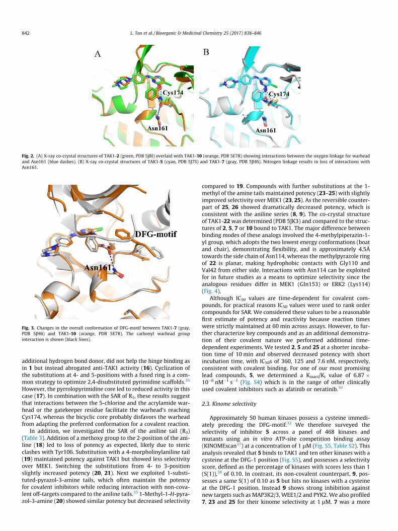
Fig. 2. (A) X-ray co-crystal structures of TAK1-2 (green, PDB 5J8I) overlaid with TAK1-10 (orange, PDB 5E7R) showing interactions between the oxygen linkage for warheadand Asn161 (blue dashes). (B) X-ray co-crystal structures of TAK1-5 (cyan, PDB 5J7S) and TAK1-7 (gray, PDB 5JH6). Nitrogen linkage results in loss of interactions withAsn161.
Fig. 3. Changes in the overall conformation of DFG-motif between TAK1-7 (gray,PDB 5JH6) and TAK1-10 (orange, PDB 5E7R). The carbonyl warhead groupinteraction is shown (black lines).
842 L. Tan et al. / Bioorganic & Medicinal Chemistry 25 (2017) 838–846
additional hydrogen bond donor, did not help the hinge binding asin 1 but instead abrogated anti-TAK1 activity (16). Cyclization ofthe substitutions at 4- and 5-positions with a fused ring is a com-mon strategy to optimize 2,4-disubstituted pyrimidine scaffolds.35
However, the pyrrolopyrimidine core led to reduced activity in thiscase (17). In combination with the SAR of R1, these results suggestthat interactions between the 5-chlorine and the acrylamide war-head or the gatekeeper residue facilitate the warhead’s reachingCys174, whereas the bicyclic core probably disfavors the warheadfrom adapting the preferred conformation for a covalent reaction.
In addition, we investigated the SAR of the aniline tail (R3)(Table 3). Addition of a methoxy group to the 2-position of the ani-line (18) led to loss of potency as expected, likely due to stericclashes with Tyr106. Substitution with a 4-morpholinylaniline tail(19) maintained potency against TAK1 but showed less selectivityover MEK1. Switching the substitutions from 4- to 3-positionslightly increased potency (20, 21). Next we exploited 1-substi-tuted-pyrazol-3-amine tails, which often maintain the potencyfor covalent inhibitors while reducing interaction with non-cova-lent off-targets compared to the aniline tails.35 1-Methyl-1-H-pyra-zol-3-amine (20) showed similar potency but decreased selectivity
compared to 19. Compounds with further substitutions at the 1-methyl of the amine tails maintained potency (23–25) with slightlyimproved selectivity over MEK1 (23, 25). As the reversible counter-part of 25, 26 showed dramatically decreased potency, which isconsistent with the aniline series (8, 9). The co-crystal structureof TAK1-22was determined (PDB 5JK3) and compared to the struc-tures of 2, 5, 7 or 10 bound to TAK1. The major difference betweenbinding modes of these analogs involved the 4-methylpiperazin-1-yl group, which adopts the two lowest energy conformations (boatand chair), demonstrating flexibility, and is approximately 4.5Åtowards the side chain of Asn114, whereas the methylpyrazole ringof 22 is planar, making hydrophobic contacts with Gly110 andVal42 from either side. Interactions with Asn114 can be exploitedfor in future studies as a means to optimize selectivity since theanalogous residues differ in MEK1 (Gln153) or ERK2 (Lys114)(Fig. 4).
Although IC50 values are time-dependent for covalent com-pounds, for practical reasons IC50 values were used to rank ordercompounds for SAR. We considered these values to be a reasonablefirst estimate of potency and reactivity because reaction timeswere strictly maintained at 60 min across assays. However, to fur-ther characterize key compounds and as an additional demonstra-tion of their covalent nature we performed additional time-dependent experiments. We tested 2, 5 and 25 at a shorter incuba-tion time of 10 min and observed decreased potency with shortincubation time, with IC50s of 360, 125 and 7.6 nM, respectively,consistent with covalent binding. For one of our most promisinglead compounds, 5, we determined a Kinact/Ki value of 6.87 �10�6 nM�1 s�1 (Fig. S4) which is in the range of other clinicallyused covalent inhibitors such as afatinib or neratinib.36
2.3. Kinome selectivity
Approximately 50 human kinases possess a cysteine immedi-ately preceding the DFG-motif.32 We therefore surveyed theselectivity of inhibitor 5 across a panel of 468 kinases andmutants using an in vitro ATP-site competition binding assay(KINOMEscan37) at a concentration of 1 lM (Fig. S5, Table S2). Thisanalysis revealed that 5 binds to TAK1 and ten other kinases with acysteine at the DFG-1 position (Fig. S5), and possesses a selectivityscore, defined as the percentage of kinases with scores less than 1(S(1)),38 of 0.10. In contrast, its non-covalent counterpart, 9, pos-sesses a same S(1) of 0.10 as 5 but hits no kinases with a cysteineat the DFG-1 position. Instead 9 shows strong inhibition againstnew targets such as MAP3K2/3, WEE1/2 and PYK2. We also profiled7, 23 and 25 for their kinome selectivity at 1 lM. 7 was a more
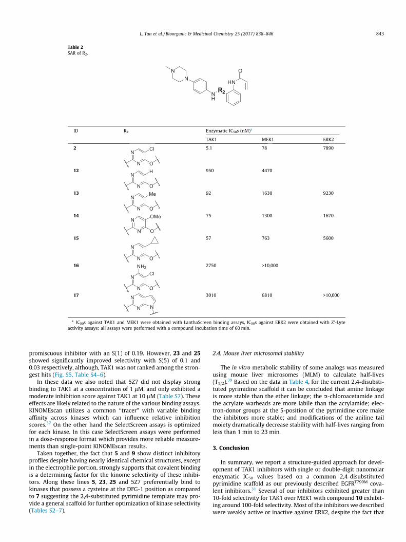
Table 2SAR of R2.
R2NH
NN
HN
O
ID R2 Enzymatic IC50s (nM)a
TAK1 MEK1 ERK2
2N
N
Cl
O
5.1 78 7890
12N
N
H
O
950 4470
13N
N
Me
O
92 1630 9230
14N
N
OMe
O
75 1300 1670
15
N
N O
57 763 5600
16
N
N
Cl
O
NH2 2750 >10,000
17 N
N N
3010 6810 >10,000
a IC50s against TAK1 and MEK1 were obtained with LanthaScreen binding assays, IC50s against ERK2 were obtained with Z’-Lyteactivity assays; all assays were performed with a compound incubation time of 60 min.
L. Tan et al. / Bioorganic & Medicinal Chemistry 25 (2017) 838–846 843
promiscuous inhibitor with an S(1) of 0.19. However, 23 and 25showed significantly improved selectivity with S(5) of 0.1 and0.03 respectively, although, TAK1 was not ranked among the stron-gest hits (Fig. S5, Table S4–6).
In these data we also noted that 5Z7 did not display strongbinding to TAK1 at a concentration of 1 lM, and only exhibited amoderate inhibition score against TAK1 at 10 lM (Table S7). Theseeffects are likely related to the nature of the various binding assays.KINOMEscan utilizes a common ‘‘tracer” with variable bindingaffinity across kinases which can influence relative inhibitionscores.37 On the other hand the SelectScreen assays is optimizedfor each kinase. In this case SelectScreen assays were performedin a dose-response format which provides more reliable measure-ments than single-point KINOMEscan results.
Taken together, the fact that 5 and 9 show distinct inhibitoryprofiles despite having nearly identical chemical structures, exceptin the electrophile portion, strongly supports that covalent bindingis a determining factor for the kinome selectivity of these inhibi-tors. Along these lines 5, 23, 25 and 5Z7 preferentially bind tokinases that possess a cysteine at the DFG-1 position as comparedto 7 suggesting the 2,4-substituted pyrimidine template may pro-vide a general scaffold for further optimization of kinase selectivity(Tables S2–7).
2.4. Mouse liver microsomal stability
The in vitro metabolic stability of some analogs was measuredusing mouse liver microsomes (MLM) to calculate half-lives(T1/2).39 Based on the data in Table 4, for the current 2,4-disubsti-tuted pyrimidine scaffold it can be concluded that amine linkageis more stable than the ether linkage; the a-chloroacetamide andthe acrylate warheads are more labile than the acrylamide; elec-tron-donor groups at the 5-position of the pyrimidine core makethe inhibitors more stable; and modifications of the aniline tailmoiety dramatically decrease stability with half-lives ranging fromless than 1 min to 23 min.
3. Conclusion
In summary, we report a structure-guided approach for devel-opment of TAK1 inhibitors with single or double-digit nanomolarenzymatic IC50 values based on a common 2,4-disubstitutedpyrimidine scaffold as our previously described EGFRT790M cova-lent inhibitors.31 Several of our inhibitors exhibited greater than10-fold selectivity for TAK1 over MEK1 with compound 10 exhibit-ing around 100-fold selectivity. Most of the inhibitors we describedwere weakly active or inactive against ERK2, despite the fact that

Table 3SAR of R3.
N
NNH
Cl
OR3
HN
O
ID R3 Enzymatic IC50s (nM)a
TAK1 MEK1 ERK2
2NN
5.1 78 7890
18b
NN
OMe 342
19b
NO4.4 8.9 361
20b
NO
3.2 11 736
21
NNO
1.7 9.5 225
22 NN
7.3 9.8 3140
23 NN
F
F 7.0 26 242
24 NNO
4.6 8.2 4620
25 NN
O
2.4 8.5 90.3
a IC50s against TAK1 and MEK1 were obtained with LanthaScreen binding assays,all assays were performed with a compound incubation time of 60 min.
b R1 of 18 is same as R1 of 7.
844 L. Tan et al. / Bioorganic & Medicinal Chemistry 25 (2017) 838–846
potent and selective ERK1/2 inhibitors have been developed andreported recently based on a very similar scaffold.34 The five newlydetermined co-crystal structures of TAK1 bound to newly synthe-sized compounds guided design choices for covalent targeting ofthe DFG-1 cysteine in TAK1 and for improving potency and selec-tivity. The inhibitors described here provide a syntheticallystraight-forward pharmacophore and a solid structural basis forfuture optimization of selective covalent TAK1 inhibitors. In theaccompanying communication we discuss the application of theseinhibitors to TAK1-centered polypharmacology.
4. Experimental
4.1. Chemistry
Unless otherwise noted, reagents and solvents were obtainedfrom commercial suppliers and were used without further purifi-cation. 1H NMR spectra were recorded on 600 or 500 MHz (VarianAS600 or Bruker A500), and chemical shifts are reported in partsper million (ppm, d) downfield from tetramethylsilane (TMS). Cou-pling constants (J) are reported in Hz. Spin multiplicities aredescribed as s (singlet), br (broad singlet), d (doublet), t (triplet),q (quartet), and m (multiplet). Mass spectra were obtained on aWaters Micromass ZQ instrument. Preparative HPLC was per-formed on a Waters Sunfire C18 column (19 � 50 mm, 5 lM) using
a gradient of 15–95% methanol in water containing 0.05% trifluo-roacetic acid (TFA) over 22 min (28 min run time) at a flow rateof 20 mL/min. Purities of assayed compounds were in all casesgreater than 95%, as determined by reverse-phase HPLC analysis.
4.1.1. 2,5-Dichloro-4-(2-nitrophenoxy)pyrimidine (3)2-Nitrophenol (840 mg, 6.0 mmol) and potassium carbonate
(800 mg, 6.0 mmol) were combined in dimethyl sulfoxide (DMSO)(10 mL) and stirred for 15 min, then 2,4,5-trichloropyrimidine(560 lL, 5.0 mmol) was added and the mixture was stirred over-night. The mixture was then diluted with ethyl acetate and washedwith water and brine, dried over Na2SO4, filtered and concentrated.The crude product was purified by column chromatography (hex-ane:ethyl acetate = 3:1) to yield 1.2 g (70%) of 3 as a white solid.MS (ESI) m/z 286 (M+H)+.
4.1.2. 5-Chloro-N-(4-(4-methylpiperazin-1-yl)phenyl)-4-(2-nitrophenoxy)pyrimidin-2-amine (4)
To 3 (570 mg, 2.0 mmol) and 2-methoxy-4-(4-methylpiperazin-1-yl)aniline (390 mg, 2.0 mmol) in sec-butanol (4 mL) was addedtrifluoroacetic acid (154 lL, 2.0 mmol) and the mixture was stirredovernight at 75 �C. The mixture was then concentrated, neutralizedwith ammonia in methanol and purified by column chromatogra-phy (dichloromethane:methanol = 10:1) to yield 265 mg (60%) of4 as a pale-yellow solid. MS (ESI) m/z 441 (M+H)+.
4.1.3. N-(2-((5-Chloro-2-((4-(4-methylpiperazin-1-yl)phenyl)amino)pyrimidin-4-yl)oxy)phenyl)acrylamide (2)
To 4 (88 mg, 0.2 mmol) in methanol (10 mL) was added 1 mLRaney nickel suspension in methanol. The reaction mixture wasstirred for 3 h under 1 atm of hydrogen. The mixture was then fil-tered with Celite, and the filtrate was concentrated and driedunder vacuum to give a crude product as a white solid. To theobtained white solid in tetrahydrofuran (3 mL) was added satu-rated NaHCO3 solution (3 mL), the stirred mixture was then cooledto 0 �C, and acryloyl chloride was added (25 lL, 0.3 mmol) drop-wise. The reaction mixture was stirred at 0 �C for 10 min, anotherbatch of acryloyl chloride was added (8 lL, 0.1 mmol) dropwise.After another 5 min the mixture was allowed to recover to roomtemperature (RT), and diluted with ethyl acetate and washed withwater and brine, dried over Na2SO4, filtered and concentrated. Thecrude product was then purified by reverse phase HPLC to give58 mg (63% for 2 steps) of 2 as a white solid. 1H NMR (600 MHz,DMSO-d6) d 9.68 (br, 1H), 9.58 (s, 1H), 9.52 (br, 1H), 8.40 (s, 1H),8.05 (m, 1H), 7.32 (dd, J = 8.4, 8.4 Hz, 2H), 7.24 (d, J = 8.4 Hz, 2H),6.72 (m, 2H), 6.55 (dd, J = 16.8, 10.8 Hz, 1H), 6.20 (d, J = 16.8 Hz,1H), 5.71 (d, J = 10.8 Hz, 1H), 3.66 (m, 2H), 3.50 (m, 2H), 3.14 (m,2H), 2.85 (s, 3H), 2.83 (m, 2H). MS (ESI) m/z 465 (M+H)+.
4.1.4. 2,5-Dichloro-N-(2-nitrophenyl)pyrimidin-4-amine (28)To 2,4,5-trichloropyrimidine (560 lL mg, 5.0 mmol) and 2-
nitroaniline (700 mg, 5.0 mmol) in sec-butanol (25 mL) was addedtrifluoroacetic acid (383 lL, 5.0 mmol) and the mixture was stirredovernight at 60 �C. The mixture was then concentrated, neutralizedwith ammonia in methanol and purified by column chromatogra-phy (hexane:ethyl acetate = 2:1) to yield 640 mg (45%) of 28 as ayellow solid. MS (ESI) m/z 285 (M+H)+.
4.1.5. 5-Chloro-N2-(4-(4-methylpiperazin-1-yl)phenyl)-N4-(2-nitrophenyl)pyrimidine-2,4-diamine (29)
To 28 (570 mg, 2.0 mmol) and 2-methoxy-4-(4-methylpiper-azin-1-yl)aniline (390 mg, 2.0 mmol) in sec-butanol (4 mL) wasadded trifluoroacetic acid (154 lL, 2.0 mmol) and the mixturewas stirred overnight at 85 �C. The mixture was then concentrated,neutralized with ammonia in methanol and purified by columnchromatography (dichloromethane:methanol = 10:1) to yield

Fig. 4. X-ray co-crystal structure of TAK1-22 (blue, PDB 5JK3), overlaid with TAK1-2(green, PDB 5J8I) and TAK1-7 (gray, PDB 5JH6) showing key interactions (bluelines). The flexibility of the piperazinyl moiety adopting different conformationsextending towards Asn114 is also seen.
L. Tan et al. / Bioorganic & Medicinal Chemistry 25 (2017) 838–846 845
680 mg (76%) of 29 as a pale-yellow solid. MS (ESI) m/z 440(M+H)+.
4.1.6. N-(2-((5-Chloro-2-((4-(4-methylpiperazin-1-yl)phenyl)amino)pyrimidin-4-yl)amino)phenyl)acrylamide (5)
To 29 (88 mg, 0.2 mmol) in methanol (10 mL) was added 1 mLRaney nickel suspension in methanol. The reaction mixture wasstirred for 3 h under 1 atm of hydrogen. The mixture was then fil-tered with Celite, and the filtrate was concentrated and driedunder vacuum to give a crude product as a white solid. To theobtained white solid in tetrahydrofuran (3 mL) was added satu-rated NaHCO3 solution (3 mL), the stirred mixture was then cooledto 0 �C, and acryloyl chloride was added (25 lL, 0.3 mmol) drop-wise. The reaction mixture was stirred at 0 �C for 10 min, anotherbatch of acryloyl chloride was added (8 lL, 0.1 mmol) dropwise.After another 5 min the mixture was allowed to recover to roomtemperature (RT), and diluted with ethyl acetate and washed withwater and brine, dried over Na2SO4, filtered and concentrated. Thecrude product was then purified by reverse phase HPLC to give55 mg (60% for 2 steps) of 5 as a white solid. 1H NMR (600 MHz,DMSO-d6) d 10.16 (s, 1H), 9.67 (br, 1H), 9.20 (br, 1H), 8.56 (s,1H), 8.09 (s, 1H), 7.74 (d, J = 7.2 Hz, 1H), 7.46 (d, J = 6.6 Hz, 1H),7.40 (d, J = 9.0 Hz, 2H), 7.32 (dd, J = 7.2, 7.2 Hz, 1H), 7.24 (dd,J = 7.8, 7.2 Hz, 1H), 6.78 (d, J = 9.6 Hz, 1H), 6.50 (dd, J = 16.8,10.8 Hz, 1H), 6.30 (d, J = 17.4 Hz, 1H), 5.79 (d, J = 10.8 Hz, 1H),3.66 (m, 2H), 3.49 (m, 2H), 3.14 (m, 2H), 2.84 (s, 3H), 2.83 (m,2H). MS (ESI) m/z 464 (M+H)+.
4.1.7. 2-((2,5-Dichloropyrimidin-4-yl)amino)phenol (30)To 2-aminophenol (550 mg, 5.0 mmol) in dioxane (10 mL) was
added 2,4,5-trichloropyrimidine (560 lL mg, 5.0 mmol), the mix-ture was stirred overnight at RT. The mixture was then diluted
Table 4Mouse liver microsomal stability.
ID MLM T1/2 (min) ID
2 6.1 145 10.8 157 5.7 1710 3 1913 8.8 20
with ethyl acetate and washed with water and brine, dried overNa2SO4, filtered and concentrated to give 1.02 g (80%) of the crudeproduct 30 as a white solid, which was directly used in the nextstep. MS (ESI) m/z 256 (M+H)+.
4.1.8. 2-((5-Chloro-2-((4-(4-methylpiperazin-1-yl)phenyl)amino)pyrimidin-4-yl)amino)phenol (31)
To 30 (510 mg, 2.0 mmol) and 2-methoxy-4-(4-methylpiper-azin-1-yl)aniline (575 mg, 3.0 mmol) in sec-butanol (5 mL) wasadded trifluoroacetic acid (230 lL, 3.0 mmol) and the mixturewas stirred overnight at 100 �C. The mixture was then concen-trated, neutralized with ammonia in methanol and purified by col-umn chromatography (dichloromethane:methanol = 10:1) to yield680 mg (76%) of 31 as a white solid. MS (ESI) m/z 411 (M+H)+.
4.1.9. 2-((5-Chloro-2-((4-(4-methylpiperazin-1-yl)phenyl)amino)pyrimidin-4-yl)amino)phenyl acrylate (7)
To 31 (82 mg, 0.2 mmol) in dimethylformamide (2 mL) wasadded diisopropylethylamine (53 lL, 0.3 mmol), the stirred mix-ture was then cooled to �60 �C, and acryloyl chloride (17.8 lL,0.22 mmol) was added dropwise. The reaction mixture was stirredat �60 �C for 10 min, allowed to recover to RT (room temperature)gradually in 30 min, and purified by reverse phase HPLC to give64 mg (TFA salt, 56% for 2 steps) of 7 as a white solid. 1H NMR(600 MHz, DMSO-d6) d 8.96 (br, 1H), 8.36 (s, 1H), 8.04 (s, 1H),7.72 (br, 1H), 7.34 (d, J = 9.0 Hz, 2H), 7.30 (m, 3H), 6.70 (d,J = 9.0 Hz, 2H), 6.37 (d, J = 16.8, 1H), 6.26 (dd, J = 17.4, 10.2 Hz,1H), 5.79 (d, J = 10.8 Hz, 1H), 3.02 (m, 4H), 2.54 (m, 4H), 2.28 (br,3H), 2.84 (s, 3H). MS (ESI) m/z 465 (M+H)+.
4.2. X-ray crystallography procedures
TAK1–TAB1 fusion protein (TAK1 kinase domain residues 31–303 fused with C-terminal TAB1 domain residues 468–497) wasexpressed in insect cells, purified and crystallized as reported pre-viously.29 Crystals grew within 2 days by vapor diffusion in hang-ing drop at 20 �C by mixing protein with equal volumes ofreservoir solution consisting of 0.65–0.75 M sodium citrate, 0.2 MNaCl, 0.1 M Tris, pH 7.0, and 5 mM adenosine. Crystals containingadenosine were then back-soaked with excess of inhibitors at250–500 lM dissolved in reservoir solution for 6–8 h. The crystalswere then flash frozen with 20% ethyleneglycol as cryoprotectantfor data collection. Diffraction data were collected at ArgonneAdvanced Photon Source (beamline 19-D) and processed withHKL2000 and HKL3000.40 The structure was determined by molec-ular replacement using Phaser41 with reported TAK1 structure(PDB code: 2YIY or 4O91) as search model. Coot was used formodel building42 and refinement was carried out using both Phe-nix, version 1.10–215543 and Refmac, version 5.8.0049.44–46, PyMol(The PyMOL Molecular Graphics System, version 1.6.0.0) andMeastro (version 1.5.014) from Schrödinger, LLC. For demonstra-tion of key interactions, LIGPLOT software was used.47
MLM T1/2 (min) ID MLM T1/2 (min)
15.3 21 <112.8 22 8.110.5 23 4.23.8 24 423 25 6.9

846 L. Tan et al. / Bioorganic & Medicinal Chemistry 25 (2017) 838–846
4.3. Enzymatic assays
Enzymatic inhibition of MEK2 and ERK1 was tested in Z’-Lyteassays with ATP concentrations near the Km for each kinase. Com-pound activity against TAK1 was tested in LanthaScreen bindingassays. All protocols are available from Life Technologies.48 TheMLM assays were previously reported and are commercially avail-able from Scripps Florida.39
Acknowledgements
NSG and PKS were supported by LINCS Grant U54-HL127365.KDW was supported by Welch Research Foundation I-1829, CPRITR1207 and CPRIT RP140233. LT and NSG were supported by NIH CA154303-05 and the Pediatric Low Grade Astrocytoma (PLGA) foun-dation. Results shown in this article are derived from work per-formed at Argonne National Laboratory, Structural Biology Centerat the Advanced Photon Source. Argonne is operated by U ChicagoArgonne, LLC, for the U.S. Department of Energy, Office of Biologicaland Environmental Research under contract DEAC02-06CH11357.
A. Supplementary data
Supplementary data associated with this article can be found, inthe online version, at http://dx.doi.org/10.1016/j.bmc.2016.11.035.
References
1. Yamaguchi K, Shirakabe K, Shibuya H, et al. Identification of a member of theMAPKKK family as a potential mediator of TGF-beta signal transduction. Science(New York, N.Y.). 1995;270:2008–2011.
2. Sato S, Sanjo H, Takeda K, et al. Essential function for the kinase TAK1 in innateand adaptive immune responses. Nat Immunol. 2005;6:1087–1095.
3. Shim J-H, Xiao C, Paschal AE, et al. TAK1, but not TAB1 or TAB2, plays anessential role in multiple signaling pathways in vivo. Genes Dev.2005;19:2668–2681.
4. Shirakabe K, Yamaguchi K, Shibuya H, et al. TAK1 mediates the ceramidesignaling to stress-activated protein kinase/c-Jun N-terminal kinase. J BiolChem. 1997;272:8141–8144.
5. Sakurai H, Shigemori N, Hasegawa K, Sugita T. TGF-beta-activated kinase 1stimulates NF-kappa B activation by an NF-kappa B-inducing kinase-independent mechanism. Biochem Biophys Res Commun. 1998;243:545–549.
6. Ninomiya-Tsuji J, Kishimoto K, Hiyama A, Inoue J, Cao Z, Matsumoto K. Thekinase TAK1 can activate the NIK-I kappaB as well as the MAP kinase cascade inthe IL-1 signalling pathway. Nature. 1999;398:252–256.
7. Sakurai H, Miyoshi H, Toriumi W, Sugita T. Functional interactions oftransforming growth factor beta-activated kinase 1 with IkappaB kinases tostimulate NF-kappaB activation. J Biol Chem. 1999;274:10641–10648.
8. Schuman J, Chen Y, Podd A, et al. A critical role of TAK1 in B-cell receptor–mediated nuclear factor jB activation. Blood. 2009;113:4566–4574.
9. Wan YY, Chi H, Xie M, Schneider MD, Flavell RA. The kinase TAK1 integratesantigen and cytokine receptor signaling for T cell development, survival andfunction. Nat Immunol. 2006;7:851–858.
10. Liu H-H, Xie M, Schneider MD, Chen ZJ. Essential role of TAK1 in thymocytedevelopment and activation. Proc Natl Acad Sci USA. 2006;103:11677–11682.
11. Sato S, Sanjo H, Tsujimura T, et al. TAK1 is indispensable for development of Tcells and prevention of colitis by the generation of regulatory T cells. IntImmunol. 2006;18:1405–1411.
12. Kawai T, Akira S. Toll-like receptors and their crosstalk with other innatereceptors in infection and immunity. Immunity. 2011;34:637–650.
13. Ma FY, Tesch GH, Ozols E, Xie M, Schneider MD, Nikolic-Paterson DJ. TGF-beta1-activated kinase-1 regulates inflammation and fibrosis in the obstructedkidney. Am J Physiol Renal Physiol. 2011;300:F1410–F1421.
14. Grillo AR, Scarpa M, D’Inca R, et al. TAK1 is a key modulator of the profibrogenicphenotype of human ileal myofibroblasts in Crohn’s disease, American journalof physiology. Gastrointest Liver Physiol. 2015;309:G443–G454.
15. Neubert M, Ridder DA, Bargiotas P, Akira S, Schwaninger M. Acute inhibition ofTAK1 protects against neuronal death in cerebral ischemia. Cell Death Differ.2011;18:1521–1530.
16. Buglio D, Palakurthi S, Byth K, et al. Essential role of TAK1 in regulating mantlecell lymphoma survival. Blood. 2012;120:347–355.
17. Cai PC, Shi L, Liu VW, et al. Elevated TAK1 augments tumor growth andmetastatic capacities of ovarian cancer cells through activation of NF-kappaBsignaling. Oncotarget. 2014;5:7549–7562.
18. Singh A, Sweeney MF, Yu M, et al. TAK1 inhibition promotes apoptosis in KRAS-dependent colon cancers. Cell. 2012;148:639–650.
19. Fan Y, Cheng J, Vasudevan SA, et al. TAK1 inhibitor 5Z-7-oxozeaenol sensitizesneuroblastoma to chemotherapy. Apoptosis. 2013;18:1224–1234.
20. Melisi D, Xia Q, Paradiso G, et al. Modulation of pancreatic cancerchemoresistance by inhibition of TAK1. J Natl Cancer Inst. 2011;103:1190–1204.
21. Sakurai H. Targeting of TAK1 in inflammatory disorders and cancer. TrendsPharmacol Sci. 2012;33:522–530.
22. Ansell SM, Hodge LS, Secreto FJ, et al. Activation of TAK1 by MYD88 L265Pdrives malignant B-cell Growth in non-Hodgkin lymphoma. Blood Cancer J.2014;4:e183.
23. Ninomiya-Tsuji J, Kajino T, Ono K, et al. A resorcylic acid lactone, 5Z-7-oxozeaenol, prevents inflammation by inhibiting the catalytic activity of TAK1MAPK kinase kinase. J Biol Chem. 2003;278:18485–18490.
24. Omori E, Matsumoto K, Zhu S, Smart RC, Ninomiya-Tsuji J. Ablation of TAK1upregulates reactive oxygen species and selectively kills tumor cells. CancerRes. 2010;70:8417–8425.
25. Schirmer A, Kennedy J, Murli S, Reid R, Santi DV. Targeted covalent inactivationof protein kinases by resorcylic acid lactone polyketides. Proc Natl Acad Sci USA.2006;103:4234–4239.
26. Wu J, Powell F, Larsen NA, et al. Mechanism and in vitro pharmacology of TAK1inhibition by (5Z)-7-Oxozeaenol. ACS Chem Biol. 2013;8:643–650.
27. Sogabe Y, Matsumoto T, Hashimoto T, Kirii Y, Sawa M, Kinoshita T. 5Z-7-Oxozeaenol covalently binds to MAP2K7 at Cys218 in an unprecedentedmanner. Bioorg Med Chem Lett. 2015;25:593–596.
28. Hornberger KR, Chen X, Crew AP, et al. Discovery of 7-aminofuro[2,3-c]pyridineinhibitors of TAK1: optimization of kinase selectivity and pharmacokinetics.Bioorg Med Chem Lett. 2013;23:4511–4516.
29. Tan L, Nomanbhoy T, Gurbani D, et al. Discovery of type II inhibitors ofTGFbeta-activated kinase 1 (TAK1) and mitogen-activated protein kinasekinase kinase kinase 2 (MAP4K2. J Med Chem. 2015;58:183–196.
30. Lebakken CS, Riddle SM, Singh U, et al. Development and applications of abroad-coverage, TR-FRET-based kinase binding assay platform. J Biomol Screen.2009;14:924–935.
31. Zhou W, Ercan D, Chen L, et al. Novel mutant-selective EGFR kinase inhibitorsagainst EGFR T790M. Nature. 2009;462:1070–1074.
32. Liu Q, Sabnis Y, Zhao Z, et al. Developing irreversible inhibitors of the proteinkinase cysteinome. Chem Biol. 2013;20:146–159.
33. For ERK2 Z’-Lyte activity assay: <http://www.thermofisher.com/content/dam/LifeTech/migration/files/drug-discovery/pdfs.par.60256.file.dat/20130430%20ssbk%20customer%20protocol%20and%20assay%20conditions.pdf>.
34. Ward RA, Colclough N, Challinor M, et al. Structure-guided design of highlyselective and potent covalent inhibitors of ERK1/2. J Med Chem.2015;58:4790–4801.
35. Tan L, Akahane K, McNally R, et al. Development of selective covalent Januskinase 3 inhibitors. J Med Chem. 2015;58:6589–6606.
36. Schwartz PA, Kuzmic P, Solowiej J, et al. Covalent EGFR inhibitor analysisreveals importance of reversible interactions to potency and mechanisms ofdrug resistance. Proc Natl Acad Sci USA. 2014;111:173–178.
37. Goldstein DM, Gray NS, Zarrinkar PP. High-throughput kinase profiling as aplatform for drug discovery. Nat Rev Drug Discovery. 2008;7:391–397.
38. Miduturu CV, Deng X, Kwiatkowski N, et al. High-throughput kinase profiling: amore efficient approach toward the discovery of new kinase inhibitors. ChemBiol. 2011;18:868–879.
39. Li X, He Y, Ruiz CH, Koenig M, Cameron MD, Vojkovsky T. Characterization ofdasatinib and its structural analogs as CYP3A4 mechanism-based inactivatorsand the proposed bioactivation pathways. Drug Metab Dispos.2009;37:1242–1250.
40. Minor W, Cymborowski M, Otwinowski Z, Chruszcz M. HKL-3000: theintegration of data reduction and structure solution–from diffraction imagesto an initial model in minutes. Acta Crystallogr D Biol Crystallogr.2006;62:859–866.
41. Bunkoczi G, Echols N, McCoy AJ, Oeffner RD, Adams PD, Read RJ. Phaser.MRage:automated molecular replacement. Acta Crystallogr D Biol Crystallogr.2013;69:2276–2286.
42. Emsley P, Cowtan K. Coot: model-building tools for molecular graphics. ActaCrystallogr D Biol Crystallogr. 2004;60:2126–2132.
43. Adams PD, Afonine PV, Bunkoczi G, et al. PHENIX: a comprehensive Python-based system for macromolecular structure solution. Acta Crystallogr D BiolCrystallogr. 2010;66:213–221.
44. Nicholls RA, Long F, Murshudov GN. Low-resolution refinement tools inREFMAC5. Acta Crystallogr D Biol Crystallogr. 2012;68:404–417.
45. Murshudov GN, Skubak P, Lebedev AA, et al. REFMAC5 for the refinement ofmacromolecular crystal structures. Acta Crystallogr D Biol Crystallogr.2011;67:355–367.
46. Vagin AA, Steiner RA, Lebedev AA, et al. REFMAC5 dictionary: organization ofprior chemical knowledge and guidelines for its use. Acta Crystallogr D BiolCrystallogr. 2004;60:2184–2195.
47. Wallace AC, Laskowski RA, Thornton JM. LIGPLOT: a program to generateschematic diagrams of protein-ligand interactions. Protein Eng.1995;8:127–134.
48. Z’-Lyte assays: <http://www.lifetechnologies.com/content/dam/LifeTech/migration/files/drug-discovery/pdfs.par.60256.file.dat/20130430%20ssbk%20customer%20protocol%20and%20assay%20conditions.pdf>; TTK assay:<http://tools.lifetechnologies.com/content/sfs/manuals/TTK_LanthaScreen_Binding.pdf>.