biomineralogy of selected skin cancer...
TRANSCRIPT

Auxiliary sciences in archaeology, preservation of relicts and environmental engineering. CD -no 23,
2017. Ed. M. Pawlikowski. -----------------------------------------------------------------------------------------------------------------
BIOMINERALOGY OF SELECTED SKIN CANCER
Biomineralogia wybranych nowotowrów skóry
Maciej Pawlikowski*. Magdalena Miler**
*/ Lab. Biomineralogy, Cath. Mineralogy, Petrography and Geochemistry, AGH –Univ. Science and Technology, 30-059 Cracow, al. Mickiewicza 30, Poland **/ Graduate of AGH –Univ. Science and Technology, 30-059 Cracow, al. Mickiewicza 30, Poland
Abstract Investigation of Carcinoma basocellulare solidum exulcerans, Carcinoma basocellulare superficiale multicentricum, and Trichoepithelioma was performed using histology and biomineralogical methods. Obtained data confirmed elevated levels of some elements in altered skin tissues. Moreover, rare micrograins of phosphates were observed. Additionally, examination of biomineralization of human tissues suggests that higher local mineralization (of tissue fluids) may lead to mistakes in DNA code at the moment of cell division. It is possible that cancer tissues are secondarily mineralized by activity of cancer cells. Further research is needed to answer questions that arose. Hey words: mineralogy, chemistry, skin cancer
Investigation was supported by grant AGH 11.11.140.319 Streszczenie Wykonano badania mineralizacji guzów nowotworowych wybranych zmian rakowych skóry. Stwierdzono w nich podwyższone zawartości niektórych pierwiastków takich jak P, Ca, S, Si, K i in. Otrzymane wyniki pozwalają na postawienie pytania czy badane nowotwory skóry s…a efektem lokalnej mineralizacji obserwowane jako podwyższone zawartości niektórych pierwiastków w tkankach czy też aktywność biologiczna stref nowotworowych powoduje koncentracje pierwiastków w tkankach. Wykonane badania sugerują, że podwyższona mineralizacja płynów ustrojowych może sprzyjać defektom powstającym w DNA w części odpowiedzialnej za mnożenie się komórek. Efektem tego zjawiska może być szybkie namnażanie komórek czyli powstawanie tkanek (guzów) nowotworowych. Słowa kluczowe: mineralogia, chemis, nowotwory skóry

Badania prowadzono w ramach badań statutowych AGH no 11.11.140.319 Introduction Number of people diagnosed with cancer, in particular different types of skin cancer, is steadily increasing (Pawlikowski 1991, 1993, Boyle et al. 2004, Deja et al. 2004, Bunyaviroch, Coleman 2006 et al.). Skin cancer is one of the forms of cancer that often ends in death. It is therefore important both to prevent this group of cancers and to develop effective treatment. Development of research techniques allows us to discover new areas of knowledge. This also applies to research in oncology, including skin cancers (Pawlikowski 1993, 1995, Devereux 1996, 2011, 2013, Męcik et al. 2000, Chichel, Skowronek 2005, Daniel et al. 2005, Harper 2004).
This work presents such new research directions in the study of skin cancer, including scanning polarization microscopy. Examinations described herein were preceded by histological identification of the samples. The aim of the study was biomineralogical diagnosis of mineralization in neoplastic skin tissue. By biomineralization we understand not only the occurrence of mineral grains or crystals, i.e. so called overt mineralization (Pawlikowski 1993, 2014, 2017), but also the presence of hidden mineralization (Pawlikowski 1987, Pawlikowski, Pfitzner 1999). Hidden mineralization affects tissues and body fluids without manifesting in the aforementioned forms, and it consists of substituting elements for atomic structures of organic compounds. It mostly affects areas with tissue changes. This type of mineralization is impossible to detect in its early stages using methods other than chemical. It is sensitive chemical methods that reveal too high or too low content of elements in the tissues, which indicate that the tissue environment deviates from normal conditions (Pawlikowski 1993). Apart from the "mineral" factors, there are others, for instance different types of radiation (Woźniak 2001, Wołowiec, Dadej 2003). Mineral, and in fact chemical "abnormality" of the environment is particularly important at the time of cell division, i.e. cell multiplication.
When cell division takes place in "abnormal" environment with excessive or insufficient "mineralization", there is a risk of an error occurring in the section of DNA responsible for the rate of cell procreation (Pawlikowski 1995). Such errors in that particular DNA section may be numerous and varied. In consequence, the cell multiplies more often than it would with the original genetic code. This defect leads to rapid growth of large number of cells and formation of tumors. The diversity of mineral (and organic) substances, and the fact that cell proliferation is directed not by one place in DNA, but its whole section, result in a huge variety of tumors both in skin and other organs and tissues. Due to the availability of the samples, this work focuses on skin tumors - Carcinoma basocellulare solidum exulcerans, Carcinoma basocellulare superficiale multicentricum. The authors are grateful to doctors Krzysztof Czajecki, MD, and Stanisław Bajcar, MD, for providing study material. Four tumors diagnosed as Carcinoma basocellulare and two cases of Trichoepithelioma tumors were obtained from the Department of Pathomorphology of the Provincial Hospital No.

2 in Rzeszow. Assessment of histological preparations was performed at the Department of Pathomorphology of the Regional Hospital No. 2 in Rzeszow and at the Department of Pathomorphology of Collegium Medicum at the Jagiellonian University in Cracow. Preparations for scanning microscopy were dewaxed in the Department of Pathomorphology of the 5th Military Clinical Hospital with Polyclinic in Cracow. Analyzed samples are presented in Tab. 1.
Tab. 1 Data concerning origin and character of samples.
Histological diagnosis
Preparation number
Patient’s gender and age
Size of the lesion
Notes
Carcinoma basocellulare solidum
256442 Woman, age 61
6 x 4 mm -follicular differentiation in places -necrotic fields present -no major calcifications
259608
Woman age 58
10 mm diameter
-clear follicular differentiation -numerous calcifications
Carcinoma basocellulare solidum exulcerans
259814 Woman, age 41
7 x 8 mm -no major calcifications
Carcinoma basocellulare superficiale multicentricum
255976 Woman, age 48
4 mm diameter
-surface covered by scab -no large calcifications
Trichoepithelioma 255750 Woman age 66
12 x 7 mm -clear follicular differentiation -visible areas of calcification
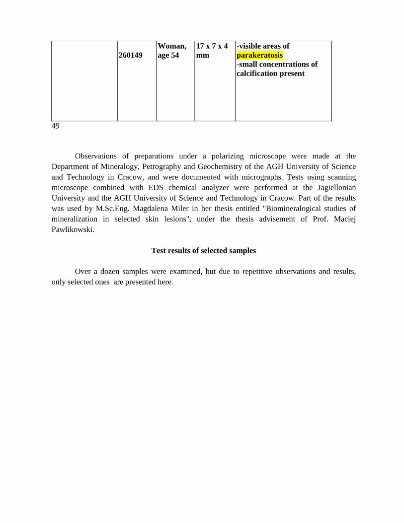
260149
Woman, age 54
17 x 7 x 4 mm
-visible areas of parakeratosis -small concentrations of calcification present
49 Observations of preparations under a polarizing microscope were made at the Department of Mineralogy, Petrography and Geochemistry of the AGH University of Science and Technology in Cracow, and were documented with micrographs. Tests using scanning microscope combined with EDS chemical analyzer were performed at the Jagiellonian University and the AGH University of Science and Technology in Cracow. Part of the results was used by M.Sc.Eng. Magdalena Miler in her thesis entitled "Biomineralogical studies of mineralization in selected skin lesions", under the thesis advisement of Prof. Maciej Pawlikowski.
Test results of selected samples
Over a dozen samples were examined, but due to repetitive observations and results, only selected ones are presented here.
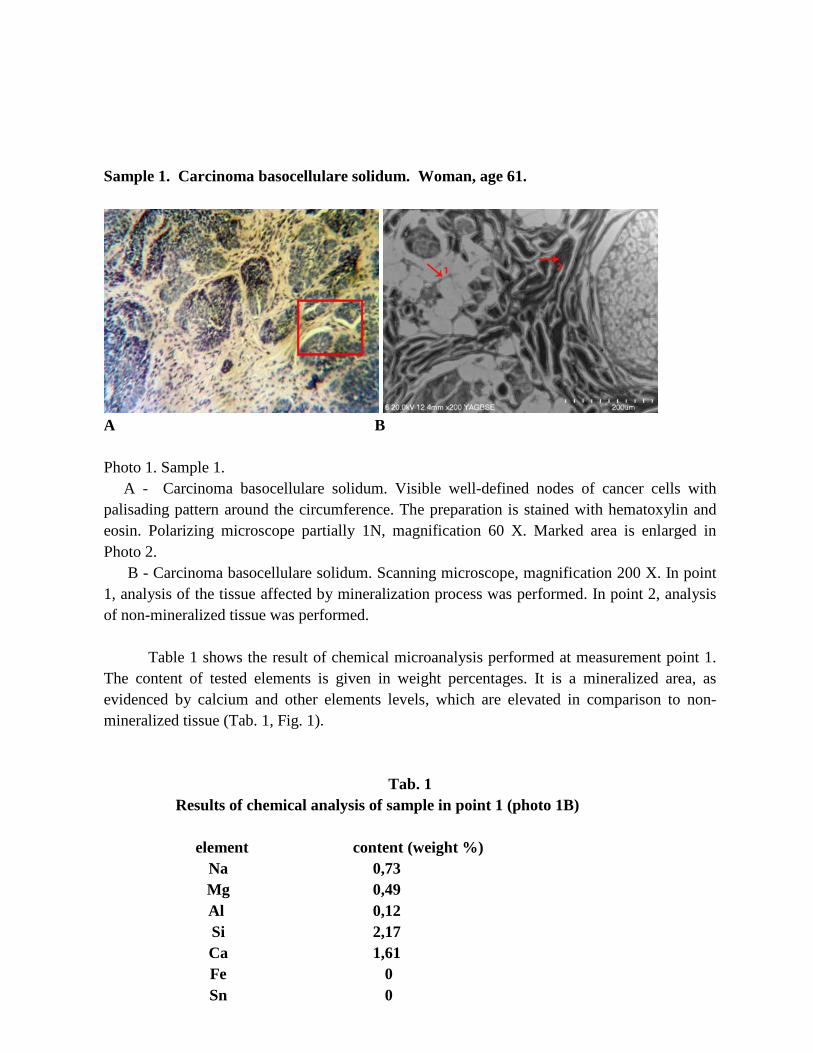
Sample 1. Carcinoma basocellulare solidum. Woman, age 61.
A B Photo 1. Sample 1. A - Carcinoma basocellulare solidum. Visible well-defined nodes of cancer cells with palisading pattern around the circumference. The preparation is stained with hematoxylin and eosin. Polarizing microscope partially 1N, magnification 60 X. Marked area is enlarged in Photo 2. B - Carcinoma basocellulare solidum. Scanning microscope, magnification 200 X. In point 1, analysis of the tissue affected by mineralization process was performed. In point 2, analysis of non-mineralized tissue was performed.
Table 1 shows the result of chemical microanalysis performed at measurement point 1. The content of tested elements is given in weight percentages. It is a mineralized area, as evidenced by calcium and other elements levels, which are elevated in comparison to non-mineralized tissue (Tab. 1, Fig. 1).
Tab. 1 Results of chemical analysis of sample in point 1 (photo 1B) element content (weight %)
Na 0,73 Mg 0,49 Al. 0,12 Si 2,17 Ca 1,61 Fe 0 Sn 0

P 0 S 1,23 K 0,5 Cl 1,45
N 0
Fig. 1. Chart of contents of analyzed elements in sample 1, point 1
Table 2 shows chemical analysis of “clean” tissue – not affected by the mineralization process (measurement point 2). In this spot there are no elevated levels of elements that could be relevant to tissue mineralization (e.g. calcium, phosphorus). Slightly elevated contents of sodium, magnesium and aluminum probably come from glass and are a result of calculation error.
Tab. 2 Results of chemical analysis of sample in point 2 (Photo 2B)
element content (weight %)
Na 0 Mg 0 Al. 0,1 Si 0 Ca 0,29 Fe 0 Sn 0 P 0 S 0,16 K 0
0
5
10
15
20
25
Na Mg Al. Si Ca Fe Sn P S K Cl N

Cl 0 N 0
Sample 1 – second area
A B Photo 3. Sample 1- second area A - Carcinoma basocellulare solidum preparation. Polarizing microscope XN, magnification 60 X. Visible tiny, gleaming concentrations of minerals near the elongated shape. Marked area is shown in photo 3B. B - Carcinoma basocellulare solidum preparation. Scanning microscope, magnification 250 X. In point 3, chemical analysis of tissue affected by mineralization was performed.
In measurement point 3, chemical analysis of mineralized area was performed. In that area (photo 3 A,B) tiny, gleaming concentrations of minerals are visible. Chemical analysis (presented in Table 3, Fig. 3) shows elevated levels of calcium and sulphur in comparison with “clean” tissue.
Tab. 3 Results of chemical analysis of sample. Sample 1 – area 2, point 3 (photo 3B). element content (weight %)
Na 0 Mg 0 Al. 0,1 Si 0 Ca 6,29

Fe 0 Sn 0 P 0 S 1,16 K 0 Cl 0 N 0
Fig. 3. Chart of analyzed elements contents in sample 1 – area 2, point 3. Sample 2 – area 1
A B Photo 4. Sample 2 – area 1 A - Carcinoma basocellulare solidum. Polarizing microscope, polarizators partially X, magnification 60X. Visible mineralization in the form of tiny, gleaming concentrations of minerals around cells. B – Carcinoma basocellulare solidum. Scanning microscope, magnification 350X. In point 4, chemical analysis was performed in mineralized areas visible in 4A.
0
5
10
15
20
25
Na Mg Al. Si Ca Fe Sn P S K Cl N

Table 4 shows the result of chemical microanalysis performed at measurement point 4. The contents of tested elements are given in weight percentages. It is a mineralized area, as evidenced by calcium and sulphur levels, which are elevated in comparison to non-mineralized tissue.
Tab. 4 Results of chemical analysis of sample 2 – area 1, point 4 (Photo 4B).
Na 0 Mg 0 Al. 0 Si 0 Ca 3,12 Fe 0 Sn 0 P 0 S 1,18 K 0 Cl 0 N 0
Sample 2 -area 2 Carcinoma basocellulare solidum. Woman, age 58. Preparation 1
A B
Photo 5. Sample 2 - area 2. A - Carcinoma basocellulare solidum. On the periphery of solid basal cell carcinoma, single

nodes of tumor cells are located within the inflammatory mass adjacent to the small vessels. Preparation stained with hematoxylin and eosin. Polarizing microscope 1N, magnification 60 X. B - Carcinoma basocellulare solidum. Enlarged fragment of the image shown in Photo 5A. In points 5 and 6 mineralized tissue analysis was performed. Scanning microscope, magnification 350 X.
Table 5 shows the result of chemical analysis of the mineralized area at measuring point
5 (Photo 5B). The analysis shows slightly increased levels of sodium, magnesium, silicon, and potassium.
Tab. 5
Results of chemical analysis of sample in point 5 (Photo 5B)
Element
Content (weight
%) Na 0,21 Mg 0,27 Al. 0 Si 0,26 Ca 1,31 Fe 0,1 Sn 0 P 3,44 S 4,54 K 0,3 Cl 0 N 0
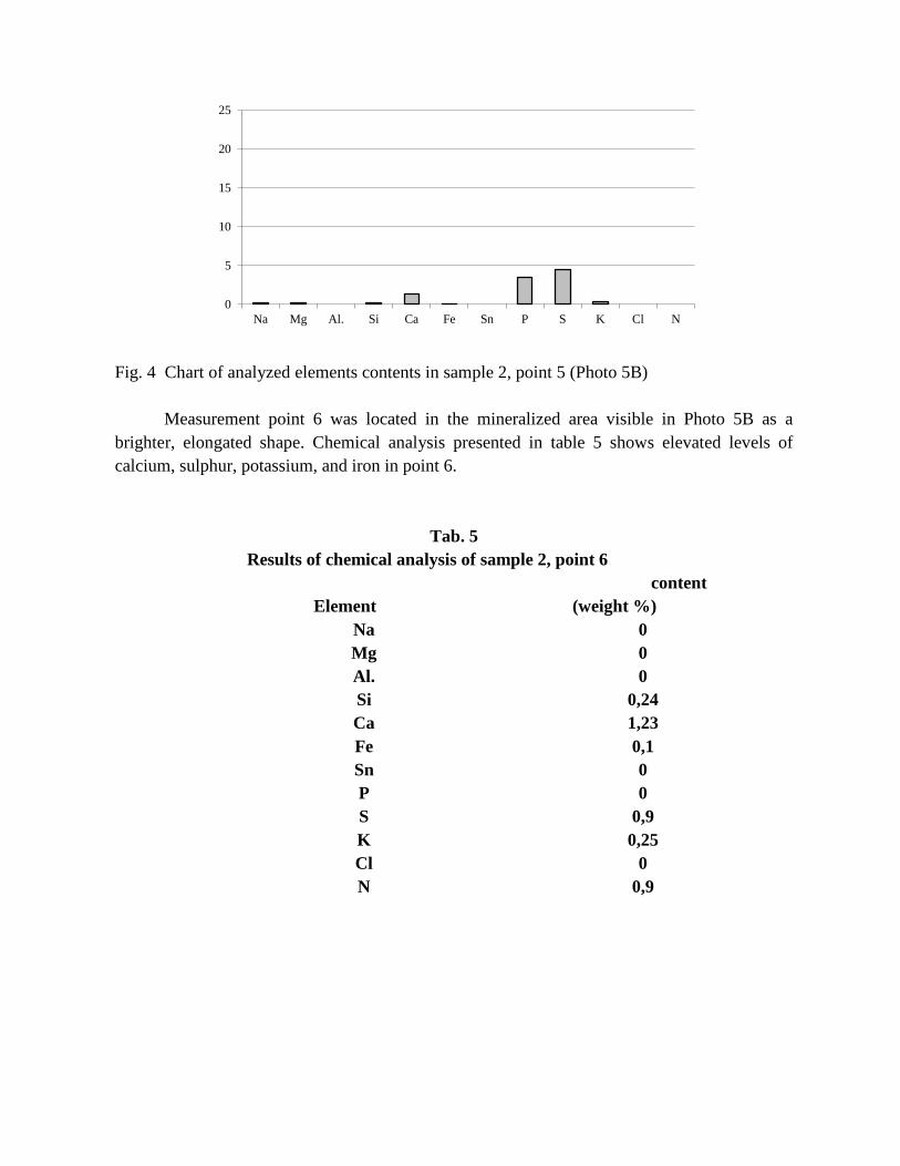
Fig. 4 Chart of analyzed elements contents in sample 2, point 5 (Photo 5B)
Measurement point 6 was located in the mineralized area visible in Photo 5B as a brighter, elongated shape. Chemical analysis presented in table 5 shows elevated levels of calcium, sulphur, potassium, and iron in point 6.
Tab. 5 Results of chemical analysis of sample 2, point 6
Element content
(weight %) Na 0 Mg 0 Al. 0 Si 0,24 Ca 1,23 Fe 0,1 Sn 0 P 0 S 0,9 K 0,25 Cl 0 N 0,9
0
5
10
15
20
25
Na Mg Al. Si Ca Fe Sn P S K Cl N

Fig. 5 Chart showing levels of analyzed elements in sample 2, point 6 (Photo 5B). Preparation 2
A B Photo 6 Preparation 2 A - Carcinoma basocellulare solidum. Polarizing microscope X N, magnification 60X. Visible spherical form of mineralization. B – SEM. Enlarged area marked in Photo 6A, with indicated spots where chemical analyses were performed (EDS).
Table 6 shows chemical analysis of the outer part of mineralized area visible in Photo 6A, B in the form of a spherical shape. Measurement point 7 contained elevated levels of calcium, sulfur, and trace amounts of potassium in comparison to healthy, non-cancerous tissue. Table 7 shows chemical analysis of the inner part of mineralized area visible in Photo 6A, B, measurement point 8. The analysis showed elevated level of calcium; it was significantly higher than in the outer part of the mineralized area (measurement point 7 - Photo 6B). Apart from calcium, slightly elevated levels of phosphorus, sulfur, and potassium were found.
0
5
10
15
20
25
Na Mg Al. Si Ca Fe Sn P S K Cl N

Tab. 6
Results of chemical analysis of sample 2, point 7 (outer part of the shape – Photo 6B)
Element Content (weight %)
Na 0 Mg 0 Al. 0 Si 0 Ca 1,18 Fe 0 Sn 0 P 0 S 2,35 K 0,08 Cl 0 N 0
Tab. 7
Results of chemical analysis of sample 2, point 8 (inner part of the shape – Photo 6B)
Element Content (weight %) Na 0 Mg 0 Al. 0 Si 1,1 Ca 14,42 Fe 0 Sn 0 P 2,2 S 0 K 0,51 Cl 0 N 0
Sample 3 Carcinoma basocellulare solidum exulcerans. Woman, age 41.

A B
C
Photo 7 Sample 3 A - Carcinoma basocellulare solidum. Visible border of tumor penetrating into dermis, with a characteristic palisading pattern of cells on the periphery. Preparation stained with hematoxylin and eosin. Polarizing microscope 1N, magnification 60X. B- Carcinoma basocellulare solidum exulcerans. Polarizing microscope XN, magnification 60X. Visible spindle-shaped concentration of minerals. Marked area is show in Photo 18. C- Carcinoma basocellulare solidum exulcerans. SEM, magnification 300X. Mineralized area analysis was performed in point 13. “Clean” tissue analysis was performed in point 14.
Table 8 shows chemical analysis of the spindle-shaped mineral concentration visible in Photos 7 B,C. Analysis of measurement point 13 showed elevated level of calcium and slightly elevated level of aluminum. Table 9 summarizes the results of element content analysis from point 14. Compared to point 13, there is a noticeable increase in the level of calcium, sulfur, and phosphorus in the tissue.
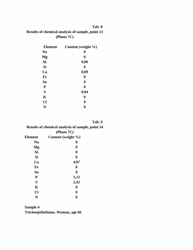
Tab. 8
Results of chemical analysis of sample, point 13 (Photo 7C)
Element Content (weight %)
Na 0 Mg 0 Al. 0,06 Si 0 Ca 0,09 Fe 0 Sn 0 P 0 S 0,04 K 0 Cl 0 N 0
Tab. 9 Results of chemical analysis of sample, point 14
(Photo 7C) Element Content (weight %)
Na 0 Mg 0 Al. 0 Si 0 Ca 4,97 Fe 0 Sn 0 P 1,12 S 2,42 K 0 Cl 0 N 0
Sample 4 Trichoepithelioma. Woman, age 66

A B Photo 8 Sample 4
A – Trichoepithelioma. Image of altered tissue structures captured with polarizing microscope. Visible mineralization in the form of elongated clusters in the marginal part of the specimen (area A) and in the form of very fine mineral clusters around cells (area B). Polarizing microscope, XN, magnification 60 X. B- SEM. Magnified area A from photo 8A. Visible tiny opaque mineral clusters. Point 9 - site of EDS chemical analysis. EDS chemical analysis performed in the spaces between mineral grains (photo 8B, point 19 – Tab. 10) shows that also in those places levels of calcium, sodium, phosphorus, sulphur, and iron are elevated.
Tab. 10
Results of chemical analysis of sample, point 19 (Photo 10B)
Element Content (weight %)
Na 0,1 Mg 0,07 Al. 0 Si 0 Ca 4,12 Fe 0.1 Sn 0 P 0,87 S 0,3 K 0 Cl 0 N 0

Summary
Two types of skin cancer - Carcinoma basocellulare and Trichoepithelioma - have been studied in order to examine the phenomenon of mineralization in these cancers.
Bio-microscopic studies allowed for performing histopathological characteristics and pre-assessment of mineralization. A more accurate qualification of the occurrence of mineral deposits in examined preparations, as well as examination of the form of mineralization, were possible under a polarizing microscope with a chemical analysis attachment (EDS).
Based on obtained results, it can be concluded that mineralization of the examined skin cancers has both hidden and overt nature.
Hidden mineralization means presence of elevated levels of certain elements (especially calcium and in some cases phosphorus) in the test tissues.
This form of mineralization doesn’t manifest as mineral grains or microcrystals. This means that the elements present in increased quantities are incorporated into the biological structures of the tissue-building compounds. Previous studies (Pawlikowski 1993, 1995, 2017, Pawlikowski, Pfitzner 1999, Harper, Moses 2006) have shown that insertions of these elements occur at sites of structural defects in tissues. By integrating into biological structures in this way, these elements change their nature and physicochemical properties, and thus their functions. Hidden mineralization was found in samples of Carcinoma basocellulare solidum and Trichoepithelioma.
Over time, hidden mineralization may, but does not have to transform into overt mineralization, which was also found in examined tumors (Pawlikowski 1979, 1981, 1995, 2011, 2013).
Overt mineralization means presence of very fine mineral grains within the tissues, in this case neoplastic tissues. This type of mineralization was found in all tested preparations.
Overt mineralization occurs in examined preparations in the following forms: - Tiny concentrations of minerals visible through tissue - Thin, mineralized margins around cells - Characteristic spherical forms - Elongated, very fine mineral grains - Irregular mineral deposits. In tissues affected by mineralization, content of calcium and,
sometimes, phosphorus is elevated in comparison to “clean” – non-mineralized - tissue. In most of the spots where chemical microanalysis of mineralized areas has been performed,
increased levels of calcium has been found, often in the absence of phosphorus. Only one of the measuring points showed presence of phosphorus in the absence of calcium. Three points contained increased levels of both calcium and phosphorus.
Increased silicon content recorded in some cases is most likely due to a calculation error, but it can not be determined without a doubt whether it has a pathological basis.
Previous studies on the mineralization of tumors indicate that this type of mineralization mainly consists of calcium phosphate, sometimes accompanied by sulfur, iron, silicon, magnesium, sodium, chlorine, and aluminum. It should be remembered that EDS method has the sensitivity to allow precise determination of elements with an accuracy of 0.0X%. This means that it is very likely that described elements are accompanied by others that are present in tissues in quantities smaller than

0.0X%.
One of the main questions resulting from presented research is: 1. Do cancerous tissues tend to concentrate certain elements? or: 2. Does "elevated mineralization” in tissue, e.g. in body fluids, occur first, and its
consequence is cancer?
Re. 1 In case of a developed tumor (e.g. skin tumor), the answer to the first question is less important from the viewpoint of the genesis of cancer. Here the question is how to get rid of the mineralization if it affects procreation of cells. Re. 2 This question is fundamental from the viewpoint of the genesis of the mechanism of cell neoplasia. It is highly likely that both hidden tissue mineralization and overt mineralization, including so-called calcifications, may favor structural defects of DNA generated during the chromosome division in the process of cell multiplication (Figure 7). The division environment in a cell with abnormal content of elements or compounds (too low or too high mineralization) may favor deformations of the DNA segment that is responsible for the rate of mitotic division (Kokot 1993, Devereux 1996, Salgia 1998, Bunyaviroch, Coleman 2006). Thus, mineralization of tissues and body fluids (caused by both external and internal factors) may promote tumor formation (Woźniak et al. 2001, Juszko-Piekut et al 2004, Yin 2007).

Fig. 7 Hypothetical DNA deformation in the segment responsible for multiplication during cell division in environment over-mineralized with PO4
3- (Pawlikowski 1995) A- Preliminary DNA splitting phase. B- Division of DNA into two parts in cellular fluid
with elevated ionic content of e.g. PO43-. Ion incorporation into DNA section
responsible for regulating cell procreation. C- Two new DNA spirals created during cell division. Left spiral, deformed by ion (or compound) incorporation goes into a state of permanent multiplication, leading to proliferation of cells and the formation of tumor.
Additional question, which should be answered by further research, is:
What factors and mechanisms lead to excessive concentration of elements (and compounds) in skin
and other organs and tissues?
The answer to this question is extremely important in terms of cancer, including skin cancer, prevention (Kokot 1993, Pawlicki 1995, Woźniak et al. 2001, Juszko-Piekut et al.). It seems that one of those mechanisms may be the transfer of elements from bones to soft tissues in the process of osteoporosis as it develops with age (Pawlikowski 2014).
123

Literature Bunyaviroch T, Coleman RE., 2006 PET evaluation of lung cancer. J Nucl Med. 47(3): 451-69. Boyle P., Doré J.-F., Autier P., Ringborg U.: 2004 Cancer of the skin: a forgotten problem in Europe. Annals of Onkology 15: 5 – 6 Chicheł A., Skowronek J.:2005 Współczesne leczenie raka skóry – dermatologia, chirurgia czy radioterapia? Współczesna Onkologia, vol 9;10: 429 – 435. Daniel L., Leśniewski – Kmak K,:2005 Leczenie długotrwale rozwijającego sięraka podstawnokomórkowego skóry, niszczącego połowętwarzy – opis przypadku. Współczesna Onkologia 2005, vol 9;10: 440 -442 Deja M., Teresiak E., Buczyńska – Górna M., Karaś A., Jenerowicz D., Bowszyc – Dmochowska M.; 2004 Analiza częstości występowania poszczególnych typów histologicznych raka podstawnokomórkowego skóry, umiejscowienia zmian oraz wieku i płci pacjentów. PostępyDermatologiiiAlergologii XXI; /5: 231 – 239 Devereux T.R., 1996 Molecular mechanisms of lung cancer. Interaction of environmental and genetic factors. Chest: 14S-19S. Harper J, Moses MA. 2006 Molecular regulation of tumor angiogenesis: mechanisms and therapeutic implications.. EXS 96: 223-268. Juszko-Piekut M., Kołosza Z., Moździerz A., 2004 The influence of selected environmental factors on lung cancer incidence in immigrant population of industrial areas. PolishJournal of EnvironmentalStudies. Vol.13, Supplement II: 174-180. Kokot F.; 1`993 Gospodarka wodno-elektrolitowa i kwasowo-zasadowa w stanach fizjologii i patologii. Warszawa, PZWL, Męcik T. J., Szczurek Z., Cieślik T., Sabat D.: 2000 Studies on the feasibility of the evaluation of neoplastic tissues calcification in oral tumors; PraceMineralogiczne, 89, Pawlicki M. 1995 Rak: nadzieje i rozczarowania Kraków: PAN.. Pawlikowski M., 1987 Mineralizacja organizmu człowieka �żyjącego (Mineralogy of human body). Prace Mineralogiczne, 79. Pawlikowski M., 1991 Mineralizacja nowotworów (Mineralization of cancer). W: A. Szymański Ed. Biomineralizacja i biomateriały. Warszawa PWN. Pawlikowski M., 1993 Kryształy w organizmie człowiek (Crystals of human body) Kraków, Wydawnictwo i Drukarnia "Secesja". Pawlikowski M., 1995 Sekrety mineralizacji tkanek. Kraków(Secrets of tissue mineralization). Wydawnictwo Centrum PPGSMiE PAN.
Pawlikowski M., Pfitzner R. 1999 Mineralizacja serca i dużych naczyń. (Mineralization of heart and big blood vessels). Wyd. IGSMiE PAN Kraków, 142 p. Pawlikowski M. 2011 Biomineralization of cancer tissues. 20th Int. Symp. Molecular and Physiological Aspects of Regulatory Processes of the Organism. Cracow. Ed. H. Lach. Wyd. Abaton. Kraków: 190-191. Pawlikowski M., 2013 Mineralization of lung cancer tumors. Auxiliary sciences in archaeology ,preservation of relicts and environmental engineering. CD -no 15, Ed. M Pawlikowski Pawlikowski M. 2014 Osteoporosis as a source of tissue mineralization. Research on osteoporosis therapy and dissolution of arterial mineralization. Jour Life Science 8, : 610-625. Pawlikowski M., 2017 Biomineralogy of angiogenezis. Arch Clin Biomed Res ; 1 (4): 161-167 Salgia R., 1998 Molecular abnormalities in lung cancer. ClinicalOncology: 1207-1217. Wołowiec J., Dadej I.: 2003 Rola UVA w patologii skóry. Postępy Dermatologii i Alergologii
XX, 3: 170 – 175 Woźniak L., et al.: 2001 Nowotwory: zarys patologii onkologicznej. Łódź,