biomechanical study of three kinds of internal fixation
TRANSCRIPT
RESEARCH ARTICLE Open Access
Biomechanical study of three kinds ofinternal fixation for the treatment ofsacroiliac joint disruption usingbiomechanical test and finite elementanalysisTao Wu1, Xuejiao Ren2, Yunwei Cui3, Xiaodong Cheng3, Shuo Peng1, Zhiyong Hou3 and Yongtai Han1*
Abstract
Background: To compare the stability of sacroiliac joint disruption fixed with three kinds of internal fixation usingboth biomechanical test and finite element analysis.
Methods: Five embalmed specimens of an adult were used. The symphysis pubis rupture and left sacroiliac jointdisruption were created. The symphysis pubis was stabilized with a five-hole plate. The sacroiliac joint disruptionwas fixed with three kinds of internal fixation in a randomized design. Displacements of the whole specimen andshifts in the gap were recorded. Three-dimensional finite element models of the pelvis, the pelvis with symphysispubis rupture and left sacroiliac joint disruption, and three kinds of internal fixation techniques were created andanalyzed.
Results: Under the vertical load, the displacements and shifts in the gap of the pelvis fixed with minimally invasiveadjustable plate (MIAP) combined with one iliosacral (IS) screw were the smallest, and the average displacements ofthe pelvis fixed with an anterior plate were the largest one. The differences among them were significant. In finiteelement analysis and MIAP combined with one IS screw fixation showed relatively best fixation stability and lowestrisks of implant failure than two IS screws fixation and anterior plate fixation.
Conclusion: The stability of sacroiliac joint disruption fixed with MIAP combined with one IS screw is better thanthat fixed with two IS screws and anterior plate under vertical load.
Keywords: Biomechanics, Internal fixation, Sacroiliac joint disruption, Finite element analyses
BackgroundSacroiliac joints (SIJs) play an important role in the pel-vic ring, which is the main structure of force transmit-ting between the upper and lower limbs [1]. Sacroiliacjoint disruption (SJD) is a severe clinical injury, causedby high-energy traumas. Although surgical treatmenthas become a gold-standard method for SJD in the re-cent years, how to select an appropriate fixation
technique remains a challenging problem for clinicalsurgeon [2].Currently, there are several internal fixation tech-
niques for SJD, including percutaneous iliosacral (IS)screw, anterior plate, posterior transiliac plating, minim-ally invasive adjustable plate (MIAP), and so on [3–5].These fixations have several advantages and disadvan-tages respectively, and none is proved to be the strongestfixation in the experiments and clinics.The percutaneous IS screws are widely used due to its
advantage of a minimal incision at present. Osterhoff etal. treated the patients with unstable pelvic fractureusing IS screws and found that IS screw fixation was a
* Correspondence: [email protected] of Bone Disease, Third Hospital of Hebei Medical University,Shijiazhuang 050051, Hebei, ChinaFull list of author information is available at the end of the article
© The Author(s). 2018 Open Access This article is distributed under the terms of the Creative Commons Attribution 4.0International License (http://creativecommons.org/licenses/by/4.0/), which permits unrestricted use, distribution, andreproduction in any medium, provided you give appropriate credit to the original author(s) and the source, provide a link tothe Creative Commons license, and indicate if changes were made. The Creative Commons Public Domain Dedication waiver(http://creativecommons.org/publicdomain/zero/1.0/) applies to the data made available in this article, unless otherwise stated.
Wu et al. Journal of Orthopaedic Surgery and Research (2018) 13:152 https://doi.org/10.1186/s13018-018-0858-2

sufficient technique [6]. However, this fixation requiresextensive experience and has a high rate of iatrogenicvascular and neural injuries. Both patients and doctorsare exposed to lots of radiation during IS screw place-ment [7]. The anterior plate is another therapeuticmethod, which can also provide biomechanical stabilityof the SIJs. Simpson et al. found that satisfactory clinicalresults were achieved by using anterior plate fixation.However, screw loosening occurred during the follow-up[8]. To avoid these limitations, Chen et al. introducedMIAP [9], which simulates the structures of the sacro-iliac joint complex. It could obtain a satisfactory resultwhen MIAP was used for treating unstable pelvic ringinjuries [5, 9]. In biomechanical test, the stability of sa-cral fracture fixed with MIAP was inferior than thatfixed with two IS screws; however, these differences werenot significant [10].Clinically, surgeons try to find the strongest internal
fixation for SJD. Stable SIJs can reduce the risk of lowback pain postoperation and ask the patients to walkearlier which could avoid lots of long-term complica-tions caused by bed rest. Therefore, we hypothesize thatSJD fixed with MIAP combined with one IS screw ismore stable than that with an anterior plate and two ISscrews. It is well known that it is very difficult to com-pare the stability of different fixations for SJD in clinicalapplications because of the variations in fracture pat-terns, bone quality, and fixation. So, biomechanical testand three-dimensional finite element analysis (FEA) arethe most commonly used methods in orthopedic bio-mechanical research. In this study, we aimed to comparethe stability of SJD fixed with three kinds of internal fix-ation using both biomechanical test and finite elementanalysis and provide a basis for the clinical application.
MethodsStructure of minimally invasive adjustable plateMIAP is made up of three parts: two similar Z-shapedbrackets and an adjustable connection bar (Fig. 1). Eachsimilar Z-shaped bracket is composed of a lower wingand a cambered wing. The lower wing of this bracket isplaced on the dorsal surface of the sacrum, and the cam-bered wing is positioned close to the dorsal surface ofthe posterior superior iliac spines, which is fixed to thesacrum and ilium using some long cancellous screws.
Preparation and preservation of specimensFive embalmed adult male cadaver pelvises (age 43.2 ±6.9 years) were used for the biomechanical test, whichwere provided by the Department of Anatomy of HebeiMedical University. The inclusion criteria for pelvicspecimens were as follows: (1) The hip joint and pubicsymphysis must be intact. (2) The soft tissues of thespecimens were removed, and the main ligaments were
left intact. (3) The specimens from patients withrheumatism, tuberculosis, anatomic variations, cancer,and other diseases were excluded. Specimens that wereproven to have osteoporosis using an osteocore 3 dualenergy X-ray osteodensitometer (Medilink Company,Parc de la Mediterranee, France) were excluded (Table 1).These specimens were stored at − 20 °C and melted atroom temperature 12 h before the test.
Modeling sacroiliac joint disruption and fixation ofspecimensFor better comparing the stability of internal fixation,the anterior and posterior pelvic rings were disrupted(Fig. 2). Anteriorly, a symphysis pubis rupture was made,which was stabilized using a five-hole plate. Posteriorly,the disruption of sacroiliac joint was manipulated bycutting the connection between the left sacroiliac joint.Three types of internal fixation were implanted ran-domly after the reduction of SJD (Table 1).Three kinds of internal fixation were used as follows:
(1) MIAP combined with one IS screw group: Toexpose the posterior side of the ilium and sacrum,choose an appropriate MIAP. Some appropriatelong cancellous screws were implanted into theilium and sacrum respectively. Afterwards, one 2.0-mm Kirschner wire was inserted through theipsilateral external surface of the ilium and into thefirst sacral vertebral body. Fluoroscopy was used toconfirm appropriate screw position. One 7.3-mmcannulated, partially threaded, and cancellous ISscrew was inserted into the first sacral vertebralbody along the Kirschner wire (Fig. 3).
(2) Two IS screws group: Two 2.0-mm Kirschner wireswere inserted into the first sacral vertebral bodyaccording to the above method. Fluoroscopy wasalso used to confirm the screws’ positions. And
Fig. 1 The structure of MIAP
Wu et al. Journal of Orthopaedic Surgery and Research (2018) 13:152 Page 2 of 8
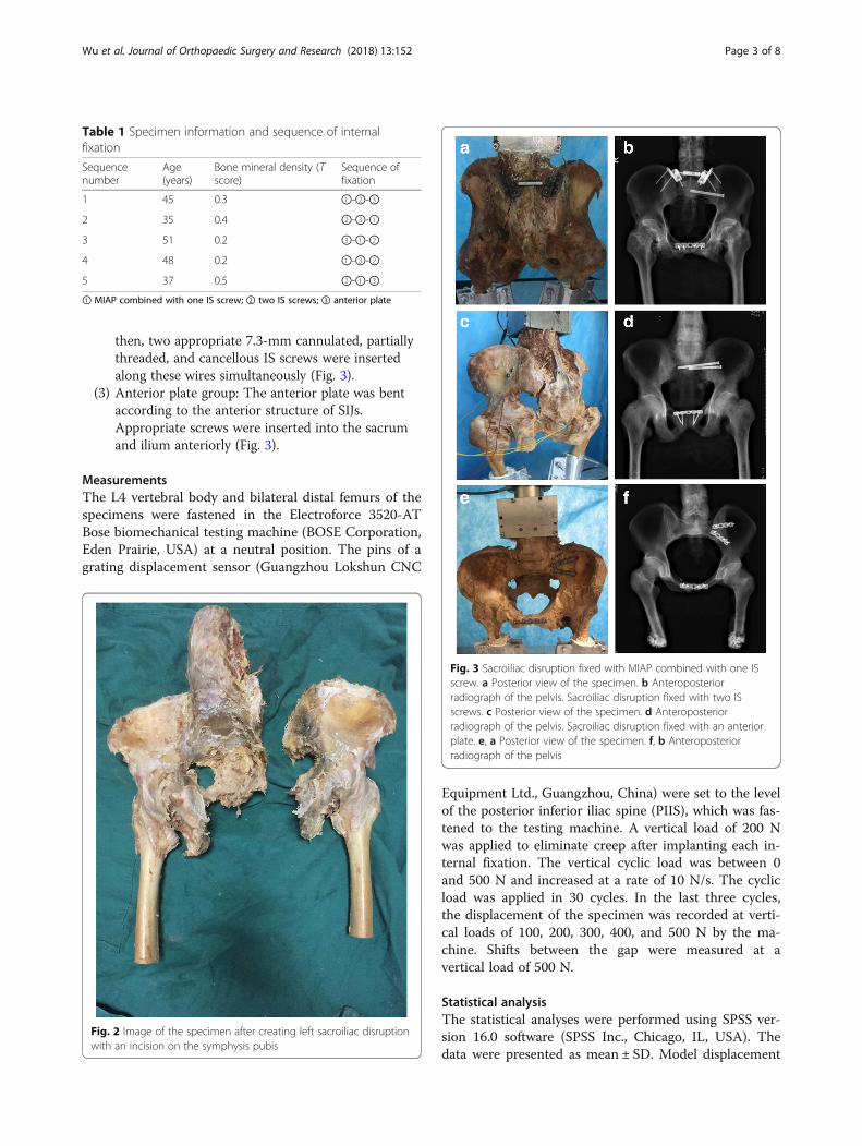
then, two appropriate 7.3-mm cannulated, partiallythreaded, and cancellous IS screws were insertedalong these wires simultaneously (Fig. 3).
(3) Anterior plate group: The anterior plate was bentaccording to the anterior structure of SIJs.Appropriate screws were inserted into the sacrumand ilium anteriorly (Fig. 3).
MeasurementsThe L4 vertebral body and bilateral distal femurs of thespecimens were fastened in the Electroforce 3520-ATBose biomechanical testing machine (BOSE Corporation,Eden Prairie, USA) at a neutral position. The pins of agrating displacement sensor (Guangzhou Lokshun CNC
Equipment Ltd., Guangzhou, China) were set to the levelof the posterior inferior iliac spine (PIIS), which was fas-tened to the testing machine. A vertical load of 200 Nwas applied to eliminate creep after implanting each in-ternal fixation. The vertical cyclic load was between 0and 500 N and increased at a rate of 10 N/s. The cyclicload was applied in 30 cycles. In the last three cycles,the displacement of the specimen was recorded at verti-cal loads of 100, 200, 300, 400, and 500 N by the ma-chine. Shifts between the gap were measured at avertical load of 500 N.
Statistical analysisThe statistical analyses were performed using SPSS ver-sion 16.0 software (SPSS Inc., Chicago, IL, USA). Thedata were presented as mean ± SD. Model displacement
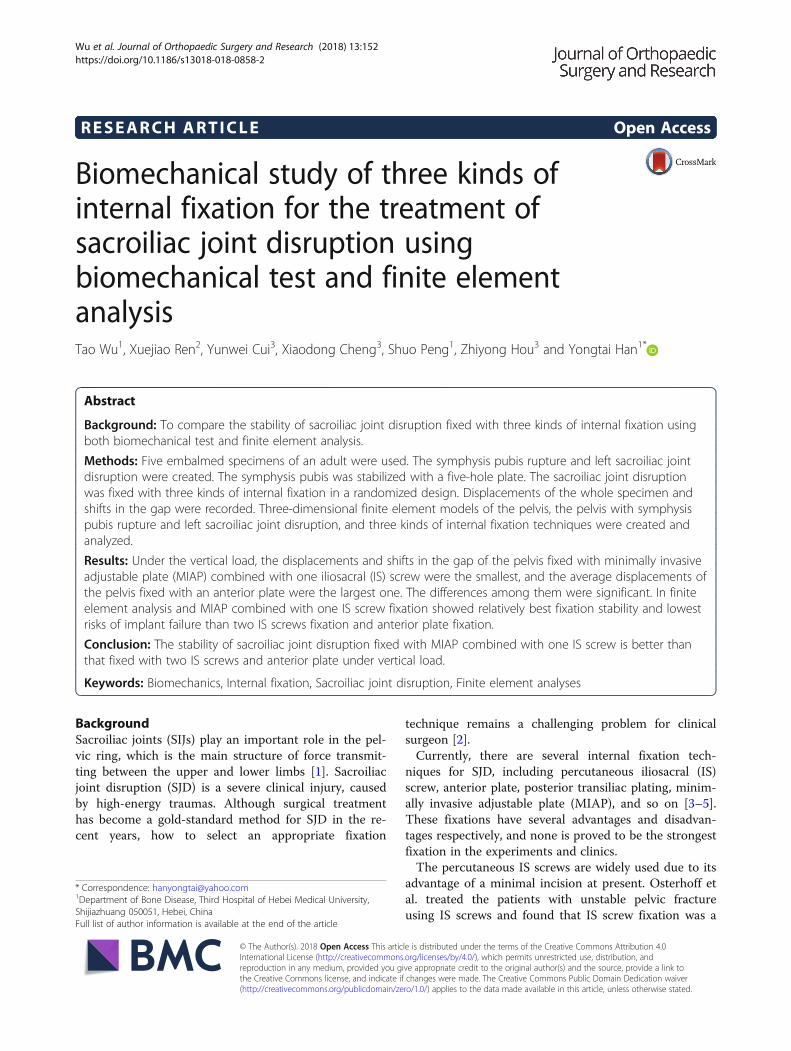
Table 1 Specimen information and sequence of internalfixation
Sequencenumber
Age(years)
Bone mineral density (Tscore)
Sequence offixation
1 45 0.3 ①-②-③
2 35 0.4 ②-③-①
3 51 0.2 ③-①-②
4 48 0.2 ①-③-②
5 37 0.5 ②-①-③
① MIAP combined with one IS screw; ② two IS screws; ③ anterior plate
Fig. 2 Image of the specimen after creating left sacroiliac disruptionwith an incision on the symphysis pubis
Fig. 3 Sacroiliac disruption fixed with MIAP combined with one ISscrew. a Posterior view of the specimen. b Anteroposteriorradiograph of the pelvis. Sacroiliac disruption fixed with two ISscrews. c Posterior view of the specimen. d Anteroposteriorradiograph of the pelvis. Sacroiliac disruption fixed with an anteriorplate. e, a Posterior view of the specimen. f, b Anteroposteriorradiograph of the pelvis
Wu et al. Journal of Orthopaedic Surgery and Research (2018) 13:152 Page 3 of 8

and shifts between the gap were compared by random-ized block design ANOVA. The Bonferroni test was usedto compare significant differences between the groups.Significance was set at P < 0.05.
Finite element models and implantsA three-dimensional finite element model was createdfrom CT scan. The intact pelvis was scanned from anadult healthy volunteer, and the DICOM format fileswere processed by MIMICS 10.01 (Materialise, Belgium).And then, these files were processed by Geomagic Stu-dio 12 (Geomagic, USA) and Abaqus 6.11-1 (SIMULIA,France). The full pelvis was composed of the left ilium,sacrum, right ilium, and symphysis pubis, and thesebones consisted of the cortical bone and cancellousbone. The anterior sacroiliac, interosseous sacroiliac,posterior sacroiliac, sacrotuberous, and sacrospinous lig-aments were also created to simulate normal condition.The linear elastic isotropic material properties wereused, and the properties of the bones and ligaments areshown in Table 2. The sacroiliac joint and symphysispubis were modeled with contact type “bonded.” In thefinite element models, the bilateral acetabulums werefully fixed and a vertical load of 500 N was applied onthe upper surface of the sacrum, which was equal to theupper body weight.Three kinds of internal fixation model, which included
percutaneous IS screws, anterior plate, and MIAP, werecreated by UG (Unigraphics NX) software according totheir structural features, and the threads of screws wereomitted so as to simplify the models.
Injured model and finite element analysesThe connection of the left symphysis pubis and leftsacroiliac joint was deleted, and the left anterior sacro-iliac, interosseous sacroiliac, posterior sacroiliac,
sacrotuberous, and sacrospinous ligaments of the pelvicmodel were also deleted, which were consistent with theinjured specimen of the biomechanical test.The injured model was fixed by MIAP combined with
one IS screw, two IS screws, and anterior plate in se-quence. The vertical force and boundary conditions werethe same in different models. In post-processing, thevon Mises of the pelvis, the maximum displacement ofthe whole pelvis, the maximum von Mises stress of in-ternal fixation, and the shifts of the gap at the level ofPIIS were calculated to compare the different kinds ofinternal fixation.
ResultsBiomechanical testAll specimens were fastened at a neutral position with-out obvious fracture or obliquity. Evulsion, loosening,and breakage of internal fixation were not observed. Thedisplacements of specimens were recorded by BOSE bio-mechanical workstation under vertical load. The shiftsbetween the gap were recorded simultaneously. Basedon load-displacement scattergraph, the smooth straightline indicated that the specimens had elastic deformation(Fig. 4).Under different vertical load, the average displace-
ments of the pelvis fixed with MIAP combined with oneIS screw were the smallest, and the average displace-ments of the pelvis fixed with an anterior plate were thelargest one. The value of the pelvis fixed with two ISscrews was in the middle. The differences among themwere significant (Table 3).Under vertical load of 500 N, the average shifts in the
SIJ gap of the pelvis fixed with MIAP combined withone IS screw were significantly the smallest one[0.619 ±0.117 mm], followed by that of fixed with two IS screws[0.893 ± 0.236 mm], and the largest one was that of fixed
Table 2 The properties of materials used in pelvic finiteelement model
Material Young’s modulus(MPa)
Poisson’sratio u
K (N/mm)
Cortical bone (ilium) 17,000 0.3
Cortical bone (sacrum) 6140 0.3
Cancellous bone (ilium) 132 0.2
Cancellous bone (sacrum) 1400 0.3
Symphysis pubis 5 0.45
Sacroiliac posterior longligament
1000
Sacroiliac posterior shortligament
400
Sacroiliac anterior ligament 700
Sacrotuberous ligament 1500
Sacrospinous ligament 1400 Fig. 4 Load-displacement scattergraph of the specimens
Wu et al. Journal of Orthopaedic Surgery and Research (2018) 13:152 Page 4 of 8

with anterior plate [1.747. ± 0.192 mm]. The differenceswere also significant (P < 0.01).
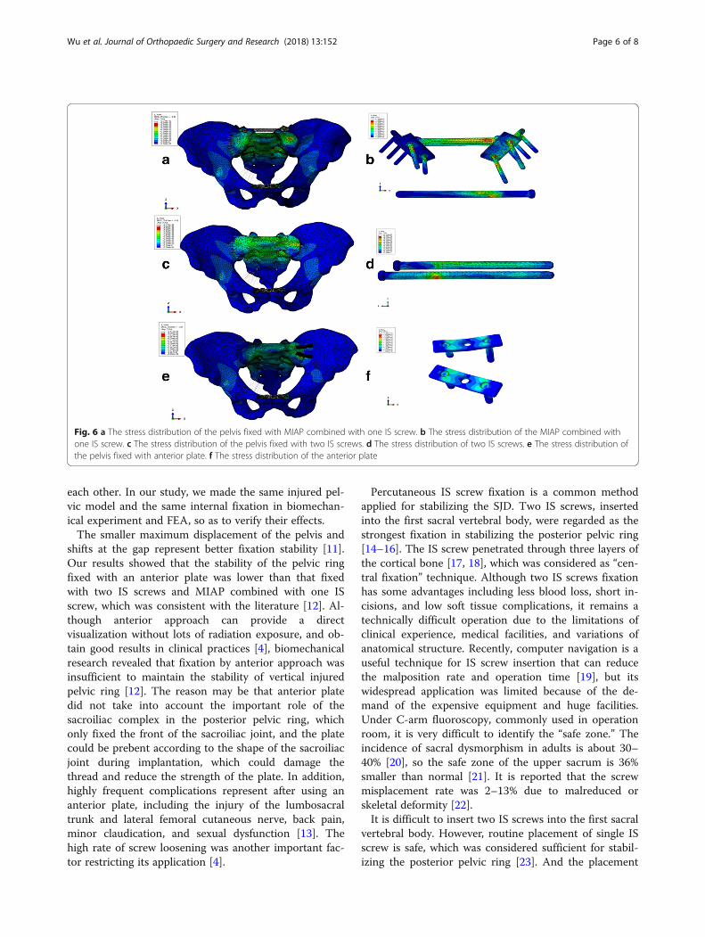
Finite element analysesUnder a vertical load of 500 N, the distribution of vonMises stresses in intact pelvic model showed that thepathway of the vertical load was from the upper surfaceof the sacrum, through the bilateral sacral wing, sacro-iliac joint, large sciatic notch, and iliac arcuate line, tothe bilateral acetabulum (Fig. 5). The maximum vonMises stresses located at the large sciatic notch and thevon Mises stresses of the anterior pelvic ring were small.Under a vertical load of 500 N, all treated models indi-
cated that the distribution of stresses had been greatlyrestored, especially for MIAP combined with one ISscrew fixation and two IS screws fixation (Fig. 6). Themaximum displacement of the model fixed with MIAPcombined with one IS screw, two IS screws, and anteriorplate was 1.265, 1.377, and 2.223 mm, respectively. Thehigher maximum von Mises stress reveals that themodel has a higher risk of broken implant. The max-imum von Mises stress of MIAP combined with one IS
screw was 374.44 MPa, located at the junction betweenthe nail and plate of MIAP. The maximum von Misesstress of two IS screws was 513.64 MPa, located at theinferior IS screw. The maximum von Mises stress of an-terior plate was 2476.57 MPa, located at the junction be-tween the nail and plate of the inferior anterior plate(Fig. 6). The shifts at the gap of the model fixed withMIAP combined with one IS screw, two IS screws, andanterior plate were 0.746, 0.897, and 1.571 mm,respectively.
DiscussionBiomechanical experiment and finite element analysisare the most commonly used methods in orthopedicbiomechanical research. Biomechanical experiment is atraditional research method, which can be analyzed in-tuitively. However, the sources of specimens are fewer,and the qualities of the bones are different, which limitits application. FEA is a new research method in recentyears. It simulates the physical condition of the bonesand can carry out microcosmic mechanical research.FEA and biomechanical experiment can complement
Table 3 Displacement of the pelvis fixed with three types of internal fixation under vertical load ( x ± s, n = 5)
Load(N)
① (mm) ② (mm) ③ (mm) F value/pvalue
p value
① vs ② ① vs ③ ② vs ③
100 0.496 ± 0.102 0.6768 ± 0.130 1.1826 ± 0.220 78.209/0.000 0.039 0.000 0.000
200 0.945 ± 0.193 1.334 ± 0.272 2.002 ± 0.296 87.893/0.000 0.004 0.000 0.000
300 1.466 ± 0.311 1.956 ± 0.342 2.832 ± 0.366 203.61/0.000 0.000 0.000 0.000
400 1.865 ± 0.369 2.478 ± 0.392 3.871 ± 0.601 207.281/0.000 0.001 0.000 0.000
500 2.477 ± 0.321 3.128 ± 0.519 4.704 ± 0.600 129.958/0.000 0.005 0.000 0.000
① MIAP combined with one IS screw; ② two IS screws; ③ anterior plate
Fig. 5 Distribution of von Mises stress in the intact pelvic model
Wu et al. Journal of Orthopaedic Surgery and Research (2018) 13:152 Page 5 of 8
each other. In our study, we made the same injured pel-vic model and the same internal fixation in biomechan-ical experiment and FEA, so as to verify their effects.The smaller maximum displacement of the pelvis and
shifts at the gap represent better fixation stability [11].Our results showed that the stability of the pelvic ringfixed with an anterior plate was lower than that fixedwith two IS screws and MIAP combined with one ISscrew, which was consistent with the literature [12]. Al-though anterior approach can provide a directvisualization without lots of radiation exposure, and ob-tain good results in clinical practices [4], biomechanicalresearch revealed that fixation by anterior approach wasinsufficient to maintain the stability of vertical injuredpelvic ring [12]. The reason may be that anterior platedid not take into account the important role of thesacroiliac complex in the posterior pelvic ring, whichonly fixed the front of the sacroiliac joint, and the platecould be prebent according to the shape of the sacroiliacjoint during implantation, which could damage thethread and reduce the strength of the plate. In addition,highly frequent complications represent after using ananterior plate, including the injury of the lumbosacraltrunk and lateral femoral cutaneous nerve, back pain,minor claudication, and sexual dysfunction [13]. Thehigh rate of screw loosening was another important fac-tor restricting its application [4].
Percutaneous IS screw fixation is a common methodapplied for stabilizing the SJD. Two IS screws, insertedinto the first sacral vertebral body, were regarded as thestrongest fixation in stabilizing the posterior pelvic ring[14–16]. The IS screw penetrated through three layers ofthe cortical bone [17, 18], which was considered as “cen-tral fixation” technique. Although two IS screws fixationhas some advantages including less blood loss, short in-cisions, and low soft tissue complications, it remains atechnically difficult operation due to the limitations ofclinical experience, medical facilities, and variations ofanatomical structure. Recently, computer navigation is auseful technique for IS screw insertion that can reducethe malposition rate and operation time [19], but itswidespread application was limited because of the de-mand of the expensive equipment and huge facilities.Under C-arm fluoroscopy, commonly used in operationroom, it is very difficult to identify the “safe zone.” Theincidence of sacral dysmorphism in adults is about 30–40% [20], so the safe zone of the upper sacrum is 36%smaller than normal [21]. It is reported that the screwmisplacement rate was 2–13% due to malreduced orskeletal deformity [22].It is difficult to insert two IS screws into the first sacral
vertebral body. However, routine placement of single ISscrew is safe, which was considered sufficient for stabil-izing the posterior pelvic ring [23]. And the placement
Fig. 6 a The stress distribution of the pelvis fixed with MIAP combined with one IS screw. b The stress distribution of the MIAP combined withone IS screw. c The stress distribution of the pelvis fixed with two IS screws. d The stress distribution of two IS screws. e The stress distribution ofthe pelvis fixed with anterior plate. f The stress distribution of the anterior plate
Wu et al. Journal of Orthopaedic Surgery and Research (2018) 13:152 Page 6 of 8

of two IS screws was shown to be clinically unreliable[24]. MIAP is a novel device for stabilizing the posteriorpelvic ring. It is easy to perform without prolonging op-eration time and radiation exposure. The MIAP simu-lates the structure of the sacroiliac complex and isfunctioned as a suspension bridge. Biomechanical stud-ies indicated that the stability of sacral fracture fixedwith MIAP was inferior to that fixed with the two ISscrews, but the difference was not significant [10]. Weproposed that MIAP combined with one IS screw fix-ation, considered as “central and rear fixation” tech-nique, should provide stronger fixation than two ISscrews. In our study, both biomechanical test and FEAshowed that the smaller maximum displacement of thepelvis and shifts at the gap were presented in the modelfixed with MIAP combined with one IS screw than thatfixed with two IS screws.The injury of the sacroiliac joint is a source of low
back pain, accounted for 15–30% of patients withchronic low back pain [25]. If the stresses on the injuredside of the sacroiliac joint increased, degeneration mayoccur. Therefore, the best choice of internal fixation isto restore the distribution of stresses. The results of FEAin this study revealed that the distribution of stress inthe pelvic model fixed with MIAP combined with one ISscrew was most similar to that in the normal pelvicmodel, which has the lowest risk of low back pain. Thelower maximum von Mises stress of fixation deviceshows a lower risk of internal fixation failure. The MIAPcombined with one IS screw had the lowest maximumvon Mises stress in these three kinds of internal fixation,which indicated that it is the best choice for stabilizingthe posterior pelvic ring.This study had several limitations. First, the biomech-
anical test had a small sample size. The power analysisof this study showed only 44.3% power to detect a sig-nificant difference at the P < 0.05 level, and it alsoshowed that a large sample size (more than 12 speci-mens) would be required to detect any difference with80% power. Artificial pelvises should be used in the bio-mechanical test in the future. Second, we implantedthree kinds of internal fixation into the same specimenin sequence for reducing the influence caused by indi-vidual difference of specimens. But the use ofpre-fixation could affect the holding power of the subse-quent fixation. Therefore, we accurately located the dir-ection and position of the screws using X-ray, in orderto minimize the influence of screw channels. Third, weused the same IS screw between the MIAP combinedwith one IS group and two IS screws group, which couldbe slight loosening around previous IS screw during thebiomechanical test. Fourth, only anterior plate on thesacroiliac joint was weak for vertically unstable pelvicring injury. Future research should add an experimental
group for comparing the stability of sacroiliac joint,which is anterior plate combined with one IS screw.Fifth, the parts of the bone were created as a linear elas-tic isotropic material in the FEA. The real structure ofthe bone is complex and different from that of FEA.And the FEA model did not contain the lumbar, femur,and muscle tissues. In future research, we should createa finite element model that is more approximate to thereal bone structure.
ConclusionThree kinds of internal fixation are all useful for thetreatment of SJD. The stability of sacroiliac joint disrup-tion fixed with MIAP combined with one IS screw isbetter than that fixed with two IS screws and anteriorplate under vertical load. We believe that this new tech-nique is an effective surgical procedure.
AbbreviationsCT: Computed tomography; DICOM: Digital imaging and communications ofmedicine; FEA: Finite element analysis; IS: Iliosacral; MIAP: Minimally invasiveadjustable plate; PIIS: Posterior inferior iliac spine; SIJs: Sacroiliac joints;SJD: Sacroiliac joint disruption
FundingThe study was funded by the Nature Science Foundation of Hebei Province(Grant No. H2016206291) and Nature Science Foundation of China (Grant No.81572162).
Availability of data and materialsThe datasets used and analyzed during the current study are available fromthe corresponding author on reasonable request.
Authors’ contributionsYTH and TW designed and participated in the whole process of the studyand drafted the manuscript. XJR and YWC carried out the FEA. XDC, SP, andZYH carried out the experimental operation. SP participated in the datacollection. XJR helped to draft the manuscript. All authors read andapproved the final manuscript.
Ethics approval and consent to participateThe ethics committee of the Third Hospital of Hebei Medical Universityapproved this study. Informed consent was obtained from the individualparticipant included in this study.
Competing interestsThe authors declare that they have no competing interests.
Publisher’s NoteSpringer Nature remains neutral with regard to jurisdictional claims inpublished maps and institutional affiliations.
Author details1Department of Bone Disease, Third Hospital of Hebei Medical University,Shijiazhuang 050051, Hebei, China. 2Department of Radiotherapy, FourthHospital of Hebei Medical University, Shijiazhuang 050011, Hebei, China.3Department of Trauma, Third Hospital of Hebei Medical University,Shijiazhuang 050051, Hebei, China.
Received: 19 April 2018 Accepted: 5 June 2018
References1. Gnat R, Spoor K, Pool-Goudzwaard A. The influence of simulated
transverses abdominis muscle force on sacroiliac joint flexibility during
Wu et al. Journal of Orthopaedic Surgery and Research (2018) 13:152 Page 7 of 8

asymmetric moment application to the pelvis. Clin Biomech (Bristol,Avon). 2015;30(8):827–31.
2. Giraldez-Sanchez MA, Lazaro-Gonzalvez A, Martinez-Reina J, Serrano-Toledano D, Navarro-Robles A, Cano-Luis P, Fraqkakis EM, Giannoudis PV.Percutaneous iliosacral fixation in external rotational pelvic fracture. Abiomechanical analysis. Injury. 2015;46(6):327–32.
3. Bousbaa H, Ouahidi M, Louaste J, Bennani M, Cherrad T, Jezzari H, KasmaouiEH, Rachid K, Amhajji L. Percutaneous iliosacral screw fixation in unstablepelvic fractures. Pan Afr Med J. 2017;27(8):244.
4. Zhang R, Yin Y, Li S, Hou Z, Jin L, Zhang Y. Percutaneous sacroiliac screwversus anterior plating for sacroiliac joint disruption: a retrospective cohortstudy. Int J Surg. 2018;50(2):11–6.
5. Wu T, Chen W, Zhang Q, Cui YW, Cheng XD, Yang YJ, Zhang YZ. Fixation ofunstable pelvic fractures with minimally invasive adjustable plate. Int J ClinExp Med. 2017;10(1):1399–404.
6. Osterhoff G, Ossendorf C, Wanner GA, Simmen HP, Werner CM.Posterior screw fixation in rotationally unstable pelvic ring injuries.Injury. 2011;42(10):992–6.
7. Hou Z, Zhang Q, Chen W, Zhang P, Jiao Z, Li Z, Smith WR, Pan J, Zhang Y.The application of the axial view projection of the S1 pedicel for sacroiliacscrew. J Trauma. 2010;69(1):122–7.
8. Simpson LA, Waddell JP, Leighton RK, Kellam JF, Tile M. Anteriorapproach and stabilization of the disrupted sacroiliac joint. J Trauma.1987;27(12):1332–9.
9. Chen W, Hou Z, Su Y, Smith WR, Liporace FA, Zhang Y. Treatment ofposterior pelvic ring disruptions using a minimally invasive adjustable plate.Injury. 2013;44(7):975–80.
10. Wu T, Chen W, Li X, Zhang Q, Lv HZ, Zhang YZ. Biomechnical comparisonof three types of internal fixation in a type C zone II pelvic fracture model.Int J Clin Exp Med. 2015;8(2):1853–61.
11. Lee CH, Hsu CC, Huang PY. Biomechanical study of different fixationtechniques for the treatment of sacroiliac joint injuries using finite elementanalyses and biomechanical test. Comput Biol Med. 2017;87(8):250–7.
12. Latenser BA, Gentilello LM, Tarver AA, Thalqott JS, Batdorf JW. Improvedoutcome with early fixation of skeletally unstable pelvic fractures. J Trauma.1991;31(1):28–31.
13. Zhou KH, Luo CF, Chen N, Hu CF, Pan FG. Minimally invasive surgery underfluoro-navigation for anterior pelvic ring fractures. Indian J Orthop. 2016;50(3):250–5.
14. Yinger K, Scalise J, Olson SA, Bay BK, Finkemeier CG. Biomechanicalcomparison of posterior pelvic ring fixation. J Orthop Trauma. 2003;17(7):481–7.
15. van Zwienen CM, van den Bosch EW, Snijders CJ, Kleinrensink GJ, van VuqtAB. Biomechanical comparison of sacroiliac screw techniques for unstablepelvic ring fractures. J Orthop Trauma. 2004;18(9):589–95.
16. van Zwienen CM, van den Bosch EW, Hoek van Dijke GA, Snijders CJ, vanVugt AB. Cyclic loading of sacroiliac screws in tile C pelvic fractures. JTrauma. 2005;58(5):1029–34.
17. Collinge C, Coons D, Aschenbrenner J. Risks to the superior glutealneurovascular bundle during percutaneous iliosacral screw insertion: ananatomical cadaver study. J Orthop Trauma. 2005;19(2):96–101.
18. Marmor M, Lynch T, Matityahu A. Superior gluteal artery injury duringiliosacral screw placement due to aberrant anatomy. Orthopedics. 2010;33(2):117–20.
19. Richter PH, Gebhard F, Dehner C, Scola A. Accuracy of computer-assisted iliosacral screw placement using a hybrid operating room.Injury. 2016;47(2):402–7.
20. Routt ML Jr, Simonian PT, Agnew SG, Mann FA. Radiographic recognition ofthe sacral alar slope for optimal placement of iliosacral screws: a cadavericand clinical study. J Orthop Trauma. 1996;10(3):171–7.
21. Gardner MJ, Morshed S, Nork SE, Ricci WM, Chip Routt ML Jr. Quantificationof the upper and second sacral segment safe zones in normal anddysmorphic sacra. J Orthop Trauma. 2010;24(10):622–9.
22. Gardner MJ, Parada S, Chip Routt ML Jr. Internal rotation and taping ofthe lower extremities for closed pelvic reduction. J Orthop Trauma.2009;23(5):361–4.
23. Osterhoff G, Ossendorf C, Wanner GA, Simmen HP, Werner CM.Percutaneous iliosacral screw fixation in S1 and S2 for posterior pelvic ringinjuries: technique and perioperative complications. Arch Orthop TraumaSurg. 2011;131(6):809–13.
24. Griffin DR, Starr AJ, Reinert CM, Jones AL, Whitlock S. Vertically unstablepelvic fractures fixed with percutaneous iliosacral screws: does posteriorinjury pattern predict fixation failure? J Orthop Trauma. 2006;20(1Suppl):S30–6.
25. Sembrano JN, Polly DW Jr. How often is low back pain not coming fromthe back? Spine (Phila Pa 1976). 2009;34(1):E27–32.
Wu et al. Journal of Orthopaedic Surgery and Research (2018) 13:152 Page 8 of 8