biomechanical evaluation of the fixation … evaluation of the fixation methods for transcondylar...
TRANSCRIPT

Biomechanical Evaluation of the Fixation Methods for Transcondylar Fracture of the Humerus: ONI Plate Versus
Conventional Plates and Screws
Yasunori Shimamuraa*, Keiichiro Nishidaa,b, Junya Imatanic, Tomoyuki Nodaa, Hiroyuki Hashizumed, Aiji Ohtsukab, and Toshifumi Ozakia
Departments of aOrthopaedic Surgery, and bHuman Morphology, Okayama University Graduate School of Medicine, Dentistry and Pharmaceutical Sciences, Okayama 700-8558, Japan, cDepartment of Orthopaedic Surgery, Okayama Saiseikai General Hospital, Okayama 700-0851, Japan, and dDepartment of Orthopaedic Surgery,
Kasaoka Daiichi Hospital, Kasaoka, Okayama 714-0043, Japan
We biomechanically evaluated the bone fixation rigidity of an ONI plate (Group I) during fixation of experimentally created transcondylar humerus fractures in cadaveric elbows, which are the most frequently observed humeral fractures in the elderly, and compared it with the rigidity achieved by 3 conventional fixation methods: an LCP reconstruction plate 3.5 using a locking mechanism (Group II), a conventional reconstruction plate 3.5 (CRP) with a cannulated cancellous screw (Group III), and a CRP with 2 cannulated cancellous screws (CS) in a crisscross orientation (Group IV). In the axial load-ing test, the mean failure loads were: Group I, 98.9±32.6; Group II, 108.5±27.2; Group III, 50.0±7.5; and Group IV, 34.5±12.2 (N). Group I fixations failed at a significantly higher load than those of Groups III and IV (p<0.05). In the extension loading test, the mean failure loads were: Group I, 34.0±12.4; Group II, 51.0±14.8; Group III, 19.3±6.0; and Group IV, 14.7±3.1 (N). Group IV fixations showed a significantly lower failure load than those of Group I (p<0.05). The fixation rigidities against mechanical loading by the ONI plate and LCP plate were comparable. These results suggested that an ONI system might be superior to the CRP and CS method, and comparable to the LCP method in terms of fixation rigidity for distal humerus fractures.
Key words: distal humerus, fracture, biomechanics, internal fixation, elderly
ractures of the distal humerus in the elderly pose a great challenge for orthopedic surgeons
throughout the world [1, 2]. Conservative treatment by cast immobilization has not been recommended because of the low bone union and high complication rate [3-5]. Among cases in which bone union is achieved, varying degrees of elbow joint contracture
are reported. On the other hand, open reduction and internal fixation is not a satisfactory method because of the high failure rate of conventional implants due to the low rigidity of bone fixation; in general, the bone fixation fails due to severe osteoporosis, the small contact area of the fracture site, and the very small size of the distal fragment, which often does not allow for insertion of a sufficient number of screws [3, 6]. In 1999, we developed an ONI transcondylar plate system (ONI plate) and in 2001 the ONI plate was made available for clinical applications. This system
F
Acta Med. Okayama, 2010Vol. 64, No. 2, pp. 115ン120CopyrightⒸ 2010 by Okayama University Medical School.
Original Article http ://escholarship.lib.okayama-u.ac.jp/amo/
Received November 17, 2009 ; accepted November 27, 2009.*Corresponding author. Phone : +81ン86ン235ン7273; Fax : +81ン86ン223ン9727E-mail : [email protected] (Y. Shimamura)

employs a characteristic structure of angular stabili-zation accomplished by a transcondylar screw which passes from the lateral epicondyle to the medial wall of the trochlea and finally locks to an ONI plate by means of an exclusive set screw. For fixation of the medial column, a 4.5mm cannulated cancellous screw is also used. In the current study, we biomechanically evaluated the bone fixation rigidity of an ONI plate during fixa-tion of experimentally created transcondylar humerus fractures in cadaveric elbows, which are the most frequently observed humeral fractures in the elderly, and compared it with the rigidity achieved by 3 con-ventional fixation methods.
Materials and Methods
Specimens. The present experiments were performed using 30 cadavers (12 men and 18 women; mean age, 78 years; range, 54-87 years) embalmed in 10オ formalin for systemic anatomy research. They had no clinical history of humeral fracture, connective tissue diseases, or paralytic dysfunctions. Thirty paired distal humeri were dissected free of soft tissue. For all specimens, the average bone mineral density (BMD) values of 2 regions of the distal humerus (a 2.4cm2 region of the lateral epicondyle and supra-condyle) were assessed using a Hologic QDR 2000 dual-energy X-ray densitometer (Hologic, Bedford,
MA), and there were no significant differences in BMD between the following 4 groups (Table 1). Preparation of the fracture fixation model.A transcondylar distal humerus fracture (extra-artic-ular, but intra-capsule metaphyseal, type13-A3.3, according to the AO comprehensive classification) was created in all 30 humeri. Using a surgical bone-saw, a 5-mm transverse gap was created in the middle of the olecranon fossa, simulating an unstable fracture according to the method reported by Korner et al. [7]. In each pair of specimens from the cadavers, four different fixation methods were applied (Fig. 1). On one side of all 30 specimens, an ONI plate and can-nulated cancellous screw (4.5mm titanium) were used for fixation (Group I). The side of the specimen to receive fixation with an ONI plate was randomly selected. The remaining 30 contralateral sides of the specimens were further allocated into 3 treatment groups: a group treated with an LCP reconstruction
116 Acta Med. Okayama Vol. 64, No. 2Shimamura et al.
Table 1 Condition of the specimens in each group
GroupNo.
Age (yrs) Mean(range)
BMD (mg/cm3)(range)
Ⅰ 71 (54/87) 0.52±0.20 (0.30/0.99)Ⅱ 68 (48/84) 0.59±0.20 (0.34/0.90)Ⅲ 74 (65/87) 0.46±0.19 (0.21/0.86)Ⅳ 75 (54/86) 0.46±0.20 (0.24/0.61)
A C DBFig. 1 Methods of osteosynthesis for each group. A, An ONI plate applied on the lateral column of the distal humerus with a cannu-lated cancellous screw (4.5mm titanium) medially; B, Two LCP reconstruction plates 3.5 (3.5mm titanium; Mathys) were prebended and adapted, dorso-laterally and ulno-medially; C, A conventional reconstruction plate 3.5 was adapted laterally and a cannulated cancellous screw (4.5mm titanium) was placed medially; D, Two cannulated cancellous screws in a crisscross orientation.

plate 3.5 (3.5mm titanium; Mathys, Bettlach, Switz-erland) using a locking mechanism (Group II, n=10); a group treated with a conventional reconstruction plate 3.5 (3.5mm stainless; Mathys) (CRP) and a can-nulated cancellous screw (CS) (4.5mm titanium) (Group III, n=10); and a group treated with two CSs in a crisscross orientation (Group IV, n=10). In Groups II and III, the lateral plates were placed adjacent to the articular cartilage of the capitellum. In Group II, plates for the medial side were pre-bended around the medial epicondyle of the humerus. All surgery was performed by an experienced senior resi-dent surgeon (Table 2). Potting and mechanical loading. After bone fixation, all proximal sides of the humeri were verti-cally fixed to the potting mass with resin to avoid any loosening during the experiments. The end of the loading jig contacting the humeral joint surface was made by resin to fit and cover a whole trochlea of each specimen (Fig. 2-A, B). For the mechanical loading test, a material testing machine (EHF-FB10KN; Shimadzu Corp, Kyoto, Japan) was used. Stiffness testing was performed under anterior and posterior bending and axial com-pression for each specimen. The specimens were loaded at a constant rate of 3mm/minute by the test-ing machine. Failure was defined as breakage of the implants, loosening of the bone-implants interface or cut-out of the screws, until the 5mm gap of the bone closed and cortical contact occurred. The maximum failure loads were measured and recorded (Fig. 3). Data analysis. For statistical analysis, the Mann-Whitney nonparametric test was performed to compare differences of failure load among the groups. A p-value of 0.05 or less was considered significant.
Results
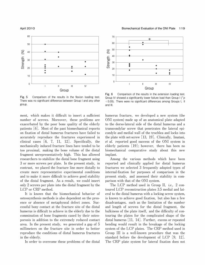
In the axial loading test, the mean failure loads were as follows: Group I, 98.9±32.6; Group II, 108.5±27.2; Group III, 50.0±7.5; and Group IV, 34.5±12.2 (N) (Fig. 4). The Group III and IV fixa-tions failed at a significantly lower load than the Group I fixations (p<0.05), but there was no signifi-cant difference in the mean failure load between Groups I and II. In flexion loading test, the mean failure loads were as follows: Group I, 26.6±11.9; Group II, 32.0±9.2; Group III, 19.3±3.1; and Group IV, 20.7±8.1 (N) (Fig. 5). There was no significant difference in mean failure load between Group I and any other group. In the extension loading test, the mean failure loads were as follows: Group I, 34.0±12.4; Group II, 51.0±14.8; Group III, 19.3± 6.0; and Group IV, 14.7±3.1 (N) (Fig. 6). There was no significant difference in mean failure load between Group I and Group II or Group III, but Group IV showed a significantly lower failure load than Group I (p<0.05).
Discussion
In tandem with worldwide increases in the elderly population, the number of osteoporosis-related frac-tures has been increasing. The first-choice treatment for cases of fractures is generally surgical interven-tion rather than more conservative treatment, since surgery facilitates an earlier return to the activities of daily life, as long as the patientʼs general condition permits. When surgery is indicated, the orthopaedic surgeon will try to stabilize the fracture as rigidly as
117Biomechanical Evaluation of the ONI PlateApril 2010
Table 2 Technique of osteosynthesis used in each group
GroupNo. n
Implant No. of screws
Lateral Medial Distal Proximal
Ⅰ 30 ONI plate 4.5-mm cannulatedcancellous screw 2 3
Ⅱ 10 3.5-mm LCPreconstruction plate
3.5-mm LCPreconstruction plate 4 6
Ⅲ 10 3.5-mm AOreconstruction plate
4.5-mm cannulatedcancellous screw 3 3
Ⅳ 10 4.5-mm cannulatedcancellous screw
4.5-mm cannulatedcancellous screw 2 0

possible, in order to provide a high level of initial stability that can withstand the early range of motion exercises and avoid joint contractures of the extremi-ties [2, 8-10]. However, the distal humerus fracture presents
several challenges that can undermine the strength of the internal fixations and ultimately the bone healing. These challenges involve the anatomy of the humerus, which presents only a small contact area at the frac-ture site, and the very small size of the distal frag-
118 Acta Med. Okayama Vol. 64, No. 2Shimamura et al.
Time (sec)
Strength (N)
Fig. 3 Failure was defined as breakage of the implants (arrow), loosening of the bone-implants interface or cut-out of the screws, until the 5mm gap of the bone closed and cortical contact occurred. The maximum failure loads were measured and recorded.
GroupI II III IV
Strength (N)
*
*250
200
150
100
50
0
Fig. 4 Comparison of the results in the axial loading test. Fixations in Groups III and IV failed at a significantly lower load than those in Group I (*p<0.05). There was no significant difference between Groups I and II.
A B
Fig. 2 The test setting. A, Stiffness testing for axial compression. Note that the loading surface was covered with resin in order to disperse the stress biomechanically; B, Stiffness testing for posterior bending. The loading point is 7cm from the end of the potting mass in all specimens.
ment, which makes it difficult to insert a sufficient number of screws. Moreover, these problems are exacerbated by the poor bone quality of the elderly patients [6]. Most of the past biomechanical reports on fixation of distal humerus fractures have failed to accurately reproduce the fractures experienced in clinical cases [6, 7, 11, 12]. Specifically, the mechanically induced fracture lines have tended to be too proximal, making the bone volume of the distal fragment unrepresentatively high. This has allowed researchers to stabilize the distal bone fragment using 3 or more screws per plate. In the present study, in contrast, we placed the fracture line more distally to create more representative experimental conditions and to make it more difficult to achieve good stability of the distal fragment. As a result, we could insert only 2 screws per plate into the distal fragment by the LCP or CRP method. It is known that the biomechanical behavior of osteosynthesis methods is also dependent on the pres-ence or absence of metaphyseal defect zones. Suc-cessful bony contact at the fracture site of the distal humerus is difficult to achieve in the elderly due to the comminution of bone fragments cased by their osteo-porosis in addition to the extremely reduced contact area. In the present study, we generated a gap of 5 millimeters on the fracture site in order to better reproduce the conditions of distal humerus fractures in the elderly. In order to overcome these problems of the distal
humerus fracture, we developed a new system (the ONI system) made up of an anatomical plate adapted to the dorso-lateral side of the distal humerus and a transcondylar screw that penetrates the lateral epi-condyle and medial wall of the trochlea and locks into the plate with set-screw [13, 19]. Clinically, Imatani, et al. reported good success of the ONI system in elderly patients [19]; however, there has been no biomechanical comparative study about this new implant. Among the various methods which have been reported and clinically applied for distal humerus fractures we selected 3 frequently adopted types of internal-fixation for purposes of comparison in the present study, and assessed their stability in com-parison with that of the ONI system. The LCP method used in Group II, i.e., 2 con-toured LCP reconstruction plates 3.5 medial and lat-eral to the distal humerus with a locking screw system, is known to achieve good fixation, but also has a few disadvantages, such as the limitation of the number and length of screws for the distal fragment, the bulkiness of the plate itself, and the difficulty of con-touring the plates for the complicated shape of the distal humerus [11, 14]. Further, excess or repeated bending would result in the breakage of the locking system of the LCP plates. The CRP method used in Group III is a well-known procedure that was the standard before the development of LCP [9, 12]. The CRP plate system for lateral fixation does not
119Biomechanical Evaluation of the ONI PlateApril 2010
I II III IV
Group
Strength (N)
60
50
40
30
20
10
0
Fig. 5 Comparison of the results in the flexion loading test. There was no significant difference between Group I and any other group.
Group
Strength (N)
*70
60
50
40
30
20
10
0I II III IV
Fig. 6 Comparison of the results in the extension loading test. Group IV showed a significantly lower failure load than Group I (*p<0.05). There were no significant differences among Groups I, II and III.

have a locking system, and the medial column is stabi-lized only by a cannulated cancellous screw. As we prospected, the stability of the CRP method is lower than that of the LCP method, and higher than that of the CS method. The CS method used in Group IV is a less invasive method because only very small incisions are required to insert the 2 screws [15]. Many surgeons have come to prefer this method because of its low-level invasive-ness and convenience [19]. However, CS has been associated with a relatively high rate of non-union in recent clinical reports [3, 9, 10], and our present results also suggest that the indication of this method for the fixation of osteoporotic bones in elderly patients might be considerably limited. Our results suggested that the fixation rigidity against mechanical loading was comparable between the ONI plate and LCP. However, in order to use LCP, surgeons must expose a large area and contour the relatively thick plate (3.0mm) over the complex figure created by the ridge of the medial column during sur-gery. In contrast, surgeons using an ONI plate need only insert a screw from the tip of the medial epicon-dyle, and the plate thickness is only 1.2mm. There-fore, the ONI system might have advantages in avoid-ing post-operative complications such as myositis ossificans, contracture of the elbow joint, and the nerve damage arising from more invasive surgical procedures [14, 16-18]. In the present study, we did not perform a cyclic loading test, since Korner, et al. had already reported that there was no remarkable difference among the methods of fixation under a cyclic loading test [11]. Another limitation of this study was the low number of samples tested, and the use of the formalin-fixed bone. However, the results of the current experimental study suggest that our new implant might have a higher rigidity for fixation of transcondylar fractures of the humerus than other conventional methods. Using this method, surgeons can expect a lower fail-ure rate of the internal fixation for this problematic fracture in the elderly.
References
1. Gupta R: Intercondylar fractures of the distal humerus in adults. Injury (1996) 27: 569-572.
2. Jupiter JB, Neff U, Holzach P and Allgower M: Intercondylar frac-
tures of the humerus. J Bone Joint Surg Am (1985) 67: 226-239. 3. Jupiter JB: The management of nonunion and malunion of the dis-
tal humerus--a 30-year experience. J Orthop Trauma (2008) 22: 742-750.
4. Korner J, Lill H, Muller LP, Hessmann M, Kopf K, Goldhahn J, Gonschorek O, Josten C and Rommens PM: Distal humerus frac-tures in elderly patients: results after open reduction and internal fixation. Osteoporos Int (2005) 16: 73-79.
5. Ackerman G and Jupiter JB: Non-union of fractures of the distal end of the humerus. J Bone Joint Surg Am (1988) 70: 75-83.
6. Wong AS and Baratz ME: Elbow fractures: distal humerus. J Hand Surg Am (2009) 34: 176-190.
7. Korner J, Diederichs G, Arzdorf M, Lill H, Josten C, Schniedser E and Linke B: A biomechanical evaluation of methods of distal humerus fracture fixation using locking compression plates versus conventional reconstruction plates. J Orthop Trauma (2004) 18: 286-293.
8. Letsch R, Schmit-Neuerburg KP, Sturmer KM and Walz M: Intraarticular fractures of the distal humerus. Surgical treatment and results. Clin Orthop Relat Res (1989) 241: 238-244.
9. Reising K, Hauschild O, Strohm PC and Suedkamp NP: Stabilisa-tion of articular fractures of the distal humerus: early experience with a novel perpendicular plate system. Injury (2009) 40: 611-617.
10. OʼDriscoll SW: Optimizing stability in distal humeral fracture fixa-tion. J Shoulder Elbow Surg (2005) 14: 186s-194s.
11. Korner J, Lill H, Muller LP, Rommens PM, Schneider E and Linke B: The LCP-concept in the operative treatment of distal humerus fractures--biological, biomechanical and surgical aspects. Injury (2003) 34: 20-30.
12. Helfet DL and RN Hotchkiss: Internal fixation of the distal humer-us: a biomechanical comparison of methods. J Orthop Trauma (1990) 4: 260-264.
13. Imatani J, Ogura T, Morito Y, Hashizume H and Inoue H: Custom AO small T plate for transcondylar fractures of the distal humerus in the elderly. J Shoulder Elbow Surg (2005) 14: 611-615 (in Japanese).
14. Schuster I, Korner J, Arzdorf M, Schwieger K, Diederichs G and Linke B: Mechanical comparison in cadaver specimens of three different 90-degree double-plate osteosyntheses for simulated C2-type distal humerus fractures with varying bone densities. J Orthop Trauma (2008) 22: 113-120.
15. Walz M and Auerbach F: Distal intraarticular humerus fractures in elderly patients. Treatment with combined percutaneous screw fix-ation and an external fixator. Unfallchirurg (2006) 109: 940-947.
16. OʼDriscoll SW: Parallel plate fixation of bicolumn distal humeral fractures. Instr Course Lect (2009) 58: 521-528.
17. Coles CP, Barei DP, Nork SE, Taitsman LA, Hanel DP and Bradford Henley M: The olecranon osteotomy: a six-year experi-ence in the treatment of intraarticular fractures of the distal humerus. J Orthop Trauma (2006) 20: 164-171.
18. McKee MD, Veillette CJ, Hall JA, Schemitsch EH, Wild LM, McCormack R, Perey B, Goetz T, Zomar M, Moon K, Mandel S, Petit S, Guy P and Leung I: A multicenter, prospective, random-ized, controlled trial of open reduction--internal fixation versus total elbow arthroplasty for displaced intra-articular distal humeral frac-tures in elderly patients. J Shoulder Elbow Surg (2009) 18:3-12.
19. Imatani J, Kondo H, Shimamura Y, Miwa T and Saiga K: Development of ONI medial plate for comminuted fracture of the distal humerus. J Elbow Surg (2007) 14: 186-187 (in Japanese).
120 Acta Med. Okayama Vol. 64, No. 2Shimamura et al.