biomaterials for cancer therapeutics || introduction to biomaterials for cancer therapeutics
TRANSCRIPT
© Woodhead Publishing Limited, 2013
3
1 Introduction to biomaterials for cancer
therapeutics
B. K. LEE , Y. H. YUN and K. PARK, Purdue University, USA
and M. STUREK, Indiana University School of Medicine, USA
DOI: 10.1533/9780857096760.1.3
Abstract : Treating cancer requires multiple levels of investigation. Anticancer agents need to be developed through in vitro testing, in vivo animal experiments, followed by clinical studies. Most anticancer drugs are highly hydrophobic and are formulated with excipient materials that increase the water solubility of the drugs, such as co-solvents, liposomes, lipids, polymer micelles, and hydrotropic agents. Anticancer drugs cause serious side effects, so there is a great need to deliver the majority of drug to target cancer cells or solid tumors more specifi cally. This requires development of formulations for targeted delivery. Delivery systems in nanosize, commonly-called nanovehicles, have been frequently used for this purpose. Nanovehicles are also used as an imaging agent for theranosis and as a photothermal agent for thermal ablation of cancer cells. Nanovehicles are engineered to be responsive to environmental changes in temperature or pH for effi cient release of a drug at a target site. While nanovehicles have increased the proportion of the drug delivered to target tumors, the absolute amount of the drug delivered is still very small. New polymeric delivery systems may be necessary to achieve the goal of targeted drug delivery. The testing of the various drugs and delivery systems in vivo is diffi cult, thus it is highly desirable to develop in vitro model systems that can simulate in vivo conditions in humans with the highest fi delity. Proper use of existing biomaterials and development of new biomaterials are necessary to achieve these goals.
Key words: poorly soluble drugs, excipients, nanovehicles, polymers, biomaterials.
1.1 Introduction
1.1.1 Historic approaches to cancer treatment
The history of cancer is extensive. Although the fi rst written document on
cancer may be traced only back to ancient Egypt, around 300 bc (Deeley,
1983), cancer must have existed since the origin of humans. Despite the
50 000 years of modern human history, it was less than 100 years ago when

4 Biomaterials for cancer therapeutics
© Woodhead Publishing Limited, 2013
cancer chemotherapy fi rst began using nitrogen mustard (Gilman, 1963).
While not successful, such an approach led to testing of another drug, meth-
otrexate, opening the door to combination chemotherapy. To alleviate the
often brutal side effects of chemotherapy, so-called targeted treatment was
developed. The targeted treatment here is based on monoclonal antibodies
that interact with specifi c target proteins on the cancer cell surface, or mol-
ecules that interact with receptors existing only on cancer cells. This is the
same as the ‘ magic bullet ’ concept by Paul Ehrlich (Winau et al ., 2004). It is
noted that the targeted treatment is different from ‘ targeted delivery ’ of a
drug to the target cancer cells only (Bae and Park, 2011). Although the goals
of the two approaches, i.e., targeted treatment and targeted drug delivery,
are the same in maximizing the drug effi cacy with minimal side effects, the
two terms need to be distinguished as their approaches are fundamentally
different.
1.1.2 Current and developing approaches to cancer treatment
As of now, it is diffi cult to cure cancer. The most effi cacious therapies are
preventing metastasis through early detection and stopping/slowing of can-
cer cells using cancer drugs, e.g., imatinib (Gleevec ® ) (Klein and Levitzki,
2007). Chemotherapy requires that the side effects of long-term treatment
are manageable. To make the cancer treatment more effective and preven-
tive, improved drug-delivery systems can be developed to make better use
of existing drugs and to promote drugs under development that have dif-
fi cult physicochemical properties. This requires advanced drug-delivery
systems that are usually based on biomaterials with various properties. For
this reason, this chapter briefl y reviews biomaterials that have been used in
cancer treatment.
1.2 Biomaterials used in cancer therapeutics
Cancer therapeutics includes preventive medicine, diagnosis, radiation
therapy, hyperthermia, photodynamic therapy, chemotherapy, and surgical
therapy. Recently, the biomaterials used in delivery of drugs (i.e., therapy)
were also used in delivery of diagnostic agents, and this led to a new dis-
cipline called theranosis (therapy and diagnosis). While there are many
biomaterials suited for targeted drug delivery and theranosis, not many of
them have evolved into testing in human patients. This is largely due to the
reluctance of the pharmaceutical industry to test new excipients that were
not used before in the clinical drug formulations approved by the Food and
Drug Administration (FDA). This is something that the drug-delivery sci-
entists must understand. When a pharmaceutical company develops a new
Introduction to biomaterials for cancer therapeutics 5
© Woodhead Publishing Limited, 2013
anticancer drug, testing its safety and effi cacy is the biggest concern. Adding
an unproven excipient (i.e., any biomaterial that is not a drug) to a poten-
tially blockbuster drug is too risky. If the formulation fails, it is not clear
whether it is due to the drug itself or due to the use of an unproven excipi-
ent. Furthermore, the cost of testing a new excipient in human clinical trials
to show its safety is not trivial. Overall, it has been very diffi cult to introduce
new biomaterials into clinical applications. Those scientists who are aware
of these limitations tend to use so-called ‘generally regard as safe’ (GRAS)
materials that are already proven to be safe for use in humans. While formu-
lation scientists continue their search for novel biomaterials for improving
cancer therapeutics, they need to consider utilizing GRAS materials that
may have better acceptance by the pharmaceutical industry. The number
of GRAS materials that can be used in cancer therapeutics is limited, and
more GRAS materials need to be identifi ed in the future to provide more
fl exibility in designing drug-delivery systems for cancer therapeutics.
In addition to cancer therapeutics, biomaterials are also important in
studying cancers using in vitro models. Before animal and clinical studies
are conducted, in vitro models are used to understand the mechanisms of
drug action and drug-delivery systems. For proper study of drug delivery to
solid tumors, development of three-dimensional (3D) tumor sphere models
is necessary. Many biomaterials have been used in development of in vitro
3D tumor spheroids and tumor spheroid-containing devices.
1.3 Materials used in anticancer formulations
1.3.1 Materials for improving drug solubility
The majority of anticancer drugs currently under clinical use are small mol-
ecules which are often not soluble in water. The poor solubility has been
the main diffi culty in formulating anticancer drugs. Many times the aqueous
solubility of a new drug candidate is so low that it cannot be developed into
a clinically useful drug. Several different approaches have been developed
to increase the water solubility of anticancer drugs. The commonly used
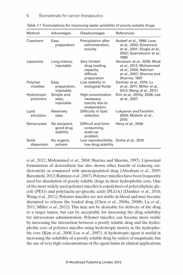
approaches are listed in Table 1.1.
One of the most widely used approaches for increasing the solubility of
poorly soluble drugs is using cosolvent systems. A poorly soluble drug is fi rst
dissolved in an organic solvent, e.g., Cremophor EL for paclitaxel and polysor-
bate for docetaxel (Loos et al ., 2003; Singla et al ., 2002; Sparreboom et al ., 1999). The solution is diluted with aqueous solution before administration by
intravenous (IV) administration. Dilution in water usually results in precipi-
tation of a poorly soluble drug, but the solution can remain stable at least for
several hours for administration without any problem. Liposome formula-
tions have also been used widely in delivering poorly soluble drugs (Modi
6 Biomaterials for cancer therapeutics
© Woodhead Publishing Limited, 2013
et al ., 2012; Mohammed et al ., 2004; Sharma and Sharma, 1997). Liposomal
formulation of doxorubicin has also shown other benefi t of reducing car-
diotoxicity as compared with unencapsulated drug (Abraham et al ., 2005;
Barenholz, 2012; Rahman et al ., 2007). Polymer micelles have been frequently
used for dissolution of poorly soluble drugs in their hydrophobic core. One
of the most widely used polymer micelles is copolymers of poly(ethylene gly-
col) (PEG) and poly(lactic-go-glycolic acid) (PLGA) (Danhier et al ., 2010;
Wang et al ., 2011). Polymer micelles are not stable in blood and may become
disrupted to release the loaded drug (Chen et al ., 2008a, 2008b; Lu et al ., 2011; Miller et al ., 2012). This may not be desirable for delivery of the drug
to a target tumor, but can be acceptable for increasing the drug solubility
for intravenous administration. Polymer micelles can become more stable
by increasing the interaction between a poorly soluble drug and the hydro-
phobic core of polymer micelles using hydrotropic moiety in the hydropho-
bic core (Kim et al ., 2008; Lee et al ., 2007). A hydrotropic agent is useful in
increasing the solubility of a poorly soluble drug by orders of magnitude, but
the use of very high concentrations of the agent limits its clinical applications
Table 1.1 Formulations for improving water solubility of poorly soluble drugs
Method Advantages Disadvantages References
Cosolvent Easy
preparation
Precipitation after
administration,
toxicity
Avdeef et al ., 1999; Loos
et al ., 2003; Simamora
et al ., 2001; Singla et al .,
2002; Sparreboom et al .,
1999
Liposome Long history,
injectable
Very limited
drug loading
capacity,
diffi cult
preparation
Abraham et al ., 2005; Modi
et al ., 2012; Mohammed
et al ., 2004; Rahman
et al ., 2007; Sharma and
Sharma, 1997
Polymer
micelles
Easy
preparation,
injectable
Low stability in
biological fl uids
Danhier et al ., 2010; Lu
et al ., 2011; Miller et al .,
2012; Wang et al ., 2011)
Hydrotropic
polymers
Simple and
easy,
injectable
High concentration
necessary,
toxicity due to
coabsorption
Kim et al ., 2010a, 2008; Lee
et al ., 2007
Lipid
emulsion
Relatively
easy
Diffi culty in lipid
selection
Lukyanov and Torchilin,
2004; M ü llertz et al .,
2010
Nanocrystal No excipient,
good drug
stability
Diffi cult and time-
consuming,
scale-up
problem
Hecq et al ., 2006
Solid
dispersion
No organic
solvent
Low reproducibility,
low drug stability
Sinha et al ., 2010
Introduction to biomaterials for cancer therapeutics 7
© Woodhead Publishing Limited, 2013
(Kim et al ., 2010a). This problem can be alleviated by using polymeric
hydrotropic agents. There are hydrotropic agents that increase the drug solu-
bility regardless of the hydrophobicity of the drug, but they are not tested
for their safety, i.e., they do not have GRAS status, making the clinical for-
mulations diffi cult. Lipid emulsion approaches exploit the hydrophobicity of
poorly soluble drugs and a variety of lipids are available for formulation of
poorly soluble drugs (Lukyanov and Torchilin, 2004; M ü llertz et al ., 2010).
The water solubility of poorly soluble drugs can also be increased by
making drug crystals into nanosize (Hecq et al ., 2006). The huge increase
in surface area results in several-fold increase in water solubility. In some
cases, this increase is enough to make a drug effi cacious and clinically viable.
The biggest advantage of using drug nanocrystals is that the drug loading
is close to 100%, even if the surface is modifi ed for improved absorption
to target tumor cells. Solid dispersion approach is based on the formation
of amorphous drug particles by rapidly cooling from the temperature that
melts both drug and polymer (Sinha et al ., 2010; Zhao et al ., 2011). Solid
dispersions are usually for oral administration of poorly soluble drugs, but
they can be applied for intravenous administration if they are prepared in
nanoscale with a controllable size and size distribution.
Cremophor EL and polysorbate were used in the development of clini-
cal products, Taxol ® and Taxotere ® , respectively. Both Cremophor EL and
polysorbate are not safe excipients. Patients must be pretreated with dex-
amethasone to avoid anaphylactic reactions by the excipients themselves.
Paclitaxel and docetaxel simply did not dissolve well in GRAS materials
known at the time of development. Despite the risks associated with using
the materials being real, the benefi t was far greater, justifying the approval
by the FDA. Development of paclitaxel/Cremophor EL and docetaxel/
polysorbate formulations was possible, because big pharmaceutical com-
panies had the incentive and resources to test them in humans. If a new
chemical entity shows unprecedented anticancer effi cacy, but has no suit-
able medium to make a formulation suitable for clinical use, then it is justifi -
able to develop and test new materials in humans even though they have
not been used before. The decision whether a new material will be tested or
not, however, depends on the pharmaceutical companies that must provide
huge resources necessary for clinical studies. This is a dilemma that drug-
delivery scientists are facing. This may be one of the main factors that slow
down the development of new drug-delivery systems.
1.3.2 Formulation for new types of anticancer agents
There are many anticancer drugs that are not small molecules and not
poorly soluble. A new class of anticancer agents includes antibodies, DNA,
siRNA, and vaccines. These molecules are not only hydrophilic, but also
8 Biomaterials for cancer therapeutics
© Woodhead Publishing Limited, 2013
very large. Proteins also need to maintain their tertiary structures to be
functional. This may be a challenge if a sustained release formulation is to
be prepared utilizing excipients such as PLGA, which degrades by hydro-
lysis although it is not water-soluble. It dissolves only in organic solvents,
and making PLGA-based sustained release formulations requires exposure
of proteins to an organic solvent, more precisely to a water‒solvent inter-
face, that can potentially denature the protein and thus its function (Tran
et al ., 2012). Delivery of nucleic acids is different from delivery of proteins,
because of the presence of the high charge density on their surface. Such
ionized macromolecules cannot cross the cell membrane, and thus they are
often condensed by positively charged polymers. The compact complex can
then be endocytosed. The nucleic acid will then have to dissociate itself from
the complex and escape from the endosome to carry out its function (Won
et al ., 2009). Both proteins and nucleic acids need to be delivered in a sus-
tained manner. On the other hand, vaccines may have to be delivered as
a bolus at certain intervals (Aki and Mooney, 2011). This type of pulsatile
delivery may require unique formulations and currently it is not easy to
prepare such systems.
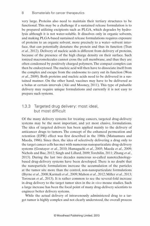
1.3.3 Targeted drug delivery: most ideal, but most diffi cult
Of the many delivery systems for treating cancers, targeted drug-delivery
systems may be the most important, and yet most elusive, formulations.
The idea of targeted delivery has been applied mainly to the delivery of
anticancer drugs to tumors. The concept of the enhanced permeation and
retention (EPR) effect was fi rst described in the 1980s (Matsumura and
Maeda, 1986). Since then, the idea of selectively delivering a drug only to
the target cancer cells has met with numerous nanoparticulate drug-delivery
systems (Goutayer et al ., 2010; Hamaguchi et al ., 2005; Maeda et al ., 2009;
Nichols and Bae, 2012; Singh and Lillard, 2009; Torchilin, 2011; Zhang et al ., 2013). During the last two decades numerous so-called nanotechnology-
based drug-delivery systems have been developed. There is no doubt that
the nanoparticle formulations increase the accumulation of the particles
at the tumor site more than the control, non-nanoparticulate formulations
(Byrne et al ., 2008; Karmali et al ., 2009; Mahon et al ., 2012; Miller et al ., 2013;
Torosean et al ., 2013). It is rather common to see the several-fold increase
in drug delivery to the target tumor sites in the in vivo mouse studies. Such
a large increase has been the focal point of many drug-delivery scientists to
engineer better delivery systems.
While the actual delivery of intravenously administered drug to a tar-
get tumor is highly complex and not clearly understood, the overall process

Introduction to biomaterials for cancer therapeutics 9
© Woodhead Publishing Limited, 2013
can be simplifi ed as shown in Fig. 1.1. The intravenously administered
nanovehicles circulate in the blood and extravasate into the surrounding
tissue when they reach a region where a tumor is growing and leaky blood
vessels are common. Since the nanovehicles can accumulate more as they
circulate in the blood longer, extended circulation has been attempted and
the current approach is to graft the nanovehicle surface with PEG, which
is known as PEGylation (Choi et al ., 2009; Pasut and Veronese, 2012; Zhu
et al ., 2010). Nanovehicles can also be used as an embolizing agent, blocking
the blood fl ow to a tumor site. For any nanovehicles to achieve their goal
of killing tumor cells, they must extravasate from blood vessels into the sur-
rounding tissue. Nanovehicles need to be made as small as possible for effi -
cient extravasation. For effi cient extravasation, nanovehicle surfaces can be
engineered to be adhesive to the surface endothelial cells, allowing them to
tumble over the endothelial cell surface until extravasation into a surround-
ing tissue. A mild increase in the local temperature, i.e., mild hyperthermia,
is known to increase the extravasation process (Li et al ., 2013).
Co-solvents
Drug formulations
IV administration
Drug crystals
Nanovehicles
Blood circulationPEGylation
Embolization
Magnetic field,Mild hyperthermia
Diffusion
Nanovehicle–tumorinteraction
Extravasation
Size <200 nm,adhesion to endothelial cells,
mild hyperthermiaTumor
Temperature-sensitivepH-sensitive
Efficient drug release
Photothermal treatment
Endocytosis,escape from endosome
Imaging
Liposomes, polymer micelles, emulsions
1.1 Simplifi ed description of nanovehicle delivery to tumor after
intravenous administration. The administered nanovehicles must go
through multiple steps to reach a target tumor. Effective nanovehicles
need to possess extra functions to release a drug at the right time
and place.

10 Biomaterials for cancer therapeutics
© Woodhead Publishing Limited, 2013
The extravasated nanovehicles must diffuse to a target tumor, and a mild
increase in temperature enhances penetration through the tissue surround-
ing a tumor. Magnetic force may be helpful for enhanced diffusion and nat-
urally magnetic nanovehicles are expected to be useful (Fang et al ., 2012;
Klostergaard and Seeney, 2012), but the magnetic approach has not been
shown to be effective or practical to date. Nanovehicles can be designed
to release a drug at a faster rate near a tumor and this can be achieved by
making the nanovehicles temperature- or pH-sensitive (Felber et al ., 2012;
Kang et al ., 2003; Wang et al ., 2013). If the surface of nanovehicles is modi-
fi ed with a ligand that binds to a receptor on the cancer cell surface, they
are endocytosed more effi ciently (Elias et al ., 2013; Mahon et al ., 2012; Wang
et al ., 2013b). The endocytosed nanovehicles must escape from endosome to
be effective (Won et al ., 2009).
While the nanoparticulate drug-delivery systems have been useful in
targeted delivery that provides signifi cant increase in drug accumulation,
the overall picture of the targeted drug delivery needs to be considered.
It is common to describe the increase in the amount of drug delivered to
a target tumor in terms of percentage increase, e.g., 100–300% increase
(Tinkov et al ., 2010; Zhu et al ., 2010). These are impressive increases, but
they must be understood in the context of the actual amount of the drug
delivered. Usually, the total amount of the drug delivered to a target tumor
by control (e.g., non-particulate) formulation is about 1–2% of the total
dose administered (Hamaguchi et al ., 2005; Wang et al ., 2013b). In this
situation, even if a 300% increase in drug accumulation at a target tumor
is observed by a particulate formulation, it still accounts for only around
5% of the total administered drug (Bae and Park, 2011). The majority of
the administered drug is still distributed to non-tumor sites. This simple
fact is often not considered. Even a small increase in the amount of a
drug delivered to a target tumor can be a viable treatment option, if the
particulate drug-delivery systems are able to cure cancer or allow patients
to live longer with no serious side effects. Unfortunately, however, this has
not been the case.
In most of the in vivo animal experiments, the decrease in the tumor mass
is measured after administration of control and test formulations. The study
is done for a month or so, the decrease in tumor size by the test formulation
becomes apparent, and this is the end point of the study. If the targeted drug
delivery is really effective, the tumor size should decrease close to zero but
this is seldom observed in the literature. If the formulation is administered
beyond 1–2 months of the test period, the animal often dies, because it is
likely that the toxicity of the anti-tumor drug has not been completely elimi-
nated. Thus, caution is necessary when we test the in vivo effi cacy of new tar-
geted drug-delivery systems. We must go beyond the fi rst several weeks of
data and thoroughly characterize toxicity profi les. Furthermore, the animal
Introduction to biomaterials for cancer therapeutics 11
© Woodhead Publishing Limited, 2013
species is a signifi cant factor for in vivo toxicology because of the variable
pharmacokinetics in different species. While mice are most commonly used,
large animal models (e.g., dog and pig) should be used because they gener-
ally mimic humans more faithfully and often meet FDA criteria for progress-
ing to human clinical studies (van der Laan et al ., 2010). Reducing toxicity
alone is a signifi cant step toward treating patients. The Doxil ® formulation
(doxorubicin in liposome formulation) is a case in point (James et al ., 1994).
It did not improve the effi cacy of doxorubicin, but it substantially reduced
cardiotoxicity, and this in itself is signifi cant enough to be approved for clini-
cal applications (Barenholz, 2012).
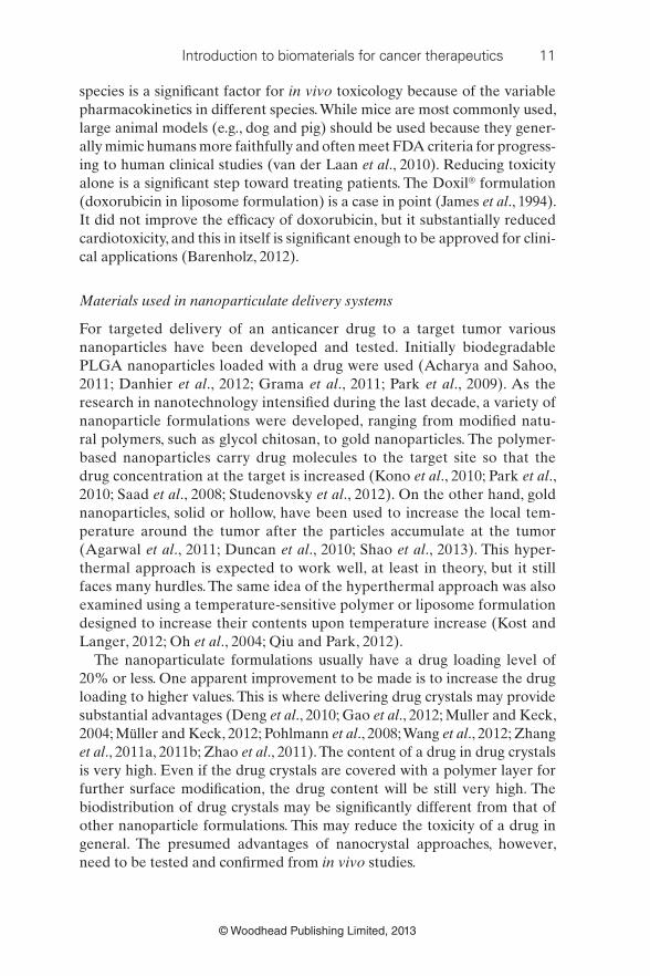
Materials used in nanoparticulate delivery systems
For targeted delivery of an anticancer drug to a target tumor various
nanoparticles have been developed and tested. Initially biodegradable
PLGA nanoparticles loaded with a drug were used (Acharya and Sahoo,
2011; Danhier et al ., 2012; Grama et al ., 2011; Park et al ., 2009). As the
research in nanotechnology intensifi ed during the last decade, a variety of
nanoparticle formulations were developed, ranging from modifi ed natu-
ral polymers, such as glycol chitosan, to gold nanoparticles. The polymer-
based nanoparticles carry drug molecules to the target site so that the
drug concentration at the target is increased (Kono et al ., 2010; Park et al ., 2010; Saad et al ., 2008; Studenovsky et al ., 2012). On the other hand, gold
nanoparticles, solid or hollow, have been used to increase the local tem-
perature around the tumor after the particles accumulate at the tumor
(Agarwal et al ., 2011; Duncan et al ., 2010; Shao et al ., 2013). This hyper-
thermal approach is expected to work well, at least in theory, but it still
faces many hurdles. The same idea of the hyperthermal approach was also
examined using a temperature-sensitive polymer or liposome formulation
designed to increase their contents upon temperature increase (Kost and
Langer, 2012; Oh et al ., 2004; Qiu and Park, 2012).
The nanoparticulate formulations usually have a drug loading level of
20% or less. One apparent improvement to be made is to increase the drug
loading to higher values. This is where delivering drug crystals may provide
substantial advantages (Deng et al ., 2010; Gao et al ., 2012; Muller and Keck,
2004; M ü ller and Keck, 2012; Pohlmann et al ., 2008; Wang et al ., 2012; Zhang
et al ., 2011a, 2011b; Zhao et al ., 2011). The content of a drug in drug crystals
is very high. Even if the drug crystals are covered with a polymer layer for
further surface modifi cation, the drug content will be still very high. The
biodistribution of drug crystals may be signifi cantly different from that of
other nanoparticle formulations. This may reduce the toxicity of a drug in
general. The presumed advantages of nanocrystal approaches, however,
need to be tested and confi rmed from in vivo studies.
12 Biomaterials for cancer therapeutics
© Woodhead Publishing Limited, 2013
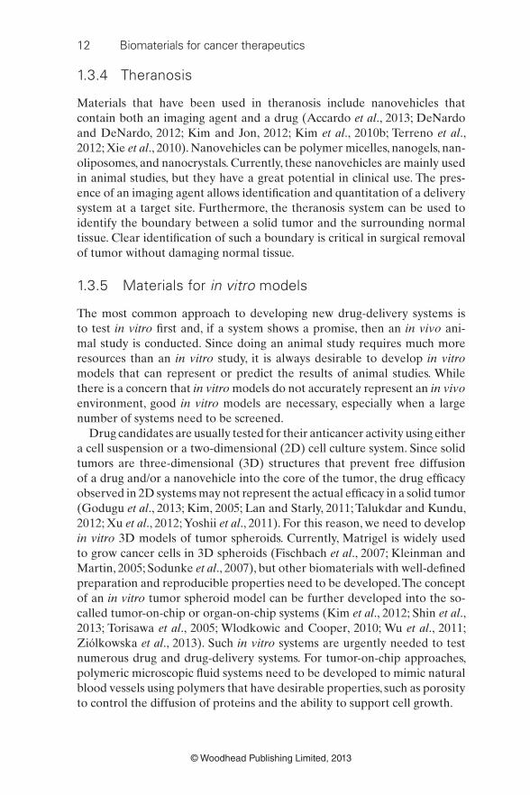
1.3.4 Theranosis
Materials that have been used in theranosis include nanovehicles that
contain both an imaging agent and a drug (Accardo et al ., 2013; DeNardo
and DeNardo, 2012; Kim and Jon, 2012; Kim et al ., 2010b; Terreno et al ., 2012; Xie et al ., 2010). Nanovehicles can be polymer micelles, nanogels, nan-
oliposomes, and nanocrystals. Currently, these nanovehicles are mainly used
in animal studies, but they have a great potential in clinical use. The pres-
ence of an imaging agent allows identifi cation and quantitation of a delivery
system at a target site. Furthermore, the theranosis system can be used to
identify the boundary between a solid tumor and the surrounding normal
tissue. Clear identifi cation of such a boundary is critical in surgical removal
of tumor without damaging normal tissue.
1.3.5 Materials for in vitro models
The most common approach to developing new drug-delivery systems is
to test in vitro fi rst and, if a system shows a promise, then an in vivo ani-
mal study is conducted. Since doing an animal study requires much more
resources than an in vitro study, it is always desirable to develop in vitro
models that can represent or predict the results of animal studies. While
there is a concern that in vitro models do not accurately represent an in vivo
environment, good in vitro models are necessary, especially when a large
number of systems need to be screened.
Drug candidates are usually tested for their anticancer activity using either
a cell suspension or a two-dimensional (2D) cell culture system. Since solid
tumors are three-dimensional (3D) structures that prevent free diffusion
of a drug and/or a nanovehicle into the core of the tumor, the drug effi cacy
observed in 2D systems may not represent the actual effi cacy in a solid tumor
(Godugu et al ., 2013; Kim, 2005; Lan and Starly, 2011; Talukdar and Kundu,
2012; Xu et al ., 2012; Yoshii et al ., 2011). For this reason, we need to develop
in vitro 3D models of tumor spheroids. Currently, Matrigel is widely used
to grow cancer cells in 3D spheroids (Fischbach et al ., 2007; Kleinman and
Martin, 2005; Sodunke et al ., 2007), but other biomaterials with well-defi ned
preparation and reproducible properties need to be developed. The concept
of an in vitro tumor spheroid model can be further developed into the so-
called tumor-on-chip or organ-on-chip systems (Kim et al ., 2012; Shin et al ., 2013; Torisawa et al ., 2005; Wlodkowic and Cooper, 2010; Wu et al ., 2011;
Zi ó lkowska et al ., 2013). Such in vitro systems are urgently needed to test
numerous drug and drug-delivery systems. For tumor-on-chip approaches,
polymeric microscopic fl uid systems need to be developed to mimic natural
blood vessels using polymers that have desirable properties, such as porosity
to control the diffusion of proteins and the ability to support cell growth.
Introduction to biomaterials for cancer therapeutics 13
© Woodhead Publishing Limited, 2013
1.4 Conclusion and future trends
Successful treatment of cancers requires development of effective drugs
and proper delivery systems that can deliver drugs selectively to target cells.
To make signifi cant progress in cancer chemotherapy, studies must be more
critical. The data obtained from in vivo animal studies need to be inter-
preted more carefully and comparison of multiple species is ideal to better
model human clinical conditions. Some formulations may show signifi cant
increase in the drug accumulated at the target site, but the effi cacy needs to
be examined in terms of its ability to shrink tumor size long term without
toxic side effects. The data observed in a single animal species (e.g., mouse)
cannot be generalized or extrapolated to humans with confi dence.
To translate the technology obtained from in vitro and in vivo animal
studies to clinical application, the current approaches need to be re-
examined, in particular using biomaterials to make nanoparticles. Better
in vitro model systems must be developed to mimic in vivo conditions
more accurately, and more importantly, more relevant to humans. In addi-
tion, better delivery systems need to be developed. Simply using a nano-
sized delivery system is not enough to achieve true targeted drug delivery.
The information currently available from the decade-long research on
nanovehicles is a good starting point to design novel biomaterials ideally
suited for treating cancers.
1.5 References Abraham, S. A., D. N. Waterhouse, L. D. Mayer, P. R. Cullis, T. D. Madden and M. B.
Bally (2005). The liposomal formulation of doxorubicin. Method Enzymol 391 ,
71–96.
Accardo, A., D. Tesauro and G. Morelli (2013). Peptide-based targeting strategies for
simultaneous imaging and therapy with nanovectors. Polym J 19 , 1–13.
Acharya, S. and S. K. Sahoo (2011). PLGA nanoparticles containing various anti-
cancer agents and tumour delivery by EPR effect. Adv Drug Deliv Rev 63 (3),
170–183.
Agarwal, A., M. A. Mackey, M. A. El-Sayed and R. V. Bellamkonda (2011). Remote
triggered release of doxorubicin in tumors by synergistic application of thermo-
sensitive liposomes and gold nanorods. ACS Nano 5 (6), 4919–4926.
Ali, O. A. and D. J. Mooney (2011). Immunologically active biomaterials for cancer
therapy. Curr Top Microbiol Immunol 344 , 279–297.
Avdeef, A., K. J. Box, J. E. A. Comer, M. Gilges, M. Hadley, C. Hibbert, W. Patterson
and K. Y. Tam (1999). pH-metric log P 11. pK a determination of water-
insoluble drugs in organic solvent–water mixtures. J Pharm Biomed Anal 20 (4), 631–641.
Bae, Y. H. and K. Park (2011). Targeted drug delivery to tumors: Myths, reality, and
possibility. J Control Release 153 , 198–205.
Barenholz, Y. (2012). Doxil® – The fi rst FDA-approved nano-drug: Lessons learned.
J.Control Release 160 (2), 117–134.

14 Biomaterials for cancer therapeutics
© Woodhead Publishing Limited, 2013
Byrne, J. D., T. Betancourt and L. Brannon-Peppas (2008). Active targeting schemes
for nanoparticle systems in cancer therapeutics. Adv Drug Deliv Rev 60 (15),
1615–1626.
Chen, H., S. Kim, W. He, H. Wang, P. S. Low, K. Park and J. X. Cheng (2008a). Fast
release of lipophilic agents from circulating PEG-PDLLA micelles revealed by
in vivo f ö rster resonance energy transfer imaging. Langmuir 24 , 5213–5217.
Chen, H., S. Kim, L. Li, S. Wang, K. Park and J. X. Cheng (2008b). Release of
hydrophobic molecules from polymer micelles into cell membranes revealed
by F ö rster resonance energy transfer imaging. Proc Natl Acad Sci USA 105 ,
6596–6601.
Choi, J. S., H. J. Yang, B. S. Kim, J. D. Kim, J. Y. Kim, B. Yoo, K. Park, H. Y. Lee and
Y. W. Cho (2009). Human extracellular matrix (ECM) powders for injectable
cell delivery and adipose tissue engineering. J Control Release 139 (1), 2–7.
Danhier, F., E. Ansorena, J. M. Silva, R. Coco, A. Le Breton and V. Pr é at (2012).
PLGA-based nanoparticles: An overview of biomedical applications. J Control Release 161 (2), 505–522.
Danhier, F., B. Ucakar, N. Magotteaux, M. E. Brewster and V. Pr é at (2010). Active
and passive tumor targeting of a novel poorly soluble cyclin dependent kinase
inhibitor. Int J Pharm 392 (1–2), 20–28.
DeNardo, G. L. and S. J. DeNardo (2012). Concepts, consequences, and implications
of theranosis. Sem Nucl Med 42 (3), 147–150.
Deng, J., L. Huang and F. Liu (2010). Understanding the structure and stability of
paclitaxel nanocrystals. Int J Pharm 390 (2), 242–249.
Duncan, B., C. Kim and V. M. Rotello (2010). Gold nanoparticle platforms as drug
and biomacromolecule delivery systems. J Control Release 148 (1), 122–127.
Elias, D. R., A. Poloukhtine, V. Popik and A. Tsourkas (2013). Effect of ligand den-
sity, receptor density, and nanoparticle size on cell targeting. Nanomedicine
9 (2), 194–201.
Fang, C., F. M. Kievit, O. Veiseh, Z. R. Stephen, T. Wang, D. Lee, R. G. Ellenbogen and
M. Zhang (2012). Fabrication of magnetic nanoparticles with controllable drug
loading and release through a simple assembly approach. J Control Release
162 (1), 233–241.
Felber, A. E., M. -H. Dufresne and J. -C. Leroux (2012). pH-sensitive vesicles, poly-
meric micelles, and nanospheres prepared with polycarboxylates. Adv Drug Deliv Rev 64 (11), 979–992.
Fischbach, C., R. Chen, T. Matsumoto, T. Schmelzle, J. S. Brugge, P. J. Polverini and
D. J. Mooney (2007). Engineering tumors with 3D scaffolds. Nat Methods 4 (10),
855–860.
Gao, L., G. Liu, J. Ma, X. Wang, L. Zhou and X. Li (2012). Drug nanocrystals: In vivo
performances. J Control Release 160 (3), 418–430.
Gilman, A. (1963). The initial clinical trial of nitrogen mustard. Am J Surg 105 (5),
574–578.
Godugu, C., A. R. Patel, U. Desai, T. Andey, A. Sams and M. Singh (2013). Algimatrix TM
based 3D cell culture system as an in-vitro tumor model for anticancer studies.
PLoS One 8 (1), 1–13.
Goutayer, M., S. Dufort, V. Josserand, A. Roy è re, E. Heinrich, F. Vinet, J. Bibette, J. L.
Coll and I. Texier (2010). Tumor targeting of functionalized lipid nanoparticles:
Assessment by in-vivo fl uorescence imaging. Eur J Pharm Biopharm 75 (2), 137–147.

Introduction to biomaterials for cancer therapeutics 15
© Woodhead Publishing Limited, 2013
Grama, C. N., D. D. Ankola and M. N. V. R. Kumar (2011). Poly(lactide-co-glycolide)
nanoparticles for peroral delivery of bioactives. Curr Opin Colloid Interface Sci 16 (3), 238–245.
Hamaguchi, T., Y. Matsumura, M. Suzuki, K. Shimizu, R. Goda, I. Nakamura, I.
Nakatomi, M. Yokoyama, K. Kataoka and T. Kakizoe (2005). NK105, a paclitaxel-
incorporating micellar nanoparticle formulation, can extend in vivo antitumour
activity and reduce the neurotoxicity of paclitaxel. Br J Cancer 92 , 1240–1246.
Hecq, J., M. Deleers, D. Fanara, H. Vranckx, P. Boulanger, S. Le Lamer and K. Amighi
(2006). Preparation and in vitro / in vivo evaluation of nano-sized crystals for
dissolution rate enhancement of ucb-35440–3, a highly dosed poorly water-
soluble weak base. Eur J Pharm Biopharm 64 (3), 360–368.
Deeley, T. J. (1983). A brief history of cancer. Clin Radiol 34 , 597–608.
James, N. D., R. J. Coker, D. Tomlinson, J. R. Harris, M. Gompels, A. J. Pinching and
J. S. Stewart (1994). Liposomal doxorubicin (doxil): An effective new treatment
for kaposi’s sarcoma in AIDS. Clin Oncol 6 , 294–296.
Kang, S. I., K. Na and Y. H. Bae (2003). Physicochemical characteristics and doxoru-
bicin-release behaviors of pH/temperature-sensitive polymeric nanoparticles.
Colloids Surf A Physicochem Eng Asp 231 (1–3), 103–112.
Karmali, P. P., V. R. Kotamraju, M. Kastantin, M. Black, D. Missirlis, M. Tirrell and E.
Ruoslahti (2009). Targeting of albumin-embedded paclitaxel nanoparticles to
tumors. Nanomedicine 5 (1), 73–82.
Kim, D. and S. Jon (2012). Gold nanoparticles in image-guided cancer therapy.
Inorganica Chim Acta 393 , 154–164.
Kim, J. B. (2005). Three-dimensional tissue culture models in cancer biology. Semin Cancer Biol 15 (5), 365–377.
Kim, J. Y., S. Kim, M. Papp, K. Park and R. Pinal (2010a). Hydrotropic solubilization
of poorly water-soluble drugs. J Pharm Sci 99 (9), 3953–3965.
Kim, K., J. H. Kim, H. Park, Y. S. Kim, K. Park, H. Nam, S. Lee, J. H. Park, R. W. Park, I.
S. Kim, K. Choi, S. Y. Kim, K. Park and I. C. Kwon (2010b). Tumor-homing mul-
tifunctional nanoparticles for cancer theragnosis: Simultaneous diagnosis, drug
delivery, and therapeutic monitoring. J Control Release 146 (2), 219–227.
Kim, S., J. Y. Kim, K. M. Huh, G. Acharya and K. Park (2008). Hydrotropic polymer
micelles containing acrylic acid moieties for oral delivery of paclitaxel. J Control Release 132 (3), 222–229.
Kim, T., I. Doh and Y. -H. Cho (2012). A 3D tumor spheroid chip with the pharmaco-
kinetic drug elimination model developed by balanced droplet dispensing. Sens Actuators B Chem 174 , 436–440.
Klein, S. and A. Levitzki (2007). Targeted cancer therapy: Promise and reality. In:
Advances in Cancer Research, F. V. W. George and K. George (eds). Academic
Press. 295–319.
Kleinman, H. K. and G. R. Martin (2005). Matrigel: Basement membrane matrix
with biological activity. Semin Cancer Biol 15 , 378–386.
Klostergaard, J. and C. E. Seeney (2012). Magnetic nanovectors for drug delivery.
Nanomedicine 8 (1), S37–S50.
Kono, K., T. Ozawa, T. Yoshida, F. Ozaki, Y. Ishizaka, K. Maruyama, C. Kojima,
A. Harada and S. Aoshima (2010). Highly temperature-sensitive liposomes
based on a thermosensitive block copolymer for tumor-specifi c chemotherapy.
Biomaterials 31 (27), 7096–7105.

16 Biomaterials for cancer therapeutics
© Woodhead Publishing Limited, 2013
Kost, J. and R. Langer (2012). Responsive polymeric delivery systems. Adv Drug Deliv Rev 64 , Supplement, 327–341.
Lan, S. -F. and B. Starly (2011). Alginate based 3D hydrogels as an in vitro co-culture
model platform for the toxicity screening of new chemical entities. Toxicol Appl Pharmacol 256 (1), 62–72.
Lee, S. C., K. M. Huh, J. Lee, Y. W. Cho, R. E. Galinsky and K. Park (2007). Hydrotropic
polymeric micelles for enhanced paclitaxel solubility: In vitro and in vivo char-
acterization. Biomacromolecules 8 (1), 202–208.
Li, L., T. L. ten Hagen, M. Bolkestein, A. Gasselhuber, J. Yatvin, G. C. van Rhoon,
A. M. Eggermont, D. Haemmerich and G. A. Koning (2013). Improved intra-
tumoral nanoparticle extravasation and penetration by mild hyperthermia. J Control Release 167 (2), 130–137.
Loos, W. J., S. D. Baker, J. Verweij, J. G. Boonstra and A. Sparreboom (2003). Clinical
pharmacokinetics of unbound docetaxel: Role of polysorbate 80 and serum
proteins. Clin Pharmacol Ther 74 (4), 364–371.
Lu, J., S. C. Owen and M. S. Shoichet (2011). Stability of self-assembled polymeric
micelles in serum. Macromolecules 44 , 6002–6008.
Lukyanov, A. N. and V. P. Torchilin (2004). Micelles from lipid derivatives of water-
soluble polymers as delivery systems for poorly soluble drugs. Adv Drug Deliv Rev 56 (9), 1273–1289.
Maeda, H., G. Y. Bharate and J. Daruwalla (2009). Polymeric drugs for effi cient
tumor-targeted drug delivery based on EPR-effect. Eur J Pharm Biopharm
71 (3), 409–419.
Mahon, E., A. Salvati, F. Baldelli Bombelli, I. Lynch and K. A. Dawson (2012).
Designing the nanoparticle – biomolecule interface for ‘ targeting and thera-
peutic delivery’. J Control Release 161 (2), 164–174.
Matsumura, Y. and H. Maeda (1986). A new concept for macromolecular therapeu-
tics in cancer chemotherapy: Mechanism of tumoritropic accumulation of pro-
teins and the antitumor agent smancs. Cancer Res 46 , 6387–6392.
Miller, T., S. Breyer, G. van Colen, W. Mier, U. Haberkorn, S. Geissler, S. Voss, M.
Weigandt and A. Goepferich (2013). Premature drug release of polymeric
micelles and its effects on tumor targeting. Int J Pharm 445 (1–2), 117–124.
Miller, T., R. Rachel, A. Besheer, S. Uezguen, M. Weigandt and A. Goepferich (2012).
Comparative investigations on in vitro serum stability of polymeric micelle for-
mulations. Pharm Res 29 , 448–459.
Modi, S., T. -X. Xiang and B. D. Anderson (2012). Enhanced active liposomal load-
ing of a poorly soluble ionizable drug using supersaturated drug solutions. J Control Release 162 (2), 330–339.
Mohammed, A. R., N. Weston, A. G. Coombes, M. Fitzgerald and Y. Perrie (2004).
Liposome formulation of poorly water soluble drugs: Optimisation of drug
loading and ESEM analysis of stability. Int J Pharm 285 (1–2), 23–34.
Muller, R. H. and C. M. Keck (2004). Challenges and solutions for the delivery of
biotech drugs – a review of drug nanocrystal technology and lipid nanopar-
ticles. J Biotechnol 113 (1–3), 151–170.
M ü ller, R. H. and C. M. Keck (2012). Twenty years of drug nanocrystals: Where are
we, and where do we go? Eur J Pharm Biopharm 80 , 1–3.
M ü llertz, A., A. Ogbonna, S. Ren and T. Rades (2010). New perspectives on lipid and
surfactant based drug delivery systems for oral delivery of poorly soluble drugs.
J Pharm Pharm 62 , 1622–1636.

Introduction to biomaterials for cancer therapeutics 17
© Woodhead Publishing Limited, 2013
Nichols, J. W. and Y. H. Bae (2012). Odyssey of a cancer nanoparticle: From injection
site to site of action. Nano Today 7 (6), 606–618.
Oh, K. S., S. K. Han, Y. W. Choi, J. H. Lee, J. Y. Lee and S. H. Yuk (2004). Hydrogen-
bonded polymer gel and its application as a temperature-sensitive drug deliv-
ery system. Biomaterials 25 (12), 2393–2398.
Park, J., P. M. Fong, J. Lu, K. S. Russell, C. J. Booth, W. M. Saltzman and T. M. Fahmy
(2009). PEGylated PLGA nanoparticles for the improved delivery of doxoru-
bicin. Nanomedicine 5 (4), 410–418.
Park, J. H., G. Saravanakumar, K. Kim and I. C. Kwon (2010). Targeted delivery of
low molecular drugs using chitosan and its derivatives. Adv Drug Deliv Rev
62 (1), 28–41.
Pasut, G. and F. M. Veronese (2012). State of the art in PEGylation: The great versa-
tility achieved after forty years of research. J Control Release 161 (2), 461–472.
Pohlmann, A. R., G. Mezzalira, C. Venturini, G. de, L. Cruz, A. Bernardi, E. J ä ger, A.
M. Battastini, N. P. da Silveira and S. S. Guterres (2008). Determining the simul-
taneous presence of drug nanocrystals in drug-loaded polymeric nanocapsule
aqueous suspensions: A relation between light scattering and drug content. Int J Pharm 359 (1–2), 288–293.
Qiu, Y. and K. Park (2012). Environment-sensitive hydrogels for drug delivery. Adv Drug Deliv Rev 64 , Supplement, 49–60.
Rahman, A. M., S. W. Yusuf and M. S. Ewer (2007). Anthracycline-induced car-
diotoxicity and the cardiac-sparing effect of liposomal formulation. Int J Nanomedicine 2 (4), 567–583.
Saad, M., O. B. Garbuzenko, E. Ber, P. Chandna, J. J. Khandare, V. P. Pozharov and
T. Minko (2008). Receptor targeted polymers, dendrimers, liposomes: Which
nanocarrier is the most effi cient for tumor-specifi c treatment and imaging? J Control Release 130 (2), 107–114.
Shao, J., R. J. Griffi n, E. I. Galanzha, J. W. Kim, N. Koonce, J. Webber, T. Mustafa, A.
S. Biris, D. A. Nedosekin and V. P. Zharov (2013). Photothermal nanodrugs:
Potential of TNF-gold nanospheres for cancer theranostics. Sci Rep 3 (1293), 1–9.
Sharma, A. and U. S. Sharma (1997). Liposomes in drug delivery: Progress and limi-
tations. Int J Pharm 154 (2), 123–140.
Shin, C. S., B. Kwak, B. Han and K. Park (2013). Development of an in vitro 3D tumor
model to study therapeutic effi ciency of an anti-cancer drug. Mol Pharm 10(6),
2167–2175.
Simamora, P., J. M. Alvarez and S. H. Yalkowsky (2001). Solubilization of rapamycin.
Int J Pharm 213 (1–2), 25–29.
Singh, R. and J. W. Lillard Jr (2009). Nanoparticle-based targeted drug delivery. Exp Mol Pathol 86 (3), 215–223.
Singla, A. K., A. Garg and D. Aggarwal (2002). Paclitaxel and its formulations. Int J Pharm 235 (1–2), 179–192.
Sinha, S., M. Ali, S. Baboota, A. Ahuja, A. Kumar and J. Ali (2010). Solid dispersion
as an approach for bioavailability enhancement of poorly water-soluble drug
ritonavir. PharmSci Tech 11 (2), 518–527.
Sodunke, T. R., K. K. Turner, S. A. Caldwell, K. W. McBride, M. J. Reginato and H.
M. Noh (2007). Micropatterns of matrigel for three-dimensional epithelial cul-
tures. Biomaterials 28 (27), 4006–4016.
Sparreboom, A., L. van Zuylen, E. Brouwer, W. J. Loos, P. de Bruijn, H. Gelderblom,
M. Pillay, K. Nooter, G. Stoter and J. Verweij (1999). Cremophor EL-mediated

18 Biomaterials for cancer therapeutics
© Woodhead Publishing Limited, 2013
alteration of paclitaxel distribution in human blood: Clinical pharmacokinetic
implications. Cancer Res 59 , 1454–1457.
Studenovsky, M., R. Pola, M. Pechar, T. Etrych, K. Ulbrich, L. Kovar, M. Kabesova
and B. Rihova (2012). Polymer carriers for anticancer drugs targeted to EGF
receptor. Macromol Biosci 12 , 1714–1720.
Talukdar, S. and S. C. Kundu (2012). A non-mulberry silk fi broin protein based 3D in vitro tumor model for evaluation of anticancer drug activity. Adv Funct Mater
22 , 4778–4788.
Terreno, E., F. Uggeri and S. Aime (2012). Image guided therapy: The advent of ther-
anostic agents. J Control Release 161 (2), 328–337.
Tinkov, S., C. Coester, S. Serba, N. A. Geis, H. A. Katus, G. Winter and R. Bekeredjian
(2010). New doxorubicin-loaded phospholipid microbubbles for targeted tumor
therapy: In-vivo characterization. J Control Release 148 (3), 368–372.
Torchilin, V. (2011). Tumor delivery of macromolecular drugs based on the EPR
effect. Adv Drug Deliv Rev 63 (3), 131–135.
Torisawa, Y. -S., H. Shiku, T. Yasukawa, M. Nishizawa and T. Matsue (2005). Multi-
channel 3-D cell culture device integrated on a silicon chip for anticancer drug
sensitivity test. Biomaterials 26 (14), 2165–2172.
Torosean, S., B. Flynn, J. Axelsson, J. Gunn, K. S. Samkoe, T. Hasan, M. M. Doyley and
B. W. Pogue (2013). Nanoparticle uptake in tumors is mediated by the inter-
play of vascular and collagen density with interstitial pressure. Nanomedicine
9 , 151–158.
Tran, V. -T., J. P. Karam, X. Garric, J. Coudane, J. P. Beno î t, C. N. Montero-Menei
and M. C. Venier-Julienne (2012). Protein-loaded PLGA–PEG–PLGA micro-
spheres: A tool for cell therapy. Eur J Pharm Sci 45 (1–2), 128–137.
van der Laan, J. W., J. Brightwell, P. McAnulty, J. Ratky, C. Stark and Steering Group
of the RETHINK Project (2010). Regulatory acceptability of the minipig in the
development of pharmaceuticals, chemicals and other products. J Pharmacol Toxicol Methods 62 , 184–195.
Wang, A., H. Gao, Y. Sun, Y. L. Sun, Y. W. Yang, G. Wu, Y. Wang, Y. Fan and J. Ma
(2013a). Temperature- and pH-responsive nanoparticles of biocompatible
polyurethanes for doxorubicin delivery. Int J Pharm 441 , 30–39.
Wang, G. D., F. P. Mallet, F. Ricard and J. Y. Heng (2012). Pharmaceutical nanocrys-
tals. Curr Opin Chem Eng 1 (2), 102–107.
Wang, H., Y. Zhao, Y. Wu, Y. L. Hu, K. Nan, G. Nie and H. Chen (2011). Enhanced
anti-tumor effi cacy by co-delivery of doxorubicin and paclitaxel with
amphiphilic methoxy PEG-PLGA copolymer nanoparticles. Biomaterials
32 (32), 8281–8290.
Wang, Z., Y. Yu, W. Dai, J. Cui, H. Wu, L. Yuan, H. Zhang, X. Wang, J. Wang, X. Zhang
and Q. Zhang (2013b). A specifi c peptide ligand-modifi ed lipid nanoparticle
carrier for the inhibition of tumor metastasis growth. Biomaterials 34 (3),
756–764.
Winau, F., O. Westphal and R. Winau (2004). Paul Ehrlich – in search of the magic
bullet. Microbes Infect 6 (8), 786–789.
Wlodkowic, D. and J. M. Cooper (2010). Tumors on chips: Oncology meets microfl u-
idics. Curr Opin Chem Biol 14 (5), 556–567.
Won, Y. Y., R. Sharma and S. F. Konieczny (2009). Missing pieces in understanding
the intracellular traffi cking of polycation/DNA complexes. J Control Release
139 (2), 88–93.

Introduction to biomaterials for cancer therapeutics 19
© Woodhead Publishing Limited, 2013
Wu, M.-H., Y.-H. Chang, Y.-T. Liu, Y.-M. Chen, S.-S. Wang, H.-Y. Wang, C.-S. Lai and
T.-M. Pan (2011). Development of high throughput microfl uidic cell culture
chip for perfusion 3-dimensional cell culture-based chemosensitivity assay.
Sens Actuators B Chem 155 (1), 397–407.
Xie, J., S. Lee and X. Chen (2010). Nanoparticle-based theranostic agents. Adv Drug Deliv Rev 62 (11), 1064–1079.
Xu, X., L. A. Gurski, C. Zhang, D. A. Harrington, M. C. Farach-Carson and X. Jia
(2012). Recreating the tumor microenvironment in a bilayer, hyaluronic acid
hydrogel construct for the growth of prostate cancer spheroids. Biomaterials
33 (35), 9049–9060.
Yoshii, Y., A. Waki, K. Yoshida, A. Kakezuka, M. Kobayashi, H. Namiki, Y. Kuroda,
Y. Kiyono, H. Yoshii, T. Furukawa, T. Asai, H. Okazawa, J. G. Gelovani and Y.
Fujibayashi (2011). The use of nanoimprinted scaffolds as 3D culture models to
facilitate spontaneous tumor cell migration and well-regulated spheroid forma-
tion. Biomaterials 32 (26), 6052–6058.
Zhang, H., C. P. Hollis, Q. Zhang and T. Li (2011a). Preparation and antitumor study
of camptothecin nanocrystals. Int J Pharm 415 (1–2), 293–300.
Zhang, J., H. Lv, K. Jiang and Y. Gao (2011b). Enhanced bioavailability after oral
and pulmonary administration of baicalein nanocrystal. Int J Pharm 420 (1),
180–188.
Zhang, Y., H. F. Chan and K. W. Leong (2013). Advanced materials and processing
for drug delivery: The past and the future. Adv Drug Deliv Rev 65 (1), 104–120.
Zhao, R., C. P. Hollis, H. Zhang, L. Sun, R. A. Gemeinhart and T. Li (2011). Hybrid
nanocrystals: Achieving concurrent therapeutic and bioimaging functionalities
toward solid tumors. Mol Pharmaceut 8 , 1985–1991.
Zhu, S., M. Hong, G. Tang, L. Qian, J. Lin, Y. Jiang and Y. Pei (2010). Partly PEGylated
polyamidoamine dendrimer for tumor-selective targeting of doxorubicin: The
effects of PEGylation degree and drug conjugation style. Biomaterials 31 (6),
1360–1371.
Zi ó lkowska, K., A. Stelmachowska, R. Kwapiszewski, M. Chudy, A. Dybko and Z.
Brz ó zka (2013). Long-term three-dimensional cell culture and anticancer drug
activity evaluation in a microfl uidic chip. Biosens Bioelectron 40 , 68–74.