biology and chemistry of endophytes† - calisfulltext.calis.edu.cn/rsc/natural product...
TRANSCRIPT

REVIEW www.rsc.org/npr | Natural Product Reports
Biology and chemistry of endophytes†
Hua Wei Zhang,* Yong Chun Song and Ren Xiang Tan
Received (in Cambridge, UK)13th July 2006First published as an Advance Article on the web 16th August 2006DOI: 10.1039/b609472b
Covering: up to May 2006. Previous review: Nat. Prod. Rep., 2001, 18, 448
This review focuses on new endophyte-related findings in biology and ecology, and also summarises thevarious metabolites isolated from endophytes.
1 Introduction2 Biology of endophytes2.1 Major groups of endophytic microbes2.2 The origin and evolution of endophytes2.3 Host range2.4 Host and tissue specificity2.5 Isolation2.6 Identification2.7 Physiological role2.8 Ecological role3 Chemistry3.1 Endophytic bacteria3.2 Endophytic actinomycetes3.3 Endophytic fungi4 Potential applicability4.1 Plant growth enhancers4.2 Phytoprotectors5 Concluding remarks6 References
1 Introduction
The term ‘endophyte’ is an all-encompassing topographical termwhich includes all organisms that, during a variable period oftheir life, symptomlessly colonise the living internal tissues of theirhosts.1 This definition is broad enough to include virtually anyorganism residing inside a plant host. Although our knowledgeof the ecology, life history and phylogeny of endophytic fungi hasaccumulated significantly over the past two decades, fundamentalquestions regarding the evolutionary origin, speciation and eco-logical role of such endophytes remain to be answered.2 This articleis a follow-up to our previous review (Nat. Prod. Rep., 2001, 18,448–459), and it focuses particularly on new endophyte-relatedfindings in biology and ecology. It also summarises the variousmetabolites isolated from endophytes and describes these underdifferent compound classes.
Institute of Functional Biomolecules, State Key Laboratory of Pharmaceu-tical Biotechnology, Nanjing University, Nanjing, 210093, P. R. China† This paper was published as a part of a special issue on natural productschemistry in China.
2 Biology of endophytes
2.1 Major groups of endophytic microbes
Endophytic microbes fall into several identifiable classes often inrelation to their plant organ source, with the major groups asfollows: 1) endophytic Clavicipitaceae; 2) fungal endophytes ofdicots; 3) endophytic fungi; 4) other systemic fungal endophytes;5) fungal endophytes of lichens; 6) endophytic fungi of bryophytesand ferns; 7) endophytic fungi of tree bark; 8) fungal endophytesof xylem; 9) fungal endophytes of root; 10) fungal endophytesof galls and cysts; 11) prokaryotic endophytes of plants (includesendophytic bacteria and actinomycetes).1,2
2.2 The origin and evolution of endophytes
Evidence of plant-associated microorganisms found in the fos-silised tissues of stems and leaves has revealed that endophyte–hostassociations may have evolved from the time that higher plants firstappeared on the Earth.3,4 The symbiosis of fungi with plants mostprobably dates back to the emergence of vascular plants.5 Carroll6
has suggested that some phytopathogens in the environment arerelated to endophytes and have an endophytic origin. In certainenvironments, some microbes appear actively to penetrate planttissues through invading openings or wounds, as well as proactivelyusing hydrolytic enzymes such as cellulase and pectinase. Somebacterial endophytes are believed to originate from the rhizosphereor phylloplane microflora,7 through penetrating and colonisingroot tissue as an access point to the xylem.8
Majewska-Sawkaa and Gentile9 have traced the presence anddistribution of Neotyphodium lolii within developing inflores-cences and embryos of perennial ryegrass (Lolium perenne L.),cultivar ‘Grassland Nui’, by in situ immunolocalisation of fungalproteins. Evidence is presented that the fungus penetrates throughthe rachilla at the base of the ovary, and localises in a very preciseand specific manner in the ovular nucellus, but never enters theembryo sac or the integuments. Young embryos do not containmycelium, but as they mature the hyphae penetrate through thescutellum from a neighbouring ‘infection layer’—a remnant ofnucellus heavily colonised by the fungus—that is readily visible asa discrete area directly adjoining the base of embryo cavity. It wasshown that N. lolii was transmitted to the embryo exclusively viasporophytic maternal tissue. In vascular tissue, bacteria can travelthroughout the host plant and hence colonise it systemically.7
This journal is © The Royal Society of Chemistry 2006 Nat. Prod. Rep., 2006, 23, 753–771 | 753

Hua Wei Zhang, born in Anhui (1978), received his BS in Food Sciences in 2000 and his MS in Food Chemistry in 2003 from NorthwestAgriculture & Forestry University, and his PhD in Natural Products Chemistry in 2006 from Nanjing University. He is now a postdoctoralfellow at the School of Chemistry and Chemical Engineering, Nanjing University. His research interest has focused on the discovery of novelnatural products produced by endophytic microorganisms, and their biological properties.
Yong Chun Song was born in Heilongjiang Province, China. She completed her BS in Agronomy in 1994 and her MS in Microbiology in 1997at Northeast Agricultural University, and her PhD in Botany in 2000 at China Agricultural University. After spending a two-year postdoctoralresearch period in Professor Tan’s group, she joined SLSNU (School of Life Sciences, Nanjing University) as Associate Professor in 2003.Her work interest includes the isolation, cultivation and identification of endophytic microorganisms from halophytes and medicinal plants.
Ren Xiang Tan was born in Jiangsu, and received his BS in Pharmacy in 1983 and his MS in Medicinal Chemistry in 1986 from ChinaPharmaceutical University. He received his PhD in Organic Chemistry in 1990 from Lanzhou University, where he spent a two-yearpostdoctoral research period. After joining SLSNU (School of Life Sciences, Nanjing University) as Associate Professor in 1992, he waspromoted to Professor of Botany (1994), Chair Professor (1999), Department Head (1996), Vice-Dean (1999) and Associate Vice-President (2002). He has been a Visiting Scholar at the Institute of Organic Chemistry, Technical University of Berlin, Berlin, Germany(Professor F. Bohlmann), the Institute of Pharmacognosy and Phytochemistry, University of Lausanne, Lausanne, Switzerland (ProfessorK. Hostettmann) and the Institution of Oceanography, University of California, San Diego, USA (Professor W. Fenical). His work interesthas focused on the structure determination and function of biomolecules originating from medicinal plants, and especially microorganismsinhabiting these plants. He has authored some 170 scientific publications and three monographs.
Hua Wei Zhang Yong Chun Song Ren Xiang Tan
In control experiments, grazing herbivores reduced the exposedbiomass of non-host plants relative to the endophyte host fescuegrass. These results demonstrate that herbivores can drive plant–microbe dynamics and, in so doing, they can modify plantcommunity structures either directly or indirectly. In hereditarysymbioses, genomes of both partners are co-inherited.10 Therefore,these symbionts are linked directly to evolutionary changes in theirhost populations. Hereditary symbionts are transmitted acrossgenerations through eggs, seeds, or clonal propagules, and rarelythrough sperm. They include a diversity of interactions and areespecially well known in arthropods and their obligate associationswith vertically transmitted bacteria.10
During the long co-evolution of endophytes and their hostplants, endophytes have adapted themselves to their specialmicroenvironments by genetic variation, including uptake of someplant DNA into their own genomes.11 This could have led to theability of certain endophytes to biosynthesise some ‘phytochem-icals’ originally associated with the host plants.12 Fungal endo-phytes have evolved two transmission modes. These are verticaland horizontal transmission, of which the former transmits thesystemic fungus from plant to offspring via host seeds, and thelatter operates by sexual or asexual spore transfer.13 In a co-evolutionary view, endophytic microbes improve the resistanceof the host plants to adversity by the secretion of bioactive sec-ondary metabolites.3 Of course, the evolved relationships between
endophytes and their hosts are complex, and involve multi-speciesinteractions, multiple levels of causation and multidirectionalflows of influence. Such interactions are affected by stochasticevents, such as abiotic and biotic challenges.13 Dong’s resultsuggested that endophytic colonisation involves an activationprocess governed by genetic determinants from both partners.14
Gentile et al.15 investigated the phylogenies of 27 Neotyphodiumspp. isolates from 10 native grass species in 22 populationsthroughout Argentina. The evolutionary relationships amongthese fungi and a worldwide collection of Epichloe endophyteswere estimated by phylogenetic analysis of sequences from variableportions (mainly introns) of genes for b-tubulin (tub2) and thetranslation elongation factor 1-a (tef1). The results showed thatmost of the Argentine endophyte isolates were interspecific hybridsof Epichloe festucae and E. typhina. Only one isolate was a hybridof different ancestry, and three isolates were apparently non-hybrid endophytes. Interspecific hybridisation promotes geneticvariation, and was clearly a persistent trait during the evolution ofendophyte colonisation of Argentine grasses.15 However, Brem16
reported that isolates inhabiting B. benekenii and B. ramosusrepresent long-standing host races or incipient species thatemerged after host shifts, and that they may have evolved throughhost-mediated reproductive isolation toward independent species.Moreover, attention to the widespread occurrence of interspecifichybrid Neotyphodium lineages, on a global scale, and the extent of
754 | Nat. Prod. Rep., 2006, 23, 753–771 This journal is © The Royal Society of Chemistry 2006
endophyte gene-flow between the Northern and Southern Hemi-spheres demonstrated that the Southern Hemisphere endophyteshave one lineage of apparently non-hybrid evolutionary originand three lineages of unique interspecific hybrid evolutionaryorigin.17 N. tembladerae appeared to be of hybrid origin, involvingE. festucae and an E. typhina genotype similar to that of isolatesfrom Poa nemoralis.
2.3 Host range
Fungal surveys of various hosts during the past 20 years havedemonstrated that the colonisation of land plants by endophytesis ubiquitous. Endophytes are detected in plants growing intropical, temperate, and boreal forests with the hosts rangingfrom herbaceous plants in various habitats including extremeArctic, alpine18 and xeric environments19 to mesic temperate andtropical forests. Endophytic fungi have been found in mossesand hepatics,20 ferns and fern allies,21 numerous angiospermsand gymnosperms including tropical palms,22,23 broad-leavedtrees,24 estuarine plants,25,26 diverse herbaceous annuals, and manydeciduous and evergreen perennials.27,28
There are approximately 250 000 different plant species on ourplanet. An estimate of 1 million endophytes seems reasonable ifeach individual higher plant hosts an average of four endophytes.29
In the past century, however, only about 100 000 fungal speciesincluding endophytic fungus were described, with an estimated900 000 fungi still unknown.30 Because numerous new endophyticspecies may exist in plants, it follows that endophytic microor-ganisms are important components of microbial biodiversity.31
For example, 21 cacti species occurring in various localitieswithin Arizona have been screened for the presence of fungalendophytes, and 900 endophyte isolates belonging to 22 fungalspecies were isolated. Cylindropuntia fulgida posessed the largestendophyte species diversity, while C. ramosissima harboured themost endophyte isolates. Alternaria sp., Aureobasidium pullulans,and Phoma spp. were isolated from several cactus species. The di-versity of the endophyte population was low, and no specific host–guest relationships were observed. However, the frequencies ofcolonisation of the few endophyte species that were recovered washigh and was comparable to that reported for tropical plant hosts.32
Endophytes are found in a wide variety of plant tissue types,such as seeds and ovules,33 fruits,34 stems,35 roots,36 leaves,37
tubers,38 buds,39 xylem,40 rachis41 and bark.42 It is now widelyaccepted that endophyte-free plants are few, and this is especiallytrue for shrubs and trees.43 Several studies have shown the presenceof fungal endophytes in host species belonging virtually to all plantdivisions, from mosses and ferns to monocotyledons. The reporteddata suggests that both bacteria and fungi are the most commonendophytic microorganisms.44,45
Endophytic bacteria reside in plant tissues mainly in intercellu-lar rather than intracellular spaces, and inside vascular tissues.46
A survey of endophytic bacteria that colonise the roots of carrots(Daucus carota) in Nova Scotia has been carried out. Amongthe 360 isolates examined, 28 bacterial genera were identified, ofwhich Pseudomonas, Staphylococcus, and Agrobacterium were themost common, constituting 31, 7 and 7% of the microbiologicalpopulations, respectively. Diversity indices showed no significantdifferences between the two separate locations.47 Endophyticfungi are known to infect hundreds of grasses worldwide. For
example, Epichloe endophytes are a group of calvicipitaceousfungi (Clavicipitaceae) that form symbiotic associations with abroad range of grasses within the Pooideae subfamily, and theyhave been the widely studied. Sexual Epichloe endophytes behaveas mutualists during the vegetative phase of plant growth andsystemically colonise the intercellular spaces of leaf primordia,leaf sheaths and culms of vegetative tissue.48 Asexual anamorphicNeotyphdium species form asymptomatic mutualistic associationswith their hosts, and are vertically transmitted through the seedfollowing colonisation of the developing ovule.48 Twenty-twocollections of Taxus baccata and one of T. brevifolia were sampledin different habitats located in central to northern Italy, and atotal of 150 fungal and 71 actinomycete strains were isolatedfrom the woody and herbaceous tissues.49 This was the first reportdescribing the presence of actinomycetes inside living tissues ofabove-ground organs of plants.
2.4 Host and tissue specificity
It is possible to isolate hundreds of endophytic species from asingle plant, and among them, at least one generally shows hostspecificity.50 Often a single woody plant will harbour more than 40fungal endophytes.51,52 Systemic grass endophytes are shown to besignificantly more host-specific than fungal and plant phylogenies,leading to host-adapted fungi that are compatible with only certainhost genotypes.11 In general, endophytic fungal communitiesdemonstrate single host specificity at the plant species level, butthis specificity can be influenced by environmental conditions.53
With the exception of Epichloe typhina, which has a very broadhost range, all other species of the Epichloe genus are relativelyhost-specific.49 Investigations on the endophytic community inQuercus ilex has revealed a higher degree of single host specificitywithin the plant’s native geographic range.54 Endophytes arealso able to colonise multiple host species belonging to differentfamilies within a given geographic site. For instance, dark septateroot endophytes (DSE) are conidial or sterile fungi that coloniseplant roots. They have been reported for nearly 600 plant species,representing about 320 genera and 100 families.55 Examination offoliose and crustose algae has revealed a wide range of alternativehosts for Acrosiphonia sporophytes.56 Phialocephala fortinii isa common root endophytic fungus with a wide geographicdistribution which occurs in both xeric and hydric sites.57
Endophytic fungi also exhibit organ and tissue specificity as aresult of their adaptation to different physiological conditions inplants.41 Fluctuations in the bacterial profile were determined bydifferent parameters (seasonal changes, plant organs, presence ofphytoplasmas), revealing influences such as temperature (warm orcold according to the season) and in the organs examined (e.g.roots or stems).58 In addition, more stressful environments drivethe selection toward higher infection frequencies of endophytesin grasses.59 For instance, summer drought exerts a selectionpressure on grass in favour of endophyte infection.60,61 Donget al.14 assessed the host range and strain specificity for endophyticcolonisation with Klebsiella pneumoniae 342 (Kp342) on five hostplants, in which Kp342 was the most efficient coloniser of the plantapoplast. The monocots inoculated in this study were colonisedendophytically in much higher numbers than the dicots. Cellsof Kp342 congregate at lateral root junctions, suggesting thatthe cells enter the plant through cracks created by lateral root
This journal is © The Royal Society of Chemistry 2006 Nat. Prod. Rep., 2006, 23, 753–771 | 755
extensions. Lasiodiplodia theobromae is a cosmopolitan funguswith a worldwide distribution in the tropics and subtropics. Astudy of the genetic diversity and gene flow between populationsof L. theobromae suggests predominant clonal reproduction withsome genotypes widely distributed within a region.62
2.5 Isolation
It is important to establish a specific protocol for the isolationof endophytes from a given plant, particularly as 90–99% ofmicroorganisms are not readily cultivable.63 The most frequentlyisolated microorganisms are endophytic fungi. Scrutiny of theliterature shows that there is little difference in the frequencyof success of various isolation protocols, and that ‘standardised’procedures inevitably fail to result in the isolation of certain specialendophyte(s).
2.5.1 Methods for the isolation of endophytes. The methodmost frequently utilised to detect and quantify endophytic fungiinvolves isolation from surface-sterilised host plant tissue. Forreports profiling inventories of endophyte species occurrence anddiversity, this is currently the most practiced approach, althoughfungal biologists recognise that some endophyte groups may beundetected or under-represented, and in particular, isolates thatare unable to sporulate in culture may need to be evaluated byother means.2 Host species, sampling strategy, host–endophyteand inter-endophyte interactions, tissue types and ages, geographicand habitat distributions, types of fungal colonisation, cultureconditions, surface sterilants, and selective media all influencethe detection and enumeration of endophytic fungi.2 Techniquesused for isolation, maintenance, identification, and preservationof grass endophytes have been reviewed by Bacon and White.64
Detailed practical information on methods for the isolation offilamentous fungi from various substrata, including techniques,selective agents, and the most common media, is available in theliterature.65,66
Surface sterilisation of plant material is usually accomplishedby treatment with a strong oxidant or general disinfectant for aperiod, followed by a sterile rinse. Household bleach (NaOCl),usually diluted in water to concentrations of 2–10%, is the mostcommonly used surface sterilant. Similarly effective oxidants in-clude 3% H2O2 and 2% KMnO4.2 Furthermore, the efficacy of sur-face sterilisation can be substantially improved by combining witha wetting agent. This is particularly appropriate for hydrophobic ordensely pubescent leaves. Ethanol (70–95%) is the most commonlyused wetting agent in this respect, however, it has limited antibioticactivity, and thus is not used alone as a surface disinfectant.65 Othersurfactants such as Tween 80 have found use as wetting agents toenhance surface sterilisation of the host plants.2
2.5.2 Isolation procedures. The host plant should be unambigu-ously identified in any endophyte-related study, and its globalposition, as defined by location, latitude, longitude and altitude,should be recorded.2 Endophytes are generally isolated aftercutting individual plant organs into segments (3–5 mm long)followed immediately by treatment with bleach.67 Alternatively,plant material is surface-treated with 70% ethanol and then driedunder a laminar flow hood.68 Two to three tissue segments areremoved every 2–3 minutes and vigorously rinsed in sterile distilled
water. These pieces then are pressed into potato dextrose agar(PDA), and the plates are incubated at room temperature for3–4 weeks. Rapidly growing fungi that appear within the first2 weeks are generally discarded since they are most probablycontaminants. After 2–4 weeks, white to off-white colonies ofendophytes become visible. Plates are prepared in triplicate toeliminate the possibility of contamination or heterokaryosis.69 Itis advisable to remove outer tissues with a sterile knife blade,and the newly formed surfaces are placed carefully onto agarplates or PDA medium co-supplemented with 200 lg mL−1
ampicillin and 200 lg mL−1 streptomycin to suppress bacterialgrowth until the mycelium or colony originating from the segmentsappears.68,70 After several days of incubation, hyphal tips of thefungal endophyte are removed and transferred to newly preparedPDA plates. Some bacterial species such as Streptomyces spp. cansurvive this treatment. For identification purposes, the endophytesare trained to sporulate on pre-treated plant materials.2
In order to isolate endophytes from plant seeds, the deglume isrequired to be removed together with contaminants associatedwith the dry glumes. This is achieved by rubbing the seedsvigorously between the hands and then rinsing the seeds for 15–20 minutes with a bleach solution.2 The isolation of endophyticbacteria is often accomplished by pasting onto LB plates thetrituration of plant tissues surface-disinfected with various disin-fectants such as sodium hypochlorite, ethanol, hydrogen peroxide,mercuric chloride, or a combination of two or more of these.71
2.6 Identification
Rigorous identification of endophytes requires microscopic exam-ination of the host tissue and relies to a significant extent on thetaxonomic expertise of the examiner. Morphological examinationis performed by scrutinising the culture, the mechanism of sporeproduction, and the characteristics of the spores. This is especiallyvaluable for isolates failing to produce spores or identifiablestructures.
Sometimes, optimisation of growth conditions aiming at induc-ing sporulation of endophytes is a trial-and-error process.2 Each ofthe isolated fungal strains is separately inoculated on PDA, CMA,CA, WSA and PCA media in Petri dishes to achieve optimumconditions for sporulation.68 Moreover, endophytic fungi thatneither grow nor sporulate in culture can only be detected andidentified by other means such as a comparison of ribosomalDNA (rDNA) gene sequences, an analysis that can be used todetermine phylogenetic relationships.72 Accordingly, endophyticisolates are often identified using a combination of morphologicaland molecular methods.73,74
Special caution has to be taken when closely related ormorphologically similar endophytic fungi are under identification.The morphological features of some fungi are usually medium-dependent, and some cultural conditions can affect substantiallyvegetative and sexual incompatibility. Thus, the morphologicalcharacter of endophytes should be coupled with the availablemolecular evidence to enable significant differentiation betweenclosely related species. For newly discovered endophytic fungi,morphology-based identification is confirmed by 18S rDNAsequence comparisons or internal transcribed spacer (ITS1 andITS2) and 5.8S rDNA sequence examinations. For instance, theproducing strain of hormonemate was identified unambiguously
756 | Nat. Prod. Rep., 2006, 23, 753–771 This journal is © The Royal Society of Chemistry 2006

as Hormonema dematioides by microscopy and ITS rDNAsequence analysis.75 An endophytic isolate from the roots ofvascular plants was characterised on the basis of cultural andmorphological properties and PCR/RFLP analysis of theITS region and a portion of the 28S subunit of rDNA.57 Therestriction digest profiles of all isolates were identical to thoseof Phialocephala fortinii for four restriction enzymes, and DNAsequences showed a low percent sequence divergence, confirmingthe reliability of the RFLP data.
Bacterial endophytic microbes are analysed by 16S rRNA-base techniques. However, several biomolecular methods generallycombined to identify unknown endophytic isolates. As an example,16S rDNA cloning and sequencing, terminal restriction fragmentlength polymorphism (T-RFLP) analysis and denaturing or tem-perature gradient gel electrophoresis (D/TGGE) were combinedto study diversity of bacterial endophytic populations in potatocultivars.48,76 Molecular techniques, as a powerful tool, are used toidentify endophytic genera and species and explore the uniquetrophic niches occupied by non-sporulating and unculturablefungi. However, the use of molecular databanks, such as GenBank,for species identification is limited in several regards.39 It maybe accepted as a reliable molecular technique once moleculardatabanks accurately reflect species collected from a broad rangeof geographic areas/environments.
2.7 Physiological role
It is generally accepted that endophytic microbial communitiesplay an important beneficial role in the physiology of hostplants. Plants infected with endophytes are often healthier thanendophyte-free ones.77 This effect may be partly due to the endo-phytes’ production of phytohormones (such as indole-3-acetic acid(IAA), cytokines, and other plant growth-promoting substanceslike vitamins) and/or partly owing to the fact that endophytescan enhance the hosts’ absorption of nutritional elements such asnitrogen68,78 and phosphorus,72,79,80 and that they regulate nutri-tional qualities such as the carbon–nitrogen ratio.74 For example,roots of Populus Esch5 explants were inoculated with Pirifor-mospora indica, and there was an increase in root biomass, withthe number of 2nd-order roots increasing significantly.81 However,Bonnet et al.82 found that selected strains of bacterial endophytesfrom the carrots in the potato bioassay had differential effects onplant growth. It emerged that 38% of the endophytes remainedgrowth-neutral, 33% promoted and 29% inhibited plant growth.
Protective effects on endophyte-infected host plants greatlyenhance their resistance to unfavourable challenges. The evidencesuggests that plants infected with endophytic fungi often havea distinct advantage against biotic and abiotic stress over theirendophyte-free counterparts.83,84 Beneficial features have beenoffered in infected plants, including drought acclimisation,85,86 im-proved resistance to insect pests87,88 and herbivores,89–92 increasedcompetitiveness,93 enhanced tolerance to stressful factors such asheavy metal presence,94,95 low pH,96 high salinity,77 and microbialinfections.77,97–104 Endophyte-infected plants also gain protectionfrom herbivores and pathogens due to the bioactive secondarymetabolites that endophytes generate in plant tissue. An increasingnumber of antimicrobial metabolites biosynthesised by endophyticmicroorganisms, such as alkaloidal mycotoxins and antibiotics,have been been detected and isolated.105–107
The endophytic strain P. indica is reported to induce resistance tofungal disease and tolerance to salt stress in barley, a monocotyle-donous plant.77 A beneficial effect on the defence status of the plantwas detected in distal leaves, demonstrating a systemic inductionof resistance by a root-endophytic fungus. The systemically altered‘defence readiness’ was associated with an elevated antioxidativecapacity due to activation of the glutathione–ascorbate cycle,and this resulted in an overall increase in grain yield.77 It hasbeen shown that symbionts mediate resistance to parasites inAcyrthosiphon pisum, rather than aphid genotype.108 In a con-trolled genetic background, it has been shown that the symbiontconfers resistance to parasite attack by inhibiting the developmentof the parasite larvae.109 Arnold et al.110 found that endophyte-mediated protection was primarily localised to endophyte-infectedtissues. Furthermore, the protection was greater in mature leaves,which bear less intrinsic defence against fungal pathogens than dothe young leaves.
The role of endophytes in influencing plant physiology hasbeen studied. Some endophyte-infected plants interact directlyor indirectly with mineral nutrient uptake to reduce or preventstress.111,112 In drought-tolerant species, endophytic fungi exerttheir action not only in the storage and secretion of sugars andalcohols,113 but also in the modification of leaf characteristics,which reduces transpiration losses.114,115 Under heavy metal stress,endophytic microbes can protect host plants by limiting heavymetal transport and metal accumulation in plant tissues.94,116
2.8 Ecological role
Endophytic microorganisms play an important role in ecologicalsystems through shaping plant communities and mediating eco-logical interactions.117 Under ambient mammalian herbivory, theabove-ground biomass of non-host plant species was lower thanwith the mammal exclusion treatment, and plant compositionshifted toward greater relative biomass of infected, tall fescuegrass. These results demonstrate that herbivores can drive plant–microbe dynamics and modify plant community structure directlyand indirectly.
In some plants, endophytic fungi perform novel ecologicalfunctions (e.g. thermotolerance of plants growing in geothermalsoils).118 Endophytes can influence community biodiversity, andmicrobial interactions have been shown to be important determi-nants of plant biodiversity.119–121 In grasses and other herbaceousplants,122,123 dominant endophytes are known to produce toxicalkaloids that deter or poison herbivores. In woody plants,endophytes may also function in specific defence roles or moregenerally act to diminish or avoid pathogen damage.124,125 Togetherwith mycorrhizal fungi, endophytes form an integral part of theextended phenotype or symbiotic community of a plant.126 The fullrange of ecological functions of the endophytes of woody plantsis poorly understood, but it is likely to be correlated with theirspecies diversity.117 A study of plant diversity in successional fieldsin the eastern USA showed that the expansion of the associationformed by tall fescue and Neotyphodium coenophialum reducedplant biodiverisity.119 The reason was ascribed to the high level ofalkaloid toxins resuling from this association, which may alter thefeeding patterns of small mammalian herbivores, birds and insects,thereby altering the community structure. Fungal endophytesof pasture grasses (mainly Festuca arundinacea) were found to
This journal is © The Royal Society of Chemistry 2006 Nat. Prod. Rep., 2006, 23, 753–771 | 757
negatively affect mycorrhizal fungi,127 and exert significant effectson components of detrital food webs, including earthworms,nematodes, collembolans, mites, and soil microflora.128 Grassendophytes had been shown to have a major impact on regulatingterrestrial food webs.129 By altering the litter quality for detri-tivores and/or the microenvironment for decomposition, foliarendophytes of grasses might influence litter decomposition andcarbon and nutrient cycling.130,131
3 Chemistry
Endophytic microorganisms are a significant reservoir of geneticdiversity, and an important source for the discovery of novel bioac-tive secondary metabolites. Endophytes are a rich source of naturalproducts displaying a broad spectrum of biological activities.3,50,132
and the phytochemistry of endophytic microbes continues toincrease in significance. As a general rule, a single endophyticstrain will produce multiple bioactives.133 The reported naturalproducts from endophytes includes antibiotics, antipathogens,immunosupressants, anticancer compounds, antioxidant agentsand other biologically active substances. This section describes thefunctional metabolites from bacterial, actinomycetous and fungalendophytes characterised since 2001.
3.1 Endophytic bacteria
3.1.1 Biomacromolecules. Polysaccharides and enzymes arecommon macromolecules from bacterial endophytes. Polysaccha-rides, such as bacterial exopolysaccharides (EPSs) and lipopoly-charrides (LPSs), play an important role in plant–bacteria in-teractions and colonisation.134 Two acidic EPSs, EPS A andEPS B, had been isolated from Burkholderia brasiliensis strainM130, associated with rice roots.135 The repeat unit of EPS Acontained two L-Rha, two D-Glc and a D-GlcA residue, andthat of EPS B posessed two L-Rha, two D-Glc, two D-GlcAand two D-Gal residues. LPSs were purified and characterisedby denaturing electrophoresis from an endophytic strain of B.cepacia in Asparagus officinalis.136 These metabolites could inducesystemic resistance in host plants against pathogens and suppressthe hypersensitive response under certain conditions.137,138
Endophytic bacteria can produce proteins and enzymes withimportant biological functions.139 A strain of Bacillus subtilis BS-2from capsicum leaves was found to produce an antifungal protein,which was thermostable and UV-tolerant.140 Pleban et al.141
isolated a chitinase with a molecular mass of 36 kDa from anendophytic B. cereus present in mustard. The enzyme exhibitedstability between pH 4.0 and 8.5, and significantly protected cottonseedlings from root rot disease caused by Rhizoctonia solani. Anextracellular pectinase from Paenibacillus amylolyticus in coffeecherries possessed thermostable properties and pH stability, andits bioactivity was hardly influenced by EDTA and any metalion.142,143 The biosynthesis of a glutamine synthetase producedby Acetobacter diazotrophicus from sugar-cane was regulatedby adenylation in response to the nitrogen source, and notstimulatively produced under diazotrophic conditions.144 Otherendophytic bacteria, such as Azozrcus sp.145 and Pseudomonasfluorescens,146 are reported to produce pectinolytic enzymes, whichassist them in penetrating plant tissues.
3.1.2 Secondary metabolites. Secondary metabolites, such asphytohormones147–149 and salicylic acid,150 are frequently isolatedfrom endophytic bacteria. An endophytic fluorescent bacteriumPseudomonas viridiflava associated with leaves of many grassspecies produces a group of novel antifungal lipopeptides namedecomycins, which contain some unusual amino acids such ashomoserine and b-hydroxyaspartic acid.151 Methylobacteriumextorquens and Pseudomanas synxantha are two endophyticbacteria from meristematic bud tissues of the Scots pine (Pi-nus sylvestris L.). They are found to produce adenine deriva-tives which may have a role as precursors in cytokinin bio-synthesis.152
3.2 Endophytic actinomycetes
3.2.1 Biomacromolecules. It is generally accepted that the acti-nomycetes (especially streptomycetes) have a particular capacityto elaborate antibiotics. Ever since Gurney and Mantle153 isolated1-N-methylalbonoursin from an Acremonium-like Streptomycessp. living in perennial ryegrass seed tissue, a broader search forendophytic actinomycetes, and particularly the identification ofbioactive macromolecules, has been carried out. For example, astrain of Nocardiopsis sp. was isolated from yam bean (Pachyrhizuserosus L. Urban) and found to excrete an a-amylase withthermostable characteristics, as indicated by the retention of 100%of residual activity at 70 ◦C and 50% of residual activity at 90 ◦Cfor 10 min.154
3.2.2 Secondary metabolites. Castillo et al.155 have isolated anendophytic Streptomyces sp. NRRL 30562 from the snakevine,which produced four novel antibiotics, munumbicins A, B, Cand D with masses of 1269.6, 1298.5, 1312.5 and 1326.5 Da,respectively. These metabolites displayed broad-spectrum activityagainst pathogenic fungi and bacteria. From the cultures ofstreptomycete NRRL 30566 of Grevillea pteridifolia growing inthe Northern Territory of Australia, several kakadumycins werepurified and characterised, each of them containing alanine andserine residues as well as an unknown amino acid.156 Biologicalassays indicated that kakadumycin A had the more potent activitythan echinomycin against B. anthracis and the malarial parasitePlasmodium falciparum, with minimum inhibitory concentrationsof 0.2–0.3 lg mL−1, and LD50 values of 7–10 ng mL−1, respectively.In addition, kakadumycin A exhibited inhibitory effects on RNAsynthesis.156 The coronamycins, a complex of novel peptides, wasisolated from a verticillate Streptomyces sp. in Monstera sp.,and displayed bioactivities against pythiaceous fungi, the humanfungal pathogen Cryptococcus neoformans, and the malarialparasite, P. falciparum.157
Two new germacrane-type sesquiterpenes, 1(10)E,5E-germacradiene-3,11-diol 1 and 1(10)E,5E-germacradiene-2,11-diol 2, were purified together with 1(10)E,5E-germacradiene-11-diol 3 from a Streptomyces griseus subsp. colonising the mangroveplant Kandelia candel.158 More recently, Lin et al.159 isolatedfour novel cyclopentenone derivatives 4–7 from an unidentifiedStreptomyces sp. endophytic on the mangrove plant Aegicerascorniculatum collected at the coastline near to Xiamen, in theFujian Province of China.
758 | Nat. Prod. Rep., 2006, 23, 753–771 This journal is © The Royal Society of Chemistry 2006

3.3 Endophytic fungi
3.3.1 Biomacromolecules. Fungal endophytes are seldom re-ported to produce polysaccharides, enzymes or proteins, incontrast to other endophytes. A mycelium-bound lipase isolatedfrom Rhizopus oryzae, an endophyte of the Mediterranean plantFoeniculum vulgare (fennel), was shown to be active over thepH range 3–8, and was thermostable, with maximal activityat 60 ◦C.160 A novel glucoamylase with a strong amylopectin-hydrolysing activity was found in the culture filtrate of endophyticAcremonium sp.,161 and this enzyme exhibited biological stabilitybetween pH 3.0–7.0 and also up to 60 ◦C.
3.3.2 Secondary metabolites. The number of secondary metabo-lites produced by fungal endophytes is larger than that of anyother endophytic microorganism class. This may of course be aconsequence of the high frequency of isolation of fungal endo-phytes from plants. Natural products from fungal endophytes havea broad spectrum of biological activity, and they can be groupedinto several categories, including alkaloids, steroids, terpenoids,isocoumarins, quinones, phenylpropanoids and lignans, phenoland phenolic acids, aliphatic metabolites, lactones, etc.
(a) Alkaloids. Most of the alkaloids have been detected inthe cultures of grass-associated endophytic fungi, such as sexualEpichloe spp. and asexual Neotyphodium spp. These metabolitesplay an important role in inhibiting herbivores and insects.Although the alkaloid levels may be influenced by environmentalfactors, the types and levels of alkaloids seem to depend mostlyon endophyte species, strain, or genotype and less on the hostgrass genotype and the environment.162 The alkaloids fromfungal endophytes include amines and amides, indole derivatives,pyrrolizidines and quinazolines.
Amines and amides. Amines and amides are common substancesproduced by fungal endophytes from tall fescue, perennial ryegrassand many temperate grasses.163 Peramine 8 is a pyrrolopyrazinealkaloid, and its biosynthetic pathway has been proposed asillustrated in Scheme 1.164 The production of this metabolite wassignificantly affected by the host plant genotype, rather than theendophytic haplotype or environmental factors.165 Ergot alkaloidsare usually detected in the cultures of endophytic fungi belonging
Scheme 1 A proposed biosynthetic pathway for peramine 8.164
to Neotyphadium spp. and Epichloe spp. found in several grassspecies.166
The three amides 9–11 were characterised as decalin tetramicacid type antibiotics. Compound 9 from an endophytic Phomasp. was shown to inhibit ras-farnesyltransferase.167 Cryptocin10, a potent antimycotic against Pyricularia oryzae and otherphytopathogens, was elaborated by Cryptosporiopsis cf. quercinapresent in the bark of the stems of Tripterygium wilfordii.168
Metabolite 11, together with tenuazonic acid 12, was purifiedfrom the culture broth of two endophytic Alternaria spp. P0506and P0535, and displayed potent activity against pathogenicGram-positive bacteria.169 Four botryane-type metabolites, L-696474 13, cytochalasin U 14, RKS-1778 15 and cytochalasinH 16, were isolated from Geniculosporium sp. 6580, an endophyticfungus from the red alga Polysiphonia sp.170 Structurally, thesecytochalasins are composed of a highly substituted isoindolonering with a benzyl group at the C-3 position. Peniprequinolone 17,gliovictin 18 and gliovictin acetate 19 were metabolised by Peni-cillium janczewskii K. M. Zalessky of the Chilean gymnospermPrumnopitys andina.171 Three new p-aminoacetophenonic acids20–22 from the mangrove fungal endophyte Streptomyces griseussubsp., seem to be precursors of the aminoacetophenone heptaeneantibiotics, such as levorin and trichomycin.172
In our laboratory, three novel alkaloids, asperfumoid 23,aspernigrin A 24 and aspernigerin 25, were isolated from As-pergillus fumigatus CY018,70 Cladosporium herbarum IFB-E002173
and Aspergillus niger IFB-E003174 respectively, all endophytesof Cynodon dactylon. Bioactivity tests indicated that compound24 inhibited Candida albicans with an MIC of 75.0 lg mL−1,and that 25 had moderate cytotoxic activity against tumourcells (nasopharynyeal epidermoid KB), cervical carcinoma Helaand human colorectal carcinoma SW1116, with corresponding
This journal is © The Royal Society of Chemistry 2006 Nat. Prod. Rep., 2006, 23, 753–771 | 759
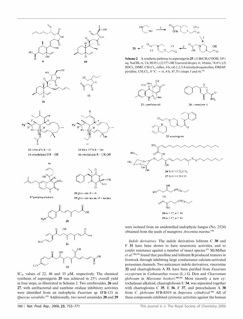
IC50 values of 22, 46 and 35 lM, respectively. The chemicalsynthesis of aspernigerin 25 was achieved in 25% overall yieldin four steps, as illustrated in Scheme 2. Two cerebrosides, 26 and27, with antibacterial and xanthine oxidase inhibitory activitieswere identified from an endophytic Fusarium sp. IFB-121 inQuercus variabilis.175 Additionally, two novel ceramides 28 and 29
Scheme 2 A synthetic pathway to aspernigerin 25. (1) BrCH2COOH, 10%aq. NaOH, rt, 1 h, 80.0%; (2) 37% HCl (several drops), rt, 10 min, 74.6%; (3)SOCl2, DMF, CH2Cl2, reflux, 4 h; (4) 1,2,3,4-tetrahydroquinoline, DMAP,pyridine, CH2Cl2, 0 ◦C → rt, 4 h, 47.3% (steps 3 and 4).173
were isolated from an unidentified endophytic fungus (No. 2524)obtained from the seeds of mangrove Avicennia marina.176
Indole derivatives. The indole derivatives lolitrem C 30 andF 31 have been shown to have neurotoxic activities, and toconfer resistance against a number of insect species.177 McMillanet al.178,179 found that paxilline and lolitrem B produced tremors inlivestock through inhibiting large conductance calcium-activatedpotassium channels. Two anticancer indole derivatives, vincristine32 and chaetoglobosin A 33, have been purified from Fusariumoxysporum in Catharanthus roseus (L.) G. Don and Chaetoniumglobosum in Maytenus hookeri.180,181 More recently a new cy-tochalasan alkaloid, chaetoglobosin U 34, was separated togetherwith chaetoglosins C 35, E 36, F 37, and penochalasin A 38from C. globosum IFB-E019 in Imperata cylindrical.182 All ofthese compounds exhibited cytotoxic activities against the human
760 | Nat. Prod. Rep., 2006, 23, 753–771 This journal is © The Royal Society of Chemistry 2006

nasopharyngeal epidermoid tumour KB cell lines with IC50 valuesof 16, 34, 52, 48, and 40 lM, respectively.
Pyrrolizidines. Pyrrolizidines, especially 1-aminopyrrolizidineswith an oxygen bridge, are a common metabolite in some grass–endophyte associations. The loline alkaloids are the only grass–endophyte-associated alkaloid class that protects endophyte-infected plants due to their anti-invertebrate and feeding deterrentactivities.183 A biosynthetic pathway for norloline 39 has beenproposed, as shown in Scheme 3. Incorporation of isotopicallylabeled L-proline and L-homoserine into 39 indicated that the A-ring carbons C1–C3 and the N1 are derived from L-homoserine,and that the B-ring carbons C5–C8 and the ring nitrogen arederived from L-proline.184
Quinazolines. Structurally, quinazolines correspond to productsof condensation of anthranilic acid with a-amino acids.185 Fourrare spiroquinazoline alkaloids, alanditrypinone 40, alantryphe-none 41, alantrypinene 42 and alantryleunone 43, were isolatedfrom the culture of the endophytic fungus Eupenicillium sp.residing in leaves of Murraya paniculata (Rutaceae).186 Thesealkaloids seem to be biosynthesised by a unique pathway because
Scheme 3 A proposed biosynthetic pathway for norloline 39.184
their precursors, anthranyllic acid and tryptophan, could not bedetected in the host plant.
(b) Steroids. Steroids, which are extensively distributed inplants, have many important physiological effects, and inter-estingly some steroidal metabolites from endophytic fungi havealso been reported. A novel ergosterol derivative, (20S,22S)-4a-homo-22-hydroxy-4-oxaergasta-7,24(28)-dien-3-one 44, was iso-lated from a strain of Gliocladium sp., an endophyte on Taxus chi-nensis (Pilg.) Rehd.187 3b-Hydroxyergosta-4,22-diene 45 togetherwith ergosterol and 3b-hydroxy-5a,8a-epi-dioxyergosta-6,22-diene
This journal is © The Royal Society of Chemistry 2006 Nat. Prod. Rep., 2006, 23, 753–771 | 761

was elaborated by the endophytic A. fumigatus CY018 found inC. dactylon.70
(c) Terpenoids. A number of terpenoid derivatives are whichproduced by fungal endophytes have been reported during 2001–2005. They involve sesquiterpenes and diterpenes, some of whichare analogues arising from metabolic degradation of terpenoidskeletons.
Sesquiterpenes. In addition to four cytochalasins, eleven novelsesquiterpenoids 46–56 were isolated from cultures of the mi-tosporic fungus Geniculosporium sp., an endophyte associated withthe red alga Polysiphonia sp.170 These 11 botryane compoundsexhibited moderate inhibitory activity against Chlorella fusca,Bacillus megaterium and Microbotryum violaceum. Macrocyclictrichothecenes are toxic sesquiterpenoids, which can cause seriousdiseases in livestock, especially during the flowering season.188
Six trichothecenes, roridins A 57, D 58, E 59 and H 60, andverrucarins A 61 and J 62, were detected in the culture of anendophytic fungus Ceratopicnidium baccharidicola from Bacchariscoridifolia.189 The production of these compounds was greater onrice culture than in liquid cultures (YES and MYRO broths). Inour laboratory, three novel cytotoxic 10,13-cyclotrichothecane-derived macrolides, myrothecines A–C (63–65), were separatedfrom Myrothecium roridum IFB-E009 and IFB-E012, endophytesassociated with the two traditional Chinese medicinal plantsTrachelospermum jasminoides and Artemisia annua, respectively.190
The absolute stereochemistry of these macrolides was establishedby a combination of NMR of a Mosher’s acid derivative followedby single-crystal X-ray diffraction analysis.
762 | Nat. Prod. Rep., 2006, 23, 753–771 This journal is © The Royal Society of Chemistry 2006

Diterpenes. Guanacastepenes A–O 66–80, a highly diversefamily of diterpenoid natural products, were identified froman unidentified endophytic fungus CR115.191,192 By comparisonwith other guanacastepenes, metabolites 66 and 73 exhibitedpronounced antibiotic activity against drug-resistant strains ofStaphylococcus aureus and Enterococcus faecalis. Scheme 4 showsputative biosynthetic relationships between these metabolites. Inaddition to important ring-generating biosynthetic transforma-tions, some oxidation/reduction and adornment reactions (e.g.methylation and acetylation) are involved in the biosynthesis ofthe individual guanacatepenes.192
(d) Isocoumarin derivatives. Over the period 2001–2005, onlythree novel isocoumarin derivatives, 81–83, have been identifiedfrom endophytic sources. These metabolites were isolated from
Geotrichum sp., an endophyte of Crassocephalum crepidioides.193
Biological assays demonstrated their antimalarial, antitubercu-lous and antifungal activities.
(e) Quinones. Two highly functionalised cyclohexenone epox-ides, jesterone 84 and hydroxyjesterone 85, were characterisedfrom a newly identified endophyte Pestalotiopsis jesteri presentin Fragraea bodenii.194 Notably, metabolite 84 displayed selectiveantimycotic activity against phytopathogens. The total synthesisof 84 was accomplished in 14 steps, as illustrated in Scheme 5.The route involved a diastereoselective epoxidation of a chiralquinone monoketal derivative and regio- and stereoselectivereduction of a quinone epoxide intermediate.195 Ambuic acid 86is a quinone epoxide metabolite with potent antifungal activityfrom Pestalotiopsis spp. and Monochaetia sp. living in Torreyataxifolia.196
Scheme 4 Putative biosynthetic relationships within the family of guanacatepetenes A–O 66–80.192
This journal is © The Royal Society of Chemistry 2006 Nat. Prod. Rep., 2006, 23, 753–771 | 763

Scheme 5 A synthetic pathway to jesterone 84. Reagents and conditions: (1) Br2, CHCl3, rt, 2.5 h, 94%; (2) NaBH4, toluene, 50 ◦C, then prenyl bromide,−30 ◦C, 4 h; (3) PhI(OAc)2, MeOH, 20 min, rt, 83%; (4) (2S,4S)-(+)-pentanediol, PPTS, benzene, 80 ◦C, 20 min, 80%; (5) KHMDS, TrOOH, THF,−35 ◦C, 15 h, 80%; (6) (E)-tributyl-1-propenylstannane, Pd(PPh3)4, toluene, 110 ◦C, 6 h, 88%; (7) HF, CH3CN, rt, 4.5 h, 82%.195
Jiang el al.197 have isolated three anthracenediones, 87–89, froman unidentified endophytic fungus, no. 1403, colonising man-groves. Xanthoviridicatins E 90 and F 91 are two novel quinone-related metabolites produced by an endophytic Penicillium chryso-genum colonising an unidentified plant. These metabolites inhibitthe cleavage reaction of HIV-1 integrase with IC50 values of 6 and5 lM, respectively.198 More recently, seven anthraquinones, 92–98,with potent cytotoxic activities against human colon (SW1116)and leukaemia (K562) cancer cell lines were separated fromPleospora sp. IFB-E006 associated with Imperata cylindrical.199
(f) Phenylpropanoids and lignans. Guignardic acid 99, thefirst member of a novel class of natural products, was detectedin the culture broth of Guignardia sp. obtained from Spondiasmombin.200 The oxidative deamination products of L-valine andL-phenylalanine (dimethylpyruvic acid and phenylpyruvic acidrespectively) are biogenetic precursors of this metabolite.
(g) Phenols and phenolic acids. Phenols and phenolic acidsfrom fungal endophytes usually have pronounced biological andantioxidant activities. Pestacin 100 and isopestacin 101, twonovel dihydroisobenzofuan-carrying phenols possessing antifun-gal and antioxidant activities, were separated from Pestalotiopsismicrospora associated with the combretaceaous plant Terminaliamorobensis of Papua New Guinea.201,202 Orsellinic acid 105 andthe three novel esersglobosumones A–C 102–104 were isolatedfrom Chaetomium globosum endophytic on Ephedra fasciulata(Mormon tea).203 Compound 102 had a moderate inhibitory effecton the cell proliferation of lung cancer, breast cancer, CNS gliomaand pancreatic carcinoma.
(h) Aliphatic compounds. Chaetomellic acid A 106, a potentand highly specific inhibitor of farnesyl-protein transferase (FP-Tase), was characterised from the endophyte Chaetomella acutisea
764 | Nat. Prod. Rep., 2006, 23, 753–771 This journal is © The Royal Society of Chemistry 2006

(MF5686).204 Several alternate syntheses of this bioactive naturalproduct had been reported. Notably, a straightforward syntheticpathway was achieved in only two steps with an 89% overall yield(Scheme 6).205
Scheme 6 A facile synthetic synthesis of chaetomellic acid A 106.Reagents and conditions: (1) (a) PPh3, AcOH, CH3(CH2)12CHO, reflux,18 h, (b) 140–150 ◦C, 30 min; (2) (a) KOH, H2O–CH3OH–THF, reflux,2 h, (b) H+/HCl.205
(i) Lactones. The seven lactones 107–113, which were originallycharacterised from a Chilean ascomycete,206,207 were re-detectedin an unidentified endophytic fungus associated with Cistussalviifolius L.208 Phomol 114 is a novel antibiotic from a Phomopsisispecies present in the medicinal plant Erythrina crista-galli.209
Microcarpalide 115, a novel microfilament-disrupting agent with
weak cytotoxicity to mammalian cells, was characterised from anunidentified fungus in Ficus microcarpa L.210 Four total synthesesof compound 115 have been described.211–214 The first convergentand stereoselective synthetic pathway of 115 was achieved byMurga et al. starting from (R)-glycidol and (S,S)-tartaric acid,as shown in Scheme 7.211
Two novel lactones, 1893A 116 and B 117, have been char-acterised from the extract of the endophyte strain no. 1893present in an estuarine mangrove on the South China Sea coast.215
Sequoiamonascins A–D 118–121 with a novel carbon skeleton, areelaborated by the fungal endophyte A. parasiticus and are reportedto display moderate activities against cancer cell lines, includingMCF7 (breast), NCI-H460 (lung), and SF-268 (CNS).216
Scheme 7 A synthetic pathway to microcarpalide 115. Reagents and conditions: (1) (a) TPSCl, Et3N, DMAP, CH2Cl2, rt, 18 h, 93%, (b) CH3(CH2)4MgBr,CuI, THF, −30 ◦C, 87%; (2) MOMCl, Et3N, DMAP, CH2Cl2, rt, 18 h, 93%; (3) TBAF, THF, 5 h, rt, 93%; (4) (COCl)2, DMSO, CH2Cl2, −78 ◦C thenN,N-diisopropylethylamine, 2 min at −78 ◦C, then rt; (5) Bu3SnCH2CH=CH2, MgBr2·Et2O, 3 A MS, CH2Cl2, 3 h at −78 ◦C, then 1.5 h at −40 ◦C, 60%yield over two steps; (6) DCC, DMAP, CH2Cl2, rt, 18 h, 86%; (7) 20 mol% catalyst A, CH2Cl2, reflux, 24 h, 67%; (8) SMe2, BF3·Et2O, −10 ◦C, 30 min,71%; (9) (CH2SH)2, BF3, CH2Cl2, 0 ◦C, 1 h, 66%.211
This journal is © The Royal Society of Chemistry 2006 Nat. Prod. Rep., 2006, 23, 753–771 | 765

Four 6H-dibenzo[b,d]pyran-6-one derivatives, alternariolmonomethyl ether (AME) 122, and graphislactones A, G andH 123–125, were isolated from Cephalosporium acremonium IFB-E007 colonising Trachelospermum jasminoides (Lindl.) Lem in ourlaboratory.217 Metabolites 122 and 123 had been isolated fromcultured lichen mycobionts of Graphis prunicola, G. cognata and G.scripta.218,219 Metabolites 122–125 were shown to be substantiallycytotoxic against SW1116 cells with IC50 values of 8.5, 14, 12, and21 lg mL−1, respectively.
(j) Miscellaneous metabolites. Sequoiatones C–F 126–129 arenovel cytotoxic metabolites isolated from Aspergillus parasiticuspresent in the coast redwood tree Sequoia sempervirens.220 Inaddition to asperfunmoid 23, asperfumin 130 was purified fromthe endophyte A. fumigatus CY018.70 Rhizotonic acid 131,
a novel anti-Helicobacter pylori metabolite, together withmonomethylsulochrin 132, was characterised from aRhizoctonia sp. (Cy064) endophytic on Cynodon dactylon.221
Four antimicrobial naphtho-c-pyrones, rubrofusarin B 133,fonsecinone A 134, asperprone B 135 and aurasperone A136, have been identified in cultures of A. niger IFB-E003obtained from C. dactylon.222 Biological assays indicate thatcompound 133 has potent cytotoxic activity against the coloncancer cell line SW1116 (IC50 4.5 lg mL−1), and compound136 exhibited inhibition on xanthine oxidase with an IC50 valueof 10.9 lM.223 Compounds 134 and 136 were re-isolated from
766 | Nat. Prod. Rep., 2006, 23, 753–771 This journal is © The Royal Society of Chemistry 2006

A. aculeatus in Melia zaedarach (Meliaceae). A new xanthene-based metabolite, paranolin 137, was characterised from anendophytic strain of Paraphaeosphaeria nolinae IFB-E011 fromArtemisia annua (Asteraceae).224 Recently, Dai et al.225 isolatedsix novel compounds from a Phomopsis sp. endophytic onAdenocarpus foliolosus, which were identified as phomosinesD–G 138–141, 6-isopropylcyclohex-1-enecarboxylic 142 and(1aS,3R,4R,4aR,6S,7R,8aS)-7-chloro-3,6-dihydroxy-3,4a,8,8-tetramethyloctahydro-1aH-naphtho[1,b]oxirene-4-carboxylicacid 143.
4 Potential applicability
4.1 Plant growth enhancers
It is generally accepted that the plant kingdom is exten-sively colonised by endophytic microorganisms which form non-pathogenic relationships with their hosts. In addition to protectingplants from biotic and abiotic influences, other nutritional benefitsextend from such associations. Although agrochemicals are amajor aspect of crop yield improvement they can have environmen-tal problems. Empirical evidence indicates that some endophyticmicrobes may act as plant fertilisers by enhancing nitrogen fixationand phosphorus assimilation.
Numerous species of endophytic rhizobacteria (PGPR) increasethe availability of nutrients in the rhizosphere, with direct benefitsto root growth and morphology, and they are beneficial to manyaspects of plant–endophyte symbioses.226 For example, nitrogen-fixing Klebsiella oxytoca VN13 and phosphorus-assimilating Xan-thomonas maltophilia VN12 have been co-mixed to form an inoc-ulant ‘Duet’ for seeds.227 The results indicate that corn inoculatedwith this ‘Duet’ generates increased yields, and possesses a higherpercentage of protein. Similarly, endophytic bacteria in rice (Oryzasativa L.) can effectively colonise host tissues and form nitrogen-fixing symbioses.228 Such applications of endophytes offers aneffective alternative to agrochemicals.
4.2 Phytoprotectors
Endophytes endow their host plants with many benefits, and theircommercial potential could reasonably receive more attention.For example, Pantoea agglomerans is an endophyte of crop plantsincluding pea, potato, sweet corn and tomato, and has been shownto effectively control bacterial plant diseases.229 Heteroconium
chaetospira, a root endophytic fungus associated with Chinesecabbage, acts as a control agent against clubroot and Verticilliumyellows.230 Chen et al.231 found six endophytic strains, Aureobac-terium saperdae, Bacillus pumilus, Phyllobacterium rubiacearum,Pseudomonas putida, P. putida, and Burkholderia solanacearum,which could significantly reduce vascular wilt in cotton caused byFusarium oxysporum f. sp. vasinfectum. A plant-growth-promotingrhizobacterium, Pseudomonas sp. strain PsJN, has been shown todisplay an antagonist effect on the in vitro growth and developmentof Botrytis cinerea, which is a fungal pathogen causing greymould diseases.232 It seems that the endophyte inhibits the growthof B. cinerea by disrupting cellular membranes and inducingcell death.233 An endophytic Streptomyces spp. obtained fromtomato (Lycopersicon esculentum), was found to effectively controlRhizoctonia solani, which is one of the most serious and widelyspread diseases in tomatoes, sometimes causing more than 70%seedling mortality.234,235
A novel application of endophytic microbes has been exploredin the field of phytoremediation to metabolise compounds as-sociated with chemical waste. Certain endophytes act as phy-toremediators by degrading compounds which present an envi-ronmental hazard.236 This ‘green’ approach to such managementis gaining public attention. For example, the newly identifiedendophytic bacterium Methylobacterium populum sp. was shownto degrade 2,4,6-trinitrotoluene (TNT), hexahydro-1,3,5-trinitro-1,3,5-triazine (HMX) and hexahydro-1,3,5-trinitro-1,3,5-trizaine(RDX).237
5 Concluding remarks
Ever since penicillin was isolated from Penicillium notatum,chemists have been engaged in the discovery of novel bioactivesfrom microbial metabolites. Despite a focused interest on syntheticproducts, bioactive natural products retain an immense impact onmodern medicine. Around 60% of the new drugs registered duringthe period 1981–2002 by the FDA as anticancer, antimigraine andantihypertensive agents are either natural products or based onnatural products.238 Endophytic microorganisms have developedthe biochemical ability to produce compounds similar or identicalto those produced by their host plants as a result of generecombination during the evolutionary process. Bioactive naturalproducts from endophytic microbes have enormous potentialas the source of new medicinal and agricultural products, andmethods to facilitate the identity of appropriate natural productsfrom this source are required. This aspect adds further weightto the preservation of plant biodiversity and greater organisationin the collection and cataloguing of endophytic microorganismsthroughout the world.
There has been an improved understanding of biosyntheticpathways to some bioactive endophytic compounds by chemicaland biochemical means,239 and recent progress in the molecularbiology of secondary metabolites offers a better insight intohow the genes for these bioactive compounds are organised.As a relatively poorly investigated group of microorganisms, therelationship between endophytes and their hosts merits improvedquantitative analysis, particularly at the molecular and geneticlevels. The cloning of the genes of endophytic metabolites hasbegun to open up attractive screening possibilities240–243 for thedirect identification of endophytic strains.
This journal is © The Royal Society of Chemistry 2006 Nat. Prod. Rep., 2006, 23, 753–771 | 767

6 References
1 J. K. Stone, C. W. Bacon and J. F. White, in An Overview of EndophyticMicrobes: Endophytism Defined, ed. C. W. Bacon and J. F. White, Jr.,M. Dekker, Inc., New York, 2000, pp. 3–5.
2 J. K. Bills, M. Christensen, M. Powell and G. Thorn, in Biodiversity ofFungi: Endophytic Fungi, ed. G. M. Mueller, G. F. Bills and M. Foster,Elsevier Academic Press, CA, USA, 2004, p. 241.
3 G. A. Strobel, Microbes Infect., 2003, 5, 535.4 C. Andrzej, Wiad. Bot., 2002, 46, 35.5 R. J. Rodriguez and R. S. Redman, Adv. Bot. Res., 1997, 24, 169.6 G. Carroll, Ecology, 1988, 69, 2.7 A. L Misko and J. J. Germida, FEMS Microbiol. Ecol., 2002, 42,
399.8 A. V. Sturz and J. Nowak, Appl. Soil Ecol., 2000, 15, 183.9 A. Majewska-Sawkaa and H. N. A. Gentile, Fungal Genet. Biol., 2004,
41, 534.10 K. Clay, J. Holah and J. A. Rudgers, Proc. Natl. Acad. Sci. U. S. A.,
2005, 102, 12465.11 K. Germaine, E. Keogh, G. Garcia-Cabellos, B. Borremans, D. Lelie,
T. Barac, L. Oeyen, J. Vangronsveld, F. P. Moore, E. R. B. Moore, C. D.Campbell, D. Ryan and D. N. Dowling, FEMS Microbiol. Ecol., 2004,48, 109.
12 A. Stierle, G. A. Strobel and D. Stierle, Science, 1993, 260, 214.13 K. Saikkonen, P. Wali, M. Helander and S. H. Faeth, Trends Plant
Sci., 2004, 9, 275.14 Y. M. Dong, A. L. Iniguez and E. W. Triplett, Plant Soil, 2003, 257,
49.15 A. Gentile, M. S. Rossi, D. Cabral, K. D. Craven and C. L. Schardl,
Mol. Phylogenet. Evol., 2005, 35, 196.16 D. Brem and A. Leuchtmann, Evolution, 2003, 57, 37.17 C. D. Moon, C. O. Miles, U. Jarlfors and C. L. Schardl, Mycologia,
2002, 94, 694.18 P. J. Fisher, F. Graf, L. E. Petrini, B. C. Sutton and P. A. Wookey,
Mycologia, 1995, 87, 319.19 T. M. Mushin, T. Booth and K. H. Zwain, Kavaka, Trans. Mycol. Soc.
India, 1989, 17, 1.20 R. Ligrone, K. Pocock and J. G. Duckett, Can. J. Bot., 1993, 71, 666.21 E. Schmid and F. Oberwinkler, New Phytol., 1993, 124, 69.22 J. Frohlich and K. D. Hyde, in Palm Microfungi, University of Hong
Kong Press, Hong Kong, China, 2000.23 K. D. Hyde, J. E. Taylor and J. Frohlich, in Genera of Ascomycetes from
Palms, University of Hong Kong Press, Hong Kong, China, 2000.24 D. J. Lodge and S. Cantrell, Mycologist, 1995, 9, 149.25 O. Petrini and P. J. Fisher, Trans. Br. Mycol. Soc., 1986, 87, 647.26 P. J. Fisher and O. Petrini, Trans. Br. Mycol. Soc., 1987, 89, 246.27 A. J. Richardson and R. S. Currah, Selbyana, J. Marie Selby Bot.
Gdns., 1995, 16, 49.28 T. S. Suryanarayanan, G. Senthilarasu and V. Muruganandam, Fungal
Diversity, 2000, 4, 117.29 R. J. Ganley, S. J. Brunsfeld and G. Newcombe, Proc. Natl. Acad. Sci.
U. S. A., 2004, 101, 10107.30 D. C. Hawksworth and A. Y. Rossman, Phytopathology, 1987, 87, 888.31 K. Clay, Nat. Toxins, 1992, 1, 147.32 T. S. Suryanarayanan, S. K. Wittlinger and S. H. Faeth, Mycol. Res.,
2005, 109, 635.33 M. R. Siegel and G. C. M. Latch, Annu. Rev. Phytopathol., 1987, 25,
293.34 L. Schena, F. Nigro, I. Pentimone, A. Ligorio and A. Ippolito,
Postharvest Biol. Technol., 2003, 30, 209.35 M. L. Gutierrez-Zmora and E. Martinez-Romero, J. Biotechnol., 2001,
91, 117.36 J. J. Germida, S. D. Siciliano, R. de Freitas and A. M. Seib, FEMS
Microbiol. Ecol., 1998, 26, 43.37 H. Smith, M. J. Wingfield and O. Petrini, For. Ecol. Manage., 1996,
89, 189.38 A. V. Sturz, B. R. Christie and B. G. Matheson, Can. J. Microbiol.,
1998, 44, 162.39 A. Ragazzi, S. Moricca, P. Capretti and I. Dellavalle, J. Phytopathol.,
1999, 147, 437.40 J. A. Hoff, N. B. Klopfenstein, G. I. McDonald, J. R. Tonn, M. S.
Kim, P. J. Zambino, P. F. Hessburg, J. D. Rogers, T. L. Peever andL. M. Carris, For. Pathol., 2004, 34, 255.
41 K. F. Rodrigue and G. J. Samuls, J. Basic Microbiol., 1999, 39, 131.42 N. S. Raviraja, J. Basic Microbiol., 2005, 45, 230.
43 M. Gennaro, P. Gonthier and G. Nicolotti, J. Phytopathol., 2003, 151,529.
44 O. Petrini, in Microbiology of the Phyllosphere, ed. J. H. Andrews andJ. van den Heuvel, Cambridge University Press, Cambridge, 1986, p.392.
45 O. Petrini, in Microbial Ecology of Leaves, ed. J. H. Andrews andS. S. Hirano, Springer, New York, 1991, p. 499.
46 A. Sessitsch, B. Reiter, U. Pfeifer and E. Wilhelm, FEMS Microbiol.Ecol., 2002, 39, 23.
47 M. A. Surette, A. V. Sturz, R. R. Lada and J. Nowak, Plant Soil, 2003,253, 381.
48 B. Scott, Curr. Opin. Mcriobiol., 2001, 4, 393.49 M. Caruso, A. L. Colombo, L. Fedeli, A. Pavesi, S. Quaroni, M.
Saracchi and G. Ventrella, Ann. Microbiol., 2000, 50, 3.50 R. X. Tan and W. X. Zou, Nat. Prod. Rep., 2001, 18, 448.51 O. Petrini, T. N. Sieber, L. Toti and O. Viret, Nat. Toxins, 1992, 1, 185.52 S. H. Faeth and K. E. Hammon, Ecology, 1997, 78, 810.53 D. C. Susan, Eur. J. Plant Pathol., 2004, 110, 713.54 P. J. Fisher, O. Petrini, L. E. Petrini and B. C. Sutton, New Phytol.,
1994, 127, 133.55 A. Jumpponen and J. M. Trappe, New Phytol., 1998, 140, 295.56 A. V. Sussmann and R. E. DeWreede, Phycologia, 2002, 41, 169.57 H. D. Addy, S. Hambleton and R. S. Currah, Mycol. Res., 2000, 104,
1213.58 S. Mocali, E. Bertelli, F. Di Cello, A. Mengoni, A. Sfalanga, F. Viliani,
A. Caciotti, S. Tegli, G. Surico and R. Fani, Res. Microbiol., 2003, 154,105.
59 A. M. Dahl Jensen and N. Roulund, Agric., Ecosyst. Environ., 2004,104, 419.
60 G. C. Lewis, C. Ravel, W. Naffaa, C. Astier and G. Charmet, Ann.Appl. Biol., 1997, 130, 227.
61 C. Leyronas and G. Raynal, Ann. Appl. Biol., 2001, 139, 119.62 S. Mohali, T. I. Burgess and M. J. Wingfield, For. Pathol., 2005, 35,
385.63 R. I. Amann, W. Ludwig and K. H. Scheidler, FEMS Microbiol. Rev.,
1995, 59, 143.64 C. W. Bacon and J. F. White, in Biotechnology of Endophytic Fungi of
Grasses, ed. C. W. Bacon and J. F. White, CRC Press, Boca Raton,FL, USA, 1994, pp. 47–56.
65 B. U. Schulz, S. Draeger and H. J. Aust, Mycol. Res., 1993, 97, 1447.66 G. F. Bills, ”Isolation and analysis of endophytic fungal communities
from woody plants’ in Endophytic Fungi in Grasses and Woody Plants:Systematics, Ecology and Evolution, ed. S. C. Redlin and L. M. Carris,American Phytopathological Society Press, St. Paul, MN, USA, 1996,pp. 31–65.
67 E. M. Clark, Jr., J. F. White and R. M. Patterson, J. Microbiol.Methods, 1983, 1, 149.
68 V. M. Reis, J. I. Baldani, V. L. D. Baldani and J. Dobereiner, Crit. Rev.Plant Sci., 2000, 19, 227.
69 K. D. Craven, J. D. Blankenship, A. Leuchtmann, K. Hignight andC. L. Schardl, Sydowia, 2001, 53, 44.
70 J. Y. Liu, Y. C. Song, Z. Zhang, L. Wang, Z. J. Guo, W. X. Zou andR. X. Tan, J. Biotechnol., 2004, 114, 279.
71 J. Hallmann, A. QuadtHallmann, W. F. Mahaffee and J. W. Kloepper,Can. J. Microbiol., 1997, 43, 895.
72 L. D Guo, K. D. Hyde and E. C. Y. Liew, New Phytol., 2000, 147, 617.73 D. P. Malinowski and D. P. Belesky, J. Plant Nutr., 1999, 22, 835.74 A. Raps and S. Vidal, Oecologia, 1998, 114, 541.75 P. Filip, , R. W. S. Weber, O. Sterner and T. Anke, Z. Naturforsch., C:
Biosci., 2003, 58, 547.76 F. G. Loiret, , E. Ortega, D. Kleiner, P. Ortega-Rodes, R. Rodes and
Z. Dong, J. Appl. Microbiol., 2004, 97, 504.77 F. Waller, B. Achatz, H. Baltruschat, J. Fodor, K. Becker, M. Fischer,
T. Heier, R. Huckelhoven, C. Neumann, D. Wettstein, P. Franken andK. H. Kogel, Proc. Natl. Acad. Sci. U. S. A., 2005, 102, 13386.
78 P. C. Lyons, Plant Physiol., 1990, 92, 726.79 L. Gasoni and B. S. Gurfinkel, Mycol. Res., 1997, 101, 867.80 D. P. Malinowski, D. K. Brauer and D. P. Belesky, J. Agron. Crop Sci.,
1999, 183, 53.81 M. Kaldorf, B. Koch, K. H. Rexer, G. Kost and A. Varma, Plant Biol.,
2005, 7, 210.82 M. A. Surette, A. V. Sturz, R. R. Lada and J. Nowak, Plant Soil, 2003,
253, 381.83 M. Bonnet, O. Camares and P. Veisseire, J. Exp. Bot., 2000, 51, 945.84 C. G. M. Latch, Agric., Ecosyst. Environ., 1993, 44, 143.
768 | Nat. Prod. Rep., 2006, 23, 753–771 This journal is © The Royal Society of Chemistry 2006

85 G. P. Cheplick, A. Perera and K. Koulouris, Funct. Ecol., 2000, 14,657.
86 J. P. J. Eerens, R. J. Lucas, S. Easton and J. G. H. White, N. Z. J. Agric.Res., 1998, 41, 219.
87 M. R. Siegel and C. L. Schardle, in Microbial Ecology of Leaves,ed. J. H. Handrews and S. S. Hirano, Springer, New York, 1990, pp.198–221.
88 J. P. Breen, Annu. Rev. Entomol., 1994, 39, 401.89 M. R. Siegel and L. P. Bush, Recent Adv. Phytochem., 1996, 30, 81.90 M. R. Siegel and L. P. Bush, in Toxin Production in Grass/Endophyte
Associations, ed. G. C. Carroll and P. Tudzynski, Springer,Berlin/Heidelberg/New York, 1997, pp. 185–208.
91 C. L. Schardl and T. D. Phillips, Plant Dis., 1997, 81, 430.92 K. Mandyam and A. Jumpponen, Stud. Mycol., 2005, 53, 173.93 N. S. Hill, D. P Belesky and W. C. Stringer, Crop Sci., 1991, 31, 185.94 F. Monnet, N. Vaillant, A. Hitmi, A. Coudret and H. Sallanon,
Physiol. Plant., 2001, 113, 557.95 C. Lodewyckx, M. Mergeay, J. Vangronsveld, H. Clijsters and D. Van
Der Lelie, Int. J. Phytorem., 2002, 4, 101.96 G. C. Lewis, Ann. Appl. Biol., 2004, 144, 53.97 M. Bacilio-Jimenez, S. Aguilar-Flores, M. V. del Valle, A. Perez, A.
Zepeda and E. Zenteno, Soil Biol. Biochem., 2001, 33, 167.98 B. Reiter, U. Pfeifer, H. Schwab and A. Sessitsch, Appl. Environ.
Microbiol., 2002, 68, 2261.99 Z. Shi, L. Hu, S. Yu, L. Xu and Y. Fan, J. Nanjing Agric. Univ., 2005,
28, 48 (in Chinese).100 H. He, X. Cat, X. Guan, F. Hu and L. Xie, Acta Phytopathol. Sin.,
2003, 33, 373 (in Chinese).101 L. Chen, H. Shi and Y. Chen, Henan Agric. Sci., 2005, 7, 54 (in
Chinese).102 X. Peng, L. Yang, Y. Chen, S. Li, B. Zhou and Z. Li, J. Fungal Res.,
2003, 11, 33 (in Chinese).103 C. Dai, B. Yu, Y. Zhao, Q. Yang and J. Jiang, Chin. J. Appl. Ecol.,
2005, 16, 1290 (in Chinese).104 B. Liu, M. Li and R. Liu, J. Northwest Univ., Nat. Sci.Ed., 2005, 8, 73
(in Chinese).105 B. A. Kunkel, P. S. Grewal and M. F. Quigley, Biol. Control, 2004, 29,
100.106 K. Saikkonen, M. Helander, S. H. Faeth, F. Schulthess and D. Wilson,
Oecologia, 1999, 121, 411.107 E. Wilhelm, W. Arthofer, R. Schafleitner and B. Krebs, Plant Cell,
Tissue Organ Cult., 1998, 52, 105.108 K. M. Oliver, N. A. Moran and M. S. Hunter, Proc. Natl. Acad. Sci.
U. S. A., 2005, 102, 12795.109 K. M. Oliver, J. A. Russell, N. A. Moran and M. S. Hunter, Proc.
Natl. Acad. Sci. U. S. A., 2003, 100, 1803.110 A. E. Arnold, L. C. Mejıa, D Kyllo, E. I. Rojas, Z. Maynard, N.
Robbins and E. A. Herre, Proc. Natl. Acad. Sci. U. S. A., 2003, 100,15649.
111 B. R. Vazquez-De-Aldana, B. Garcia-Criado, I. Zabalgogeazcoa andA. Garcia-Ciudad, J. Plant Nutr., 1999, 22, 163.
112 D. P. Malinowski, G. A. Alloush and D. P. Belesky, Plant Soil, 2000,227, 115.
113 M. D. Richardson, G. S. Chapman, C. S Hoveland and C. W. Bacon,Crop Sci., 1992, 32, 145.
114 M. D. Richardson, C. W. Bacon and C. S. Hoveland, in Proc. Int. Symp.Neotyphodium/Grass Interact., Louisiana Agricultural ExperimentalStation, Baton Rouge, LA, USA, 1990, pp. 189–93.
115 A. A. Elmi, C. P. West, R. T. Robbin and T. L. Kirkpatrick, GrassForage Sci., 2000, 31, 166.
116 J. Liao, X. Lin and Z. Cao, Soils, 2003, 35, 37 (in Chinese).117 R. J. Ganley, S. J. Brunsfeld and G. Newcombe, Proc. Natl. Acad. Sci.
U. S. A., 2004, 101, 10107.118 R. J. Ganley, R. S. Redman, K. B. Sheehan, R. G. Stout, R. J.
Rodriquez and J. M. Henson, Science, 2002, 298, 1581.119 K. Clay and J. Holah, Science, 1999, 285, 1742.120 K. Clay, Am. Zool., 2001, 41, 810.121 M. Ernst, K. W. Mendgen and S. G. R. Wirsel, Mol. Plant–Microbe
Interact., 2003, 16, 580.122 H. H. Wilkinson, M. R. Siegel, J. D. Blankenship, A. C. Mallory, L. P.
Bush and C. L. Schardl, Mol. Plant–Microbe Interact., 2000, 13, 1027.123 K. Braun, J. Romero, C. Liddell and R. Creamer, Mycol. Res., 2003,
107, 980.124 J. D. Miller, S. Mackenzie, M. Foto, G. W. Adams and J. A. Findlay,
Mycol. Res., 2002, 106, 471.
125 A. E. Arnold, L. C. Mejia, D. Kyllo, E. I. Rojas, Z. Maynard, N.Robins and E. A. Herre, Proc. Natl. Acad. Sci. U. S. A., 2003, 100,15649.
126 T. G. Whitham, W. P. Young, G. D. Martinsen, C. A. Gehring, J. A.Schweitzer, S. M. Shuster, G. M. Wimp, D. G. Fischer, J. K. Baileyand R. L. Lindroth, Ecology, 2003, 84, 559.
127 B. Z. Guo, J. W. Hendrix, A.-Q. An and R. S. Ferris, Mycology, 1992,84, 882.
128 K. Clay, in Neotyphodium/Grass interactions, ed. C. W. Bacon andN. S. Hill, Plenum Press, New York, 1997, pp. 93–108.
129 M. Omacini, E. J. Chaneton, C. M. Ghersa and C. B. Muller, Nature,2001, 409, 78.
130 M. Omacini, E. J. Chaneton, C. M. Ghersa and P. Otero, Oikos, 2004,104, 581.
131 C. A. Yong, M. K. Bryant, M. J. Christensen, B. A. Tapper, G. T.Bryan and B. Scott, Mol. Genet. Genomics, 2005, 274, 13.
132 G. A. Strobel, B. Daisy, U. Castillo and J. Harper, J. Nat. Prod., 2004,67, 257.
133 G. A. Strobel, A. Stierle, D. Stierle and W. M. Hess, Mycotaxon, 1993,47, 71.
134 J. A. Leigh and D. L. Coplin, Annu. Rev. Microbiol., 1992, 46, 307.135 K. A. Mattos, C. Jones, N. Heise, J. O. Previato and L. Mendoca-
Previato, Eur. J. Biochem., 2001, 268, 3174.136 H. S. Coventry and I. A. Dubery, Physiol. Mol. Plant Pathol., 2001,
58, 149.137 R. Van Peer and B. Schippers, Neth. J. Plant Pathol., 1992, 98,
129.138 M. A. Newman, M. J. Daniels and J. M. Dow, Mol. Plant–Microbe
Interact., 1997, 10, 926.139 A. S. Sahai and M. S. Manocha, FEMS Microbiol. Rev., 1993, 11, 317.140 H. He, X. Cai, X. Guan, F. Hu and L. Xie, Acta Phytopathol. Sin.,
2003, 33, 373 (in Chinese).141 S. Pleban, L. Chernin and I. Chet, Lett. Appl. Microbiol., 1997, 25,
284.142 B. Reinhold-Hurk and T. Hurek, Trends Microbiol., 1998, 6, 139.143 C. C. H. Sakiyama, E. M. Paula, P. C. Pereira, A. C. Borges and D. O.
Silva, Lett. Appl. Microbiol., 2001, 33, 117.144 A. Ureta and S. Nordlund, FEMS Microbiol. Lett., 2001, 202, 177.145 T. Hurek, B. Reinhold-Hurek, M. Van Montagu and E. Kellenberger,
J. Bacteriol., 1994, 176, 1913.146 A. Quadt-Hallmann, N. Benhamou and J. W Kloepper, Can. J. Mi-
crobiol., 1997, 43, 577.147 M. A. Holland and J. C. Polacco, Plant Physiol., 1992, 98, 942.148 E. G. Ivanova, N. V. Doronina and Y. A. Trotsenko, Microbiology,
2001, 70, 392.149 R. L. Koenig, R. O. Morris and J. C. Polacco, J. Bacteriol., 2002, 184,
1832.150 L. C. Van Loon, P. A. H. M. Bakker and C. M. J. Pieterse, Annu. Rev.
Phytopathol., 1998, 36, 453.151 C. M. Miller, R. V. Miller, D. Garton-Kinney, B. Redgrave, J. Sears,
M. Condron, D. Teplow and G. A. Strobel, J. Appl. Microbiol., 1998,84, 937.
152 A. M. Pirttila, P. Joensuu, H. Pospiech, J. Jalonen and A. Hohtola,Physiol. Plant., 2004, 121, 305.
153 K. A. Gurney and P. G. Mantle, J. Nat. Prod., 1993, 56, 1194.154 T. L. M. Stamford, N. P. Stamford, L. C. B. B. Coelho and J. M.
Araujo, Bioresour. Technol., 2001, 76, 137.155 U. Castillo, G. A. Strobel, E. J. Ford, W. M. Hess, H. Porter, J. B.
Jensen, H. Albert, R. Robison, M. A. Condron, D. B. Teplow, D.Stevens and D. Yaver, Microbiology, 2002, 148, 2675.
156 U. Castillo, J. K. Harper, G. A. Strobel, J. Sears, K. Alesi, E. Ford, J.Lin, M. Hunter, M. Maranta, H. Ge, D. Yaver, J. B. Jensen, H. Porter,R. Robison, D. Millar, W. M. Hess, M. Condron and D. Teplow,FEMS Microbiol. Lett., 2003, 224, 183.
157 D. Ezra, U. F. Catillo, G. A. Strobel, W. M. Hess, H. Porter, J. Jensen,M. Condron, D. Teplow, J. Sears, M. Maranta, M. Hunter, B. Weberand D. Yaver, Microbiology, 2004, 150, 785.
158 S. Guan, S. Grabley, I. Groth, W. Lin, A. Christner, D. Guo and I.Sattler, Magn. Reson. Chem., 2005, 43, 1028.
159 W. H. Lin, L. Y. Li, H. Z Fu, I. Sattler, X. S. Huang and S. Grabley,J. Antibiot., 2005, 58, 594.
160 M. Torres, M. M. Dolcet, N. Sala and R. Canela, J. Agric. Food Chem.,2003, 51, 3328.
161 Y. Marlida, N. Saari, Z Hassan, S. Radu and J. Baker, Food Chem.,2000, 71, 221.
This journal is © The Royal Society of Chemistry 2006 Nat. Prod. Rep., 2006, 23, 753–771 | 769

162 M. R. Siegel, G. C. M. Latch, L. P. Bush, F. F. Fannin, D. D. Rowan,B. A. Tapper, C. W. Bacon and M. C. Hohnson, J. Chem. Ecol., 1990,16, 3301.
163 H. H. Wilkinson, M. R. Siegel, J. D. Blankenship, A. C. Mallory,L. P. Bush and C. L. Schardl, Mol. Plant–Microbe Interact., 2000, 13,1027.
164 A. Tanaka, B. A. Tapper, A. Popay, E. J. Parker and B. Scott, Mol.Microbiol., 2005, 57, 1036.
165 S. H. Faeth, L. P. Bush and T. J. Sullivan, J. Chem. Ecol., 2002, 28,1511.
166 B. R. Vazquez de Aldana, I. Zabalgogeazcoa, A. Garcıa Ciudad andB. Garcıa Criado, J. Sci. Food Agric., 2003, 83, 347.
167 T. Ishii, K. Hayashi, T. Hida, Y. Yamamoto and Y. Nozaki, J. Antibiot.,2000, 53, 765.
168 J. Y. Li, G. A. Strobel, J. Harper, E. Lobkovaky and J. Clardy, Org.Lett., 2000, 23, 767.
169 V. Hellwig, T. Grothe, A Mayer-Bartschmid, R. Endermann, F. U.Geschke, T. Henkel and M. Stadler, J. Antibiot., 2002, 55, 881.
170 K. Krohn, J. Dai, U. Florke, H. J. Aust, S. Drager and B. Schulz,J. Nat. Prod., 2005, 68, 400.
171 G. Schmeda-Hirschmann, E. Hormazabal, L. Astudillo, J. Rodrigueand C. Theoduloz, World J. Microbiol. Biotechnol., 2005, 21, 27.
172 S. H. Guan, S. Isabel, W. H. Lin, D. A. Guo and S. Grabley, J. Nat.Prod., 2005, 68, 1198.
173 Y. H. Ye, H. L. Zhu, Y. C. Song, J. Y. Liu and R. X. Tan, J. Nat. Prod.,2005, 68, 1106.
174 L. Shen, Y. H. Ye, X. T. Wang, H. L. Zhu, C. Xun, Y. C. Song, H. Liand R. X. Tan, Chem. Eur. J., 2006, 12, 4393.
175 R. G. Shu, F. W. Wang, Y. M. Yang, Y. X. Liu and R. X. Tan, Lipids,2004, 39, 667.
176 H. J. Li, J. H. Yao, Y. G. Chen, Y. C. Lin and L. L. P. Virjmoed, ActaSci. Nat. Univ. Sunyatseni, 2003, 42, 132 (in Chinese).
177 O. J.-P. Ball, G. M. Barker, R. A. Prestidge and J. M. Sprosen, J. Chem.Ecol., 1997, 23, 1435.
178 L. K. McMillan, R. L. Carr, C. A Yong, J. W. Astin, R. G. Lowe,E. J. Parker, G. B. Jameson, S. C. Finch, C. O. Miles, O. B. McManus,W. A. Schmalhofer, M. L. Garcia, G. J. Kaczorowski, M. Goetz, J. S.Tkacz and B. Scott, Mol. Genet. Genomics, 2003, 270, 9.
179 J. E. Dalziel, S. C. Finch and J. Dunlop, Toxicol. Lett., 2005, 155, 421.180 L. Q. Zhang, B. Guo, H. Y. Li, S. R. Zeng, H. Shao, S. Gu and R. C.
Wei, Chin. Tradit. Herb. Drugs, 2000, 31, 805 (in Chinese).181 L. Q. Zhang, H. K. Wang, H. Shao, Y. M. Shen, S. R. Zeng, C. D. Xu,
Q. Xuan and R. C. Wei, Chin. Pharm. J., 2002, 37, 172 (in Chinese).182 G. Ding, Y. C. Song, J. R. Chen, H. M. Ge, X. T. Wang and R. X.
Tan, J. Nat. Prod., 2006, 69, 302.183 C. T. Dougherty, F. W. Knapp, L. P. Bush, J. E. Maul and J. Van
Willigen, J. Med. Entomol., 1998, 35, 798.184 J. D. Blankenship, J. B. Houseknecht, S. Pal, L. P. Bush, R. B.
Grossman and C. L. Schardl, ChemBioChem, 2005, 6, 1016.185 S. Johne, Prog. Chem. Org. Nat. Prod., 1984, 46, 159.186 F. A. Proenca Barros and E. Rodrigues-Filho, Biochem. Syst. Ecol.,
2005, 33, 257.187 J. H. Zhang, S. X. Guo, J. S. Yang and P. G. Xiao, Acta Bot. Sin.,
2002, 44, 1239 (in Chinese).188 G. G. Habermehl, L. Busam, P. Heydel, D. Mebs, C. Tokarnia, J.
Dobereiner and M. Spraul, Toxicon, 1985, 23, 731.189 I. Rizzo, E. Varsavky, M. Haidukowski and H. Frade, Toxicon, 1997,
35, 753.190 L. Shen, R. H. Jiao, Y. H. Ye, X. T. Wang, C. Xu, Y. C. Song, H. L.
Zhu and R. X. Tan, Chem. Eur. J., 2006, 12, 5596.191 S. F. Brady, M. P. Singh, J. E. Janso and J. Clardy, J. Am. Chem. Soc.,
2000, 122, 2116.192 S. F. Brady, S. M. Bondi and J. Clardy, J. Am. Chem. Soc., 2001, 123,
9900.193 P. Kongsaeree, S. Prabpai, N. Sriubolmas, C. Vongvein and S.
Wiyakrutta, J. Nat. Prod., 2003, 66, 709.194 J. Y. Li and G. A. Strobel, Phytochemistry, 2001, 57, 261.195 Y. Hu, C. Li, B. A Kulkarin, G. A. Strobel, E. Lobkovsky, R. M.
Torczynski and J. A. Porco, Jr., Org. Lett., 2001, 3, 1649.196 J. Y. Li, J. K. Harper, D. M. Grant, B. O. Tombe, B. Bashyal, W. M.
Hess and G. A. Strobel, Phytochemistry, 2001, 56, 463.197 G. C. Jiang, Y. C. Lin, S. N. Zhou, L. L. P. Vrijmoed and E. B. G.
Jones, Acta Sci. Nat. Univ. Sunyatseni, 2000, 39, 68 (in Chinese).198 S. B. Singh, D. L. Zink, Z. Guan, J. Collado, F. Pelaez, P. J. Felock
and D. J. Hazuda, Helv. Chim. Acta, 2003, 86, 3380.
199 H. M. Ge, Y. C. Song, C. Y. Shan, Y. H. Ye and R. X. Tan, PlantaMed., 2005, 71, 1065.
200 K. F. Rodrigues-Heerklotz, K. Drandarov, J. Heerklotz, M. Hesse andC. Werner, Helv. Chim. Acta, 2001, 84, 3766.
201 G. A. Strobel, E. Ford, J. Worapong, J. K. Harper, A. M. Arif, A. M.Grant, P. C. W. Fung and R. M. W. W. Chau, Phytochemistry, 2002,60, 179.
202 J. K. Harper, A. M. Arif, E. J. Ford, G. A. Strobel, J. A. Porco, Jr.,D. P. Tomer, K. L. Oneill, E. M. Heider and D. M. Grant, Tetrahedron,2003, 59, 2471.
203 B. P. Bashyal, E. M. Kithsiri Wijeratne, S. H. Faeth and A. A. LeslieGunatilaka, J. Nat. Prod., 2005, 68, 724.
204 R. B. Lingham, K. C. Silverman, G. F. Bills, C. Cascales, M. Sanchez,R. G. Jankins, S. E. Gartner, I. Martin, M. T. Diez, F. Pelaez, S.Monchales, Y. L. Kong, R. W. Burg, M. S. Meinz, L. Huang, M.Nallin-Omstead, S. D. Mosser, M. D. Schaber, C. A. Omer, D. L.Pompliano, J. B. Gibbs and S. B. Singh, Appl. Microbiol. Biotechnol.,1993, 40, 370.
205 S. B. Desai and N. P. Argade, J. Org. Chem., 1997, 62, 4862.206 B. Kopcke, M. Johansson, O. Sterner and H. Anke, J. Antibiot., 2002,
55, 36.207 M. Johansson, B. Kopcke, H. Anke and O. Sterner, J. Antibiot., 2002,
55, 104.208 B. Kopcke, R. W. S. Weber and H. Anke, Phytochemistry, 2002, 60,
709.209 D. Weber, O. Sternere, T. Anke, S. Gorzalczancy, V. Martino and C.
Acevedo, J. Antibiot., 2004, 57, 559.210 A. S. Ratnayake, W. Y. Yoshida, S. L. Mooberry and T. Hemscheidt,
Org. Lett., 2001, 3, 3479.211 J. Murga, E. Falomir, J. Garcia-Fortanet, M. Carda and J. A. Marco,
Org. Lett., 2002, 4, 3447.212 M. K. Gurjar, R. Nagaprasad and C. V. Ramana, Tetrahedron Lett.,
2003, 44, 2873.213 S. Ghosh, R. V. Rao and J. Shashidhar, Tetrahedron Lett., 2005, 46,
5479.214 K. Ishigami, H. Watanabe and T. Kitahara, Tetrahedron, 2005, 61,
7546.215 G. Chen, Y. Lin, L. Wen, L. L. P. Vrijmoed and E. B. Gareth Jones,
Tetrahedron, 2003, 59, 4907.216 D. B. Stierle, A. A. Stierle and T. Bugni, J. Org. Chem., 2003, 68, 4966.217 H. W. Zhang, Y. C. Song, W. Y. Huang, J. R. Chen and R. X. Tan,
Helv. Chim. Acta, 2005, 88, 2861.218 T. Tanahashi, M. Kuroishi, A. Kuwahara, N. Nagakura and N.
Hamada, Chem. Pharm. Bull., 1997, 45, 1183.219 T. Tanahashi, Y. Takenaka, N. Nagakura and N. Hamada, Phyto-
chemistry, 2003, 62, 71.220 A. A. Stierle, D. B. Stierle and T. Bugni, J. Nat. Prod., 2001, 64, 1350.221 Y. M. Ma, Y. Li, J. Y. Liu, Y. C. Song and R. X. Tan, Fitoterapia, 2004,
75, 451.222 Y. C. Song, H. Li, Y. H. Ye, C. Y. Shan, Y. M. Yang and R. X. Tan,
FEMS Microbiol. Lett., 2004, 241, 67.223 F. R. Campos, A. Barison, C. Daolio, A. G. Ferreira and E. Rodrigues-
Fo, Magn. Reson. Chem., 2005, 43, 962.224 H. M. Ge, Y. C. Song, J. R. Chen, S. Hu, J. Y. Wu and R. X. Tan, Helv.
Chim. Acta, 2006, 89, 502.225 J. Q. Dai, K. Krohn, U. Florke, D. Gehle, H.-J. Aust, S. Drarger, B.
Schulz and J. Rheinheimer, Eur. J. Org. Chem., 2005, 5100.226 J. K. Vessey, Plant Soil, 2003, 255, 571.227 N. Kozyrovska, G. Kovtunovych, E. Gromosova, P. Kuharchuk and
V. Kordyum, Resour., Conserv. Recycl., 1996, 18, 79.228 J. R. Stoltzfus, R. So, P. P. Malarvithi, J. K. Ladha and E. J. de Bruijin,
Plant Soil, 1997, 194, 25.229 T. F. Hsieh, H. C. Huang and R. S. Erickson, J. Phytopathol., 2005,
153, 608.230 K. Narisawa, K. T. Ohki and T. Hashiba, Plant Pathol., 2000, 49, 141.231 C. Chen, E. M. Bauske, G. Musson, R. Rodriguez-Kabana and J. W.
Kloepper, Biol. Control, 1995, 5, 83.232 E. A. Barka, S. Gognies, J. Nowak, J.-C. Audran and A. Belarbi, Biol.
Control, 2002, 24, 135.233 L. Cao, Z. Qiu, J. You, H. Tan and S. Zhou, Lett. Appl. Microbiol.,
2004, 39, 425.234 L. A. Newman and C. M. Reynolds, Trends Biotechnol., 2005, 23, 6.235 T. Barac, S. Taghavi, B. Borremans, A. Provoost, L. Oeyen, J. V.
Colpaert, J. Vangronsveld and D. Lelie, Nat. Biotechnol., 2004, 22,583.
770 | Nat. Prod. Rep., 2006, 23, 753–771 This journal is © The Royal Society of Chemistry 2006

236 C. Lodewyckx, S. Taghavi, M. Mergeay, J. Vangronsveld, H. Clijstersand D. Van Der Lelie, Int. J. Phytorem., 2001, 3, 173.
237 S. D. Siciliano, N. Fortin, A. Mihoc, G. Wisse, S. Labelle, D. Beaumier,D. Ouellette, R. Roy, L. G. Whyte, M. K. Banks, P. Schwab, L. Leeand C. W. Greer, Appl. Environ. Microbiol., 2001, 67, 2469.
238 D. J. Newman, G. M. Cragg and K. D. Hyde, J. Nat. Prod., 2003, 66,1022.
239 C. A. Young, Y. Itoh, R Johnson, I. Garthaite, C. O. Miles, S. C.Munday-Finch and B. Scott, Curr. Genet., 1998, 33, 368.
240 P. Tudzynski, K. Holter, T. Correia, C. Arntz, N. Grammel and U.Keller, Mol. Gen. Genet., 1999, 261, 133.
241 C. A. Young, L. McMillan, E. Telfer and B. Scott, Mol. Microbiol.,2001, 39, 754.
242 J. H. Wang, C. Machado, D. G. Panaccione, H. F. Tsai and C. L.Schardl, Fungal Genet. Biol., 2004, 41, 189.
243 T. Haarmann, C. Machado, Y. Lubbe, T. Correia, C. L. Schardl,D. G. Panaccione and P. Tudznski, Phytochemistry, 2005, 66,1312.
This journal is © The Royal Society of Chemistry 2006 Nat. Prod. Rep., 2006, 23, 753–771 | 771