biology 113 laboratory skeletal system · pdf filebio 113 skeletal lab fall 2011 page 1 of 28...
TRANSCRIPT
BIO 113 Skeletal Lab Fall 2011 Page 1 of 28
BIOLOGY 113 LABORATORY Skeletal System Objectives
• Distinguish between the axial and appendicular skeleton. • Distinguish between the cranium and facial skeleton. • Locate and name the bones of the skull and their major features. • Locate and name the major sutures of the cranium. • Identify the features of the vertebral column. • Name the parts of a vertebra. • Distinguish between a cervical, thoracic and lumbar vertebra. • Locate and name the parts of the thoracic cage. • Distinguish between true and false ribs. • Locate and identify the bones of the upper limb (including the pectoral girdle) and their
major features. • Locate and identify the bones of the lower limb (including pelvic girdle) and their major
features. 1. THE ORGANIZATION OF THE SKELETON Using your textbook and the specimens and models available study the following: AXIAL SKELETON: • Skull: Cranium Facial bones • Hyoid Bone • Vertebral Column: Vertebrae Intervertebral disks Sacrum Coccyx • Thoracic Cage: Ribs Sternum APPENDICULAR SKELETON: • Upper Limb: Pectoral Girdle: • Scapula • Clavicle Humerus Radius Ulna Carpals
BIO 113 Skeletal Lab Fall 2011 Page 2 of 28
Metacarpals Phalanges • Lower Limb: Pelvic Girdle: • Coxal Bones Femur Tibia Fibula Patella Tarsals Metatarsals Phalanges Note: On yourself palpate and identify as many bones of your own skeleton as possible. Using the glossary in your textbook and/or the internet define and then locate the following bony surface markings:
• Condyle -
• Crest -
• Epicondyle -
• Facet -
• Fissure-
• Fontanel -
• Foramen -
• Fossa -
• Fovea -
• Tubercle -
• Tuberosity -
• Head -
• Trochanter -

BIO 113 Skeletal Lab Fall 2011 Page 3 of 28
Complete the following: 1. Label the major bones of the axial skeleton.

BIO 113 Skeletal Lab Fall 2011 Page 4 of 28
2. Label the major bones of the appendicular skeleton.
3. Which of the following bones are part of the axial skeleton and which are part of the appendicular skeleton: Hyoid Bone _______________ Cervical Vertebra _______________
Ulna _____________________ Clavicle ______________________
Sternum __________________ Carpal Bone ___________________
Patella ___________________ Sacrum _______________________

BIO 113 Skeletal Lab Fall 2011 Page 5 of 28
4. Complete the following statements:
a. The extra bones that sometimes develop between the flat bones of the skull are called
____________________.
b. ____________________ is another term for brain case.
c. The ____________________ located at the distal end of the vertebral column is composed
of several fused vertebrae.
d. The true ribs are attached to the ____________________ anteriorly.
e. The thoracic cage is composed of ____________________ pairs of ribs.
f. The scapulae and clavicles together form the ____________________.
g. The humerus, radius and ____________________ articulate to form the elbow joint.
h. The wrist is composed of eight bones called the ____________________.
i. The coxal bones are attached posteriorly to the ____________________.
j. The coxal bones, sacrum and coccyx form the ____________________.
k. The ____________________ covers the anterior surface of the knee.
l. The bones that articulate with the distal ends of the tibia and fibula are called the
____________________.
m. The finger and toe bones are called ____________________.
5. Match the terms in the first column A with the definitions in second. Place the letter of your choice in the space provided.
Condyle A nearly flat surface Crest B deep depression Facet C rounded process Fontanel D opening or passageway Foramen E line of union Fossa F narrow, ridge-like projection Suture G soft region between bones of
immature skull

BIO 113 Skeletal Lab Fall 2011 Page 6 of 28
6. Match the terms in Column A with the definitions in Column B. Place the letter of your choice in the space provided.
2. THE SKULL Using your textbook and any other materials provided, the human skulls and the skull models available, learn the following: • The Skull Bones (22 bones in total): Cranium (8 bones in total): • Single Bones (4) 1. Occipital Bone 2. Frontal Bone 3. Sphenoid Bone 4. Ethmoid Bone • Paired Bones (2 pairs) 1. Parietal Bones 2. Temporal Bones Facial Skeleton (14 bones in total): • Single Bones (2) 1. Mandible 2. Vomer • Paired Bones (6 pairs)
1. Maxillary Bones 2. Zygomatic Bones 3. Nasal Bones 4. Lacrimal Bones 5. Palatine Bones 6. Inferior Nasal Conchae
• The Sutures: Coronal Suture Sagittal Suture Lambdoid Suture Squamous Suture
Fovea A tube-like passageway
Head B tiny pit or depression
Meatus C small, knoblike process (bump)
Sinus D thorn-like projection
Spine E enlargement at end of the bone
Trochanter F hollow space within bone
Tubercle G relatively large process (big bump)

BIO 113 Skeletal Lab Fall 2011 Page 7 of 28
Complete the following: 1. Label the structures on the frontal view of the skull.. 2. Label the structures on the posterior view of the skull.

BIO 113 Skeletal Lab Fall 2011 Page 8 of 28
3. Label the structures on the lateral view of the skull.
4. Label the structures on the inferior view of the skull.

BIO 113 Skeletal Lab Fall 2011 Page 9 of 28
5. Label the structures on the floor of the cranial cavity.
6. List the bones of the skull that contribute to the formation of the eye socket. 7. List the bones of the skull that can be palpated. (Palpate them on yourself to make your list.) 8. List the four prominent sutures of the skull and the bones that are joined to form these sutures. 9. Name the three cranial bones that contain sinuses.

BIO 113 Skeletal Lab Fall 2011 Page 10 of 28
10. Name the pair of facial bones that contain sinuses. 11. Match the bones in first column with the characteristics in second column. Place the letter of your choice in the space provided. A Inferior Nasal Concha forms bridge of nose
B Mandible only moveable bone in facial skeleton
C Maxillary Bone contains coronoid process
D Nasal Bone creates prominence of cheek
E Zygomatic bone contains socket of upper teeth
forms anterior portion of zygomatic arch
scroll-shaped bone
forms anterior roof of mouth
contains mental foramen 3. THE VERTEBRAL COLUMN AND THORACIC CAGE Using your textbook, the articulated skeletons, individual bones and models available, examine the vertebral column and locate the following parts and features: • Curvatures: Cervical Thoracic * Lumbar Sacral (Pelvic) * * denotes the primary curvatures • Vertebrae: Type and number of each Size Differences Parts:
• Body • Pedicles • Vertebral Foramen • Lamina • Spinous Process • Vertebral Arch • Transverse Process • Superior Articulating Process • Intervertebral Formen
• Cervical Vertebrae:

BIO 113 Skeletal Lab Fall 2011 Page 11 of 28
Transverse Foramen Bifid Process
Atlas Axis (odontoid process or dens)
• Thoracic Vertebra • Lumbar Vertebra Using your textbook and atlas, the articulated skeletons, individual bones and models available, examine the sacrum and coccyx and locate the following features: • Sacrum:
Superior Articulating Process Anterior Sacral Foramen Dorsal Sacral Foramen Sacral Promontory Sacral Canal Median Sacral Crest Sacral Hiatus Apex and Base
• Coccyx Using your textbook and atlas, the articulated skeletons, individual bones and models available, examine the thoracic cage of the human skeleton and locate the following parts: Thoracic Vertebrae Ribs:
• Head • Neck • Tubercle • Angle • Body • Costal Groove • Rib Differences (length, curvature) • True Ribs versus False Ribs
Costal Cartilages and Costal Margin Sternum
• Manubrium • Body • Xiphoid Process • Suprasternal Notch (jugular notch) • Sternal Angle • Clavicular Notch • Articular Facets

BIO 113 Skeletal Lab Fall 2011 Page 12 of 28
Complete the following: 1. Label the parts and features of the vertebral column. 2. Label the superior features of a typical vertebra.
BIO 113 Skeletal Lab Fall 2011 Page 13 of 28
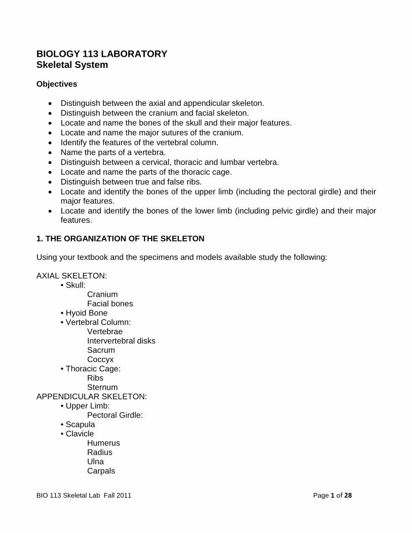
3. Label the lateral features of typical vertebrae.
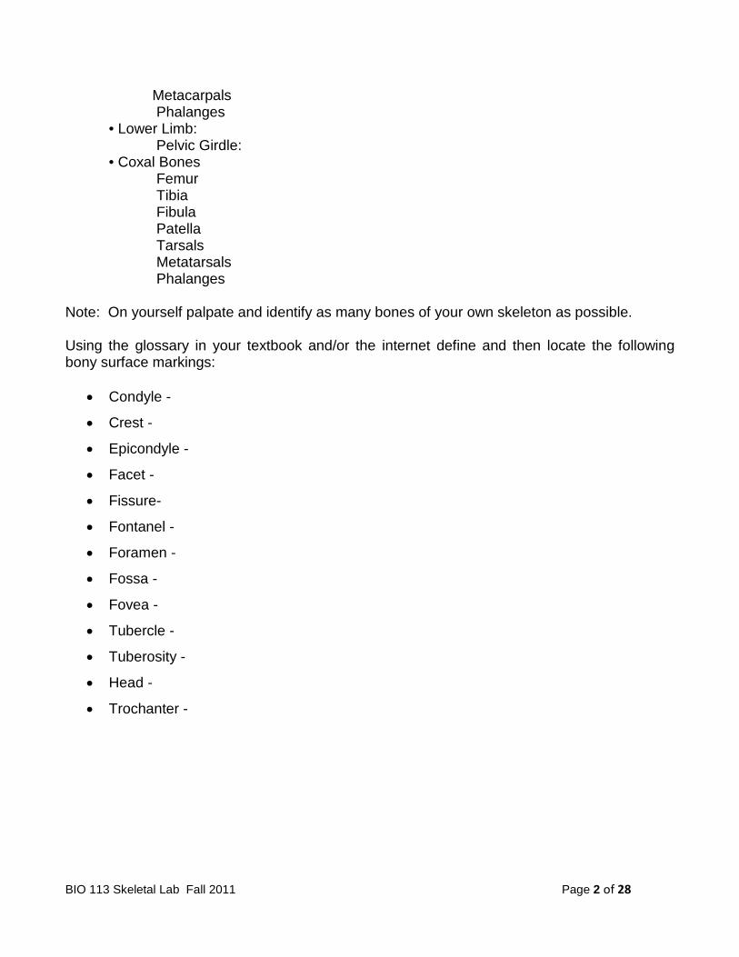
4. Label the superior features of (a) the atlas and (b) the axis.

BIO 113 Skeletal Lab Fall 2011 Page 14 of 28
5. Label the parts of the (a) cervical, (b) thoracic and (c) lumbar vertebrae.

BIO 113 Skeletal Lab Fall 2011 Page 15 of 28
6. Label the parts of the sacrum: anterior view and posterior view.
7. Label the parts and features of the thoracic cage.

BIO 113 Skeletal Lab Fall 2011 Page 16 of 28
8. Complete the following statements:
a) The vertebral column encloses and protects the ____________________.
b) The number of separate bones in the vertebral column of an infant is
____________________.
c) The thoracic and pelvic curvatures of the vertebral column are called
____________________ curves.
d) The ____________________ of the vertebrae supports the weight of the head and trunk.
e) The ____________________ separate adjacent vertebrae, and they soften the forces
created by walking.
f) The pedicles, laminae and spinous processes of a vertebra form the
____________________.
g) The intervertebral foramina provide passageways for ____________________.
h) Transverse foramina of cervical vertebrae serve as passageways for
____________________.
i) The first vertebra is called the ____________________.
j) When the head is moved from side to side, the first vertebra pivots around the
____________________ of the second vertebra.
k) The second vertebra is called the ____________________.
l) The ____________________ have the largest and strongest bodies.
m) The number of vertebrae that fuse to form the sacrum is ____________________.
n) The joint between a coxal bone of the pelvis and the sacrum is called the
____________________ joint.
o) The upper, anterior margin of the sacrum that projects forward is called the
____________________.
p) An opening called the ____________________ exists at the tip of the sacral canal.
9. Complete the following statements: a) Men have 12 pairs of ribs and women have ____________________ pairs of ribs.
b) The last 2 pairs of ribs that have no cartilaginous attachments to the sternum are sometimes
called the ____________________ ribs.

BIO 113 Skeletal Lab Fall 2011 Page 17 of 28
c) The tubercles of the ribs articulate with the ____________________ of the thoracic
vertebrae.
d) Costal cartilages are composed of ____________________ tissue.
e) The manubrium articulates with the ____________________ on its superior border.
4. THE BONES OF THE UPPER LIMB Using your textbook, the articulated and disarticulated skeletons and models available, find and learn the following: • Pectoral Girdle (Shoulder Girdle): Clavicle: • Medial and Lateral Ends Scapula • Borders: superior, vertebral (medial) and axiallary (lateral)
• Angles: superior, inferior and lateral • Spine • Head • Acromion Process • Coracoid Process • Glenoid Cavity • Subscapular Fossa • Supraspinous Fossa • Infraspinous Fossa
• Humerus Head Anatomical Neck Surgical Neck Greater Tubercle (lateral) Lesser Tubercle (medial and anterior) Intertubercular Groove (Bicipital Groove) Deltoid Tuberosity Capitulum (lateral) Trochlea (medial) Medial and Lateral Epicondyles Coronoid Fossa (anterior) Olecranon Fossa (posterior)
• Radius: Head Radial Tuberosity Styloid Tuberosity Ulnar Notch
• Ulna: Trochlear Notch

BIO 113 Skeletal Lab Fall 2011 Page 18 of 28
Olecranon Process Coronoid Process Head Radial Notch Styloid Process
• Wrist and Hand: Carpal Bones (8):
• Scaphoid • Lunate • Triquetral • Pisiform • Trapezium • Trapezoid • Capitate • Hamate
Metacarpals (5) Phalanges (14)
• proximal • middle • distal
Complete the following: 1. Label the bones and features of the pectoral girdle and clavicle.

BIO 113 Skeletal Lab Fall 2011 Page 19 of 28
2. Label (a) the anterior surface and (b) the posterior surface of the right scapula.
3. Label (a) the anterior surface and (b) the posterior surface of the right humerus.

BIO 113 Skeletal Lab Fall 2011 Page 20 of 28
4. Label the major features of the (a) anterior surface and (b) posterior surface of the right radius and ulna.
5. Label the bones of the hand and wrist.

BIO 113 Skeletal Lab Fall 2011 Page 21 of 28
6. Match the terms in Column A with the definitions in Column B. Place the letter of your choice in the space provided.
A Humerus coronoid fossa
B Radius deltoid tuberosity
C Ulna greater tubercle
intertubercular groove
olecranon fossa
radial notch
radial tuberosity
trochlea
trochlear notch
ulnar notch 7. Complete the following statements: a) The pectoral girdle is an incomplete ring because it is open in the back between the
____________________.
b) The medial ends of the clavicles articulate with the ____________________.
c) The lateral ends of the clavicles articulate with ____________________.
d) A clavicle is structurally weak as a result of its ____________________.
e) The ____________________ divides the scapula into unequal portions.
f) The tip of the shoulder is due to the ____________________ of the scapula.
g) At the head of the scapula, the ____________________ curves forward and downward
below the clavicle.
h) The glenoid cavity of the scapula articulates with the ____________________ of the
humerus.
8. Describe the position of the ulna relative to the radius when the palm of the hand is up and when the palm is down.

BIO 113 Skeletal Lab Fall 2011 Page 22 of 28
5. THE BONES OF THE LOWER LIMB Using your textbook and atlas, articulated and disarticulated skeletons and models available, and your learning exercise material study the bones of the lower limb to locate the following: • Pelvic Girdle (Coxal Bones) Acetabulum Ilium • Iliac Crest • Iliac Fossa Sacroiliac Joint
Anterior Superior Iliac Spine (ASIS) Posterior Superior Iliac Spine (PSIS) Anterior Inferior Iliac Spine (AIIS) Posterior Inferior Iliac Spine (PIIS) Greater Sciatic Notch Ischium Ischial Tuberosity Ischial Spine Lesser Sciatic Notch Pubis Body Superior and Inferior Rami Pubic Crest Symphysis Pubis Pubic Arch Obturator Foramen
• Femur: Head Neck Foven Capitis Greater (lateral) and Lesser (posteromedial) Trochanters Linea Aspera Lateral and Medial Condyles Lateral and Medial Epicondyles Popliteal Surface Patellar Surface Intercondylar notch Intertrochanteric Line (anterior) Intertrochanteric Crest (posterior) Gluteal Tuberosity
• Patella • Tibia:
Lateral and Medial Condyles Tibial Tuberosity (TibTub) Anterior Crest (border or margin) Medial Malleolus Fibular Notch

BIO 113 Skeletal Lab Fall 2011 Page 23 of 28
• Fibula: Head Shaft Lateral Malleolus
• Tarsal Bones: Talus Calcaneus Navicular Cuboid Lateral Cuneiform Intermediate Cuneiform Medial Cuneiform
• Metatarsal Bones • Phalanges:
Proximal Middle Distal
Complete the following: 1. Label the anterior features of the bony pelvis.

BIO 113 Skeletal Lab Fall 2011 Page 24 of 28
2. Label (a) the lateral and (b) the medial features of the right coxal bone.

BIO 113 Skeletal Lab Fall 2011 Page 25 of 28
3. Label the features of (a) the anterior surface and (b) the posterior surface of the right femur.

BIO 113 Skeletal Lab Fall 2011 Page 26 of 28
4. Label the anterior features of the right tibia and fibula.
BIO 113 Skeletal Lab Fall 2011 Page 27 of 28
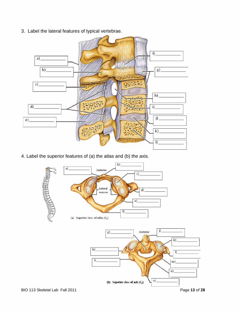
5. Label the features of the medial surface of the left foot.
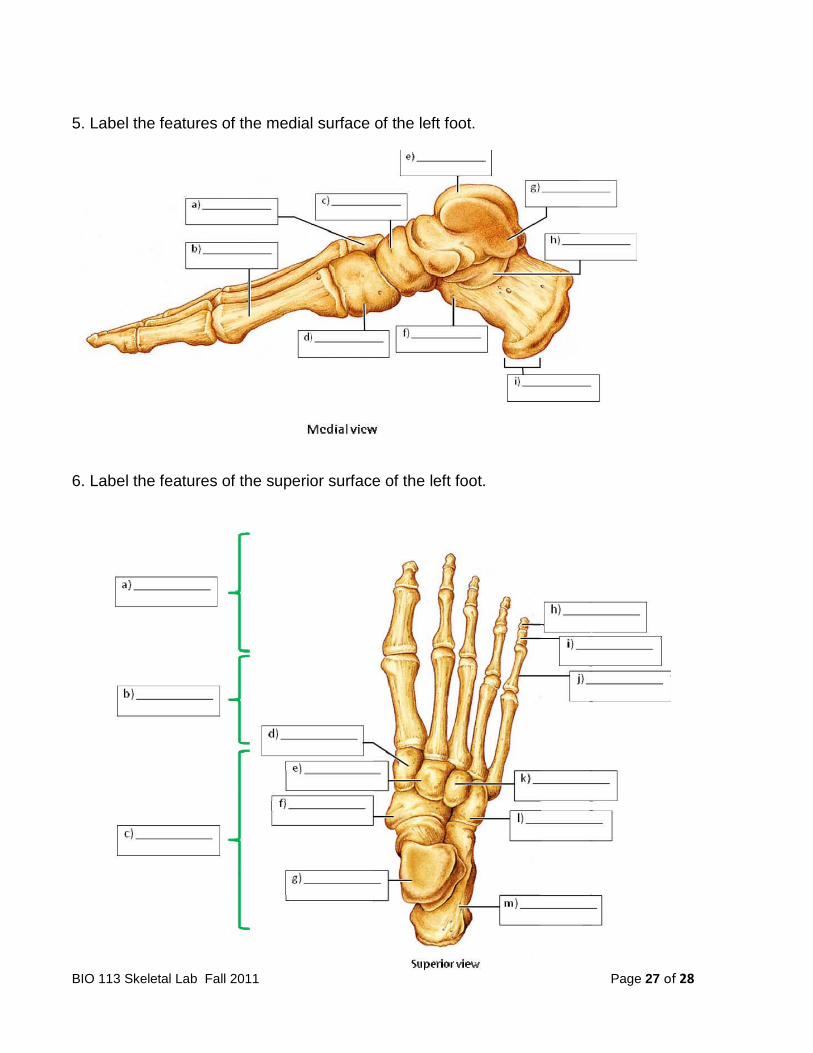
6. Label the features of the superior surface of the left foot.

BIO 113 Skeletal Lab Fall 2011 Page 28 of 28
7. Complete the following statements:
a) The pelvic girdle consists of two ____________________.
b) The head of the femur articulates with the ____________________ of the hip bone.
c) The ____________________ is the largest portion of the coxal bone.
d) The distance between the ____________________ represents the shortest diameter of the
pelvic outlet.
e) The pubic bones come together anteriorly to form the joint called ____________________.
f) The ____________________ is the portion of the coxal bone that causes the prominence of
the hip.
g) When a person sits, the ____________________ of the ischium supports the weight of the
body.
h) The angle formed by the pubic bones below the symphysis pubis is called the
____________________.
i) ____________________ is the largest foramen in the skeleton.
j) The ilium joins the sacrum at the ____________________.
8. Match the terms in the first column with the definitions in the second column. Place the letter of your choice in the space provided.
A Femur middle phalanx
B Fibula lesser trochanter
C Metatarsals medial malleolus
D Patella fovea capitis
E Phalanges calcaneus
F Tarsals lateral cuneiform
G Tibia tibial tuberosity
talus
linea aspera
lateral malleolus