biological photochemistry: the fate of electronic excited states in proteins, dna, and the role of...
Post on 22-Dec-2015
219 views
TRANSCRIPT

Biological Photochemistry: The fate of electronic excited states
in proteins, DNA, and the role of quenching
Robert J. StanleyRobert J. Stanley
DOE Workshop on Aqueous ScintillatorsJanuary 19, 2010
emple Chemistry DepartmentPhiladelphia, PAwww.chem.temple.edu

Electronic excited states in Biology
• Chemiluminescence– Bioluminescence – charge transfer? radicals?
• Photoinduced electron transfer– Photosynthesis– DNA repair
• Photochemistry– DNA damage– photosensors

DNA…a polymer of nucleotides connected by DNA…a polymer of nucleotides connected by phosphodiester linkagesphosphodiester linkages
5’
3’
Nucleic acid bases A, T, C, & G
Voet and Voet, Biochemistry, 2nd Ed. Wiley, New York, 1995

B-DNA is double-stranded (ds) DNA,B-DNA is double-stranded (ds) DNA,
forming the famous double helixforming the famous double helix
(1954 - Watson, Crick, Franklin)(1954 - Watson, Crick, Franklin)Watson-Crick base pairing
(complementarity)

DNA absorbs UV radiationDNA absorbs UV radiation
240 260 280 300 320 340 360 380 400
0.0
0.2
0.4
0.6
0.8 5'-CTCCPACTTGC-3' 5'-GCAAGTTGGAG-3' dsDNA
Abs
orba
nce
(cor
rect
ed)
Wavelength (nm)
P=6MAP
* transition

Quenching of excited states can be desirous or devastating in living systems: DNA
• UV light absorbed by DNA is rapidly transformed into heat – Conical intersections in the potential surfaces of excited
and ground state nucleic acid bases leads to ultrafast degradation of light into heat (10-12 sec.) …GOOD!
• Excited native DNA bases (Guanine, Adenine, Thymine, Cytosine) can be either excited state donors or acceptors– sequence dependent reaction– *G8-oxo-G– T-T T<>T pyrimidine dimerization– Cancer, apoptosis…BAD
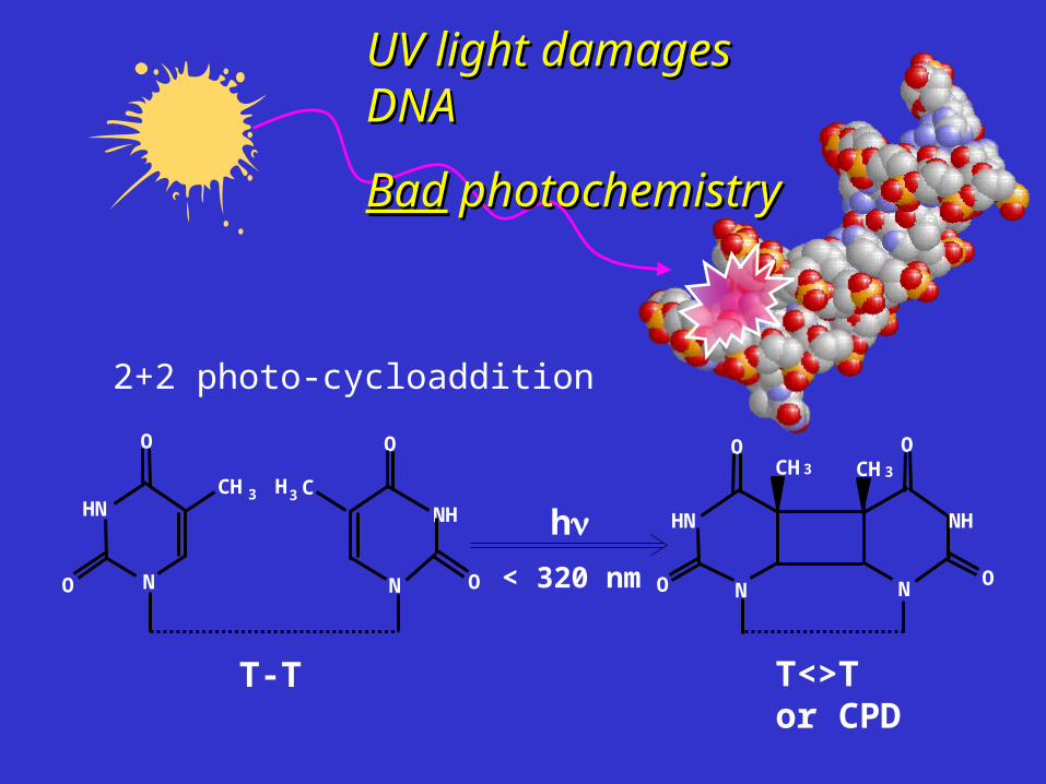
UV light damages DNAUV light damages DNA
BadBad photochemistry photochemistry
h
N
NH
O
T-T
O
O O
HN
N
CH 3
N
HN
O
O N
O
O
T<>Tor CPD
CH3 CH3
NH
C3H
< 320 nm
2+2 photo-cycloaddition

If DNA damage is left unrepaired If DNA damage is left unrepaired then mutations, cell death, and cancer then mutations, cell death, and cancer
can developcan develop
http://toms.gsfc.nasa.gov/ery_uv/euv.html

Förster orDexter Transfer(singlets)
Triplet Energy Transfer
Fluorescence
D*A
hD
DA
3
3 1
*
or
ISCD A DA
D A D A
DA*
hA
BrightDarkBright or Dark
Pathways involving energy transfer
D = G*, A*, C*, T*
A = G, A, C, T

ConicalIntersection
Intramolecularvibrationalrelaxation
Fluorescence
D*ABrightDark
hD
DA
“Structural” quenching pathways
DhotA

PhotoinducedElectron Transfer(PET)
Exciplex (EX) formation(charge transfer) Fluorescence
D*A
hD
DA
or D A D A
Pathways involving electron transfer
or D A D A
BrightDarkBright or Dark
hEX?

•Repair of the thymidines is Repair of the thymidines is
direct: direct:
T<>TT<>T T-T without modifying T-T without modifying
the DNA backbonethe DNA backbone
•Wide spread: E. coli, Frogs, Wide spread: E. coli, Frogs, Rice, Kangaroos…Humans (no!)Rice, Kangaroos…Humans (no!)
Enzymatic Repair of CPDs by Enzymatic Repair of CPDs by DNA PhotolyaseDNA Photolyase uses uses blue light as an energy source (Good photochemistry)blue light as an energy source (Good photochemistry)
Sancar, A. Structure and function of DNA photolyase. Biochemistry 33, 2-9 (1994).
Possible Applications:•Photosomes® (AGI Dermatics)•transgenic crops (wheat?)
Mees, A., et al (2004) Science 306, 1789-1793.

• PL functions efficiently with PL functions efficiently with only only FADFAD (required for (required for repair repair andand binding binding
• PL binds the CPD with high PL binds the CPD with high affinity (no light required):affinity (no light required):
KKAA = 10 = 109 9 MM-1 -1 for dsDNA with CPDfor dsDNA with CPD
DNA Photolyase (PL) is aDNA Photolyase (PL) is a flavoprotein flavoprotein (Vitamin B(Vitamin B22) that binds and repairs CPDs) that binds and repairs CPDs
Park, H.-W., Kim, S.-T., Sancar, A., and Deisenhofer, J. (1995) Science 268, 1866-72.

Biochemistry 2Biochemistry 2ndnd Ed., Voet and Voet, J. Wiley & Sons Ed., Voet and Voet, J. Wiley & Sons
Flavin Structure and Flavin Structure and Oxidation StatesOxidation States
•Flavins can transfer 1 Flavins can transfer 1 oror 2 electrons (unlike 2 electrons (unlike nicotinamide) and are nicotinamide) and are used in a large number of used in a large number of redox reactions in the cellredox reactions in the cell
•Surprisingly, flavins Surprisingly, flavins are a major biological are a major biological chromophore (DNA chromophore (DNA repair, circadian rhythms, repair, circadian rhythms, phototropism, etc.)phototropism, etc.)
FADH—
—

Photolyase functions by Photoinduced Electron Photolyase functions by Photoinduced Electron Transfer from the FAD to the CPDTransfer from the FAD to the CPD
•A large separation between the A large separation between the FADHFADH-- and the CPD (~16 and the CPD (~16 ÅÅ) ) would give a slow electron would give a slow electron transfer rate (ktransfer rate (keTeT, from Marcus , from Marcus
theory)theory)
kTGreT eek 4/)(2 2
Orbital overlap x Driving force
•Slow electron transfer would Slow electron transfer would compete poorly with compete poorly with 11FADHFADH—— deactivation (about 5 ns)deactivation (about 5 ns)
but but repairrepair > 0.7! > 0.7!
There’s a cavity in the protein
FAD

What happens to substrate conformation What happens to substrate conformation upon binding to Photolyase?upon binding to Photolyase?
Base Flipping
Photolyase
Minor disruption
Moderate disruption
Severe disruption
AA
T<>T

Fluorescent reporter approach to probing Fluorescent reporter approach to probing double helical structuredouble helical structure
Base Flipping5’
5’
3’
3’
5’
5’
3’
3’
5’-probe approach:
Base Flipping5’
5’
3’
3’
5’
5’
3’
3’
3’-probe approach:
The fluorescence quantum yield of the reporter decreases when base
stacked…but why?

6MAP is an attractive new fluorescent 6MAP is an attractive new fluorescent adenosine analogueadenosine analogue
Properties:1
fl = 0.2
ex = 330 nm ( ~ 8,500 M-1cm-1)
em= 430 nm (large Stokes shift)
1Hawkins, et al, “Synthesis and Fluorescence Characterization of Pteridine Adenosine Nucleoside Analogs for DNA Incorporation.” Anal. Biochem.298, 231-240 (2001).
4-amino-6-methyl-8-(2-deoxy--D-ribofuranosyl)-7(8H)-pteridone
310 320 330 340 350 360 370 380 390 400 410
0.00
0.01
0.02
0.03
ss-3'-6MAP ds-3'-6MAP-TT ds-3'-6MAP-CPD ss-5'-6MAP ds-5'-6MAP-TT ds-5'-6MAP-CPD
Abs
orba
nce
(cor
rect
ed)
Wavelength (nm)
K. Yang, S. Matsika, and R.J. Stanley, Biochemistry 2007
N N
H
O
O
ThymineR
N8
7
6
N 4
N
2
N
N
O
6MAP
H H
CH3
3
CH3C 1'
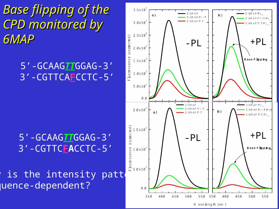
Base flipping of the Base flipping of the CPD monitored by CPD monitored by 6MAP6MAP
5’-GCAAGTTGGAG-3’3’-CGTTCAFCCTC-5’
5’-GCAAGTTGGAG-3’3’-CGTTCFACCTC-5’
Why is the intensity pattern sequence-dependent?
-PL +PL
-PL +PL

These data are consistent with disruption of base These data are consistent with disruption of base stacking due to base flipping of the CPD by stacking due to base flipping of the CPD by
PhotolyasePhotolyase
Photolyase
Mees et al, Science v. 306, 1789-1793 (2004)
?

Is the fluorescence quantum yield modulation of Is the fluorescence quantum yield modulation of 6MAP due to PET?6MAP due to PET?
Stern-Volmer quenching of 6MAP by G,A,C, and T:what is the rate of quenching, kq?
What are the redox potentials?Cyclic voltammetry of 6MAP in aprotic organic solvents
submitted to Biochemistry

The quenching of 6MAP* proceeds through The quenching of 6MAP* proceeds through nucleobase oxidation:nucleobase oxidation:
6MAP*:NMP6MAP*:NMP6MAP6MAP¯:NMP¯:NMP++
(Scandola-Balzani relation)
FBA NB GET(eV) Eact(eV)
6MAP
G -0.63 0.000
A -0.16 0.003
C 0.021 0.048
dT -0.009 0.032
submitted to Biochemistry
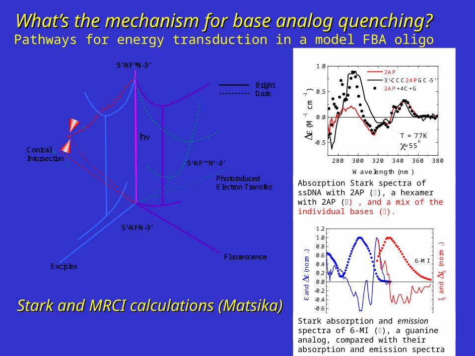
What’s the mechanism for base analog quenching?What’s the mechanism for base analog quenching?Pathways for energy transduction in a model FBA oligo
ConicalIntersection
PhotoinducedElectron Transfer
ExciplexFluorescence
5’-NF*N-3’
BrightDark
5’-NFN-3’
5’-NF +N--3’
hConicalIntersection
PhotoinducedElectron Transfer
ExciplexFluorescence
5’-NF*N-3’
BrightDark
5’-NFN-3’
5’-NF +N--3’
h
280 300 320 340 360 380
-0.5
0.0
0.5
1.0
(
M-1 c
m-1)
Wavelength (nm)
2AP 3'-CCC2APGC-5' 2AP+4C+G
T = 77K=55
Absorption Stark spectra of ssDNA with 2AP (), a hexamer with 2AP () , and a mix of the individual bases ().
300 350 400 450
-0.6-0.4-0.20.00.20.40.60.81.01.2
I F a
nd
I F (
norm
.)
and
(
norm
.)
Wavelength (nm)
6-MI
Stark absorption and emission spectra of 6-MI (), a guanine analog, compared with their absorption and emission spectra ().
Stark and MRCI calculations (Matsika)Stark and MRCI calculations (Matsika)

380 400 420 440 460 480 500 520 540
0
200000
400000
600000
800000
1000000
1200000
1400000
1600000
1800000
Wavelength (nm)
ss-6MAP/PLox
ss-6MAP/TT/PLox
ss-6MAP/T<>T/PLox
380 400 420 440 460 480 500 520 540
0
200000
400000
600000
800000
1000000
1200000
1400000
1600000
1800000
Wavelength (nm)
Another possibility: Another possibility: 6MAP emission overlaps the absorption of the 6MAP emission overlaps the absorption of the FAD: FRET from 6MAP*FAD: FRET from 6MAP*FAD?FAD?
Yang et al, JPC B (2007)

60
6 60
ETDA
R
R r
R0 the Förster distance where ET = 0.5
rDA the distance between a donor (fluorescent analogue) and an acceptor (FAD in photolyase)
Fluorescence Energy Transfer Efficiency Fluorescence Energy Transfer Efficiency

R0 (Å) = 6/142 )(211.0 Jn D
The Förster distanceThe Förster distance
2 : the orientation factor;n : the refractive index of the medium;D : the fluorescence quantum yield of the donor; J : the overlap integral.

dF
dFJ
D
AD
)(
)()( 4
FD(): the fluorescence intensity of the donor as a function of wavelength.
εA(): the molar extinction coefficient of the acceptor at that wavelength;
The Overlap IntegralThe Overlap Integral
350 400 450 500
FD
A
Wavelength (nm)

22 )coscos3(cos ADT
θT: mD, mA
θD: mD , rDA
θA: mA, rDA
The Orientation FactorThe Orientation Factor
R
N8
7
6
N
4 N
2
N
H2N
O
6MAP in 3'-6MAP
3
H3C
8
76
5a
9a9
N5
4a
10a
N10 4
NH
2N
1
H3C
H3C
O
O
R
FADox in Photolyase
mD mArDA

The transition dipole moment directionThe transition dipole moment direction6MAP was calculated from TD-DFT6MAP was calculated from TD-DFT
Yang et al, JPC B (2007)
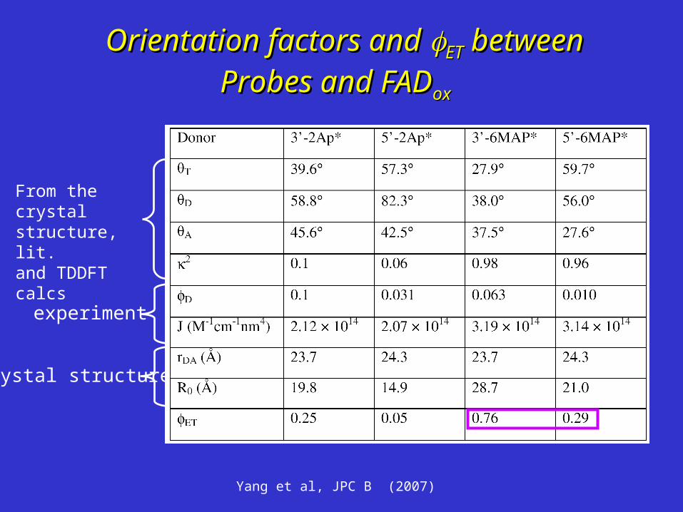
Orientation factors and Orientation factors and ETET between between
Probes and FADProbes and FADoxox
From the crystal structure, lit. and TDDFT calcs
crystal structure
experiment
Yang et al, JPC B (2007)

FRET efficiency vs. orientationFRET efficiency vs. orientation
60 70 80 90 100 1100.0
0.2
0.4
0.6
0.8
1.0
3'-6MAP/FAD (m1)
5'-6MAP/FAD (m1)
FRET
(deg.)
)6( MAPxtal
Yang et al, JPC B (2007)

NO FRET!NO FRET!
400 450 500 550 600
-0.04
-0.02
0.00
0.02
0.04
I [I
(6M
AP
/T<
>T
/PL)
-I(6
MA
P/T
<>
T)]
Wavelength (nm)
• The FAD is quenched 100x in the protein (acceptor is dark)
• A work-around : time-resolved FRET?
• Quenching mechanism is different for the two probes
• photoinduced electron transfer vs. ultrafast internal conversion?
• Does FAD* undergo PET to tryptophan???
Yang et al, JPC B (2007)

Can we identify the kinetics and mechanism of repair? Two color pump probe femtosecond spectroscopy:
•What is the electron transfer
lifetime (eT)?
•Does repair proceed by a concerted or sequential mechanism?
PLred : T<>T
1
5
4
3
2
6
1PLred : T<>T
PLsq• : T<>T •
PLsq• : T|_|T •
PLsq• : T-T •
PLred + T-T
PLred : T-T
7
eT
krec
1
2
kbeT
kdiss
kic, krad
h
c
MacFarlane and Stanley (2003) Biochemistry 42, 8558-8568

Ti:sapphire
CW Nd:YAG
ISO
Ti:Sapphire amplifier
Mode and wavelengthmonitor
Lasercontrol
YLF laser
CCD
Mo
no
chro
ma
to
r
Delay stagecontroller
SynchronizationDelay
Generator
ChopperController
M3
M1
M2
M4
M5
M6M
M12
M13
M7
M10
M9
M8M11
M15
M14
L1
L5L2
L4 L3
L6
L7 L8
W1 W3
W2
P1
B1
F1
F2
Transient absorption measurement layout
BBO
CaF2
Sample

0 1000 2000 3000
-0.003
-0.002
-0.001
0.000
0.001
0.002
0.003
1:5 PLred
_:(T<>T)5
A26
5
Time (ps)
PLred
_
0 20 40 60 80 100
pseT 2032
PET to the CPD substrate quenches the PET to the CPD substrate quenches the FADHFADH excited state in ~ 30 ps excited state in ~ 30 ps
MacFarlane and Stanley (2003) Biochemistry 42, 8558-8568
1
1 1
(3 ns)~ 0.01
(3 ns) (0.032 ns)
radfl
rad ET
k
k k

What’s are the intermediates?What’s are the intermediates?
A(A(,t) = ,t) = ccii(t)(t)ii(() = C(E - ) = C(E - 00))
where Ewhere Eii(() = True spectra of the intermediates) = True spectra of the intermediates
00(() = Ground state absorption spectrum) = Ground state absorption spectrum
• ConstructConstruct C(t) = CC(t) = C00eeKt Kt (from the K matrix)(from the K matrix)
• CalculateCalculate EEii ( () = C) = C-1-1A(A(,t),t)• MinimizeMinimize {{A(A(,t) – C(E- ,t) – C(E- 00)} using K )} using K
matrixmatrix
PLred : T<>T or T-T 1
4
3
2
1PLred : T<>T
PLsq• : T<>T •
PLsq• : T-T •
keT
krec
krepair
h krad
A unidirectional sequential model:A unidirectional sequential model:
rec
repairet
et
rec
k
kk
khv
khvhv
K
000
00
00
0

01000
20003000
400500
600700
0
0.005
0.01
0.015
0.02
0.025
0.03
Wavelength (nm)Time (ps)
de
lta
A
Pl-red+(TTT<>TT)The broadband The broadband
transient absorption transient absorption data:data:
01000
20003000
400
500
600
7000
0.005
0.01
0.015
0.02
0.025
0.03
0.035
Wavelength (nm)Time (ps)
de
lta A
Pl-red+(TTTTT)

400 450 500 550 600 650 700
0
0.5
1
1.5
2
2.5
3
3.5
4
4.5x 10
4 Intermediate Spectra: PLred-CPD
Wavelength (nm)
Ext
inct
ion
(M
-1 c
m-1
)Spectrotemporal intermediates in the repair reaction:Spectrotemporal intermediates in the repair reaction:
E spectraE spectra
• Fitting the data does not rule out a sequential bond breaking Fitting the data does not rule out a sequential bond breaking mechanism...mechanism...• More complicated kinetics cannot be ruled out!More complicated kinetics cannot be ruled out!
PLSQ
PLred : T<>T or T-T 1
4
3
2
1PLred : T<>T
PLsq• : T<>T •
PLsq• : T-T •
53 ps
2753 ps
540 psh
620 ps
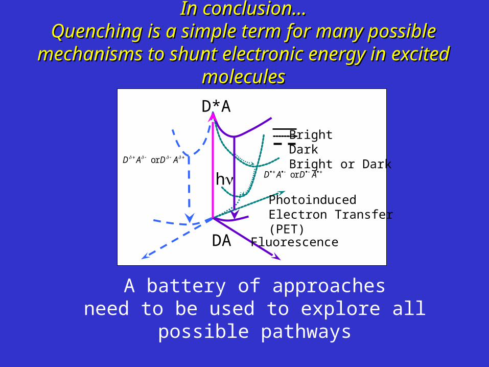
In conclusion…In conclusion…Quenching is a simple term for many possible mechanisms Quenching is a simple term for many possible mechanisms
to shunt electronic energy in excited moleculesto shunt electronic energy in excited molecules
PhotoinducedElectron Transfer(PET)
Fluorescence
D*A
h
DA
or D A D A
or D A D A
BrightDarkBright or Dark
A battery of approachesneed to be used to explore all possible
pathways

The Charge Separation Investigation Team
Goutham Kodali• Stark spectroscopy• Computational chemistry• “Vector dude”
Salim Siddiqui, M.D., Ph.D.•Stark spectroscopy•Computational chemistry
Dr. Zhanjia Hou•Ultrafast spectroscopy•Single molecule spectroscopy
Madhavan Narayanan•Ultrafast spectroscopy•Protein Chemistry
Dr. Alex MacFarlane IV•Ultrafast spectroscopy•Electric field effects

The GroupThe Group
CollaboratorsCollaboratorsProf. Aziz Sancar (UNC)Mary Hawkins (NIH)Prof. Spiridoula Matsika
FundingFundingNSF Molecular Biosciences, REUPetroleum Research Fund
Gone, but not forgotten..Gone, but not forgotten..

A closer look at the damage…A closer look at the damage…5’-GCTTAATTCG-3’5’-GCTTAATTCG-3’
3’-CGAA3’-CGAATTTTAAGC-5’AAGC-5’
Crystal structure: Park et al, PNAS 99, 15965-15970 (2002).
5’3’
AA
Base stacking is weakenedWatson-Crick base pairing is distorted
2.4Å1.9Å

DNA Photolyase (PL) binds its CPD DNA Photolyase (PL) binds its CPD substrate by base flippingsubstrate by base flipping
Mees, A., et al (2004) Science 306, 1789-1793.
Flavin Adenine Dinucleotide
CPD

350 400 450 500
0.0
0.2
0.4
0.6
0.8
1.0
Nor
mal
ized
Abs
orba
nce
FADox
(A)
3'-6map (D)
Nor
mal
ized
Flu
ores
ence
Wavelength (nm)
Spectral overlaps of probes and FADSpectral overlaps of probes and FAD
Does FRET explain the intensity pattern
difference?
S0S2 S0S1