biological mass spectrometry - rutgers...
TRANSCRIPT

Biological Mass Spectrometry
April 30, 2014

Mass SpectrometryHas become the method of choice for
• precise protein and nucleic acid mass determination in a very wide mass range
• peptide and nucleotide sequencing
• identification of protein post-translational modifications
• protein structural changes, folding and dynamics
• identification of subpicomole quantities of proteins
• identification of isotope labelling.

Ionization• Transfer of ions from solution to gas phase is a
desolation process (removal of water)
• This process requires energy.
• the transfer of Na+ ion from solution to gas phase requires 98 kcal/mol of energy
• Two types commonly used for protein mass spectrometry - (1) matrix-assisted laser desorption ionization and (2) Electrospray ionization
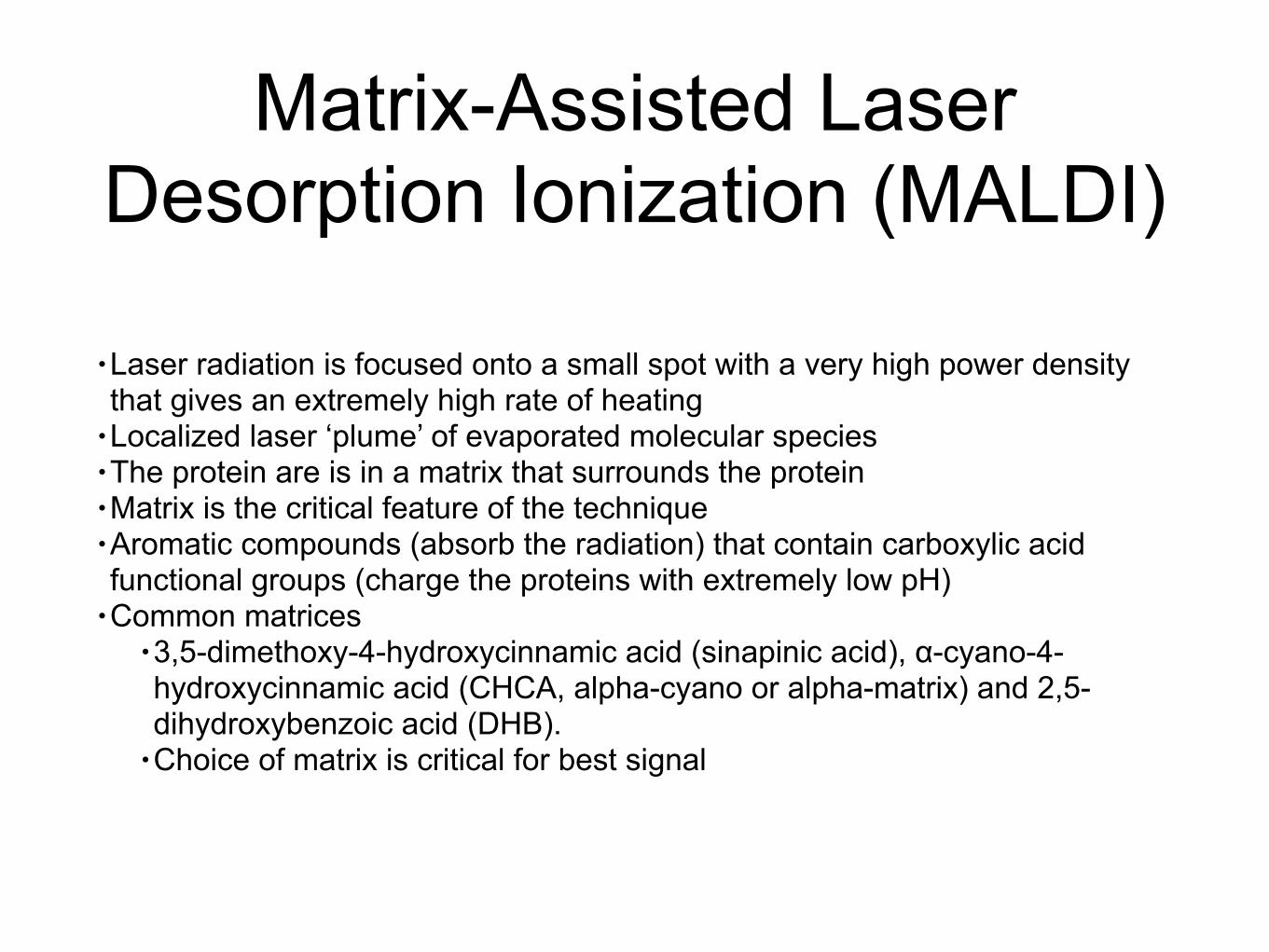
Matrix-Assisted Laser Desorption Ionization (MALDI)
• Laser radiation is focused onto a small spot with a very high power density that gives an extremely high rate of heating
• Localized laser ‘plume’ of evaporated molecular species • The protein are is in a matrix that surrounds the protein • Matrix is the critical feature of the technique • Aromatic compounds (absorb the radiation) that contain carboxylic acid functional groups (charge the proteins with extremely low pH)
• Common matrices • 3,5-dimethoxy-4-hydroxycinnamic acid (sinapinic acid), α-cyano-4-hydroxycinnamic acid (CHCA, alpha-cyano or alpha-matrix) and 2,5-dihydroxybenzoic acid (DHB).
• Choice of matrix is critical for best signal

MALDI
Absorption of laser UV Ionization of protein by matrix Ions placed into the gas phase

Time of Flight (TOF)Ions can travel in a linear fashion and be detected by the detector at the opposite end as an ion source (Time of flight)
MALDI In Practice
http://www.jove.com/video/50635/matrix-assisted-laser-desorptionionization-time-flight-maldi-tof-mass

Mass to Charge•Common to get multiple charged species for the same molecule [M+H]+, [M+2H]2+, [M+3H]3+, etc !
•Mass is referred to mass/charge (m/Z) !
•Number of protons that attach to a peptide or protein correlates with the total number of basic amino acids (Arg, Lys, His) plus the N-terminal amino group !
•The distribution of charge states thus depends on pH, temperature and any denaturing agent

Electrospray Ionization (ESI)• ESI produces intact ions from sample molecules directly
• Ions are formed by applying a 1-5 kV voltage to a sample solution emerging from a capillary tube, at a low flow rate (1-20 nl/min).
• The solvent evaporates from the droplets as they move from the atmospheric pressure of the ionization region into the vacuum chamber containing the mass analyzer.
• Evaporation of the solvent is aided either by a counter-current flow of drying gas or by heating the tube
• ESI is one of the most gentle ionization methods available, yielding no molecular fragmentation in practice
• Considered the more accurate than MALDI for small proteins

Mass Resolution•Separate mass signals is affected by the resolving power of the mass spectrometer. •Resolution R in mass spectrometry is defined as R = m/Δm, where Δm is the mass difference of two neighboring masses, m and m + Δm, of equal intensity, with signal overlap of 10%.
•one can resolve 100,000Da from 100,001Da then the resolution is 10 parts per million (ppm)
•Two definitions •10% valley definition in which the two adjacent peaks each contribute 5% to the valley in between them.
•fullwidth, half-maximum (FWHM) mass of the peak (in daltons) divided by the width (in daltons) measured at the half-height of the peak
•FWHM is approximately twice that of the 10% valley definition

Mass Accuracy
• Difference between measure and calculated mass
• The accuracy is stated as a percentage of the measured mass (10,000 ± 0.01%) or ppm (10,000 ± 100ppm)
• Therefore, as the mass increases so does the absolute mass error

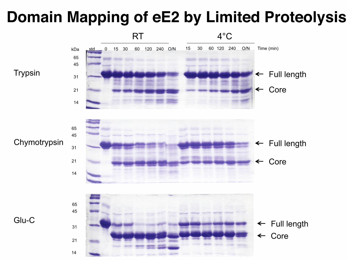
Domain Elucidation by Limited Proteolysis and Mass Spectrometry
What is a potential problem with this model?

Domain Elucidation• In order to obtain high-resolution structures, it appears important to work with compact, well-defined proteins.
• degrees of conformational motion in a protein increases the chance that it will crystallize into well-ordered lattices.
• A precisely defined folding domain also results in a protein with reduced tendencies for aggregation
• Limited proteolysis exposure of protein to trace amounts of proteases with different specificities
• Trypsin cleaves C-terminal to Arg/Lys • Glu-C (V8 protease) C-terminal to Glu/Asp • Chymotrypsin C-terminal to Tyr, Phe, Trp
• Why these enzymes? • Fragments are determined by mass spectrometry

mRNA Cap-Binding Protein
Full length (residues1-217)
Folded region construct (28-217)
std 0 15 30 60 120 240 O/N 15 30 60 120 240 O/N
14
31
45
21
65
14
31
45
21
65
14
31
45
21
65
kDa Time (min)
4°CRT
Trypsin
Chymotrypsin
Glu-C
Domain Mapping of eE2 by Limited Proteolysis
Full length
Core
Full length
Core
Full lengthCore

Structure Determination HCV E2 Core Bound to Fab
* Trypsin * Chymotrypsin * Glu-C
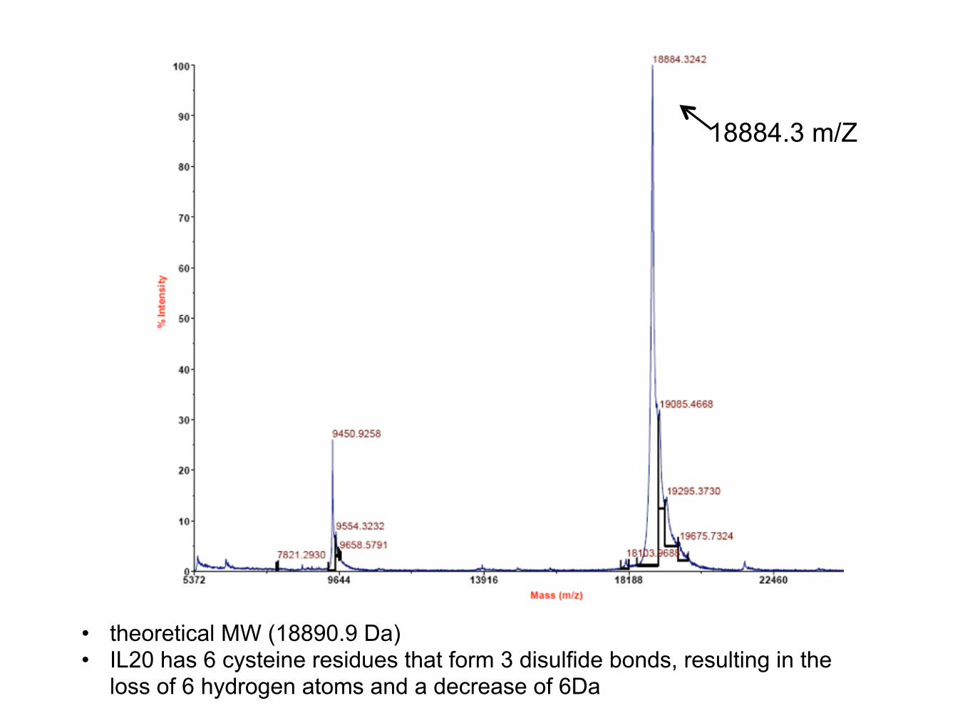
18884.3 m/Z
• theoretical MW (18890.9 Da) • IL20 has 6 cysteine residues that form 3 disulfide bonds, resulting in the
loss of 6 hydrogen atoms and a decrease of 6Da

1 4 9 5 4 2 7 0 3 4 3 9 1 1 4 5 1 1 9 4 6 3 2 7 4 7 5 3 5 4M a s s (m /z )
7 3 0 .2
0
1 0
2 0
3 0
4 0
5 0
6 0
7 0
8 0
9 0
1 0 0
% In
te
ns
ity
4700 Linear S pec #1[BP = 45911.8, 730]45633 .5039
22865 .6230
Mass Spectrometry
Produced in HEK293T GNTI- (N-acetylglucosaminyltransferase I) cell line
Mass (m/Z)
45,481.8 m/Z
Predicted protein mass 32,107.51 Da
!Glycosylation (Man5GlcNAc2) 1216Da x 11 sites= 13,376 Da
!Total= 45,483.5 Da