biogeochemistry: methane and microbes
TRANSCRIPT
© 2006 Nature Publishing Group
NEWS & VIEWS NATURE|Vol 440|13 April 2006
878
Methane is not only a fossil fuel but also a keyplayer in the carbon cycle. About 1% of thecarbon dioxide annually fixed by photosyn-thesis is converted back to carbon dioxide bymicroorganisms via methane, which amountsto 1 billion tonnes of methane formed andconsumed per year. Moreover, methane is apowerful greenhouse gas, and its concentra-tion in the atmosphere has increased continu-ously over the past hundred years.
It is therefore news in itself that Raghoe-barsing and colleagues have identified a hitherto unknown microbial sink for methane(page 918 of this issue)1. Their discovery of the microbial consortium that mediates theprocess is also exciting — the microorganismsinvolved couple the oxidation of methane todenitrification (nitrate reduction to nitrogen)in the absence of oxygen (Fig. 1), which from amechanistic viewpoint is a challenging reac-tion (alongside benzene, methane is by far themost unreactive hydrocarbon).
Raghoebarsing et al. isolated the methane-oxidizing microbial consortium from theanoxic sediments of a freshwater canal thatwas subject to agricultural run-off with a highnitrogen load (Fig. 2). The sediment was satu-rated with methane and contained nitrate at aconcentration of 1 mM. The culture obtainedgrew on methane and nitrate (or nitrite) as thesole energy sources, with an estimated dou-bling time of more than one month. After 16months, the culture mainly consisted of twodifferent types of microorganism, a bacterium(80%) and an archaeon (10%).
Sequence analysis of the microorganisms’genomes revealed that the dominant bac-terium belonged to a novel phylum withoutany documented cultured species, and that the archaeon was distantly related to archaeainvolved in the anaerobic oxidation ofmethane with sulphate. Similar sequences to those identified by Raghoebarsing et al.1
have been retrieved from the denitrifyingzone of freshwater sediments of Lake Biwa,Japan, and from contaminated ground-water in the United States1, where methanedisappears in the nitrate zone of the ground-water2. So the anaerobic oxidation of meth-ane coupled to denitrification is probably
of widespread ecological importance.Why did it take until now to identify this
process? The main reason is probably that itproceeds at much lower rates than the anaero-bic oxidation of other organic compounds or ofhydrogen sulphide with nitrate. So the processis evident only in anoxic environments with
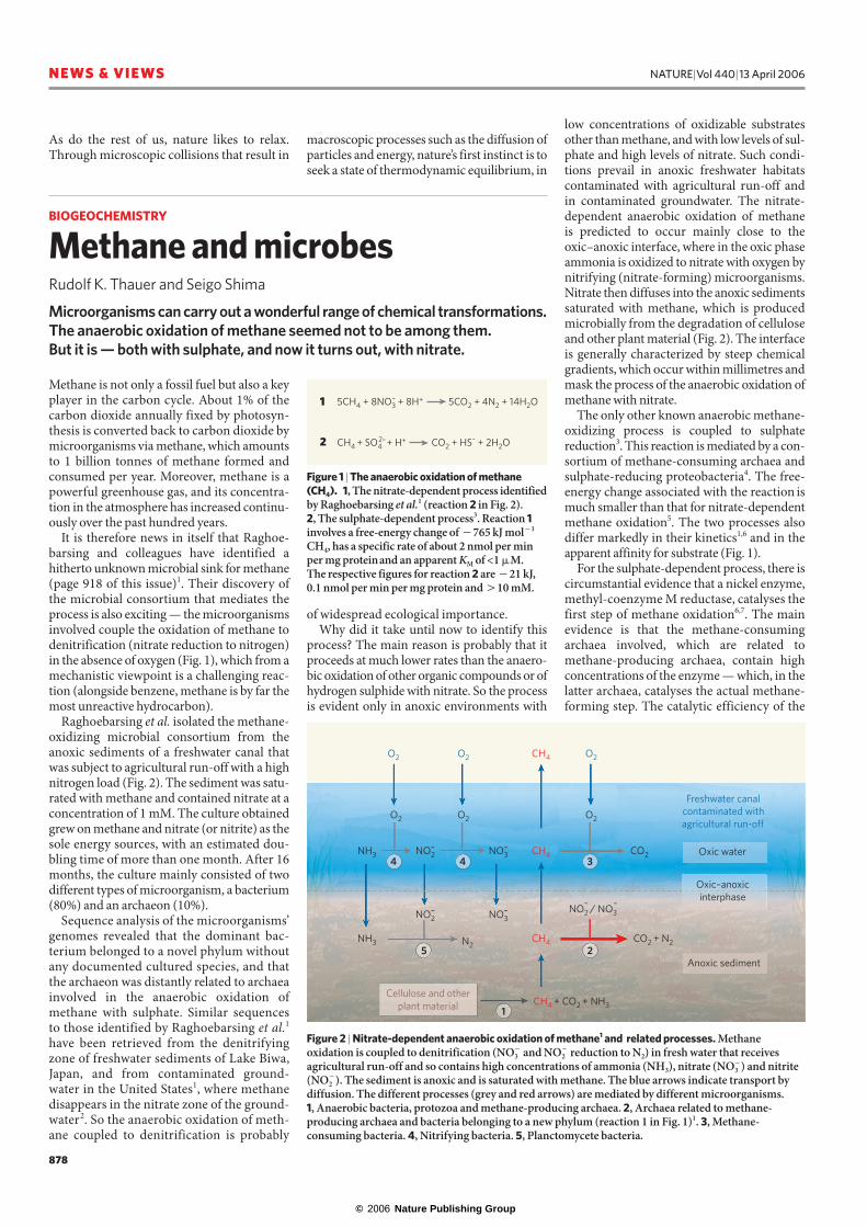
low concentrations of oxidizable substratesother than methane, and with low levels of sul-phate and high levels of nitrate. Such condi-tions prevail in anoxic freshwater habitatscontaminated with agricultural run-off and in contaminated groundwater. The nitrate-dependent anaerobic oxidation of methane is predicted to occur mainly close to theoxic–anoxic interface, where in the oxic phaseammonia is oxidized to nitrate with oxygen bynitrifying (nitrate-forming) microorganisms.Nitrate then diffuses into the anoxic sedimentssaturated with methane, which is producedmicrobially from the degradation of celluloseand other plant material (Fig. 2). The interfaceis generally characterized by steep chemicalgradients, which occur within millimetres andmask the process of the anaerobic oxidation ofmethane with nitrate.
The only other known anaerobic methane-oxidizing process is coupled to sulphate reduction3. This reaction is mediated by a con-sortium of methane-consuming archaea andsulphate-reducing proteobacteria4. The free-energy change associated with the reaction ismuch smaller than that for nitrate-dependentmethane oxidation5. The two processes alsodiffer markedly in their kinetics1,6 and in theapparent affinity for substrate (Fig. 1).
For the sulphate-dependent process, there iscircumstantial evidence that a nickel enzyme,methyl-coenzyme M reductase, catalyses thefirst step of methane oxidation6,7. The mainevidence is that the methane-consumingarchaea involved, which are related tomethane-producing archaea, contain highconcentrations of the enzyme — which, in thelatter archaea, catalyses the actual methane-forming step. The catalytic efficiency of the
NH3
NH3
N2
O2 O2 O2
O2 O2 O2
NO2 CO2
CO2 + N2
–
NO2–
NO3–
NO3– NO2 / NO3
– –
CH4
CH4
CH4
CH4 + CO2 + NH3
Oxic water
1
2
3
5
4 4
Freshwater canal contaminated with agricultural run-off
Oxic–anoxic interphase
Anoxic sediment
Cellulose and other plant material
Figure 2 | Nitrate-dependent anaerobic oxidation of methane1 and related processes. Methaneoxidation is coupled to denitrification (NO3
� and NO2� reduction to N2) in fresh water that receives
agricultural run-off and so contains high concentrations of ammonia (NH3), nitrate (NO3�) and nitrite
(NO2�). The sediment is anoxic and is saturated with methane. The blue arrows indicate transport by
diffusion. The different processes (grey and red arrows) are mediated by different microorganisms. 1, Anaerobic bacteria, protozoa and methane-producing archaea. 2, Archaea related to methane-producing archaea and bacteria belonging to a new phylum (reaction 1 in Fig. 1)1. 3, Methane-consuming bacteria. 4, Nitrifying bacteria. 5, Planctomycete bacteria.
BIOGEOCHEMISTRY
Methane and microbes Rudolf K. Thauer and Seigo Shima
Microorganisms can carry out a wonderful range of chemical transformations.The anaerobic oxidation of methane seemed not to be among them. But it is — both with sulphate, and now it turns out, with nitrate.
Figure 1 | The anaerobic oxidation of methane(CH4). 1, The nitrate-dependent process identifiedby Raghoebarsing et al.1 (reaction 2 in Fig. 2). 2, The sulphate-dependent process3. Reaction 1involves a free-energy change of �765 kJ mol�1
CH4, has a specific rate of about 2 nmol per minper mg proteinand an apparent KM of <1 �M. The respective figures for reaction 2 are �21 kJ,0.1 nmol per min per mg protein and �10 mM.
5CH4 + 8NO3 + 8H+ 5CO2 + 4N2 + 14H2O
CH4 + SO4 + H+ CO2 + HS– + 2H2O2–
–1
2
As do the rest of us, nature likes to relax.Through microscopic collisions that result in
macroscopic processes such as the diffusion ofparticles and energy, nature’s first instinct is toseek a state of thermodynamic equilibrium, in
© 2006 Nature Publishing Group
NATURE|Vol 440|13 April 2006 NEWS & VIEWS
879
used by the vesicles for identification, dockingand fusion.
By labelling a vesicle-membrane proteincalled synaptotagmin with a fluorescent tag andusing STED microscopy, Jahn and colleaguescould follow the fusion of individual vesicles atthe plasma membrane (Fig. 2 on page 937).Their study provides some of the most com-pelling evidence to date that at least some mem-brane constituents remain grouped togetherafter vesicles fuse with the plasma membrane(rather than diffusing freely within the mem-brane like a drop of water on water), which isconsistent with the kiss-and-run theory.
Although STED is not the first technique to break the diffraction limit, it is arguably the one best suited for biological imaging. For nearly two decades, near-field scanning optical microscopy has yielded images with aspatial resolution down to about 15 nm. Scan-ning is usually achieved by allowing light toescape through a tiny aperture in a metal-coated tip5,6. Materials with negative refractiveindices also hold promise for ultra-high-resolu-tion optical imaging7,8. However, these tech-niques generally require the sample to beimmediately next to the imaging element. Imag-ing of buried interfaces in live cells would beprohibitively complicated using such strategies.
By comparison, STED instrumentation isdirectly compatible with existing approaches
BIOLOGICAL IMAGING
The diffraction barrier brokenGarth J. Simpson
The conventional optical limitations of fluorescence microscopy have been defied, to achieve nanoscale resolution of individual vesicle organelles at the junctions of neuronal cells.
Traditional optical microscopy cannot easilydistinguish objects separated by less thanabout half the wavelength of visible light. Forexample, measurements of two fluorescentparticles closer together than this ‘diffractionbarrier’ generally produce one indistinct,bright blob. In practice, this equates to a maxi-mum resolution corresponding to distances ofaround 200 nm for biological imaging usingvisible light. However, Stefan Hell and col-leagues have developed a technique that over-comes the diffraction barrier and can resolveobjects separated by less than about 40 nm(refs 1–3). On page 935 of this issue, Jahn, Helland colleagues4 use the technique to providethe first in vitro images of the movements ofsingle neuronal vesicles — the membrane-bound, bubble-like organelles that mediatecommunication between neurons. Theirimages help to settle a question that has beenexercising cell biologists for some time.
The latest approach uses stimulated emis-sion depletion (STED) microscopy1–3, a technique that relies on the overlap of two light beams in the focal region. The first beam excites molecules just as in traditionalfluorescence microscopy — as the moleculeabsorbs the energy from the light, it is pro-moted up to a higher energetic state, and as it relaxes back to the ground state, it releasesthe energy in the form of light. The secondbeam, at a different wavelength, suppressesthis fluorescence by ‘stimulated emission’, inwhich molecules are actively pumped downout of the excited state by light (in essence,absorption driven backwards). In sufficientlyintense optical fields, stimulated emissionbecomes more efficient than fluorescence and
is the dominant effect, drastically reducing the fluorescence.
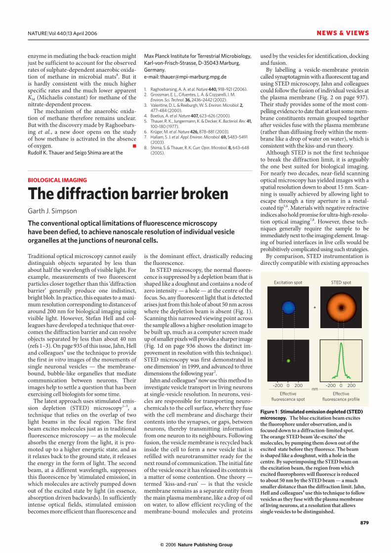
In STED microscopy, the normal fluores-cence is suppressed by a depletion beam that isshaped like a doughnut and contains a node ofzero intensity — a hole — at the centre of thefocus. So, any fluorescent light that is detectedarises just from this hole of about 50 nm acrosswhere the depletion beam is absent (Fig. 1).Scanning this narrowed viewing point acrossthe sample allows a higher-resolution image tobe built up, much as a computer screen madeup of smaller pixels will provide a sharper image(Fig. 1d on page 936 shows the distinct im-provement in resolution with this technique).STED microscopy was first demonstrated inone dimension1 in 1999, and advanced to threedimensions the following year2.
Jahn and colleagues4 now use this method toinvestigate vesicle transport in living neuronsat single-vesicle resolution. In neurons, vesi-cles are responsible for transporting neuro-chemicals to the cell surface, where they fusewith the cell membrane and discharge theircontents into the synapses, or gaps, betweenneurons, thereby transmitting informationfrom one neuron to its neighbours. Followingfusion, the vesicle membrane is recycled backinside the cell to form a new vesicle that isrefilled with neurotransmitter ready for thenext round of communication. The initial fateof the vesicle once it has released its contents isa matter of some contention. One theory —termed ‘kiss-and-run’ — is that the vesiclemembrane remains as a separate entity fromthe main plasma membrane, like a drop of oilon water, to allow efficient recycling of themembrane-bound molecules and proteins
Figure 1 | Stimulated emission depleted (STED)microscopy. The blue excitation beam excites the fluorophore under observation, and isfocused down to a diffraction-limited spot. The orange STED beam ‘de-excites’ themolecules, by pumping them down out of theexcited state before they fluoresce. The beam is shaped like a doughnut, with a hole in thecentre. By superimposing the STED beam on the excitation beam, the region from whichexcited fluorophores will fluoresce is reduced to about 50 nm by the STED beam — a muchsmaller distance than the diffraction limit. Jahn,Hell and colleagues4 use this technique to followvesicles as they fuse with the plasma membrane of living neurons, at a resolution that allowssingle vesicles to be distinguished.
Excitation spot
+
Effectivefluorescence spot
Effectivefluorescence profile
nm–200 2000 –200 2000
STED spot
enzyme in mediating the back-reaction mightjust be sufficient to account for the observedrates of sulphate-dependent anaerobic oxida-tion of methane in microbial mats8. But it is hardly consistent with the much higher specific rates and the much lower apparent KM (Michaelis constant) for methane of thenitrate-dependent process.
The mechanism of the anaerobic oxida-tion of methane therefore remains unclear. But with the discovery made by Raghoebars-ing et al., a new door opens on the study of how methane is activated in the absence of oxygen. ■
Rudolf K. Thauer and Seigo Shima are at the
Max Planck Institute for Terrestrial Microbiology,Karl-von-Frisch-Strasse, D-35043 Marburg,Germany.e-mail: [email protected]
1. Raghoebarsing, A. A. et al. Nature 440, 918–921 (2006).2. Grossman, E. L., Cifuentes, L. A. & Cozzarelli, I. M.
Environ. Sci. Technol. 36, 2436–2442 (2002).3. Valentine, D. L. & Reeburgh, W. S. Environ. Microbiol. 2,
477–484 (2000).4. Boetius, A. et al. Nature 407, 623–626 (2000).5. Thauer, R. K., Jungermann, K. & Decker, K. Bacteriol. Rev. 41,
100–180 (1977).6. Krüger, M. et al. Nature 426, 878–881 (2003).7. Hallam, S. J. et al. Appl. Environ. Microbiol. 69, 5483–5491
(2003). 8. Shima, S. & Thauer, R. K. Curr. Opin. Microbiol. 8, 643–648
(2005).