biodiversity of rhizobia that nodulate melilotus indicus l ...staff.usc.edu.eg › uploads ›...
TRANSCRIPT

7 Egypt. J.Microbiol. 50, pp. 101-117 (2015)
# Corresponding author email: [email protected]
Tel.: +2 01007500881,
Fax: +2 048 2235689,
T
Biodiversity of Rhizobia That Nodulate Melilotus
indicus L. in Egyptian Soils
Nadia H. El-Batanony, Ayat M. Bdwy* , H.E. Hassan *
and M.E. El-Lithy*#
Environmental Studies & Research Institute (ESRI), University of
Sadat City, Sadat City and *Botany Department, Faculty of
Science, Menoufia University, Shebin El-Kom, Menoufia, Egypt.
HE OBJECTIVES of this work were to describe the biodiversity
…. and the phylogeny of the selected rhizobial isolates nodulating
wild legume Melilotus indicus L. (M. indicus) from 14 different
Egyptian soils. These isolates were characterized morphologically and
physiologically on the basis of their tolerance to NaCl and pH.
Furthermore, the DNA of each rhizobial isolate was analyzed by rep-
PCR amplification fingerprinting using REP, ERIC and BOX A1R
primers. Thirty seven rhizobial isolates were obtained from the root
nodules of M. indicus. These isolates didn`t absorb Congo- red (CR)
when incubated in dark; grew poorly, or not at all, on peptone glucose
agar medium containing bromocresol purple (BCP) and acidified the
medium suggesting fast-growing rhizobia. Five isolates tolerated NaCl
up to 7%. Rhizobial isolates showed a wide diversity in their pH
tolerance. Moreover, PCR with REP and ERIC primer pairs yielded
multiple distinct DNA products for each isolate of size ranged from
approximately 177 to 3773 bp and 200 to 2921 bp, respectively. BOX
A1R primer did not reveal any polymorphism for the isolates. We can
conclude that rhizobia isolated from M. indicus from Egyptian soils
are both phenotypically and genetically diverse.
Keywords: Genetic Diversity, Legume Nodules, Melilotus, Rhizobia.
In fact, rhizobial distribution and survival in natural habitats are due to the impact
of the environmental factors prevailing in such habitats and the existence of the
proper host. Also, the existence of diverse rhizobia helps the host legumes to be
adapted to many different habitats (Bala & Giller, 2006). While, the great
diversity and the vast geographic distribution of the legumes shaped their distinct
rhizobial populations and drove their diversification (Drew & Ballard, 2010).
Therefore, the diversity of rhizobia present in certain ecosystems is a result of
interactions between rhizobia, their host legumes, biotic and abiotic factors of the
ecosystem (Yan et al., 2014).
In the Rhizobium-legume symbiosis, the process of N2 fixation is strongly
related to the physiological status of the host plant. Therefore, competitiveness
and persistence of rhizobial strains are not expected to be expressed in full

NADIA H. EL-BATANONY et al.
Egypt. J. Microbiol. 50 (2015)
102
capacity for nitrogen fixation if restrictive factors, such as salinity, unfavorable
soil pH, impose limitations on vigor and growth of the host legume. A given
stress may also have more than one effect: e.g., salinity may act as a water stress,
which affects photosynthetic rate, or may affect nodule metabolism directly
(Zahran, 1999). Such stresses suppress growth and symbiotic power of most
rhizobia (Gálvez, 2005).
The different responses of rhizobial strains to stress factors could be
considered as basic criteria for differentiation and identification of these bacteria
(Zahran et al., 2012). Regarding rhizobia, soil pH and salinity are the main
ecological factors determining their diversity (Adhikari et al., 2012). It is
expected that the genetic variation for traits in nature reflect their adaptation to
specific environments (Koornneef et al., 2004).
Studying rhizobial biodiversity enables isolation of an effective isolates from
wild legumes that could be used in reforestation programs. Also, it`s a good
strategy to improve quality and productivity of leguminous crops (food and feed)
when inoculated with such isolates (Zahran et al., 2012). Rhizobia of wild
legumes may have better traits such as phosphorous solublization, producing
plant growth promoting (PGP) compounds as indole acetic acid (IAA) and/or
hydrogen cyanide (HCN) and/or possessing antibiosis effect (Alikhania &
Yakhchali, 2009 and Arora et al., 2001). Also, being tolerant to different stressful
conditions more than the specific rhizobia of the cultivated leguminous crops
(Abdel-Salam et al., 2010 and Zahran et al., 2012). Such bacteria are very
important from both economical and environmental points of view (Kesari et al.,
2013).
Several molecular techniques have been readily developed to examine
rhizobial biodiversity. These include: RAPD-PCR (random amplified
polymorphic DNA-polymerase chain reaction), PCR amplification of REP
(repetitive extragenic palindrome), ERIC (enterobacterial repetitive intergeneric
consensus) and BOX element or RFLP (random amplified length polymorphism)
of amplified ribosomal genes (16S, 23S or 16S-23S inter generic spacer - IGS),
and nod or nif genes sequencing (De Bruijn, 1992; Edulamudi et al., 2015;
Laguerre et al., 1994 and Versalovic et al., 1994). Also, amplified ribosomal DNA
restriction analysis (ARDRA) of both 16S and 23S rDNA (Shamseldin et al., 2005)
have been used to study the variation that exist for rhizobial isolates.
In Egypt, wild legumes, such as M. indicus plants, are widely distributed
through the Mediterranean costal region, the Nil Delta and the deserts where
there is a remarkable diversity of such plant species. M. indicus, sometimes
incorrectly written M. indica, is a yellow-flowered herb native to northern Africa,
Europe and Asia, as forage and as a soil improver (Velázquez et al., 2010).
Nevertheless, little information is known about the diversity of the endophytic
rhizobia associated with M. indicus plants and their importance to legume
establishment and growth.
BIODIVERSITY OF RHIZOBIA THAT NODULATE …
Egypt. J.Microbiol. 50 (2015)
103
Accordingly, the present work aimed to describe the biodiversity and the
phylogeny of endophytic rhizobia nodulating wild legume M. indicus from 14
different Egyptian soils.
Material and Methods
Plant sampling and nodules collection
Fourteen locations were selected to study endophytic rhizobial diversity that
exists in root nodules of M. indicus (L.) All. These locations were pinpointed by
GPS (Magellan GPS 310), which distributed from East to West Mediterranean
sea coast and within Nile Delta, (Table 1). Plants were sampled at the flowering
and/or fruiting stage from the 14 locations. The collected plants were identified
according to Täckholm (1974) and Boulos (2000). Nodules were collected and
preserved according to Somasegaran & Hoben (1994).
TABLE 1. Locations of the collected plants, date of collection, latitude, longitude,
and isolates number (no)/location. Alex=Alexandria.
No Locations Date Latitude Longitude
Isolates no/ location
1 Abo Shnar 1/4/2014 N 31 17 01 E 34 09 94 1.2, 1.2*
2 Rafah 1/4/2014 N 31 08 67 E 33 51 58 8.1, 8.7
3 Arish 1/4/2014 N 30 54 12 E 32 24 09 9.1, 9.17, 9.19
4 Port Said 31/3/2014 N 30 42 27 E 32 16 12 4.8, 4.17, 4.21
5 Ismailia 13/4/2014 N 30 31 49 E 32 09 49 11.1, 11.2
6 Ismailia 31/3/2014 N 30 29 86 E 32 07 13 3.6, 3.12*
7 Kafr Al-Shaykh 19/4/2014 N 31 05 01 E 30 57 42 13.1, 13.2
8 Rasheed 19/4/2014 N 31 22 19 E 30 24 57 16.3, 16.4
9 40Km West Alex 20/4/2014 N 30 55 39 E 29 28 09 14.1, 14.2, 15.1, 15.2
10 55Km West Alex 14/4/2014 N 30 54 59 E 29 26 52 6.2
11 El Dabaa 14/4/2014 N 31 00 57 E 28 33 46 7.1, 7.9
12 Shebeen El-Kom
(Unifarm) 4/4/2014 N 30 55 77 E 31 01 74 10.1, 10.11
13 El Sadat City
(Unifarm)
7/3/2014 N 30 23 07 E 30 30 55 12.1, 12.2, 12.3, 12.4
14 Faiyum (Tamya) 25/4/2014 N 29 29 07 E 30 54 40 2.2, 2.15, 2.36, 2.4, 2.7, 2.8
Isolation and purification of rhizobia from nodules
Endophytic bacteria were isolated from root nodules according to Vincent
(1970) onto yeast extract mannitol agar (YEMA) containing Congo red (CR).
The well purified single colony was streaked onto peptone glucose agar plates
containing bromocresol purple (BCP) to confirm rhizobial selection. Well
isolated colonies were re-streaked onto YEMA for better purification then single
colony was selected and streaked on YEMA slants containing 1g calcium
carbonate per liter then stored at 4°C.

NADIA H. EL-BATANONY et al.
Egypt. J. Microbiol. 50 (2015)
104
Growth characterization of rhizobial isolates
The morphological traits (Elevation, margin, transparency, color, diameter
and texture) and the production of acid or alkali were recorded by growing the
isolates respectively on YEMA supplemented with CR and on YEMA
supplemented with Bromothymol blue (BTB) (Somasegaran & Hoben, 1994 and
Shetta et al., 2011). Gram-stain reaction of the different rhizobial isolates and cell
morphology were examined (Vincent, 1970).
Physiological characterization of rhizobial isolates
Several experiments were performed in broth tubes or agar plates inoculated
with an exponentially growing liquid culture. All the experiments were carried
out in triplicate otherwise it will be indicated. Bacterial growth was quantified as
0 (no growth), 1 (low growth), 2 (medium growth) or 3 (high growth = the
control) otherwise it will be specified.
NaCl tolerance
Rhizobial isolates were grown on YEM broth at different concentrations of
NaCl ranged from 1 to 7% (w/v) to test their salt tolerance. Growth was
determined after 72 h of incubation at 28-30°C by measuring the turbidity at
550nm using spectrophotometer (Metertek SP-850) (Singh et al., 2008).
pH tolerance
The ability of isolates to grow in acidic or alkaline media was determined on
YEMA Petri dishes, where pH was adjusted either to 4, 5, 6, 9, 10 or 11.
Bacterial growth was recorded (from 0 to 3), after 4 days of incubation at 28-
30°C, compared to the control that was adjusted to pH 6.8 (Beauregard et al.,
2004).
Molecular characterization of rhizobial isolates
DNA isolation and gel electrophoresis
DNA was isolated from rhizobial cells either grown on solid or liquid media.
The Stewart & Via (1993) DNA isolation protocol adopted originally for plant
tissues was adapted in this work for rapid extraction of small quantities DNA.
The concentration and purity of DNA were estimated spectrophotometrically at
260 and 280 nm, respectively.
PCR amplification with REP and ERIC primers
REP and ERIC fingerprinting were performed with primers REP 1R-I and
REP 2-I and ERIC 1R and ERIC 2, respectively (De Bruijn, 1992). The PCR
reactions for REP and ERIC primers were carried out in 25 µl volume with the
following modifications: 1 µl of each two opposing primers (50 pmol/µl)
(Metabion international AG, Germany); 0.4 µl of 50 mM dNTP Mix
(AllianceBio); 2.5 µl polymerase reaction buffer (10 x) "complete": 160 mM
(NH4)2SO4, 670 mM Tris-HCl pH 8.8, 0.1 % Tween-20, 25 mM MgCl2
(BIORON GmbH); 0.8 µl (5 U/µl) Taq DNA polymerase (BIORON GmbH);
1.5µl of DNA 50 ng/µl and finally sterile milli-Q water to complete the volume.

BIODIVERSITY OF RHIZOBIA THAT NODULATE …
Egypt. J.Microbiol. 50 (2015)
105
The cycles used were as follows: 1 cycle at 95°C for 6 or 7 min, 30 cycles at
94°C for 1 min, at 40 or 52°C for 1 min, and at 65°C for 8 min; 1 cycle at 65°C
for 16 min; and a final soaking at 4°C, for REP or ERIC primers, respectively.
PCR amplification with specific BOX A1R primer
The DNA of each bacterium was amplified by PCR with primer BOX A1R as
described by Kaschuk et al. (2006).
DNA and the amplified fragments were separated on 1% agarose gel
(BIO-RAD) at 85 V for 3 h. GeneRulerTM
1Kb Plus DNA Ladder, ready-to-
use, (Fermentas) was used as molecular marker. All the PCR reactions were
carried out in an TC-96/T/H(a) BIOER TECHNOLOGY CO., LTD, thermal
cycler and the amplified fragments were separated by horizontal
electrophoresis SGE-020-02, C.B.S.*SCIENTIFIC (20 cm * 20 cm). Gels
were stained with ethidium bromide, visualized under UV light (UVP,
PhotoDoc-It, TM
Imaging System Digital, UK) and photographed with a
Canon Power Shot A720 IS camera. The gel photos were analyzed using
UVIsoft UVIbandmap Windows Application Vll.ll.
Results
Isolation and purification of rhizobia from nodules
A total of 37 rhizobial pure isolates were selected from the endophytic
bacteria that exist in the root nodules of M. indicus plants. They showed little or
no CR absorption when incubated in dark. The rhizobial isolates grow poorly, or
not at all, on peptone glucose agar medium containing BCP, heavy growth is
indicative of contamination (Somasegaran & Hoben, 1994).
Growth characterization of rhizobial isolates
The colony shape of the obtained 37 isolates was either dome (30 isolates) or flat
(seven isolates). The colonies were round with smooth edges. Thirty two isolates
were opaque while five isolates were translucent. Colony color was milky (27
isolates), white (4 isolates), yellow (2 isolates) or watery (4 isolates) while colony
size ranged from 0.1cm to 0.7cm in diameter. The texture varied from being not
sticky (8 isolates), semi sticky (2 isolates) to sticky (27 isolates) colonies.
The green color of agar media (YEMA containing BTB) changed to yellow color
as all the selected isolates acidified the medium assuming fast-growing rhizobia.
All the isolates were gram-negative being short rod in shape with pink color
under light microscope.
Physiological characterization of rhizobial isolates
A number of experiments were carried out in order to characterize the
rhizobia isolated from root nodules of Melilotus indicus (L.) physiologically.
Such experiments will be used for further clustering to discriminate between the
rhizobial isolates for their similarities as well as their natural variation.

NADIA H. EL-BATANONY et al.
Egypt. J. Microbiol. 50 (2015)
106
NaCl tolerance
All isolates grew in the control cultures (28-30ºC, pH 6.8 and 0.01% NaCl).
Fig.1 showed the frequency distribution of the rhizobial isolates grew at different
NaCl concentrations. Most of the isolates were able to grow and showed good
salt tolerance efficiency up to 3% w/v NaCl. While, 81%, 70.2% and 32.4% of
the isolates could grow up to 4%, 5% and 6% NaCl, respectively. Isolates 9.1,
9.17, 10.11, 15.1 and 15.2 were able to tolerate a maximum concentration up to
7% NaCl.
Fig. 1. Frequency distribution of the different isolates showing the effect of different
NaCl concentrations.

BIODIVERSITY OF RHIZOBIA THAT NODULATE …
Egypt. J.Microbiol. 50 (2015)
107
When analyzed separately, the dendrogram obtained from numerical analysis
of the isolates according to their tolerance to different NaCl concentrations
showed a wide diversity among them (Fig. 2). All the isolates were placed in two
distinctive major groups (group I and II). Group I differentiated into two
subgroups; subgroup 1 include isolates (12.1, 12.2, 7.9, 12.4 and 7.1), that were
able to grow and tolerate NaCl concentration up to 3% and few of them had the
same origin. While, subgroup 2 include isolates (13.1, 14.2, 8.1, 13.2 and 12.3)
that were able to grow and tolerate NaCl concentration from 3% to 5% and few
of them had the same origin. Group II separated to two faraway subgroups;
subgroup 1 and subgroup 2. The latter was divided into many subgroups, where
isolate 6.2 was the most deviating one. Also, in this subgroup 2, isolates (2.7, 2.8,
2.15, 2.36, 2.2, 2.4 and 4.8) were closely clustered. Such isolates were able to
grow and tolerate NaCl concentration up to 6% and had the same origin.
Fig. 2. UPGMA dendrogram of physiological relationships among the rhizobial
isolates determined by NaCl tolerance. Rectangles indicating closely
clustered groups (gp.) and/or subgroups (subgp.).
NADIA H. EL-BATANONY et al.
Egypt. J. Microbiol. 50 (2015)
108
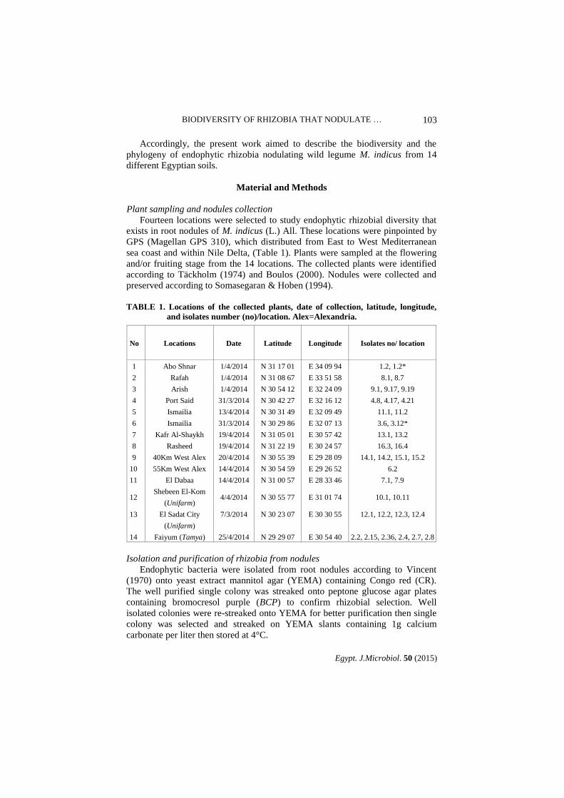
pH - tolerance
As shown in Fig. 3, the rhizobial isolates showed a wide diversity in their pH
tolerance. All the isolates grew well at pH 6.8 being the control cultures. At pH 9,
89.2% of the isolates gave high growth while 10.8% of the isolates gave moderate
growth. At pH 10, 70.3% and 2.7% of the isolates showed moderate and low growth,
respectively. At pH 11, 68% of the isolates gave low growth. 86.5% and 70.3% of the
isolates were unable to survive at pH 4 and 5, respectively. While, 13.5% of the
isolates gave low growth at pH 4, and 16.2%, 5.4% and 8.1% of isolates showed low,
moderate and high growth at pH 5, respectively.
Fig. 3. Frequency distribution of the different isolates showing the effect of different
pH.
BIODIVERSITY OF RHIZOBIA THAT NODULATE …
Egypt. J.Microbiol. 50 (2015)
109
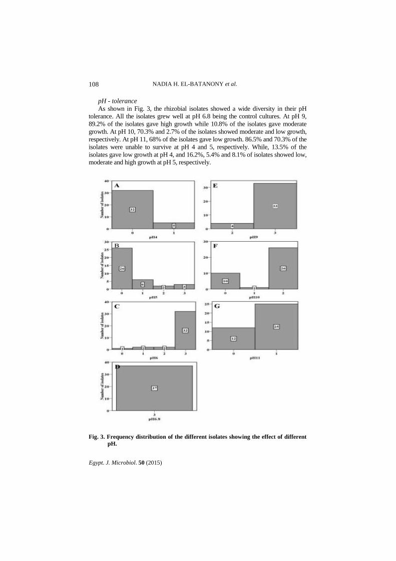
Cluster analysis of the rhizobial isolates according to their tolerance to the
different pHs showed a diversity among them (Fig. 4). Two isolates (7.9 and
12.4) were clustered separately in group II away from group I. The latter was
subdivided into two subgroups, where subgroup 1 comprised of 7 clusters.
Cluster 1 include isolates (2.7, 2.8, 1.2*, 2.36, 2.4, 2.2, 2.15, 14.2, 15.1, 7.1, 13.2,
4.8 and 4.17) that had isolates with the same origin and able to grow at pH values
above 6.8 till 11. Cluster 2 comprised of five isolates (11.2, 13.1, 10.1, 10.11 and
11.1), that were able to grow at pH values from 5 to 11 and few of them had the
same origin. Cluster 6 comprised of six isolates (9.19, 12.3, 3.6, 9.1, 9.17 and
3.12*) that had isolates with the same origin (few) and able to grow at pH values
6, 6.8 and 9 (Fig. 4).
Fig. 4. UPGMA dendrogram of physiological relationships among the rhizobial
isolates determined by pH tolerance. Rectangles indicating closely clustered
groups (gp.) and/or subgroups (subgp.).
NADIA H. EL-BATANONY et al.
Egypt. J. Microbiol. 50 (2015)
110
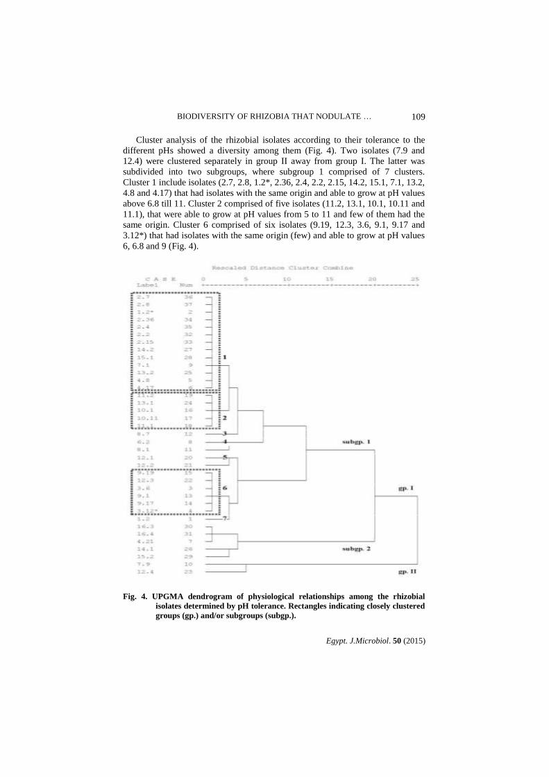
Molecular characterization of rhizobial isolates Genomic DNA was extracted from the 37 rhizobial isolates. This DNA was used as
a template for amplification of rep PCR primers (REP, ERIC and BOX). Such PCR products were used to discriminate between the isolates and to obtain a picture about the diversity and the natural variation that might exist between the rhizobial isolates.
PCR amplification with REP and ERIC primers PCR with REP and ERIC primer pairs of DNA from the isolates yielded
multiple distinct DNA products of size ranged from 177 to 3773 bp with the REP primer and from 200 to 2921 bp with the ERIC primer.
High resolution REP and ERIC PCR fingerprints of the 37 isolates were
generated. Isolates (4.8, 7.1, 10.11, 11.1, 15.2 and 16.4) and (2.7, 2.15, 3.6, 7.1, 7.9, 8.1, 8.7, 10.11, 11.2, 13.2 and 16.4) did not produce a PCR amplification profile with REP and ERIC PCR respectively, while the remaining 31 and 26 isolates were resolved in distinctive profiles with REP and ERIC PCR, respectively.
REP- PCR analysis of the 31 isolates revealed a relatively high level of
genetic diversity for isolate 12.2. On the other hand, isolates (3.6, 9.1 and 10.1), (9.19, 14.1 and 14.2;), (2.15 and 4.17) and (2.36, 2.4, 2.7 and 2.8;) showed 100% homology except 14.2 being 85% (Fig. 5).
Fig. 5. Electrophoretic patterns for the different rhizobial isolates generated by rep-
PCR using REP primer (A). Dendrogram of the 31 rhizobial isolates
originated by UPGMA cluster analysis based on REP primer (B).
BIODIVERSITY OF RHIZOBIA THAT NODULATE …
Egypt. J.Microbiol. 50 (2015)
111
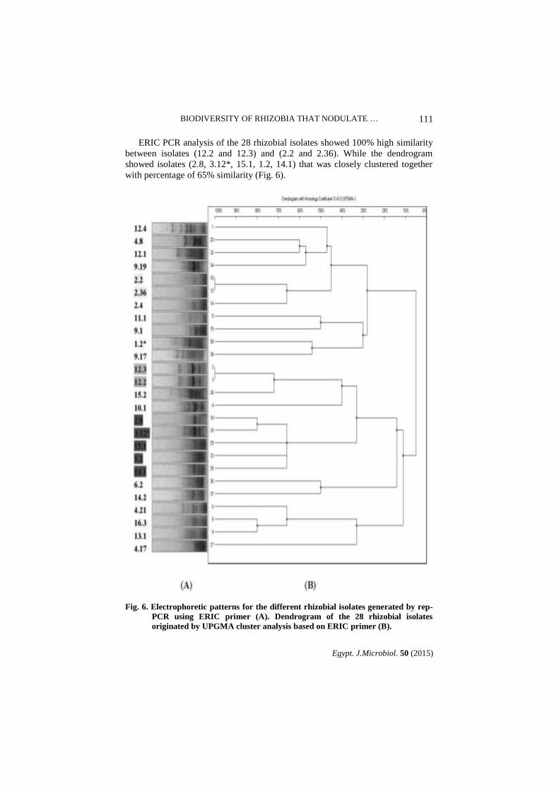
ERIC PCR analysis of the 28 rhizobial isolates showed 100% high similarity
between isolates (12.2 and 12.3) and (2.2 and 2.36). While the dendrogram
showed isolates (2.8, 3.12*, 15.1, 1.2, 14.1) that was closely clustered together
with percentage of 65% similarity (Fig. 6).
Fig. 6. Electrophoretic patterns for the different rhizobial isolates generated by rep-
PCR using ERIC primer (A). Dendrogram of the 28 rhizobial isolates
originated by UPGMA cluster analysis based on ERIC primer (B).
NADIA H. EL-BATANONY et al.
Egypt. J. Microbiol. 50 (2015)
112
PCR amplification with specific BOX A1R primer
BOX A1R primer did not reveal any polymorphism in all isolates (data not shown).
Discussion
Colonies of the obtained isolates showed little or no CR absorption when
incubated in dark and grow poorly on peptone glucose agar medium containing
BCP. Therefore, it is designated as colonies of a typical Rhizobium species. This
judgment coincides with that obtained by others (Deshwal & Chaubey, 2014 and
Somasegaran & Hoben, 1994).
In this study, the colonies of the 37 rhizobial isolates showed large variation
in their morphological traits similar to those obtained by Singh et al. (2008) and
Zahran et al. (2012). Regarding the pH-reaction, the 37 rhizobial isolates showed
an acid reaction assuming that these isolates are fast-growing rhizobia. In this
respect, the obtained results coincide with that obtained by many workers
(Baoling et al., 2007; Hatice et al., 2009; Somasegaran & Hoben, 1994 and
Vincent, 1970).
Saline conditions may limit Rhizobium-legume association by affecting
survival and proliferation of Rhizobium spp. in soil and rhizosphere; inhibiting
the early stages of infection process; affecting root nodule development and
reducing the host growth (Evans, 2015 and Graham, 1991). In the present work,
most of the rhizobial isolates showed high capacity for salt tolerance as 26 and 5
out of 37 isolates could withstand up to 5% and 7% NaCl, respectively. This
NaCl tolerance agreed with previous reports (Abdel-Wahab et al., 2002; Al-
Shaharani & Sheetta, 2011; Legesse & Assefa, 2014; Zahran et al., 2003 and
Zahran et al., 2012). Nevertheless, other Rhizobium strains from arid saline areas
were highly salt-tolerant and withstand high NaCl levels up to 5-10% (Abdel-
Wahab et al., 2002 and Zahran et al., 2003).
The dendrogram obtained from the numerical analysis of NaCl tolerance of
the 37 isolates showed a wide diversity among them. Only, few isolates grouped
together as they were able to tolerate NaCl concentration up to 3% or 5% and few
of them had the same origin.
Extreme pH can be a major factor limiting fast growing groups. Slight
variation in pH of the medium might have significant effects on the growth of
bacteria or organism (Adhikari et al., 2012; Singh et al., 2008 and Yan et al.,
2014). In this study, the majority of isolates could tolerate pH up to 10, while 25
out of 37 isolates gave poor growth at pH 11. On the other hand, at acidic
conditions (pH 4 and 5), only 5 and 11 isolates could grow, respectively. Such
results agreed with studies on rhizobial strains (Guerrouj et al., 2013; Legesse &
Assefa, 2014; Youseif et al., 2014 and Zahran et al., 2012), who showed that
even pH 10 was not inhibitory to rhizobial strains and these strains were sensitive
to acidic conditions (pH 3.5-4.0).

BIODIVERSITY OF RHIZOBIA THAT NODULATE …
Egypt. J.Microbiol. 50 (2015)
113
Both REP- and ERIC-like sequences (elements) are present in the genomes of
gram-negative soil bacteria, such as rhizobia (De Bruijn, 1992). In the present
work, both REP and ERIC-PCR profiles enabled strain differentiation and
demonstrated a considerable degree of genetic diversity among the rhizobial
isolates. The isolates were clustered on the bases of fingerprinting similarity
where few of them had the same origin. Also, our results support the conclusion
of De Bruijn, (1992), Edulamudi et al. (2015), Evans (2015) and Laguerre et al.
(1994) that the REP and ERIC PCR could become a powerful tool for the
molecular genetic analysis of bacteria and for bacterial taxonomy. It allows the
fingerprinting of individual genera, species, and strains and could help to
determine phylogenetic relationships. Also, these findings are in agreement with
the results obtained by other authors who studied diversity among natural
rhizobial populations in different world regions (Adiguzel et al., 2009 and
Granada et al., 2014).
It could be concluded that rhizobia isolated from root nodules of M. indicus
from Egyptian soils are both phenotypically and genetically diverse. Utilization
of both salt and pH tolerant rhizobial isolates may contribute to the reclamation
of salt affected soils which occupy large worldwide areas.
References
Abdel-Salam, M.S., Ibrahim, S.A., Abd-El-Halim, M.M., Badawy, F.M. and Abo-
Aba, S.E.M. (2010) Phenotypic characterization of indigenous Egyptian rhizobial
strains for abiotic stresses performance. J. Am. Sci. 6, 498-503.
Abdel-Wahab, S.M., El-Bakry, A.A., Tantawy, H. and El-Batanony, N.H. (2002) Phenotypic characterization of some wild rhizobial isolates isolated from some wild
legumes in Egypt. El-Azhar J. Microbiol. 58, 87-102.
Adhikari, D., Kaneto, M., Itoh, K., Suyama, K., Pokharel, B.B. and Gaihre, Y.K.
(2012) Genetic diversity of soybean-nodulating rhizobia in Nepal in relation to
climate and soil properties. Plant Soil, 357,131–145.
Adiguzel, A., Ogutcu, H., Baris, O., Karadayi, M., Medine, G. and Sahin, F. (2009)
Isolation and characterization of Rhizobium strains from wild vetch collected from
high altitudes in Erzurum-Turkey. Rom Biotechnol Lett. 15, 5017- 5024.
Alikhani, H.A. and Yakhchali, B. (2009) Potential use of Iranian rhizobial strains as
plant growth promoting rhizobacteria (PGPR) and effects of selected strains on growth
characteristics of wheat, corn and alfalfa. Desert, 14, 27-35.
Al-Shaharani, T.S. and Shetta, N.D. (2011) Evaluation of growth, nodulation and
nitrogen fixation of two Acacia species under salt stress. World Appl Sci J.13, 256-
265.
Arora, N.K., Kang, S.C. and Maheshwari, D.K. (2001) Isolation of siderophore-
producing strains of Rhizobium meliloti and their biocontrol potential against
Macrophomina phaseolina that causes charcoal rot of groundnut. Curr. Sci. 81, 673-
677.

NADIA H. EL-BATANONY et al.
Egypt. J. Microbiol. 50 (2015)
114
Bala, A. and Giller, K.E. (2006) Relationships between rhizobial diversity and host
legume nodulation and nitrogen fixation in tropical ecosystems. Nutr. Cycl
Agroecosyst. 76, 319–330.
Baoling, H., Chengqun, L. and Liqin, F. (2007) A rhizobia strain isolated from root
nodule of gymnosperm Podocarpus macrophyllus. Sci. Chin. Ser C-Life Sci. 50, 1-6.
Beauregard, M.S., Zheng, W. and Seguin, P. (2004) Diversity of Trifolium ambiguum
nodulating rhizobia from the lower Caucasus. Biol. Fertil. Soils, 40, 128–135.
Boulos, L. (2000) “Flora of Egypt”. Al-Hadara Publishing Cairo.
De Bruijn, F.J. (1992) Use of repetitive (Repetitive Extragenic Palindromic and
Enterobacterial Repetitive Intergeneric Consensus) sequences and the polymerase
chain reaction to fingerprint the genomes of Rhizobium meliloti isolates and other soil
bacteria. Appl. Environ. Microb. 58, 2180-2187.
Deshwal, V.K. and Chaubey, A. (2014) Isolation and characterization of Rhizobium
leguminosarum from root nodule of Pisum sativum L. JAIR, 2, 464- 467.
Drew, E.A. and Ballard, R.A. (2010) Improving N2 fixation from the plant down:
Compatibility of Trifolium subterraneum L. cultivars with soil rhizobia can influence
symbiotic performance. Plant Soil, 327, 261–277.
Edulamudi, P., Masilamani, A.J.A., Divi, V.R.S.G., Zakkula, V. and Konada, V.M.
(2015) Genetic characterization of rhizobia associated with horse gram [Macrotyloma
uniflorum (Lam.) Verdc.] based on RAPD and RFLP. Br. Microbiol. Res. J. 5, 340-
350.
Evans, A.T. (2015) Phylogeny and diversity of rhizobial bacteria. J. Phylogen. Evolution
Biol. 3,1.
Gálvez, M.D. (2005) Nodule metabolism in Pisum sativum L. in response to water stress:
carbon/nitrogen interactions and the possible molecules involved in the modulation of
the response, Ph.D. Thesis, Public University of Navarre.
Graham, P.H. (1991) Stress tolerance in Rhizobium and Bradyrhizobium and nodulation
under adverse soil conditions. Can. J. Microb. 38, 475-484.
Granada, C.E., Strochein, M., Vargas, L.K., Bruxel, M., Saccol de Sá, E.l. and
Passaglia, L.M.P. (2014) Genetic diversity and symbiotic compatibility among
rhizobial strains and Desmodium incanum and Lotus spp. Plants. Genet. Mol. Biol. 37,
396-405.
Guerrouj, K., Pe´rez-Valera, E., Chahboune, R., Abdelmoumen, H., Bedmar, E.J.
and El Idrissi, M.M. (2013) Identification of the rhizobial symbiont of Astragalus
glombiformis in Eastern Morocco as Mesorhizobium camelthorni. Antonie van
Leeuwenhoek, 104,187–198.

BIODIVERSITY OF RHIZOBIA THAT NODULATE …
Egypt. J.Microbiol. 50 (2015)
115
Hatice, O., Ahmed, A., Medine, G., Mehmet, K. and Fikrettin, S. (2009) Molecular
characterization of Rhizobium strains isolated from wild Chickpeas collected from
high altitudes in Erzurum-Turkey. Rom. Biotechnol . Lett. 14, 4294-4300.
Kaschuk, G., Hungria, M., Santos, J.C.P. and Berton-Junior, J.F. (2006) Differences
in common bean rhizobial populations associated with soil tillage management in
southern Brazil. Soil Tillage Res. 87, 205–217.
Kesari, V., Ramesh, A.M. and Rangan, L. (2013) Rhizobium pongamiae sp. nov. from
root nodules of Pongamia pinnata. Biomed Res Int. 1-9.
Koornneef, M., Alonso-Blanco, C. and Vreugdenhil, D. (2004) Naturally occurring
genetic variation in Arabidopsis thaliana. Annu. Rev.Plant .Biol. 55, 141 – 172.
Laguerre, G., Allard, M., Revoy, F. and Amarger, N. (1994) Rapid identification of
rhizobia by restriction fragment length polymorphism analysis of PCR-amplified 16S
rRNA genes. Appl. Environ. Microb. 60, 56-63.
Legesse, S. and Assefa, F. (2014) Symbiotic and phenotypic characteristics of rhizobia
nodulating faba bean (Vicia Faba) from Tahtay Koraro, northwestern zone of Tigray
Regional State, Ethiopia. Int. J. Emerg. Eng. Res. Technol. 2, 15-23.
Shamseldin, A.A.Y., Vinuesa, P., Thierfelder, H. and Werner, D. (2005) Rhizobium
etli and Rhizobium gallicum nodulate Phaseolus vulgaris in Egyptian soils and display
cultivar-dependent symbiotic efficiency. Symbiosis, 38, 145-161.
Shetta, N.D., Al-Shaharan, I.T.S. and Abdel-Aal, M. (2011) Identification and
characterization of Rhizobium associated with woody legume trees grown under Saudi
Arabia condition. Am. J. Agric. Environ. Sci. 10(3) 410–418.
Singh, B., Kaur, R. and Singh, K. (2008) Characterization of Rhizobium strain isolated
from the root of Trigonella foenum graecum (fenugreek). Afr. J. Biotechnol. 7, 3671-
3676.
Somasegaran, P. and Hoben, H.J. (1994) “Handbook for Rhizobia”, Springer-Verlag,
New York, USA.
Stewart, C.N. and Via, L.E. (1993) A rapid CTAB DNA isolation technique useful for
RAPD fingerprinting and other PCR applications. Biotechniques, 14, 748-750.
Täckholm, V. (1974) “Student's Flora of Egypt”, Cooperative Printing, Beirut, 2nd ed.
Cairo University, Cairo.
Velázquez, E., García-Fraile, P., Ramírez-Bahena, M. H., Peix, A. and Rivas, R.
(2010) In:“Proteobacteria Forming Nitrogen Fixing Symbiosis with Higher Plants.
Proteobacteria: Phylogeny, Metabolic Diversity and Ecological Effects”. Sezenna,
M.L. (Ed.), pp. 37–56. Nova Science Publishers Inc., New York, USA,
Versalovic, J., Schneider, M., De Bruijn, F.J. and Lupski, J.R. (1994) Genomic
fingerprinting of bacteria using repetitive sequence-based polymerase chain reaction.
Meth. Mol. Cell Biology, 5, 25-40.

NADIA H. EL-BATANONY et al.
Egypt. J. Microbiol. 50 (2015)
116
Vincent, J.M. (1970) “A Manual for the Practical Study of the Root Nodule Bacteria”.
IBP15. Blackwell Scientific Publications. Oxford and Edinburgh. U. K.
Yan, J., Han, X.Z., Ji, Z.J., Li, Y., Wang, E.T., Xie, Z.H. and Chen, W.F. (2014)
Abundance and diversity of soybean-nodulating rhizobia in black soil are impacted by
land use and crop management. App1. Environ. Microbial. 80, 5394–5402.
Youseif, S.H., Abd El-Megeed, F.H., Ageez, A., Zeinat, K., Mohamed, Z.K.,
Shamseldin, A. and Saleh, S. A. (2014) Phenotypic characteristics and genetic
diversity of rhizobia nodulating soybean in Egyptian soils. Eur . J. Soil. Biol. 60, 34-
43.
Zahran, H.H. (1999) Rhizobium-legume symbiosis and nitrogen fixation under severe
conditions and in an arid climate. Microbiol. Mol. Biol. Rev. 63, 968-989.
Zahran, H.H., Abdel-Fattah, M., Ahmad, M.S. and Zaki, A.Y. (2003) Polyphasic
taxonomy of symbiotic rhizobia from wild leguminous plants growing in Egypt. Folia.
Microbiol. 348, 510-520.
Zahran, H.H., Abdel-Fattah, M., Yasser, M.M., Mahmoud, A.M. and Bedmar, E.J.
(2012) Diversity and environmental stress responses of rhizobial bacteria from
Egyptian grain legumes. Aust. J. Basic Appl. Sci. 6, 571-583.
(Received 24 /6/2015;
accepted 6 /9/2015)

BIODIVERSITY OF RHIZOBIA THAT NODULATE …
Egypt. J.Microbiol. 50 (2015)
117
تنوع الريزوبيا المعزولة من نبات الحندقوق المر في األراضي
المصرية
نادية حامد البتانوني، آيات مصطفي بدوي*
، حسن الطنطاوي حسن*
محمد عزت و
الليثي*
و مدينة السادات -مدينة السادات جامعة - معهد الدراسات والبحوث البيئية*
قسم
.مصر - نوفيةالم - جامعة المنوفية -كلية العلوم -النبات
وصف التنوع البيولوجي لبكتريا الريزوبيا المعزولة من نبات ىإليهدف البحث
ىالمستو ىكان مختلف من األراضى المصرية علم 41الحندقوق المر من
.لهذه العزالت ىالمظهر الخارج ىلباإلضافة إ ىو الوراث ىالفسيولوج
وفسيولوجيا على ىرجمظهرها الخا ىالعزالت بناءا علحيث تم وصف هذه
وعالوة على ذلك تم . أساس تحملها لدرجة الملوحة ودرجة األس الهيدروجينى
تحليل الحامض النووى لهذه العزالت عن طريق استخدام عالمات وراثية مختلفة
. rep-PCRباستخدام
. لنبات الحندقوق المر عزلة ريزوبيا من العقد الجذرية 73تم الحصول على
التجارب الفسيولوجية كانت جميع العزالت سريعة النمو ، بينما كانت ىلإوإستنادا
وأظهرت . من كلوريد الصوديوم %3 ىعزالت فقط متحملة لدرجة ملوحة حتسبع
. هيدروجينى المختلفةعزالت الريزوبيا تنوعا واسعا فى تحملها لدرجات األس ال
عالوة على ذلك فإن دراسة تحليل الحامض النووى لهذه العزالت عن طريق
أنتج العديد من قطع الحامض rep-PCRاستخدام عالمات وراثية مختلفة باستخدام
قاعدة فى حالة 7337لى إ 433الحجم والتى تتراوح ما بين النووى المتميزة فى
لم . ERICقاعدة فى حالة الـ 0204لى إ 022بين ا ، بينما تتراوح م REPالـ
. كداللة وراثية مع جميع العزالت A1R BOXتظهر أية فروق باستخدام
لذلك يمكننا أن نستنتج أنه يوجد تباين و تنوع في المظهر الخارجي والوراثي
. بين عزالت الريزوبيا المعزوله من نبات الحندقوق من األماكن المختلفه في مصر