biodiversity of pigmented fungi isolated from marine
TRANSCRIPT

HAL Id: hal-01657071https://hal.univ-reunion.fr/hal-01657071
Submitted on 6 Dec 2017
HAL is a multi-disciplinary open accessarchive for the deposit and dissemination of sci-entific research documents, whether they are pub-lished or not. The documents may come fromteaching and research institutions in France orabroad, or from public or private research centers.
L’archive ouverte pluridisciplinaire HAL, estdestinée au dépôt et à la diffusion de documentsscientifiques de niveau recherche, publiés ou non,émanant des établissements d’enseignement et derecherche français ou étrangers, des laboratoirespublics ou privés.
Biodiversity of Pigmented Fungi Isolated from MarineEnvironment in La Réunion Island, Indian Ocean: New
Resources for Colored MetabolitesMireille Fouillaud, Mekala Venkatachalam, Melissa Llorente, Hélène Magalon,
Pascale Cuet, Laurent Dufossé
To cite this version:Mireille Fouillaud, Mekala Venkatachalam, Melissa Llorente, Hélène Magalon, Pascale Cuet, etal.. Biodiversity of Pigmented Fungi Isolated from Marine Environment in La Réunion Island,Indian Ocean: New Resources for Colored Metabolites. Journal of Fungi, MDPI, 2017, 3 (3),�10.3390/jof3030036�. �hal-01657071�

FungiJournal of
Article
Biodiversity of Pigmented Fungi Isolated fromMarine Environment in La Réunion Island, IndianOcean: New Resources for Colored Metabolites
Mireille Fouillaud 1,2,* , Mekala Venkatachalam 1 , Melissa Llorente 1, Helene Magalon 3,Pascale Cuet 3 and Laurent Dufossé 1,2
1 Laboratoire de Chimie des Substances Naturelles et des Sciences des Aliments—LCSNSA EA 2212,Université de La Réunion, 15 Avenue René Cassin, CS 92003, F-97744 Saint-Denis CEDEX 9,Ile de La Réunion, France; [email protected] (M.V.); [email protected] (M.L.);[email protected] (L.D.)
2 Ecole Supérieure d’Ingénieurs Réunion Océan Indien—ESIROI, 2 Rue Joseph Wetzell,F-97490 Sainte-Clotilde, Ile de La Réunion, France
3 UMR ENTROPIE and LabEx CORAIL, Université de La Réunion, 15 Avenue René Cassin, CS 92003,F-97744 Saint-Denis CEDEX 9, Ile de La Réunion, France; [email protected] (H.M.);[email protected] (P.C.)
* Correspondence: [email protected]; Tel.: +2-62-48-33-62
Received: 31 May 2017; Accepted: 28 June 2017; Published: 2 July 2017
Abstract: Marine ecosystems cover about 70% of the planet surface and are still an underexploitedsource of useful metabolites. Among microbes, filamentous fungi are captivating organisms used forthe production of many chemical classes of secondary metabolites bound to be used in various fieldsof industrial application. The present study was focused on the collection, isolation, screening andgenotyping of pigmented filamentous fungi isolated from tropical marine environments around LaRéunion Island, Indian Ocean. About 150 micromycetes were revived and isolated from 14 marinesamples (sediments, living corals, coral rubble, sea water and hard substrates) collected in fourdifferent locations. Forty-two colored fungal isolates belonging to 16 families, 25 genera and 31 specieswere further studied depending on their ability to produce pigments and thus subjected to molecularidentification. From gene sequence analysis, the most frequently identified colored fungi belongto the widespread Penicillium, Talaromyces and Aspergillus genera in the family Trichocomaceae(11 species), then followed by the family Hypocreaceae (three species). This study demonstratesthat marine biotopes in La Réunion Island, Indian Ocean, from coral reefs to underwater slopesof this volcanic island, shelter numerous species of micromycetes, from common or uncommongenera. This unstudied biodiversity comes along with the ability for some fungal marine inhabitants,to produce a range of pigments and hues.
Keywords: fungi; biodiversity; Indian Ocean; Marine; coral reef; genotyping; pigment production
1. Introduction
With the growing demand for natural compounds in the industrial sector, marine derived fungiappear to present many interests. Filamentous fungi are ubiquitous in nature due to their huge capacityof adaptation and their ability to produce an assortment of new secondary metabolites. Literature nowabundantly reports the significant involvement of fungi in the industry, through the production ofvarious useful substances, such as antibiotics, immunosuppressants, anti-cancer drugs, plant hormones,enzymes, acids and also natural pigments [1–5]. Both the pigments and enzymes equally find theirusages in food and beverages, animal feeds, pharmaceuticals, cosmetics, textile, leather, pulp andpaper industries, biofuel production, and environment bioremediation [6].
J. Fungi 2017, 3, 36; doi:10.3390/jof3030036 www.mdpi.com/journal/jof

J. Fungi 2017, 3, 36 2 of 22
Nevertheless, the distribution of the marine-derived fungal species and their contribution tomarine biotopes are still in infancy, and more has to be explored [7–11]. The highest diversity ofmarine fungi seems to appear in tropical regions, mainly in tropical mangroves, which are extensivelystudied because of their high richness in organic matters and especially lignocellulosic materials,favorable to the development of a wide range of heterotrophic microorganisms [11–14]. Anyway,many marine ecological niches are still unexplored and it seems plausible that unique features of marineenvironments can be the inducers of unique substances, biosynthesized by marine or marine-derivedmicroorganisms [15,16].
Considering the immense genetic and biochemical diversity of these fungi, partially derived fromthe specificity of the biotopes they are facing, marine-derived fungi are regarded as a potential brightsource of new molecules with likely application in pigment production [17,18]. Many genera producingpigments have then been isolated either from water, sediments, and decaying organic residues, or fromliving organisms such as invertebrates, plants or algae. Fungi belonging to genera such as Aspergillus,Penicillium, Paecilomyces, Eurotium, Alternaria, Fusarium, Halorosellinia, Monodictys and Microsphaerospsishave already been identified from marine biotopes [19–21]. They are therefore able to exhibit brightcolors, from yellow to black, mainly belonging to polyketides. Indeed, polyketides pigments andparticularly azaphilones and anthraquinones seem to dominate marine natural products of fungalorigin [22]. Colored compounds, usually described as secondary metabolites, do not seem to be directlyinvolved in the primary growth of the fungus in which they occur [23]. However they may play someimportant roles in the resistance to a variety of adverse environmental factors (desiccation, exposureat extreme temperatures, irradiations and photo-oxidation) or in ecological interactions with otherorganisms (macroorganisms such as sponges, corals or other microbial communities) [24]. For thisreason, many fungal secondary metabolites exhibit useful biological activities and are of interest to thepharmaceutical, food, and agrochemical industries [16,25].
This study initiated the search for filamentous fungi in some tropical marine biotopes of coralreefs and underwater slopes of the volcano from La Réunion Island. Fungal isolates from samples ofsediments, seawater, hard substrates, coral rubbles or living coral individuals (Pocillopora sp.) werecharacterized both by phenotypic and molecular ways. The production of pigments of quinoid-typeproduced from the mycelia cultured in liquid media was used as a first approach to screen for thepigment production. This work reveals a part of the mycofloral biodiversity in La Réunion Islandtropical marine environment and its potentiality to propose new pigment sources to expand in anindustrial setting.
2. Materials and Methods
2.1. Samples Collection
La Réunion Island lies in the Indian Ocean and is located 800 km east of Madagascar (21◦06′54.5′ ′ Sand 55◦32′11.0′ ′ E) (Figure 1a). This tropical island arose two million years ago from a volcanic hotspot (Piton de La Fournaise) and is known for its rainforested interior and its fringing reefs holdingmost of the marine wealth.

J. Fungi 2017, 3, 36 3 of 22J. Fungi 2017, 3, 36 3 of 22
Figure 1. (a) La Réunion island location (Indian Ocean, 21°06′54.5′′ S and 55°32′11.0′′ E); (b) geolocation of sampling sites around La Réunion Island (West: La Saline; and East: Sainte Rose and Tremblet); and (c) geolocation of the three sampling spots at La Saline fringing reef: Trou d’eau (TDE inner reef and TDE outer slope ) and Planch’Alizé (PA) (back arrow represents the main water flow).
A first set of samples was collected on the fringing reef from La Saline, which lies on the dry west coast of the island. It is more than 9 km long and ranges in width from 50 m in its northern part to 600 m in the south [26]. For the purpose of research, samples were collected from three sampling spots on the west coast that cover the sites of Trou d’Eau (TDE) (inner reef flat at −1 m depth and outer slope at −17 m) and Planch’ Alizé (PA) (inner reef flat, −1 m) (Figure 1b,c). Planch’ Alizé is considered as a sheltered site, located downstream of seawater flowing over the Trou d’Eau (Figure 1c). The outer slope is found at the outer edge of the reef, closest to the open ocean, and is characterized by spurs and grooves extending downward to the sand bottom, while the inner reef flat displays wide transversal strips of branched coral colonies alternating with narrow detrital channels perpendicular to the reef flat [27–31]. Low water flow and high solar radiation contribute to heating the reef water during the day, inducing important daily sea surface temperature variations. This area is also heavily laden with organic and mineral matter coming from nearby human activity (seaside area).
A second set of samples was collected in Sainte Rose area (south-east) on the submerged lava flows (Figure 1b). Indeed, the Piton de la Fournaise is one of the most active effusive volcanoes in the world with 27 eruptions between 1998 and 2007 and a mean frequency, over a century, of an eruptive phase every nine months. Submerged lava flows appear on the south-east part of the island when, during eruptions, the pool of lava overflows the active volcano mouth and pours down on the slopes of the volcano, to the sea. These costal marine ecosystems facing the deep ocean, are then regularly subjected to natural hazards such as being covered by incandescent lava flows, temporary
Figure 1. (a) La Réunion island location (Indian Ocean, 21◦06′54.5′ ′ S and 55◦32′11.0′ ′ E); (b) geolocationof sampling sites around La Réunion Island (West: La Saline; and East: Sainte Rose and Tremblet);and (c) geolocation of the three sampling spots at La Saline fringing reef: Trou d’eau (TDE inner reefand TDE outer slope ) and Planch’Alizé (PA) (back arrow represents the main water flow).
A first set of samples was collected on the fringing reef from La Saline, which lies on the dry westcoast of the island. It is more than 9 km long and ranges in width from 50 m in its northern part to600 m in the south [26]. For the purpose of research, samples were collected from three sampling spotson the west coast that cover the sites of Trou d’Eau (TDE) (inner reef flat at −1 m depth and outerslope at −17 m) and Planch’ Alizé (PA) (inner reef flat, −1 m) (Figure 1b,c). Planch’ Alizé is consideredas a sheltered site, located downstream of seawater flowing over the Trou d’Eau (Figure 1c). The outerslope is found at the outer edge of the reef, closest to the open ocean, and is characterized by spurs andgrooves extending downward to the sand bottom, while the inner reef flat displays wide transversalstrips of branched coral colonies alternating with narrow detrital channels perpendicular to the reefflat [27–31]. Low water flow and high solar radiation contribute to heating the reef water during theday, inducing important daily sea surface temperature variations. This area is also heavily laden withorganic and mineral matter coming from nearby human activity (seaside area).
A second set of samples was collected in Sainte Rose area (south-east) on the submerged lavaflows (Figure 1b). Indeed, the Piton de la Fournaise is one of the most active effusive volcanoes in theworld with 27 eruptions between 1998 and 2007 and a mean frequency, over a century, of an eruptivephase every nine months. Submerged lava flows appear on the south-east part of the island when,during eruptions, the pool of lava overflows the active volcano mouth and pours down on the slopes

J. Fungi 2017, 3, 36 4 of 22
of the volcano, to the sea. These costal marine ecosystems facing the deep ocean, are then regularlysubjected to natural hazards such as being covered by incandescent lava flows, temporary changes inphysicochemical conditions of water bodies and exceptional rises of temperature. Besides, this area ispoorly inhabited and urbanized and, as a consequence, the amount of organic matter poured in thesea is reduced compared to other coastal ecosystems. It provides a natural laboratory to study thecolonization of a blank substrate and the evolution of the biodiversity all around, during the followingyears. Samples were then obtained from sediments extracted from 1977 lava flow (−25 m depth) and2004 lava flow (−70 m), as well as from surrounding free water at −70 m.
Seawater, sediments, parts of living corals and hard substrates (volcanic rocks or coral rubbles)were collected in sterile bottles, during the months of April and May 2012, stored in a cooling box(4 ◦C), brought to the laboratory, and treated immediately for the fungal isolation.
2.2. Culture and Purification of Fungi
To cultivate the revivable fungi from the collected seawater, 100 mL of water was filtered using a0.45 µm sterile cellulose-nitrate filter (Sartorius Stedim, Göttingen, Germany). The filters were thenplaced in Petri plates containing malt extract agar (MEA) and Sabouraud agar (BD Difco, FranklinLakes, NJ, USA) prepared with natural seawater collected near La Saline, and beforehand sterilized at121 ◦C, 15 min.
The other samples such as sediments, hard substrates and parts of living/dead coral were treatedseparately. The samples were first washed with 70% alcohol and rinsed in sterile seawater. Then theywere ground using sterile pestle and mortar. Ground material (5 g) was taken from each sample andadded to 15 mL of sterile diluent (1.6 g of tryptone (Sigma- Aldrich, T-9410, Saint Louis, MO, USA),0.05 g of tween 20, 1 L of sterile seawater of pH = 7.5). After stirring for 20 min at 150 rpm on a shakingtable (Edmunt Bühler GmbH, VKS 75 Control, Hechningen, Germany), the suspension was diluted byemploying serial decimal dilution method up to 10−3 [32]. Each diluted sample (1 mL) was poured onPetri plates containing MEA and Sabouraud agar prepared with natural seawater.
All the platings were performed in triplicates and incubated at 25 ◦C for 21 days. During thisperiod, the plates were checked each day for the appearance of new colonies. Each new colonywas individually isolated and cultured on new MEA solid medium. During the growth period,the production of colors was observed.
All the isolated fungi were cultured using monospore technique for future experiments andlong-term storage. The fungi grown after 5 days were scraped and transferred into a sterile vialcontaining a cryoprotectant medium composed of 15% skimmed milk and 2% glycerol for long termstorage at −80 ◦C [33,34]. In total, 42 fungal isolates were then selected for pigment production basedon the visual appearance of the thalli grown on solid culture media.
2.3. Fungal Identification
2.3.1. Fungal DNA Extraction
To extract DNA from the 42 purified isolates, a small amount of mycelium along with spores wascultivated on potato dextrose agar (PDA) at 25 ◦C under day light exposure. After 5 days of growth,the fungal mycelium was scraped and DNA was extracted using DNeasy Blood and Tissue kit (Qiagen,Hilden, Germany) following the manufacturer’s instructions. DNA amount and purity contained ineach extract were evaluated by measuring the absorbances at 230, 260 and 280 nm (Nanodrop 2000,Thermo Scientific, Waltham, MA, USA) and calculating the ratio A260/A280 and A260/A230. DNAs werestored at −20 ◦C prior to use for amplification studies [35].
2.3.2. Primers, PCR Amplification and Sequencing
The choice of PCR primers was made based on observed phenotypic characteristics for molecularidentification. Aspergillus species were amplified for calmodulin gene using primers Cmd5/Cmd6

J. Fungi 2017, 3, 36 5 of 22
and Penicillium species for β-tubulin using primers T10/Bt2b [36]. To amplify and sequence the DNAfrom Trichoderma and Hypocreales species, EF-1H/EF-2T primer pair was used to amplify a fragmentof the translation elongation factor 1 alpha gene (Tef1) [37]. For uncharacterized fungi, the fragmentscontaining ITS region were amplified using ITS1-F_KYO2/ITS2 or ITS3_KYO2/ITS4, and, whennecessary, the large subunit rDNA was also amplified using V9G/LR3 primer pair (Table 1) [36–38].
Table 1. PCR amplification and the sequencing primers used for the identification of fungal isolates.
Primers Direction Sequences (5’→ 3’) Note Hybrid. T ◦C Refs.
ITS1-F_KYO2 Forward TAGAGGAAGTAAAAGTCGTAA
Small sub-unit, ITS 1, 5.8S,ITS 2, Largest sub unit
rDNA
56
[36]ITS2_KYO2 Reverse TTYRCTRCGTTCTTCATC 47
ITS3_KYO2 Forward GATGAAGAACGYAGYRAA 47
ITS 1 Forward TCCGTAGGTGAACCTGCGG 55
[39]ITS 2 Reverse GCTGCGTTCTTCATCGATGC 55
ITS 3 Forward GCATCGATGAAGAACGCAGC 55
ITS 4 Reverse TCCTCCGCTTATTGATATGC 55
V9G Forward TTACGTCCCTGCCCTTTGTA Large sub unit D1/D2 forbasidiomycetous yeast
55
[38]LR3 Reverse TGACCATTACGCCAGCATCC 57
Cmd 5 Forward CCGAGTACAAGGARGCCTTC Calmodulin, specific forAspergillus
52
Cmd 6 Reverse CCGATRGAGGTCATRACGTGG 52
T 10 Forward ACGATAGGTTCACCTCCAGAC β- tubulin, specific forPenicillium
55[38]
Bt2b Reverse ACCCTCAGTGTAGTGACCCTTGGC 55
EF1-728F Forward CATCGAGAAGTTCGAGAAGG Elongation factor 1 forTrichoderma
55[38]
TEF1-LLErev Reverse AACTTGCAGGCAATGTGG 55
PCR reactions were carried out in a total volume of 30 µL: 1× of MasterMix (Applied Biosystems,Foster city, CA, USA), 0.5 µM of forward and reverse primers and at least 1.3 ng/µL of genomicDNA. Amplifications were carried out on a thermal cycler GeneAmp® PCR System 9700 (AppliedBiosystems) according to the following program: 94 ◦C for 5 min + 35 × (94 ◦C for 30 s, 55 ◦C (or52 ◦C for the primers of calmodulin: Cmd5/Cmd6) for 60 s, 72 ◦C for 60 s) + 72 ◦C for 5 min for finalelongation step.
2.3.3. Sequence Analysis
Amplicons were sequenced in both directions (GENOSCREEN, Lille, France). The obtainedelectropherograms were read and corrected with Chromas software (version 2.13, Technelysium ptyLtd., South Brisbane, Australia). The extracted sequences for each gene were separately used toperform nucleotide searches using online BLAST algorithm, provided by NCBI (http://www.ncbi.nlm.nih.gov/BLAST/). BLAST results were sorted based on the maximum identity to the query sequenceand considered as the best hit. Sequence-based identities with a cutoff of 97% or above and querycoverage >90% were considered as significant [40,41]. Because of low recovery rates and concordancevalues, some isolates were amplified and sequenced a second time, with additional sets of primers,mainly among the isolates of the genera Penicillium and Trichoderma.
2.4. Culture Conditions for Pigment Production and Separation of Biomass from Liquid Medium
2.4.1. Culture Conditions
Erlenmeyer flasks (250 mL) containing 80 mL of potato dextrose broth (PDB) medium wereautoclaved at 121 ◦C for 15 min. Then, 120 mg of mycelia from interesting fungal species grownon PDA Petri plates were transferred into the sterile flasks and incubated at 25 ◦C under daylightexposure, with an agitation of 150 rpm for 10 days (Multitron Pro, Infors HT, Bottmingen, Switzerland).
J. Fungi 2017, 3, 36 6 of 22
2.4.2. Separation of Biomass from Culture Liquid
After the end of the fermentation period, the culture medium containing extracellular pigmentswas separated from mycelia by vacuum filtration using Whatman filter paper No. 2 (Merck, Darmstadt,Germany). Thus, liquid medium and biomass were treated separately. The wet mycelium was furtherused for the extraction of pigment content.
2.5. Production of Pigments
2.5.1. Determination of Pigments Production in Liquid Cultures
Chromophore is a chemical group that absorbs light of specific frequency and confers color to amolecule. Widespread polyketide pigments such as anthraquinones or azaphilones are often highlysubstituted aromatic molecules, with fused benzene rings [42]. Thus the majority of the commonchromophores from fungi absorb in the UV region (one or several peaks between 200–300 nm),whereas absorbance in the visible region (400–700 nm) highly depends on the nature and the numberof substituted groups.
To compare the pigment production of all isolates cultured in PDB medium, the amount ofpigments produced in liquids was expressed as mg equivalent (mg eq.) of a chosen commercialstandard per liter of culture medium (mg eq. purpurin L−1). Purpurin was chosen as a polyketidepigment in orange-red hue, which absorbs in the UV area (250–270 nm) as many polyketides [43],and also in the visible range 458–520 nm [44]. Thus, the absorbance of an authentic coloredstandard purpurin (Sigma-Aldrich) was estimated at different concentrations using an UV-visiblespectrophotometer (Shimadzu UV-1800 Spectrophotometer). Then, in regard with the diversity ofpigments content in the fungal cultures and as a preliminary approach, the absorbance of each samplewas measured at 254 nm and the amount of pigments produced was expressed in “mg equivalentpurpurin L−1” (Figure S1). In addition, for each isolate, the intracellular (IC) pigments (extracted fromthe biomass) and extracellular (EC) contents (liquid from culture, separated from the biomass) werescanned between 200 and 600 nm with a UV-1800 spectrophotometer (Shimadzu UV spectrophotometer,Shimadzu Corporation, Kyoto, Japan) in a quartz cell of 10 mm path length.
2.5.2. Extraction of Pigments
IC pigments contained in the wet mycelium were extracted using a methanol: water combination(1:1 v/v) as conventional extraction method. The mixture was immersed in an ultrasonic bath at 45 ◦Cfor 30 min. The suspension was allowed to stir overnight at room temperature on a shaking table(VKS 75 Control, Edmunt Bühler GmbH). On the following day, it was filtered through Whatman filterpaper No. 2 to recover the solvent containing the pigments extracted from biomass.
To compare the amount of pigments produced within the cells with the one diffused into theextracellular medium, we performed the nonparametric Mann–Whitney–Wilcoxon test as our data didnot follow the normal distribution using the R software (R Development Core Team 2016) [45].
3. Results
3.1. Diversity of Isolated Fungi
More than 150 isolates were first recovered from the 14 samples collected among four locations.Among them, 42 were selected for identification, according to their capacity to develop colored myceliaor to secrete colored compounds in the media.
After sequencing, the 42 colored isolates were assigned to 16 families, 25 genera and 31 species(accession numbers mentioned in Table 2). The vast majority of the isolates have been identified withmore than 98% concordance rate and recovered with high precision at the species level. However,few fungi (Acremonium sp., Periconia spp. and Biscogniauxia sp.) were identified to the genus level only,according to the gene chosen. The genetic characterization with these primers partially failed for two

J. Fungi 2017, 3, 36 7 of 22
isolates (Whalleya microplaca B and Wallemia sebi). Wallemia sebi was only characterized according tomorphological criteria.
Table 2. Fungal isolates from La Réunion Island marine biotopes from different sample types andsampling sites: Trou d’Eau (TDE); Planch’ Alizé (PA); Lava flow corresponds to 1977 lava flow in SainteRose/Tremblet area.
Family Fungal Species Sampling Site Gene Accession Number
Water Bodies
Davidiellaceae Cladosporium Cladosporioides Lava flow (−70 m) JF949719.1
Didymellaceae Peyronellaea glomerata (syn: Phoma glomerata) Lava flow (−70 m) JQ936163.1
Nectriaceae Nectria haematococca A Lava flow (−70 m) XM_003053163.1
Pleosporales IncertaeSedis
Periconia sp. A Lava flow (−70 m) HQ608027.1
Periconia sp. B Lava flow (−70 m) HQ608027.1
Sporidiobolaceae Rhodosporidium paludigenum Lava flow (−70 m) AF444493.1
Stachybotryaceae Myrothecium atroviride Lava flow (−70 m) AJ302002.1
Teratospheriaceae Hortaea werneckii (syn: Cladosporium werneckii) Lava flow (−70 m) JN997372.1
Trichocomaceae
Aspergillus sydowii B Lava flow (−70 m) KC253961.1
Emericella qinqixianii TDE outer slope AB249008.1
Penicillium brocae NRRL 32599 TDE outer slope DQ123642.1
Penicillium viticola B TDE inner reef AB606414.1
Talaromyces rotundus TDE inner reef EU497950.1
Talaromyces verruculosus PA inner reef KC416631.1
Wallemiaceae Wallemia sebi Lava flow (−70 m) Morphological Identification
Living Coral Pocillopora sp.
Hypocreaceae Acremonium sp. PA inner reef FJ770373.1
Hypocrea koningii TDE inner reef JX174420.1
Trichocomaceae
Aspergillus creber A TDE inner reef JN854049.1
Aspergillus creber B TDE inner reef JN854049.1
Aspergillus sydowii A PA inner reef JN854052.1
Eurotium amstelodami TDE outer slope FR727111.1
Penicillium viticola C PA inner reef AB606414.1
Coral Rubbles
Chaetomiaceae Chaetomium globosum or Chaetomium murorum TDE outer slope JN209898.1
Trichocomaceae
Penicillium herquei TDE outer slope JN246042.1
Talaromyces albobiverticillius B TDE outer slope JN899313.1
Talaromyces albobiverticillius C TDE outer slope JN899313.1
Hard Substrate/Rock Substrate
Nectriaceae
Fusarium equiseti A Lava flow (−25 m) JQ936153.1
Fusarium equiseti B Lava flow (−25 m) JF311925.1
Fusarium equiseti C Lava flow (−25 m) JQ936153.1
Nectria haematococca B Lava flow (−25 m) XM_003053163.1
Pleosporaceae Epicoccum sorghi (syn: Phoma sorghina;Peyronellaea stemphylioides) Lava flow (−25 m) KC106717.1
Sordariomycetes Nigrospora sphaerica (or Env. sample frommarine air) TDE outer slope KC505176.1
Sporidiobolaceae Rhodotorula mucilaginosa TDE outer slope KC515367.1
TrichocomaceaePenicillium citrinum Lava flow (−25 m) EU030332.1
Penicillium viticola A Lava flow (−25 m) AB606414.1
Ustilaginaceae Sporisorium exsertum TDE outer slope JN367293.1
Xylariaceae
Biscogniauxia sp. PA inner reef FJ884075.1
Whalleya microplaca A TDE outer slope JQ760548.1
Sordariomycete (or Whalleya microplaca B) TDE outer slope FJ416301.1
Sediments
Didymosphaeriaceae Paraconiothyrium variabile TDE outer slope JQ936271.1
Hypocreaceae Trichoderma atroviride TDE outer slope KC008065.1
Trichocomaceae Talaromyces albobiverticillius A TDE outer slope JN899313.1

J. Fungi 2017, 3, 36 8 of 22
Pigment producing fungi (42) were isolated from all types of samples: sediments (3), living coralPocillopora sp. (7), unidentified coral rubbles (4), hard substrates (reef basis or volcanic rocks) (13) andseawater (15) (Table 2).
The most represented fungi, in the selection of colored micromycetes, belonged to the familyTrichocomaceae with Penicillium, Talaromyces and Aspergillus genera (11 species); and then came theHypocreaceae with Trichoderma, Hypocrea and Acremonium.
A high diversity of pigmented isolates was observed from the so-called “hard substrates” (rockybasis on which the coral colonies recruit, or submerged lava flows). Some Nigrospora, Sporisorium,Whalleya, and Rhodotorula isolates were collected from the outer slope at Trou d’Eau (TDE), althoughthey are rather rarely isolated from marine environment. In Sainte Rose, Penicillium species (P. citrinum,P. viticola) as well as Fusarium equiseti, Epicoccum sorghi, Nectria haematococca, were successfully revivedfrom lava flow, sampled at −25 m.
From our study, the coral rubbles (dead parts of corals) contained colored Penicillium or relatedspecies: P. herquei and two isolates of Talaromyces albobiverticillius (B and C), as well as an isolate ofChaetomium globosum. Coral rubbles or hard substrates naturally appear diversely colored underwater.Indeed, they support the colonization by multiple organisms (colored algae or other aquatic organisms),visually detectable when sampling.
The revivable colored fungi sheltered by the living coral Pocillopora sp. belonged to the generaAspergillii (A. creber, A. sydowii and Eurotium amstellodami (the teleomorphic form of A. amstellodami),as well as to Penicillium (P. viticola), Hypocrea (H. koningii) and Acremonium.
Some fungal species were identified from different types of samples in the same area. At TDEouter slope, Talaromyces albobiverticillius A came from sediment and T. albobiverticillius B and C wererevived from coral rubbles. Nectria haematococca was found in lava substrate (−25 m) (isolate B) as wellas seawater (−70 m near lava flow) in the same area (isolate A).
Some similar species also appeared in separate locations: Aspergillus sydowii was found near lavaflow on the east coast (seawater, −70 m) (isolate B) and also in living Pocillopora colonies (isolate A),from the west coast back reef (PA site). Penicillium viticola was isolated from the west coast on livingPocillopora sp. coral in PA (isolate C), from seawater in TDE back reef (isolate B), as well as from lavaflow hard substrate (−25 m), on the east coast (isolate A).
These fungi found in several samples and/or in different locations may be considered as frequentin this marine environment.
3.2. Pigment Production
3.2.1. In Culture Broth
The majority of the isolates produced pigments after four days of fermentation in PDB. The colorsof the broth (biomass plus liquid culture medium) always darkened over time, which indicated theirpotential for pigment production (Figure 2).

J. Fungi 2017, 3, 36 9 of 22J. Fungi 2017, 3, 36 9 of 22
Figure 2. Colors observed in potato dextrose broth cultures from (a) Talaromyces albobiverticillius B, (b) T. albobiverticillius C, and (c) Aspergillus creber B, after four and seven days.
Overall, it was observed that the coloring trend was not directly related to the genus. Even if dominant colors such as yellow, red, brown, purple, orange, pink and green were observed in flasks, the hues were extremely diverse according to the species, even to the isolates (Table 3).
Table 3. Dominant colors of culture broth 1, extracellular (EC) 2 and intracellular (IC) 3 pigments from fungal isolates.
Fungal Isolates Approximate Hues Fungal Isolates Approximate HuesIsolates with Intense Hues (Purple/Red/Maroon) Isolates with Orange Hues
Broth EC IC Broth EC IC Acremonium sp. Penicillium viticola A Talaromyces albobiverticillius A Penicillium viticola B Talaromyces albobiverticillius B Epicoccum sorghi Talaromyces albobiverticillius C Penicillium brocae NRRL 32599 Aspergillus sydowii A Penicillium herquei Aspergillus creber A Aspergillus sydowii B Aspergillus creber B Chaetomium globosum or C. murorum Emericella qinqixianii Penicillium viticola C Trichoderma atroviride Penicillium citrinum Biscogniauxia sp. Hypocrea koningii Paraconiothyrium variabile Myrothecium atroviride
Isolates with Yellow Hues Isolates with Green/Brown Hues Broth EC IC Broth EC IC
Peyronellaea glomerata Talaromyces verruculosus Eurotium amstelodami Talaromyces rotundus Rhodosporidium paludigenum Wallemia sebi Periconia sp. A Sporisorium exertum Periconia sp. B Hortea werneckii Rhodotorula mucilaginosa Whalleya microplaca A Fusarium equiseti A Whalleya microplaca B Fusarium equiseti B Nigrospora sphaerica or Env. sample Fusarium equiseti C from marine air Nectria haematococca A Cladosporium cladosporioïdes Nectria haematococca B
1 Culture broth: mycelium + liquid medium; 2 EC: filtrate from liquid culture medium; 3 IC: intracellular extract of fungal pigments.
Figure 2. Colors observed in potato dextrose broth cultures from (a) Talaromyces albobiverticillius B, (b)T. albobiverticillius C, and (c) Aspergillus creber B, after four and seven days.
Overall, it was observed that the coloring trend was not directly related to the genus. Even ifdominant colors such as yellow, red, brown, purple, orange, pink and green were observed in flasks,the hues were extremely diverse according to the species, even to the isolates (Table 3).
Table 3. Dominant colors of culture broth 1, extracellular (EC) 2 and intracellular (IC) 3 pigments fromfungal isolates.
Fungal Isolates Approximate Hues Fungal Isolates Approximate Hues
Isolates with Intense Hues (Purple/Red/Maroon) Isolates with Orange Hues
Broth EC IC Broth EC ICAcremonium sp. Penicillium viticola ATalaromyces albobiverticillius A Penicillium viticola BTalaromyces albobiverticillius B Epicoccum sorghiTalaromyces albobiverticillius C Penicillium brocae NRRL 32599Aspergillus sydowii A Penicillium herqueiAspergillus creber A Aspergillus sydowii BAspergillus creber B Chaetomium globosum or C. murorumEmericella qinqixianii Penicillium viticola CTrichoderma atroviride Penicillium citrinumBiscogniauxia sp. Hypocrea koningiiParaconiothyrium variabileMyrothecium atroviride
Isolates with Yellow Hues Isolates with Green/Brown Hues
Broth EC IC Broth EC ICPeyronellaea glomerata Talaromyces verruculosusEurotium amstelodami Talaromyces rotundusRhodosporidium paludigenum Wallemia sebiPericonia sp. A Sporisorium exertumPericonia sp. B Hortea werneckiiRhodotorula mucilaginosa Whalleya microplaca AFusarium equiseti A Whalleya microplaca BFusarium equiseti B Nigrospora sphaerica or Env. sampleFusarium equiseti C from marine airNectria haematococca A Cladosporium cladosporioïdesNectria haematococca B
1 Culture broth: mycelium + liquid medium; 2 EC: filtrate from liquid culture medium; 3 IC: intracellular extract offungal pigments.

J. Fungi 2017, 3, 36 10 of 22
Indeed, some similar-looking fungi, identified under a unique accession number (i.e., sharingthe same sequence for the considered gene), nevertheless developed different color-phenotypes,while cultured under the same culture conditions. As an example A. creber A developed a red hue,clearly different from the green-like color of A. creber B (“broth” column in Table 3). Moreover, if thesame coloring trend was applicable to all the three isolates of T. albobiverticillius (A–C) or P. viticola(A–C), different shades of red or yellow-orange hues, respectively, were noticed (Figure 3).
J. Fungi 2017, 3, 36 10 of 22
Indeed, some similar-looking fungi, identified under a unique accession number (i.e., sharing the same sequence for the considered gene), nevertheless developed different color-phenotypes, while cultured under the same culture conditions. As an example A. creber A developed a red hue, clearly different from the green-like color of A. creber B (“broth” column in Table 3). Moreover, if the same coloring trend was applicable to all the three isolates of T. albobiverticillius (A–C) or P. viticola (A–C), different shades of red or yellow-orange hues, respectively, were noticed (Figure 3).
Oppositely, no clear difference could be visually established among the pale pink shades of the three isolates of F. equisetti (A–C) or the two N. haematococca isolates (A and B).
Figure 3. Colors observed from culture filtrates from three isolates of Penicillium viticola (A–C) (seven-day cultures in potato dextrose broth).
3.2.2. Pigmented Contents from Mycelium
For all pigment-producing isolates, the intracellular pigments (from mycelium) were extracted from the biomass. The approximate colors visualized after extraction are presented in Table 3. The pigments from most of the extracellular fungal culture filtrates were of dominating red, orange, yellow, green, brown, pink and violet. However, after extraction from biomass, many intracellular samples were uncolored, especially for isolates producing extracellular culture filtrates of pink, yellow and green color. This is probably characteristic of isolates essentially secreting water-soluble colored molecules in the culture media.
Instead, many dark colored cultures, mainly in the shades of red or maroon extracellular pigments, gave dark pigmented intracellular extracts from the biomass, indicating that the pigment was also highly concentrated inside the mycelium. These mainly concerned the isolates included in the group “isolates with intense hues”, and in the group “isolates with orange hues” to a lesser extent (Table 3). Thus, isolates appeared with different status and varying capacities, towards pigment production.
3.3. Spectrophotometric Characterization of Pigments
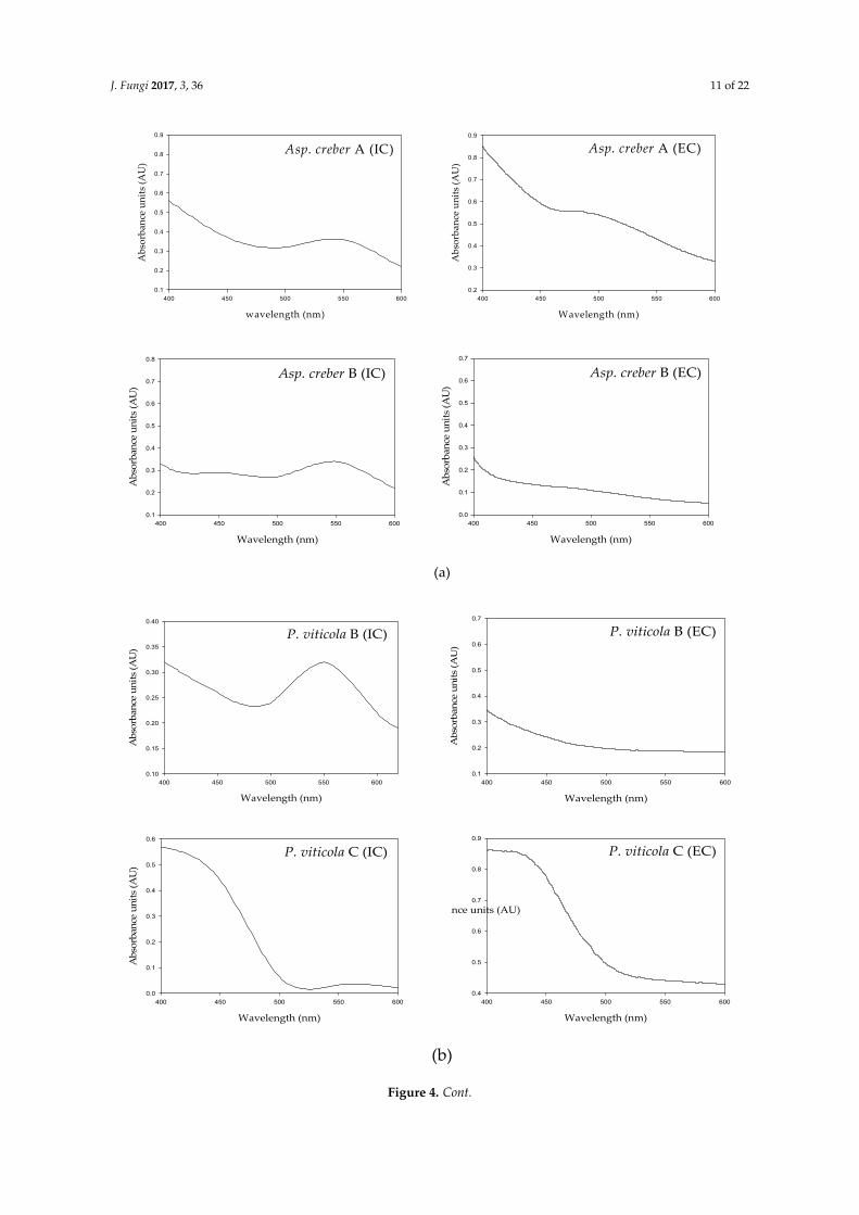
As shown in Figure 4, the absorbance spectra of intra- and extracellular solutions from a single isolate revealed quite similar profiles characterized by a strong absorbance in the UV region and also an area of absorbance in the visible range of wavelengths. The values were principally located in the 400–480 nm area for pale yellow to yellow-orange pigments. The maximal absorbances spread in the 500–550 nm region for red colors.
Figure 3. Colors observed from culture filtrates from three isolates of Penicillium viticola (A–C)(seven-day cultures in potato dextrose broth).
Oppositely, no clear difference could be visually established among the pale pink shades of thethree isolates of F. equisetti (A–C) or the two N. haematococca isolates (A and B).
3.2.2. Pigmented Contents from Mycelium
For all pigment-producing isolates, the intracellular pigments (from mycelium) were extractedfrom the biomass. The approximate colors visualized after extraction are presented in Table 3.The pigments from most of the extracellular fungal culture filtrates were of dominating red, orange,yellow, green, brown, pink and violet. However, after extraction from biomass, many intracellularsamples were uncolored, especially for isolates producing extracellular culture filtrates of pink, yellowand green color. This is probably characteristic of isolates essentially secreting water-soluble coloredmolecules in the culture media.
Instead, many dark colored cultures, mainly in the shades of red or maroon extracellular pigments,gave dark pigmented intracellular extracts from the biomass, indicating that the pigment was alsohighly concentrated inside the mycelium. These mainly concerned the isolates included in the group“isolates with intense hues”, and in the group “isolates with orange hues” to a lesser extent (Table 3).Thus, isolates appeared with different status and varying capacities, towards pigment production.
3.3. Spectrophotometric Characterization of Pigments
As shown in Figure 4, the absorbance spectra of intra- and extracellular solutions from a singleisolate revealed quite similar profiles characterized by a strong absorbance in the UV region and alsoan area of absorbance in the visible range of wavelengths. The values were principally located in the400–480 nm area for pale yellow to yellow-orange pigments. The maximal absorbances spread in the500–550 nm region for red colors.
J. Fungi 2017, 3, 36 11 of 22J. Fungi 2017, 3, 36 11 of 22
(a)
(b)
Asp. creber A (EC)
Wavelength (nm)
400 450 500 550 600
Abs
orba
nce
units
(AU
)
0.2
0.3
0.4
0.5
0.6
0.7
0.8
0.9
Asp. creber A (IC)
wavelength (nm)
400 450 500 550 600
Abs
orba
nce
units
(AU
)
0.1
0.2
0.3
0.4
0.5
0.6
0.7
0.8
0.9
Asp. creber B (IC)
Wavelength (nm)
400 450 500 550 600
Abs
orba
nce
units
(AU
)
0.1
0.2
0.3
0.4
0.5
0.6
0.7
0.8
Asp. creber B (EC)
Wavelength (nm)
400 450 500 550 600
Abs
orba
nce
units
(AU
)
0.0
0.1
0.2
0.3
0.4
0.5
0.6
0.7
P. viticola B (IC)
Wavelength (nm)
400 450 500 550 600
Abs
orba
nce
units
(AU
)
0.10
0.15
0.20
0.25
0.30
0.35
0.40
P. viticola B (EC)
Wavelength (nm)
400 450 500 550 600
Abs
orba
nce
units
(AU
)
0.1
0.2
0.3
0.4
0.5
0.6
0.7
P. viticola C (IC)
Wavelength (nm)
400 450 500 550 600
Abs
orba
nce
units
(AU
)
0.0
0.1
0.2
0.3
0.4
0.5
0.6
P. viticola C (EC)
Wavelength (nm)
400 450 500 550 600
ance units (AU)
0.4
0.5
0.6
0.7
0.8
0.9
Figure 4. Cont.

J. Fungi 2017, 3, 36 12 of 22
J. Fungi 2017, 3, 36 12 of 22
(c)
Figure 4. Intracellular (IC) and extracellular (EC) UV-visible spectra of: (a) Aspergillus creber A and B; (b) Penicillium viticola B and C; and (c) Talaromyces albobiverticillius A–C cultures in potato dextrose broth (7 days).
However, slight variations were noticed between extra- and intracellular liquids: in A. creber A as an example, extracellular maximum absorbance was around 470 nm (yellow-orange hue) instead of 550 nm (red shade) for intracellular liquid (Figure 4a). These slight variations however indicate that intra- and extracellular solutions may contain different assortments of colored compounds, in different proportions, resulting in different hues (Figure 5).
T. albobiverticillius A (IC)
Wavelength (nm)
400 450 500 550 600
Abs
orba
nce
units
(AU
)
0.1
0.2
0.3
0.4
0.5
0.6
0.7
0.8
0.9
T. albobiverticillius A (EC)
Wavelength (nm)
400 450 500 550 600
Abs
orba
nce
units
(AU
)
0.0
0.1
0.2
0.3
0.4
T. albobiverticillius B (IC)
Wavelength (nm)
400 450 500 550 600
Abs
orba
nce
units
(AU
)
0.1
0.2
0.3
0.4
0.5
0.6
0.7
0.8
T. albobiverticillius B (EC)
Wavelength (nm)
400 450 500 550 600
Abs
orba
nce
units
(AU
)
0.2
0.3
0.4
0.5
0.6
0.7
0.8
0.9
T. albobiverticillius C (IC)
Wavelength (nm)
400 450 500 550 600 650
Abs
orba
nce
units
(AU
)
0.05
0.10
0.15
0.20
0.25
0.30
0.35
T. albobiverticillius C (EC)
Wavelength (nm)
400 450 500 550 600
Abs
orba
nce
units
(AU
)
0.4
0.5
0.6
0.7
0.8
0.9
1.0
1.1
Figure 4. Intracellular (IC) and extracellular (EC) UV-visible spectra of: (a) Aspergillus creber A andB; (b) Penicillium viticola B and C; and (c) Talaromyces albobiverticillius A–C cultures in potato dextrosebroth (7 days).
However, slight variations were noticed between extra- and intracellular liquids: in A. creber A asan example, extracellular maximum absorbance was around 470 nm (yellow-orange hue) instead of550 nm (red shade) for intracellular liquid (Figure 4a). These slight variations however indicate thatintra- and extracellular solutions may contain different assortments of colored compounds, in differentproportions, resulting in different hues (Figure 5).

J. Fungi 2017, 3, 36 13 of 22J. Fungi 2017, 3, 36 13 of 22
Figure 5. Colors observed in different fungal species: (a) obverse face on PDA; (b) reverse face on PDA; (c) culture in PDB (seven days); (d) extract of intracellular pigments (Ethanol/water 50/50) (IC); and (e) filtrate from liquid culture (EC).
Differences were also observed among the spectral profiles of different isolates belonging to the same species. As shown from the intracellular profiles of A. creber A and B, P. viticola B and C, and T. albobiverticillius A–C (Figure 4a–c, respectively, and Table 4), maximal absorbance areas differed in the visible region (510–560 nm for P. viticola B and 420–450 nm for P. viticola C; and 422–525 nm for T. albobiverticillius A, 500 nm for T. albobiverticillius B and 520–580 nm for T. albobiverticillius C), but, for A. creber A and B, the spectra looked similar (Figure 4a,b). Similar variation was stated between the extracellular profiles.
These results clearly imply that isolates from a same species produce and secrete different pigments and therefore have different behavior towards colored compound production.
Figure 5. Colors observed in different fungal species: (a) obverse face on PDA; (b) reverse face on PDA;(c) culture in PDB (seven days); (d) extract of intracellular pigments (Ethanol/water 50/50) (IC); and (e)filtrate from liquid culture (EC).
Differences were also observed among the spectral profiles of different isolates belonging tothe same species. As shown from the intracellular profiles of A. creber A and B, P. viticola B and C,and T. albobiverticillius A–C (Figure 4a–c, respectively, and Table 4), maximal absorbance areas differedin the visible region (510–560 nm for P. viticola B and 420–450 nm for P. viticola C; and 422–525 nm forT. albobiverticillius A, 500 nm for T. albobiverticillius B and 520–580 nm for T. albobiverticillius C), but,for A. creber A and B, the spectra looked similar (Figure 4a,b). Similar variation was stated between theextracellular profiles.
These results clearly imply that isolates from a same species produce and secrete differentpigments and therefore have different behavior towards colored compound production.
J. Fungi 2017, 3, 36 14 of 22
Table 4. Summary of main peaks (λmax) noticed in 10-days old culture of Talaromyces albobiverticilliusisolates A–C cultivated in liquid medium (potato dextrose broth).
T. albobiverticillius SamplePeaks in the UV Region (nm) Peaks in the Visible Region (nm)
200–250 250–300 300–400 >400
AIC 235 286 362 422, 425, 511, 525EC 265 365 458, 469.8, 480
BIC 232 268, 292 410, 440, 460, 500EC 288 412, 524, 532
CIC 222 283 385 520–580EC 283 370, 385 436
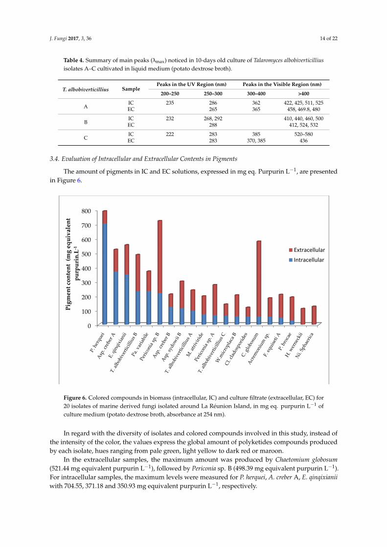
3.4. Evaluation of Intracellular and Extracellular Contents in Pigments
The amount of pigments in IC and EC solutions, expressed in mg eq. Purpurin L−1, are presentedin Figure 6.
J. Fungi 2017, 3, 36 14 of 22
Table 4. Summary of main peaks (λmax) noticed in 10-days old culture of Talaromyces albobiverticillius isolates A–C cultivated in liquid medium (potato dextrose broth).
T. albobiverticillius Sample Peaks in the UV Region (nm) Peaks in the Visible Region (nm)
200–250 250–300 300–400 >400
A IC 235 286 362 422, 425, 511, 525 EC 265 365 458, 469.8, 480
B IC 232 268, 292 410, 440, 460, 500 EC 288 412, 524, 532
C IC 222 283 385 520–580 EC 283 370, 385 436
3.4. Evaluation of Intracellular and Extracellular Contents in Pigments
The amount of pigments in IC and EC solutions, expressed in mg eq. Purpurin L−1, are presented in Figure 6.
In regard with the diversity of isolates and colored compounds involved in this study, instead of the intensity of the color, the values express the global amount of polyketides compounds produced by each isolate, hues ranging from pale green, light yellow to dark red or maroon.
Figure 6. Colored compounds in biomass (intracellular, IC) and culture filtrate (extracellular, EC) for 20 isolates of marine derived fungi isolated around La Réunion Island, in mg eq. purpurin L−1 of culture medium (potato dextrose broth, absorbance at 254 nm).
In the extracellular samples, the maximum amount was produced by Chaetomium globosum (521.44 mg equivalent purpurin L−1), followed by Periconia sp. B (498.39 mg equivalent purpurin L−1). For intracellular samples, the maximum levels were measured for P. herquei, A. creber A, E. qinqixianii with 704.55, 371.18 and 350.93 mg equivalent purpurin L−1, respectively.
The amount of intracellular content was significantly lower than the one of the extracellular content in this population (n = (20,20), V = 10, P = 6 × 10⁻⁴). However, looking at each isolate separately, the amount of intracellular pigments was significantly higher than the extracellular one
0
100
200
300
400
500
600
700
800
Pigm
ent c
onte
nt (
mg
equi
vale
ntpu
rpur
in.L
-1 Extracellular
Intracellular
Figure 6. Colored compounds in biomass (intracellular, IC) and culture filtrate (extracellular, EC) for20 isolates of marine derived fungi isolated around La Réunion Island, in mg eq. purpurin L−1 ofculture medium (potato dextrose broth, absorbance at 254 nm).
In regard with the diversity of isolates and colored compounds involved in this study, instead ofthe intensity of the color, the values express the global amount of polyketides compounds producedby each isolate, hues ranging from pale green, light yellow to dark red or maroon.
In the extracellular samples, the maximum amount was produced by Chaetomium globosum(521.44 mg equivalent purpurin L−1), followed by Periconia sp. B (498.39 mg equivalent purpurin L−1).For intracellular samples, the maximum levels were measured for P. herquei, A. creber A, E. qinqixianiiwith 704.55, 371.18 and 350.93 mg equivalent purpurin L−1, respectively.

J. Fungi 2017, 3, 36 15 of 22
The amount of intracellular content was significantly lower than the one of the extracellularcontent in this population (n = (20,20), V = 10, P = 6 × 10⁻⁴). However, looking at each isolateseparately, the amount of intracellular pigments was significantly higher than the extracellular one forP. herquei (704.55 vs. 84 mg equivalent purpurin L−1), and A. creber A (371.18 vs. 151.11) and B (125.25vs. 86.35), and E. qinqixianii (350.93 vs. 202.74).
4. Discussion
4.1. Biodiversity of Marine-Derived Fungi around La Réunion Island
From the sampling in La Réunion Island marine biotopes, 31 different species distributed in25 genera were identified as pigment producers. The identification of isolates collected in coral reefsand lava flows of La Réunion Island coincides with identifications conducted from various marineenvironments. Indeed, the majority of the studied fungi, such as those sampled from north of theIndian Ocean, belong to the phylum Ascomycetes. The fungi of the genus Aspergillus, particularlyA. sydowii, are also found in the Caribbean corals (Porites lobata), Polynesia, and in sediments off thecoast of India [46,47]. Penicillium citrinum was isolated from the red algae Actinotrichia fragilis, fromsponges, and the species was also found on other substrates such as hard substrate or water [48–51].The genera Penicillium, Cladosporium, Chaetomium, and Fusarium, and species Nigrospora oryzae andHortea werneckii, have been identified in marine sediments collected at different depths in the centralbasin of the Indian Ocean and considered to be coral pathogens [8,52]. Fungi, namely Alternaria sp.,Acremonium sp. and Rhodotorula mucilaginosa, were isolated from salt lakes in Antarctica, as wereP. chrysogenum and P. crustosum [53]. Rhodotorula mucilaginosa was also found in the sediments ofcentral Indian basin [52].
The diversity within the isolated fungal population was crucial while comparing the abilityof pigment production [54]. However, in our samples, the highest diversity of pigmented fungiwas revealed from the water column (13 species) and from hard substrates (limestone or lava flow)(11 species). If the water column can be suspected of carrying a multitude of fungal propagulesoriginating from terrestrial environments, hard substrates are probably more representative of marineand marine-derived biodiversity.
Our study demonstrates that the living coral Pocillopora sp. shelters fungi from the generaAspergillus (A. creber, A. sydowii and Eurotium amstellodami) and Penicillium (P. viticola), as well asHypocrea koningii and Acremonium sp. Widely disseminated on land, this mainly saprophytic genusAcremonium sp., has already been isolated from marine environments (sea fans, sea water, seacucumbers, and intertidal sediment samples) [55–57]. These fungi, were extracted from the inner partsof the coral structure. They are then supposed to be at least endophytic species for this coral genus.
The coral rubbles (dead part of corals) from our samples contained colored Penicillium (P. herquei)or related species T. albobiverticillius as well as an isolate of Chaetomium globosum. Chaetomium globosumis a common fungal species from soil and environment.
Most of the fungi we identified can also be found on land, in soil, on plants or insects, but some ofthem have rarely been isolated from marine environments such as Whalleya microplaca, Biscogniauxia sp.,Paraconiothyrium variabile, Myrothecium atroviride, Nectria haematococca, Peyronellaea glomerata, Epicoccumsorghi, Sporisorium exsertum and Periconia sp.. From our study, the genera Aspergillus and Penicillium orthe close ones such as Talaromyces, Emericella or Eurotium (from the Trichocomaceae family) are muchmore diverse than others in these tropical marine biotopes (12 different species), and are representedin several types of samples and locations. These aerobic and xerophilic species are well-known forpopulating dry and/or salty biotopes. However, their ability to subsist or develop underwater, withwidely varying oxygenation conditions is less known. These cosmopolitan fungi are well-knownto produce a wide range of secondary metabolites such as polyketide-based pigments in solid andliquid cultures. Overall, in our study, some fungal species (T. albobiverticillius or N. haematococca)were identified from different types of samples in the same area. Some others (A. sydowii, P. viticola)

J. Fungi 2017, 3, 36 16 of 22
appeared in separate locations. These fungi found in several sample types and/or in different locationsmay be considered as frequent in marine environment around La Réunion Island.
4.2. Qualitative Aspect of the Pigment Production
For marine-derived isolates, two statuses lead to particular behaviors and products: the challengeof facing unusual living conditions (exogenous fungi) and the use of specific procedures naturallyadapted to the marine niches (for instance fungal endophytes of marine microorganisms, i.e.,indigenous micromycetes, naturally selected by aquatic environments).
Overall, in unusual biotopes (sometimes extreme), the fungal species with pigmented cell walls(in the spores and/or mycelium), are clearly able to tolerate dehydration-hydration cycles or high solarradiations, better than the moniliaceous fungi, whose cells are devoid of pigments. These aromaticcompounds, as melanin, sporopollenin (brown product of oxydative polymerization of β-carotene) orcycloleucomelone (terphenylquinone), often show significant antioxidant activities, and are bound toprotect the biological structures, giving them an excellent durability and a high potential for survivalin hostile environments [58,59].
From the available literature, the microorganisms of the genus Trichoderma are frequent in marineenvironments and some terrestrial strains are able to produce anthraquinone-like compounds [60].Indeed, isolates of the family Hypocreaceae (Trichoderma, Hypocrea and Acremonium) are alsorepresented in our study and exhibit orange to purple hues. Some strains of the common soil fungusCladosporium cladosporioides, also isolated from our samples with green shades, have already beenstudied for their production of intracellular melanin [61].
The most important colored compounds produced by Aspergillus and Emericella species arerespectively, hydroxyanthraquinones and azaphilone pigments, exhibiting a very wide range of hues.Furthermore, A. sydowii and Eurotium amstelodami isolated from La Réunion Island showed red andyellow colors respectively, as produced by their terrestrial counterparts [62].
Penicillium species and related ones seem to adjust easily to multiple conditions and tobe a source of original compounds as they appear among the most chemically inventive fungi.In Penicillium and Talaromyces species, polyketide-based pigments are also very common, and,particularly, the azaphilones, such as the derivatives of monascorubrin and rubropunctatin [63].Monascus-like azaphilone pigments such as N-glutarylmonascorubramine, N-glutarylrubropunctamine,monascorubramine homologues PP-V [(10Z)-12-carboxyl-monascorubramine] and PP-R [(10Z)-7-(2-hydroxyethyl)-monascorubramine] are frequently identified in their cultures [64,65]. However thecommercial production of red anthraquinoid pigments (Arpink Red™, Natural Red™) has alreadybeen carried out with P. oxalicum var. armeniaca [1]. The most common hues produced by bothgenera include yellow, red, orange and reddish-brown. Nevertheless, it was found that the yellowpigments seem predominant in most of the Penicillium species, while Talaromyces species mainlyproduce red pigments with few synthesizing yellow compounds of azaphilone series [66]. The coloredmolecules sometimes demonstrate mycotoxic activities such as rubratoxins A and B, rugulovasins andluteoskyrin [67].
Some strains of the widespread Acremonium sp. produce the yellow oosporein (chaetomidin)(biquinone, benzoquinone) and also some toxic compounds as diterpene glycosides [68].
Chaetomium globosum, isolated from the coral rubbles biosynthesizes maroon pigments in theculture conditions of our experiment. Many members of the family produce metabolites withantifungal properties. C. globosum is already known to biosynthesize yellow azaphilones namedchaetoviridins (A–D), antifungal compounds involved in the induction of chlamydospores-likecells [69]. It also produces nitrogenous azaphilones (4′-epi-N-2-hydroxyethyl-azachaetoviridin A,and N-2-butyric-azochaetoviridin E) and isochromophilone XIII, with orange to red hues. Some strainsgenerate pigmented chaetoglobins, chaetoglobosins, chaetomugilins, and seco-chaetomugilins,while others can secrete a purple pigment called cochliodinol [70–73].

J. Fungi 2017, 3, 36 17 of 22
Associated with lava flows, Fusarium equiseti belongs to a group of widespread plant pathogens,but marine-derived Fusarium strains are also frequent in mangroves or associated with marineorganisms. These are already known to produce original colored anthraquinoid compounds (5-acetyl-2-methoxy-1,4,6-trihydroxy-anthraquinone;6,8-dimethoxy-1-methyl-2-(3-oxobutyl)-anthraquinone andfusaquinones) [19]. Among the Fusarium secondary metabolites, numerous polyketide pigments havealready been identified, such as naphthoquinone pigments which are the most abundant (bikaverin,nor-bikaverin, javanicin, anhydrojavanicin, fusarubin, anhydrofusarubin, bostrycoidin, and novarubin)and the hydroxyanthraquinones emodin, physcion, dermolutein, chrysophanol, erythroglaucin,dermocybin, dermorubin, tritisporin, cynodontin, helminthosporin or aurofusarin (review in [19,21]).All these molecules develop a palette of colors, ranging from yellow to purple or brown. Some speciesare also able to produce orange carotenoids (neurosporaxanthin by F. fujikuroi) [74]. The putativecarcinogen, fusarin C, apicidin F, fujikurins, the perithecal pigments fusarubins as well as the mycelialpigment bikaverin are also produced in the family.
From our work, Periconia sp. A isolate produced an impressive violet hue in PDB culture. Periconiais a cosmopolitan genus, often found in soil, and decaying herbs and forages. Some Periconia strainswere nevertheless identified from marine environments (P. abyssa (deep sea), P. byssoides (sea slugAplysia kurodai)) [75–77]. They attract interest because of the production of promising anti-cancer drugs,such as the carbosugar pericosine A. Some strains may produce an unidentified hepatoxin.
4.3. Quantitative Aspect of Pigment Production
As a promising factor, several of the marine-derived fungi isolated in this study had the ability togrow and biosynthesize pigments in unsalted synthetic conditions (e.g., Czapek Dox medium, PDB).During the period of fermentation, the pigment production started between Day 1 and Day 4 for themajority of isolates such as Aspergillus, Eurotium, Fusarium, Nigrospora, Pencillium and Talaromyces.For some fungi, the detection of the pigment production was notably delayed (e.g., Acremonium,Epicoccum, or Myrothecium). This might be due to the low level of pigment producing ability of thefungi or due to unfavorable environmental conditions for pigment production such as pH, temperature,nutrient sources, osmolarity and illumination conditions [78].
Considering the visual observation of pigment color in flasks and the respective UV-visiblespectra, fungi belonging to the same species may produce different colored mixtures (e.g., Aspergilluscreber A and B or Talaromyces alboverticillius A–C). They may then belong to different varieties and thusproduce pigments of distinct natures. The slight variations observed between intra- and extra-cellularsolutions also indicate that the solutions may contain different assortments of colored compounds, indifferent proportions, resulting in different hues.
From these findings, it is understood that a higher quantity of pigments has been mainly purifiedfrom extracellular filtrates in a significant manner (11/21 isolates). In our experimental conditions,the maximum pigment production was obtained in the extracellular samples for C. globosum andPericonia sp. B. On average, the values measured in the cells were significantly lower; indicatingthat pigments secretion in the liquid medium seems a widespread behavior in the conditions of theexperiment. Only the isolate P. herquei had a very high level of intracellular pigment biosynthesis(704.55 mg equivalent purpurin L−1). Nevertheless, for high intracellular pigment production frombiomass, A. creber A and E. qinqixianii present a true production potential. On the other hand,the extraction of intracellular colored compounds appeared sometimes not completely effective.The fungal biomass was still colored even after extraction. The efficiency of the extraction processcould probably be improved to recover higher pigment quantities from intracellular samples [79].
This work highlights different behaviors of fungal isolates towards the secretion of coloredmolecules compared to internal storage. Anyway, the production of secondary metabolites oftenoccurs after fungal growth has ceased, as a result of nutrient limitation coupled with excess carbonavailability. This makes it possible to manipulate their formation [80,81].

J. Fungi 2017, 3, 36 18 of 22
5. Conclusions
Marine and marine-derived fungi are promising resources for the production of new metabolitesof interest, and, among them, pigments are attractive [82–84]. The potential of marine-derivedmicroorganisms to produce unique and original molecules may come from specific metabolic orgenetic adaptation appearing to meet very specific combinations of physical and chemical parameters(high salinity, low O2 penetration, low temperature, limited light access and high pressure). Based onthis statement, our study explores, for the first time, the biodiversity of fungi from marine environmentsaround La Réunion Island, Indian Ocean, along with the ability of the isolates to produce pigments.The potentiality of these marine derived isolates to secrete pigments or to concentrate coloredcompounds inside the cells was highlighted. Several isolates collected from lava flows, hard substratessediments and corals (living or dead) turned out to be the interesting producers of intense colorson PDA culture medium. The main types identified, Aspergillus, Penicillium and related genera,are also found in other marine regions (such as Polynesia or along the coast of India). However,a great biodiversity (31 species) emphasizes the range of possible hues and molecules susceptible to beisolated. The majority of the isolates, probably marine optional, may also be able to grow in syntheticmedia, devoid of sea salts and may show the competence of producing pigments in an industrialscale. The most promising pigmented products, probably of intense red or purple hues, which seem toconsist in mixtures, will be subjected to purification and further analyses by analytical techniques suchas liquid chromatography–mass spectrometry/time-of-flight (LC-MS/TOF) and Nuclear MagneticResonance (NMR). The interesting isolates will also be subjected to further analyses to determine theirability as antibiotics or for enzyme production.
Supplementary Materials: The following are available online at www.mdpi.com/2309-608X/3/3/36/s1.
Acknowledgments: The authors are grateful to Regional Council of La Réunion Island for financial support.Thanks are also given to BIOLAVE program and Quod Jean Pascal.
Author Contributions: Mireille Fouillaud conceived, designed and performed the experiments. Mireille Fouilaudand Pascale Cuet collected the samples. Melissa Llorente, Hélène Magalon and Mekala Venkatachalam performedthe molecular analysis and analyzed the genetic data. Mireille Fouillaud, Mekala Venkatachalam and LaurentDufossé contributed to write the paper. The authors would also like to thank Cathie Milhau from ESIROI andPatricia Clerc from LCSNSA, of Université de La Réunion, for their logistic and technical help; and Gary Maresfor his timely help on data analysis.
Conflicts of Interest: The authors declare no conflict of interest.
References
1. Dufossé, L.; Galaup, P.; Yaron, A.; Arad, S.M.; Blanc, P.; Chidambara Murthy, K.N.; Ravishankar, G.A.Microorganisms and microalgae as sources of pigments for food use: A scientific oddity or an industrialreality? Trends Food Sci. Technol. 2005, 16, 389–406. [CrossRef]
2. Mayer, A.M.; Rodriguez, A.D.; Taglialatela-Scafati, O.; Fusetani, N. Marine pharmacology in 2009–2011:Marine compounds with antibacterial, antidiabetic, antifungal, anti-inflammatory, antiprotozoal,antituberculosis, and antiviral activities; affecting the immune and nervous systems, and other miscellaneousmechanisms of action. Mar. Drugs 2013, 11, 2510–2573. [PubMed]
3. Mapari, S.A.S.; Nielsen, K.F.; Larsen, T.O.; Frisvad, J.C.; Meyer, A.S.; Thrane, U. Exploring fungal biodiversityfor the production of water-soluble pigments as potential natural food colorants. Curr. Opin. Biotechnol.2005, 16, 231–238. [CrossRef] [PubMed]
4. Hohmann, C.; Schneider, K.; Bruntner, C.; Irran, E.; Nicholson, G.; Bull, A.T.; Jones, A.L.; Brown, R.; Stach, J.E.;Goodfellow, M.; et al. Caboxamycin, a new antibiotic of the benzoxazole family produced by the deep-seastrain Streptomyces sp. Ntk 937. J. Antibiot. 2009, 62, 99–104. [CrossRef] [PubMed]
5. Costantino, V.; Fattorusso, E.; Mangoni, A.; Perinu, C.; Cirino, G.; De Gruttola, L.; Roviezzo, F. Tedanol:A potent anti-inflammatory ent-pimarane diterpene from the caribbean sponge Tedania ignis. Bioorg. Med.Chem. 2009, 17, 7542–7547. [CrossRef] [PubMed]

J. Fungi 2017, 3, 36 19 of 22
6. Bonugli-Santos, R.C.; dos Santos Vasconcelos, M.R.; Passarini, M.R.Z.; Vieira, G.A.L.; Lopes, V.C.P.;Mainardi, P.H.; dos Santos, J.A.; de Azevedo Duarte, L.; Otero, I.V.R.; da Silva Yoshida, A.M.; et al.Marine-derived fungi: Diversity of enzymes and biotechnological applications. Front. Microbiol. 2015,6, 269. [CrossRef] [PubMed]
7. Panno, L.; Bruno, M.; Voyron, S.; Anastasi, A.; Gnavi, G.; Miserere, L.; Varese, G.C. Diversity, ecological roleand potential biotechnological applications of marine fungi associated to the seagrass Posidonia oceanica.New Biotechnol. 2013, 30, 685–694. [CrossRef] [PubMed]
8. Cathrine, S.J.; Raghukumar, C. Anaerobic denitrification in fungi from the coastal marine sediments off Goa,India. Mycol. Res. 2009, 113, 100–109. [CrossRef] [PubMed]
9. Holguin, G.; Vazquez, P.; Bashan, Y. The role of sediment microorganisms in the productivity, conservation,and rehabilitation of mangrove ecosystems: An overview. Biol. Fertil. Soils 2001, 33, 265–278. [CrossRef]
10. Bugni, T.S.; Ireland, C.M. Marine-derived fungi: A chemically and biologically diverse group ofmicroorganisms. Nat. Prod. Rep. 2004, 21, 143–163. [CrossRef] [PubMed]
11. Jones, E.B.G.; Pang, K.L. Marine Fungi: And Fungal-Like Organisms; De Gruyter: Berlin, Germany, 2012.12. Kathiresan, K.; Bingham, B.L. Biology of mangroves and mangrove ecosystems. In Advances in Marine
Biology; Southward, A., Young, C., Fuiman, L., Tyler, P., Eds.; Academic Press: San Diego, CA, USA, 2001;Volume 40, pp. 81–251.
13. Kohlmeyer, J.; Kohlmeyer, E. Marine Mycology; Elsvier Inc.: London, UK, 1979; p. 704.14. Jones, E.B.G. Marine fungi: Some factors affecting biodiversity. Fungal Divers. 2000, 4, 53–73. [CrossRef]15. Saleem, M.; Nazir, M. Bioactive natural products from marine-derived fungi: An update. In Studies in
Natural Products Chemistry; Atta-ur-Rahman, Ed.; Elsevier: Amsterdam, The Netherlands, 2015; Volume 45,pp. 297–361.
16. Imhoff, J.F. Natural products from marine fungi—Still an underrepresented resource. Mar. Drugs 2016, 14,19. [CrossRef] [PubMed]
17. Debashish, G.; Malay, S.; Barindra, S.; Joydeep, M. Marine enzymes. Adv. Biochem. Eng./Biotechnol. 2005, 96,189–218.
18. Zhang, C.; Kim, S.K. Application of marine microbial enzymes in the food and pharmaceutical industries.Adv. Food Nutr. Res. 2012, 65, 423–435. [PubMed]
19. Fouillaud, M.; Venkatachalam, M.; Girard-Valenciennes, E.; Caro, Y.; Dufossé, L. Anthraquinones andderivatives from marine-derived fungi: Structural diversity and selected biological activities. Mar. Drugs2016, 14, 64. [CrossRef] [PubMed]
20. Fouillaud, M.; Venkatachalam, M.; Girard-Valenciennes, E.; Caro, Y.; Dufossé, L. Marine-derivedfungi producing red anthraquinones: New resources for natural colors? In Proceedings of the 8thInternational Conference of Pigments in Food, “Coloured Foods for Health Benefits”, Cluj-Napoca, Romania,28 June—1 July 2016.
21. Caro, Y.; Venkatachalam, M.; Lebeau, J.; Fouillaud, M.; Dufossé, L. Pigments and colorants from filamentousfungi. In Fungal Metabolites; Merillon, J.-M., Ramawat, G.K., Eds.; Springer: Cham, Switzerland, 2016;pp. 1–70.
22. Ebel, R. Natural product diversity from marine fungi. In Comprehensive Natural Products II: Chemistry andBiology; Mander, L., Liu, H.-W., Eds.; Elsevier: Oxford, UK, 2010; Volume 2, pp. 223–262.
23. Calvo, A.M.; Wilson, R.A.; Bok, J.W.; Keller, N.P. Relationship between secondary metabolism and fungaldevelopment. Microbiol. Mol. Biol. Rev. 2002, 66, 447–459. [CrossRef] [PubMed]
24. Margalith, P. Pigment Microbiology; Springer: London, UK; New York, NY, USA, 1992; p. 156.25. Demain, A.L.; Fang, A. The natural functions of secondary metabolites. In History of Modern Biotechnology I;
Fiechter, A., Ed.; Springer: Berlin/Heidelberg, Germany, 2000; pp. 1–39.26. Réunion’s Coral Reef. Available online: https://en.wikipedia.org/wiki/R%C3%A9union%27s_coral_reef
(accessed on 1 May 2017).27. Peyrot-Clausade, M.; Chazottes, V.; Pari, N.; Peyrot-Clausade, M.; Chazottes, V.; Pari, N. Bioerosion in the
carbonate budget of two indo-pacific reefs: La Réunion (Indian Ocean) and moorea (Pacific Ocean). Bull. Geol.Soc. Denmark 1999, 1999, 1–30.
J. Fungi 2017, 3, 36 20 of 22
28. Conand, C.; Chabanet, P.; Cuet, P.; Letourneur, Y. The carbonate budget of a fringing reef in La ReunionIsland (Indian Ocean): Sea urchin and fish bioerosion and net calcification. In Proceedings of the 8thInternational Coral Reef Symposium, Panama City, Panama, 24–29 June 1997; Lessios, H.A., Macintyre, I.G.,Eds.; pp. 953–958.
29. Naim, O.; Cuet, P.; Mangar, V. Coral reefs of the mascarene archipelago. In Coral Reefs of the Indian Ocean:Their Ecology and Conservation; McClanahan, T.R., Sheppard, C., Obura, D.O., Eds.; Oxford University Press:New York, NY, USA, 2000; pp. 353–381.
30. Turner, J.; Klaus, R. Coral reefs of the mascarenes, western indian ocean. Philos. Trans. R. Soc. Lond. A Math.Phys. Eng. Sci. 2005, 363, 229–250. [CrossRef] [PubMed]
31. Montaggioni, L.; Faure, G. Les Récifs Coralliens des Mascareignes (Océan Indien); Université Francaise de l’OcéanIndien, Centre Universitaire de La Réunion: Réunion, France, 1980; p. 151.
32. Sanders, E.R. Aseptic laboratory techniques: Plating methods. J. Vis. Exp. JoVE 2011, 63, e3064. [CrossRef][PubMed]
33. Jong, S.; Dugan, F.; Edwards, M. ATCC Filamentous Fungi, 19th ed.; Rockville, MD American Type CultureCollection: Manassas, VA, USA, 1996.
34. Dahmen, H.; Staub, T.; Schwinn, F. Technique for long-term preservation of phytopathogenic fungi in liquidnitrogen. Phytopathology 1983, 73, 241–246. [CrossRef]
35. Knebelsberger, T.; Stoger, I. DNA extraction, preservation, and amplification. Methods Mol. Biol. 2012, 858,311–338. [PubMed]
36. Toju, H.; Tanabe, A.S.; Yamamoto, S.; Sato, H. High-coverage its primers for the DNA-based identification ofascomycetes and basidiomycetes in environmental samples. PLoS ONE 2012, 7, e40863. [CrossRef] [PubMed]
37. Samson, R.A.; Visagie, C.M.; Houbraken, J.; Hong, S.B.; Hubka, V.; Klaassen, C.H.W.; Perrone, G.; Seifert, K.A.;Susca, A.; Tanney, J.B.; et al. Phylogeny, identification and nomenclature of the genus aspergillus. Stud. Mycol.2014, 78, 141–173. [CrossRef] [PubMed]
38. Samson, R.A.; Yilmaz, N.; Houbraken, J.; Spierenburg, H.; Seifert, K.A.; Peterson, S.W.; Varga, J.; Frisvad, J.C.Phylogeny and nomenclature of the genus talaromyces and taxa accommodated in penicillium subgenusbiverticillium. Stud. Mycol. 2011, 70, 159–183. [CrossRef] [PubMed]
39. White, T.J.; Bruns, T.; Lee, S.; Taylor, J. Amplification and direct sequencing of fungal ribosomal RNA genesfor phylogenetics. PCR Protoc. Guide Methods Appl. 1990, 18, 315–322.
40. Romanelli, A.M.; Sutton, D.A.; Thompson, E.H.; Rinaldi, M.G.; Wickes, B.L. Sequence-based identification offilamentous basidiomycetous fungi from clinical specimens: A cautionary note. J. Clin. Microbiol. 2010, 48,741–752. [CrossRef] [PubMed]
41. Sutton, D.A.; Marín, Y.; Thompson, E.H.; Wickes, B.L.; Fu, J.; García, D.; Swinford, A.; de Maar, T.; Guarro, J.Isolation and characterization of a new fungal genus and species, aphanoascella galapagosensis, fromcarapace keratitis of a galapagos tortoise (chelonoidis nigra microphyes). Med. Mycol. 2013, 51, 113–120.[CrossRef] [PubMed]
42. Zhou, H.; Li, Y.; Tang, Y. Cyclization of aromatic polyketides from bacteria and fungi. Nat. Prod. Rep. 2010,27, 839–868. [CrossRef] [PubMed]
43. Caro, Y.; Anamale, L.; Fouillaud, M.; Laurent, P.; Petit, T.; Dufosse, L. Natural hydroxyanthraquinoidpigments as potent food grade colorants: An overview. Nat. Prod. Bioprospect. 2012, 2, 174–193. [CrossRef]
44. Machatová, Z.; Barbieriková, Z.; Poliak, P.; Jancovicová, V.; Lukeš, V.; Brezová, V. Study of naturalanthraquinone colorants by epr and uv/vis spectroscopy. Dyes Pigments 2016, 132, 79–93. [CrossRef]
45. Geyer, C.J. Fuzzy p-Values and Ties in Nonparametric Tests. Avaliable online: http://www.stat.umn.edu/geyer/fuzz (accessed on 30 June 2017).
46. Golubic, S.; Radtke, G.; Le Campion-Alsumard, T. Endolithic fungi in marine ecosystems. Trends Microbiol.2005, 13, 229–235. [CrossRef] [PubMed]
47. Priess, K.; Le Campion-Alsumard, T.; Golubic, S.; Gadel, F.; Thomassin, B. Fungi in corals: Black bands anddensity-banding of Porites lutea and P. lobata skeleton. Mar. Biol. 2000, 136, 19–27. [CrossRef]
48. Nicoletti, R.; Trincone, A. Bioactive compounds produced by strains of penicillium and Talaromyces of marineorigin. Mar. Drugs 2016, 14, 37. [CrossRef] [PubMed]
49. Tsuda, M.; Kasai, Y.; Komatsu, K.; Sone, T.; Tanaka, M.; Mikami, Y.; Kobayashi, J.i. Citrinadin a, a novelpentacyclic alkaloid from marine-derived fungus Penicillium citrinum. Org. Lett. 2004, 6, 3087–3089.[CrossRef] [PubMed]

J. Fungi 2017, 3, 36 21 of 22
50. Malmstrøm, J.; Christophersen, C.; Frisvad, J.C. Secondary metabolites characteristic of Penicillium citrinum,Penicillium steckii and related species. Phytochemistry 2000, 54, 301–309. [CrossRef]
51. Endo, A.; Kuroda, M.; Tsujita, Y. Ml-236a, ml-236b, and ml-236c, new inhibitors of cholesterogensis producedby Penicillium citrinum. J. Antibiot. 1976, 29, 1346–1348. [CrossRef] [PubMed]
52. Singh, P.; Raghukumar, C.; Verma, P.; Shouche, Y. Assessment of fungal diversity in deep-sea sediments bymultiple primer approach. World J. Microbiol. Biotechnol. 2012, 28, 659–667. [CrossRef] [PubMed]
53. Brunati, M.; Rojas, J.L.; Sponga, F.; Ciciliato, I.; Losi, D.; Gottlich, E.; de Hoog, S.; Genilloud, O.; Marinelli, F.Diversity and pharmaceutical screening of fungi from benthic mats of antarctic lakes. Mar. Genom. 2009, 2,43–50. [CrossRef] [PubMed]
54. Yahr, R.; Schoch, C.L.; Dentinger, B.T. Scaling up discovery of hidden diversity in fungi: Impacts of barcodingapproaches. Philos. Trans. R. Soc. B 2016, 371, 20150336. [CrossRef] [PubMed]
55. An, X.; Feng, B.-M.; Chen, G.; Chen, S.-F.; Wang, H.-F.; Pei, Y.-H. Isolation and identification of two newcompounds from marine-derived fungus Acremonium fusidioides rz01. Chin. J. Nat. Med. 2016, 14, 934–938.[CrossRef]
56. Afiyatullov, S.S.; Kalinovsky, A.I.; Antonov, A.S.; Zhuravleva, O.I.; Khudyakova, Y.V.; Aminin, D.L.;Yurchenko, A.N.; Pivkin, M.V. Isolation and structures of virescenosides from the marine-derived fungusAcremonium striatisporum. Phytochem. Lett. 2016, 15, 66–71. [CrossRef]
57. Gallardo, G.L.; Butler, M.; Gallo, M.L.; Rodríguez, M.A.; Eberlin, M.N.; Cabrera, G.M. Antimicrobialmetabolites produced by an intertidal acremonium furcatum. Phytochemistry 2006, 67, 2403–2410. [CrossRef][PubMed]
58. Hiort, J.; Maksimenka, K.; Reichert, M.; Perovic-Ottstadt, S.; Lin, W.H.; Wray, V.; Steube, K.; Schaumann, K.;Weber, H.; Proksch, P.; et al. New natural products from the sponge-derived fungus Aspergillus niger. J. Nat.Prod. 2004, 67, 1532–1543. [CrossRef] [PubMed]
59. Pagano, M.C.; Rosa, L.H. Fungal molecular taxonomy. In Fungal Biomolecules; John Wiley & Sons, Ltd.:Chichester, UK, 2015; pp. 311–321.
60. Slater, G.; Haskins, R.; Hogge, L.; Nesbitt, L. Metabolic products from a Trichoderma viride pers. Ex fries.Can. J. Chem. 1967, 45, 92–96. [CrossRef]
61. Duran, N.; Teixeira, M.F.; De Conti, R.; Esposito, E. Ecological-friendly pigments from fungi. Crit. Rev. FoodSci. Nutr. 2002, 42, 53–66. [CrossRef] [PubMed]
62. Butinar, L.; Frisvad, J.C.; Gunde-Cimerman, N. Hypersaline waters–a potential source of foodborne toxigenicAspergilli and Penicillia. FEMS Microbiol. Ecol. 2011, 77, 186–199. [CrossRef] [PubMed]
63. Woo, P.C.; Lam, C.W.; Tam, E.W.; Lee, K.C.; Yung, K.K.; Leung, C.K.; Sze, K.H.; Lau, S.K.; Yuen, K.Y.The biosynthetic pathway for a thousand-year-old natural food colorant and citrinin in Penicillium marneffei.Sci. Rep. 2014, 4, 6728. [CrossRef] [PubMed]
64. Arai, T.; Koganei, K.; Umemura, S.; Kojima, R.; Kato, J.; Kasumi, T.; Ogihara, J. Importance of the ammoniaassimilation by Penicillium purpurogenum in amino derivative monascus pigment, PP-V, production. AMBExpress 2013, 3, 19. [CrossRef] [PubMed]
65. Ogihara, J.; Kato, J.; Oishi, K.; Fujimoto, Y. Pp-r, 7-(2-hydroxyethyl)-monascorubramine, a red pigmentproduced in the mycelia of Penicillium sp. AZ. J. Biosci. Bioeng. 2001, 91, 44–47. [CrossRef]
66. Frisvad, J.C.; Yilmaz, N.; Thrane, U.; Rasmussen, K.B.; Houbraken, J.; Samson, R.A. Talaromyces atroroseus, anew species efficiently producing industrially relevant red pigments. PLoS ONE 2013, 8, e84102. [CrossRef][PubMed]
67. Yilmaz, N.; Houbraken, J.; Hoekstra, E.S.; Frisvad, J.C.; Visagie, C.M.; Samson, R.A. Delimitation andcharacterisation of Talaromyces purpurogenus and related species. Persoonia 2012, 29, 39–54. [CrossRef][PubMed]
68. Thomson, R.H. Naturally Occurring Quinones IV: Recent Advances; Blackie Academic & Professional: London,UK; New York, NY, USA, 1997.
69. Takahashi, M.; Koyama, K.; Natori, S. Four new azaphilones from Chaetomium globosum var. Flavo-viridae.Chem. Pharm. Bull. 1990, 38, 625–628. [CrossRef]
70. McMullin, D.R. Structural Characterization of Secondary Metabolites Produced by Fungi Obtained fromDamp Canadian Building. Ph.D. Thesis, Ottawa-Carleton University, Ottawa, ON, Canada, 2008.

J. Fungi 2017, 3, 36 22 of 22
71. McMullin, D.R.; Sumarah, M.W.; Miller, J.D. Chaetoglobosins and azaphilones produced by Canadian strainsof Chaetomium globosum isolated from the indoor environment. Mycotoxin Res. 2013, 29, 47–54. [CrossRef][PubMed]
72. Ming Ge, H.; Yun Zhang, W.; Ding, G.; Saparpakorn, P.; Chun Song, Y.; Hannongbua, S.; Xiang Tan, R.Chaetoglobins A and B, two unusual alkaloids from endophytic Chaetomium globosum culture. Chem. Commun.2008, 5978–5980. [CrossRef] [PubMed]
73. Brewer, D.; Jerram, W.A.; Taylor, A. The production of cochliodinol and a related metabolite by chaetomiumspecies. Can. J. Microbiol. 1968, 14, 861–866. [CrossRef] [PubMed]
74. Prado-Cabrero, A.; Schaub, P.; Diaz-Sanchez, V.; Estrada, A.F.; Al-Babili, S.; Avalos, J. Deviation of theneurosporaxanthin pathway towards beta-carotene biosynthesis in Fusarium fujikuroi by a point mutation inthe phytoene desaturase gene. FEBS J. 2009, 276, 4582–4597. [CrossRef] [PubMed]
75. Kohlmeyer, J. New genera and species of higher fungi from the deep sea (1615–5315 m). Revue de Mycologie1977, 41, 189–206.
76. Usami, Y.; Ichikawa, H.; Arimoto, M. Synthetic efforts for stereo structure determination of cytotoxic marinenatural product pericosines as metabolites of Periconia sp. From sea hare. Int. J. Mol. Sci. 2008, 9, 401–421.[CrossRef] [PubMed]
77. Dighton, J.; White, J.F. The Fungal Community: Its Organization and Role in the Ecosystem, 3rd ed.; CRC Press:Boca Raton, FL, USA, 2005.
78. Ogbonna, C.N. Production of food colourants by filamentous fungi. Afr. J. Microbiol. Res. 2016, 10, 960–971.79. Kaufmann, B.; Christen, P. Recent extraction techniques for natural products: Microwave-assisted extraction
and pressurised solvent extraction. Phytochem. Anal. PCA 2002, 13, 105–113. [CrossRef] [PubMed]80. Debbab, A.; Aly, A.H.; Proksch, P. Bioactive secondary metabolites from endophytes and associated marine
derived fungi. Fungal Divers. 2011, 49, 1–12. [CrossRef]81. Gunatilaka, A.L.; Wijeratne, E.K. Natural Products from Bacteria and Fungi; Elsevier: Amsterdam,
The Netherlands, 2000.82. Kim, S.-K. Marine Microbiology: Bioactive Compounds and Biotechnological Applications; Wiley & Sons: Weinheim,
Germany, 2013; p. 550.83. Kjer, J.; Debbab, A.; Aly, A.H.; Proksch, P. Methods for isolation of marine-derived endophytic fungi and
their bioactive secondary products. Nat. Protoc. 2010, 5, 479–490. [CrossRef] [PubMed]84. Rateb, M.E.; Ebel, R. Secondary metabolites of fungi from marine habitats. Nat. Prod. Rep. 2011, 28, 290–344.
[CrossRef] [PubMed]
© 2017 by the authors. Licensee MDPI, Basel, Switzerland. This article is an open accessarticle distributed under the terms and conditions of the Creative Commons Attribution(CC BY) license (http://creativecommons.org/licenses/by/4.0/).