bioconjugatesfortreatment of metastatic melanoma
TRANSCRIPT

Bioconjugates for treatment of metastaticmelanoma - difficulties and challenges
Ildikó Szabó
02.05.2019.

Anatomy of skin, melanocytes
https://commons.wikimedia.org/wiki/File:Layers_of_the_skin.jpg
https://www.dreamstime.com/stock-illustration-melanocyte-melanin-melanogenesis-melanin-producing-cells-melanin-pigment-responsible-skin-color-image54608996

Melanoma
https://www.wellnesscommunitydc.org/skin-cancer-stages/
Treatment!Good
prognosis
• develops in melanocytes
• melanocyte form moles by aggregation
• moles begin to grow and divide in an uncontrolled way
• most serious form of cancer, it can grows very quickly if left untreated
• spread to lower part of skin enter bloodstream and lymphatic system forming metastasis

The ABCDE’s of melanoma
Forrás: https://www.health.harvard.edu/cancer/melanoma-overview

Cancer statistics
https://careinthesun.org/skin-cancer/skin-cancer-statistics/
https://www.mysocietysource.org/Pages/newsdetails.aspx?ItemID=416
• Cancer of the skin is most common of all cancers
• Melanoma account only 1% of skin cancers
• Rates of melanoma have been rising• Ageing population
Importance of the sun-bathing and usage of sun-beds
Overexposure UVB radiation:
• Sunburn
• Skin cancer due to direct DNA
damaging effect

Treatment of Melanomafacts, posibilities, difficulties
• For determination of melanoma biopsy is applied
• Based on the laboratory examination:
o Cancerous or not
o How deep has it grown
• Early detected melanoma can be effectively treated
o surgery, biopsy
o Sentinel lymph node is cancer cell free
• In case of metastasis:
o One or more lymph node contain cancer cells→ quickly get to
other organs
o Chemotherapy, immunotherapy
o The overall success in metastatic melanoma is quite limited

Possibilities of melanoma treatmenttargeted tumor therapy

Possibilities of melanoma treatmentcancer cell specific cell surface molecules

Phage display technology
• Principle screening for specific peptides that bind to target from a library of phage
particles
• Peptides binding to individual targets can be identified by affinity selection
(biopanning)
• Phage-displayed peptide library can be used:
o B-cell and T-cell epitop mapping
o selection of bioactive peptides bound to receptors or proteins
o selection of disease specific antigen mimics
o selection of peptides bound to non-protein targets
o selection of cell specific peptides
o selection of organ-specific peptides
o development of peptide mediated drug delivery systems
• Targeting peptides have potential use in basic research and translational medicine.
Smith, G.P. et al Science (1985) 228, 1315-1317 Wu, C-H. et al. J. Med. Sci (2016) 23, 8-22.

Possibilities of melanoma treatmenttargeting proteoglycans

CSPG4/NG2 proteoglican
• Melanoma-associated chondroitin sulphate proteoglycan (MCSP)
• Transmembrane proteoglican („single-pass, type-I transmembrane
proteoglycan)
• Highly immunogenic tumor antigen of melanoma tumor cells
• It has been subsequently detected in various species (e.g. Human,
mouse, rat)
Iozzo, R.V.; Schaefer, L. Matrix Biol (2015) 42, 11-55.
Structure of CSPG4/NG2
N-terminal domain: two laminin-like globular
(LG) repeats; mediate ligandbinding, cell–matrix and cell–cell interactions, interaction with integrins and receptor tyrosinekinase
Central subdomain (D2): 15 tandem repeats of
a new module called CSPG (cell–matrix interaction, bind to collagen V és VI, FGF and PDGF
Intracellular domain (D3): bind to integrins,
galectin and numerous proteas cleavage sites

Guan, Y-Y et al. Biomaterials (2014) 35, 3060-3070
Burg, MA et al. Cancer Research (1999) 59, 2869-2874.
Specific NG2-binding peptides
TAASGVRSMHLTLRWVGLMS
have been used phage display to isolate peptides that bind to the NG2proteoglycan and home to NG2-expressing tumor neovasculature
• have high affinity and specificity to
NG2 proteoglycan
• Binding to BSA is minimal compared
to the proteoglycan
• Peptides bind to similar sites on NG2
„Peptide 1” „Peptide 2”
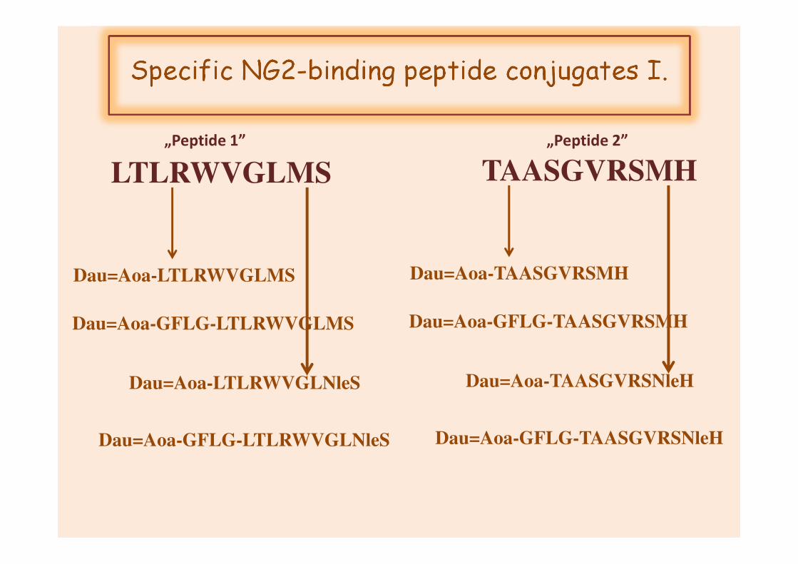
Specific NG2-binding peptide conjugates I.
TAASGVRSMH
Dau=Aoa-TAASGVRSMH
Dau=Aoa-GFLG-TAASGVRSMH
Dau=Aoa-TAASGVRSNleH
Dau=Aoa-GFLG-TAASGVRSNleH
LTLRWVGLMS
Dau=Aoa-LTLRWVGLMS
Dau=Aoa-GFLG-LTLRWVGLMS
Dau=Aoa-LTLRWVGLNleS
Dau=Aoa-GFLG-LTLRWVGLNleS
„Peptide 1” „Peptide 2”

Conjugates tR (min)a Mcalc Mmeasb
SzI-1 Dau=Aoa-TAASGVRSMH-NH2 10.6 1596.6 1596.5
SzI-10 Dau=Aoa-GFLG-TAASGVRSMH-NH2 13.5 1970.5 1970.9
SzI-2 Dau=Aoa-LTLRWVGLMS-NH2 14.4 1756.1 1756.2
SzI-3 Dau=Aoa-TAASGVRSNleH-NH2 13.9 1578.1 1578.8
SzI-4 Dau=Aoa-GFLG-TAASGVRSNleH-NH2 13.7 1952.5 1953.1
SzI-5 Dau=Aoa-LTLRWVGLNleS-NH2 15.1 1737.5 1737.2
SzI-6 Dau=Aoa-GFLG-LTLRWVGLNleS-NH2 18.1 2112.3 2112.4
Dau=Aoa-GFLG-LTLRWVGLMS-NH2 - 2130.8 -
Chemical characterization of drugcontaining NG2 conjugates
a Analitical RP-HPLC, Agilent Eclipse XDB C8, 5 µm, 80Å, 4.6 x 150 mm, HPLC coloumn, gradient: 5% B, 2 min; 5-100% B, 20 min. b Bruker Daltonics Esquire 3000plus (Bremen, Germany) ion trap mass spectrometer. Spectra were acquired in the50–2000 m/z range

Determination of in vitro cytostatic effect of drug containing NG2 conjugates
MTT-assay
Treatment(24h, 37°C, CM,concentration:0.16/0.8 – 100 µM;Washing, culturing in FCSM, 48h, ,
37°C )
(24h, 37°C)
MTT-assay (3.5h, 37°C)Determination of IC50 values
A2058

Results
In vitro cytostatic effect of drug containing NG2 conjugates
IC50 (µM)
A2058
SzI-1 Dau=Aoa-TAASGVRSMH-NH2 62,8±22,1*
SzI -2 Dau=Aoa-LTLRWVGLMS-NH2 4,3±1,9
SzI -3 Dau=Aoa- TAASGVRSNleH-NH2 22,1 és >100*
SzI -4 Dau=Aoa- GFLG-TAASGVRSNleH-NH2 2,3±1,3
SzI -5 Dau=Aoa-LTLRWVGLNleS-NH2 17,5±3,3
SzI -6 Dau=Aoa-GFLG-LTLRWVGLNleS-NH2 26,5±17,6*
SzI -10 Dau=Aoa-GFLG-TAASGVRSMH-NH2 5,2±2,4
Dau Dau·HCl <0.16

TAASGVRSMH
SzI-1 SzI-10+GFLG
(62.8±22.1µM) (5.2±2.4µM)
TAASGVRSNleHMet→Nle
SzI-3 SzI-4+GFLG
(22.1 és >100µM) (2.3±1.3µM)
ø
Conclusion I.
Structure-activity relationship
LTLRWVGLMS
SzI-2+GFLG
(4.3±1.9µM)
LTLRWVGLNleSMet→Nle
SzI-5 SzI-6+GFLG
(17.5±3.3µM) (26.5±17.6µM)øø
„Peptide 1”
„Peptide 2”

Specific NG2-binding peptide conjugates II.
GFLG-TAASGVRSNleH
Dau=Aoa-GFLG-ASGVRSNleH
Dau=Aoa-GFLG-GVRSNleH
Dau=Aoa-GFLG-ARASNleHSTGV-NH2
(scr)
LTLRWVGLMS
Dau=Aoa-GFLG-LRWVGLMS
Dau=Aoa-GFLG-WVGLMS
Dau=Aoa-VGLMWSLTRL-NH2
(scr)
„Peptide 1” „Peptide 2”
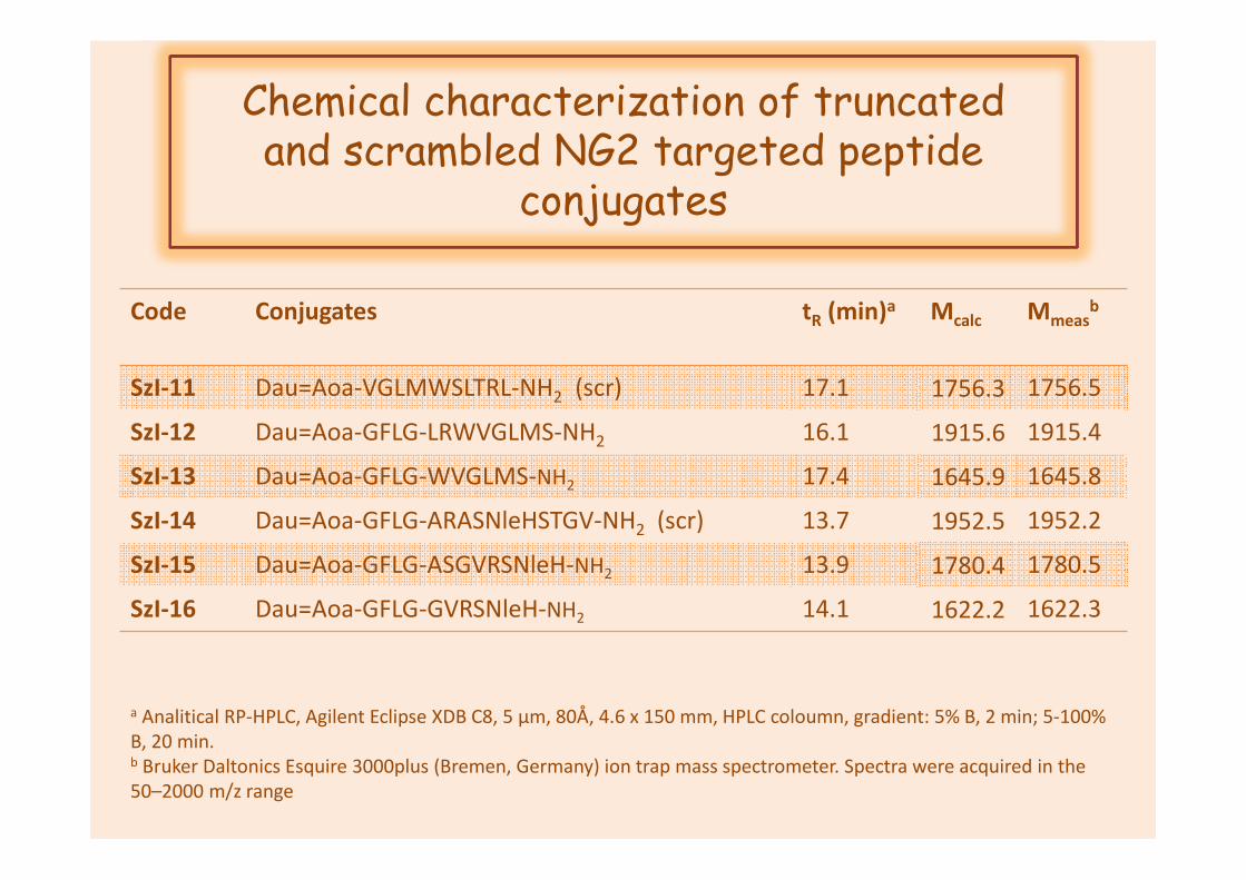
Chemical characterization of truncatedand scrambled NG2 targeted peptide
conjugates
Code Conjugates tR (min)a Mcalc Mmeasb
SzI-11 Dau=Aoa-VGLMWSLTRL-NH2 (scr) 17.1 1756.3 1756.5
SzI-12 Dau=Aoa-GFLG-LRWVGLMS-NH2 16.1 1915.6 1915.4
SzI-13 Dau=Aoa-GFLG-WVGLMS-NH2 17.4 1645.9 1645.8
SzI-14 Dau=Aoa-GFLG-ARASNleHSTGV-NH2 (scr) 13.7 1952.5 1952.2
SzI-15 Dau=Aoa-GFLG-ASGVRSNleH-NH2 13.9 1780.4 1780.5
SzI-16 Dau=Aoa-GFLG-GVRSNleH-NH2 14.1 1622.2 1622.3
a Analitical RP-HPLC, Agilent Eclipse XDB C8, 5 µm, 80Å, 4.6 x 150 mm, HPLC coloumn, gradient: 5% B, 2 min; 5-100% B, 20 min. b Bruker Daltonics Esquire 3000plus (Bremen, Germany) ion trap mass spectrometer. Spectra were acquired in the50–2000 m/z range

Results-preliminary dataIn vitro cytostatic effect of drug containing NG2
conjugates
IC50 (µM)
A2058 A431
SzI-2 Dau=Aoa-GFLG-LTLRWVGLMS-NH2 4,3±1,9 14.0±0.0
SzI-11 Dau=Aoa-VGLMWSLTRL-NH2 (scr) 2.9 3.2
SzI -12 Dau=Aoa-GFLG-LRWVGLMS-NH2 2.8 4.8
SzI -13 Dau=Aoa-GFLG-WVGLMS-NH2 15.3 15.7
SzI-4 Dau=Aoa-GFLG-TAASGVRSNleH-NH2 2,3±1,3 n.d.
SzI -14 Dau=Aoa-GFLG-ARASNleHSTGV-NH2 (scr) <0.8 3.6
SzI -15 Dau=Aoa-GFLG-ASGVRSNleH-NH2 2.3 2.9
SzI -16 Dau=Aoa-GFLG-GVRSNleH-NH2 3.2 3.0
Dau Dau·HCl <0.16 Dau

Conclusion II.
Structure-activity relationship
-TA -ASGFLG-TAASGVRSNleH
SzI-15
(2,3µM) øSzI-4
(2,3±1,3µM)
GFLG-ASGVRSNleH GFLG-GVRSNleH
SzI-16
(3,2µM) øscr
GFLG-ARASNleHSTGV
SzI-14
(<0.8µM)
LTLRWVGLMS
SzI-12
(2.8µM)
-LT; +GFLG
øSzI-2
(4.3±1.9µM)
GFLG-LRWVGLMS GFLG-WVGLMS
SzI-13
(15.3µM)scr
VGLMWSLTR
SzI-11
(2.9µM)
-LR
ø
„Peptide 1”
„Peptide 2”

Possibilities of melanoma treatmenttargeting cell surface receptors

Possibilities of melanoma treatmentMelanocortin-1 receptor (MC1R)
• GPCR; 5 subtypes with specific
distribution pattern in human tissues1,2
• MC1R is expressed in melanocytes and
melanomas3,4
• High level of MC1R gene expression is
characteristic for primary and metastatic
melanomas5
• MC1R is a highly specific marker of
melanoma
• promising candidate for targeted drug
delivery to melanoma cells
• MC1R ligands has specific internalization
into the cells
1Chhajlini, V. et al FEBS Lett. (1992) 309,417-420 4Roberts, D.W. et al Pigment Cell Res (2006) 19, 76-892Gantz, I. et al J. Biol. Chem (1993) 268, 8246-8250 5Salazar-Onfray, F. et al Br. J. Cancer (1993) 87, 414-4223Schwahn, D.J. et al Pigment Cell Res (2001) 14, 32-39
http://rspb.royalsocietypublishing.org/content/272/1573/1633

α-MSHα-Melanocyte Stimulating Hormone
• Ac-SYSMEHFRWGKPV-NH2
• Produced in adenohypophysis
• regulation of skin pigmentation
• >80% of human melanoma tumor samples obtained from patients with metastatic melanoma bear α-MSH receptors
• Superagonist α-MSH analog: [Nle4, D-Phe7] α-MSH
• Increased stability, resistant to enzymatic degradation
• increased receptor affinity (<nM)
• Ligand for targeted tumor therapy (radionucleotides, toxins, drugs, etc)
Siegrist, W. et al. (1989) Cancer Res, 49, 6352–6358.
Tatro, JB et al. (1990) J Clin Investig, 85,1825–1832.
Cone, RD et al. Ann NY Acad Sci (1993) 680, 342–363.
Saywer, TK et al. (1980) PNAS, 77, 5754–5758.
Giblin, MF et al. (1998) PNAS, 95, 12814–12818.
Morandini, R et al. (1994) Int J Cancer, 56, 129–133.

α-MSH
DiagnosisTumor imaging
treatmentRadiolabeled MSH
Application of α-MSH in melanoma cancer
Drug-MSH conjugates
treatment

Application of α-MSH in melanoma cancer
• First conjugates: Daunomycin-β-MSH,
• Melanotropin fragments have significant biological
activity (nitrosurea, melphalan)
• Specific receptor recognition
• Hormon-receptor complex is rapidly internalized
• Receptor may undergo recycling
Vargha, JM et al. (1977) Nature, 267, 56–58.
Garcia-Borron, J et al. (1992) Biochem (Life Sci. Adv), 11, 273–277.
Orlow, SJ et al. (1990) J Cell Physiol, 142, 129–136.
Ala-Glu-Lys-Lys-Asp-Glu-Gly-Pro-Tyr-Arg-Met-Glu-His-Phe-Arg-Trp-Gly-Ser-Pro-Pro-Lys-Asp

Application of α-MSH as targeting unit
• Central fragment containing conjugate
has selective and specific cytotoxic
effect
• The effect is mediated by MC1R
Morandini, R. et al Int. J. Cancer (1994) 56, 129-133
Ac-SYSMEHFRWGKPV-NH2
E5HFRWG10
Melphalan
H. Süli-Vargha; J. Botyánszky; K. Medzihradszky

Code Conjugates tR (min)a Mcalc Mmeasb
SzI-7 Dau=Aoa-SYSNleEHFRWGKPV-NH2 12.8 2185.8 2186.1
SzI-8 Ac-SYSNleEHFRWGK(Dau=Aoa)PV-NH2 13.1 2228.0 2228.5
SzI-9 Dau=Aoa-SYSNleEHFRWGK(Dau=Aoa)PV-NH2 13.0 2768.0 2768.4
Chemical characterization of drugcontaining α-MSH conjugates
a Analitical RP-HPLC, Agilent Eclipse XDB C8, 5 µm, 80Å, 4.6 x 150 mm, HPLC coloumn, gradient: 5% B, 2 min; 5-100% B, 20 min. b Bruker Daltonics Esquire 3000plus (Bremen, Germany) ion trap mass spectrometer. Spectra were acquired in the50–2000 m/z range
Ac-SYSMEHFRWGKPV-NH2
Dau=Aoa-SYSNleEHFRWGKPV
Ac-SYSNleEHFRWGK(Dau=Aoa)PV
Dau=Aoa-SYSNleEHFRWGK(Dau=Aoa)PV

Determination of in vitro cytostatic effect of drug containing α-MSH conjugates
MTT-assay
Treatment(24h, 37°C, CM,concentration:0.8 – 100 µM;Washing, culturing in FCSM, 48h, ,
37°C )
(24h, 37°C)
MTT-assay (3.5h, 37°C)Determination of IC50 values
A2058 A431

In vitro efficacy of daunomicin containingpeptide conjugates
IC50 (µM)
A2058 A431
SzI -7 Dau=Aoa-SYSNleEHFRWGKPV-NH2 9.8±5.4 25.0±11.2
SzI -8 Ac-SYSNleEHFRWGK(Dau=Aoa)PV-NH2 3.2±0.4 8.8±5.9
SzI -9 Dau=Aoa-SYSNleEHFRWGK(Dau=Aoa)PV-NH2 3.0±0.8 16.5±1.6
Dau Dau·HCl <0.16 0.5±0.4
IC50 (µM)
B16 M24 WM983b
SzI -7 Dau=Aoa-SYSNleEHFRWGKPV-NH2 2.9±0.6 12.8±1.6 9.9±1.5
SzI -8 Ac-SYSNleEHFRWGK(Dau=Aoa)PV-NH2 2.8±0.7 11.5±0.4 7.9±0.7
SzI -9 Dau=Aoa-SYSNleEHFRWGK(Dau=Aoa)PV-NH2 2.0±0.7 11.0±0.8 3.6±0.2

Dau=Aoa-SYSNleEHFRWGKPV-NH2SzI-7
(9.8±5.4µM)
Ac-SYSNleEHFRWGK(Dau=Aoa)PV-NH2 SzI-8(3.2±0.4µM)
Dau=Aoa-SYSNleEHFRWGK(Dau=Aoa)PV-NH2SzI-9
(3.0±0.8µM)
Conclusion III.
Structure-activity relationship

In vivo antitumor activity of drugcontaining α-MSH conjugates
Conditions:adult BALB/c male mice (28-32g) ;(i.p.) administration;25 mg DAU content/kg;3 mice/ groupThe toxicity was evaluated on the basis of life span, behavior and looking of the mice, and body weight. Parameters were followed for 14 days.
After 14 days following, significant change in
body weight, behaviour, and also in general
looking was not observed.
↓
not toxic for the animals →that can be further
investigated their antitumor activity in Vivo.
SzI-8

In vivo antitumor activity of drugcontaining α-MSH conjugates
• B16 (s.c.) injected into C57BL/6 male mice (20-28g), 7 animal/group
• i.p. administration. • doses:
control group (solvent); free DAU group (1 mg/kg,
treatment on day 9 and 17); SZI-7, SZI-8 and SZI-9 groups (10
mg/kg DAU content, treatmenton day 9, 13, 15, 17 after cellsinoculation).
• Termination: 20 days after cellinoculation,
• Determination: Animal weight and tumor volumes
SZI-8> SZI-7>> SZI-9
Conjugate SZI-8 showed higher anti-tumor activity in comparison with free Dau
Ctr Dau SzI-7 SzI-8 SzI-9
Antitumor activity of conjugates SzI-7,-8,-9 and
free Dau in s.c. B16 melanoma model

Summary
Melanoma
localization Rapid progression
TIME

Thank You for Your Attention!