biochimica et biophysica acta - kuhnlab.bmb.msu.edu · d. canella et al. / biochimica et biophysica...
TRANSCRIPT
Biochimica et Biophysica Acta 1799 (2010) 454–462
Contents lists available at ScienceDirect
Biochimica et Biophysica Acta
j ourna l homepage: www.e lsev ie r.com/ locate /bbagrm
DNA binding by the Arabidopsis CBF1 transcription factor requires thePKKP/RAGRxKFxETRHP signature sequence
Donatella Canella a,b,1, Sarah J. Gilmour a, Leslie A. Kuhn b,c,d, Michael F. Thomashow a,⁎a MSU-DOE Plant Research Laboratory, Michigan State University, East Lansing, MI 48824, USAb Department of Biochemistry and Molecular Biology, Michigan State University, East Lansing, MI 48824, USAc Department of Computer Science and Engineering, Michigan State University, East Lansing, MI 48824, USAd Department of Physics and Astronomy, Michigan State University, East Lansing, MI 48824, USA
⁎ Corresponding author. Tel.: +1 517 355 2299; fax:E-mail address: [email protected] (M.F. Thomash
1 Current address: Université de Lausanne, Centre InCH-1015 Lausanne-Dorigny, Lausanne, Switzerland.
1874-9399/$ – see front matter © 2009 Elsevier B.V. Adoi:10.1016/j.bbagrm.2009.11.017
a b s t r a c t
a r t i c l e i n f oArticle history:Received 16 October 2009Accepted 23 November 2009Available online 27 November 2009
Keywords:AP2/ERF domainArabidopsisCBF/DREB1 transcription factorDNA bindingSignature sequence
The CBF/DREB1 transcriptional activators are key regulators of plant freezing tolerance. They are members ofthe AP2/ERF multi-gene family, which in Arabidopsis comprises about 145 members. Common to theseproteins is the AP2/ERF DNA-binding domain, a 60-amino-acid fold composed of a three-stranded β-sheetfollowed by a C-terminal α-helix. A feature that distinguishes the CBF proteins from the other AP2/ERFproteins is the presence of “signature sequences,” PKKP/RAGRxKFxETRHP (abbreviated PKKPAGR) andDSAWR, which are located immediately upstream and downstream, respectively, of the AP2/ERF DNA-binding domain. The signature sequences are highly conserved in CBF proteins from diverse plant speciessuggesting that they have an important functional role. Here we show that the PKKPAGR sequence of AtCBF1is essential for its transcriptional activity. Deletion of the sequence or mutations within it greatly impairedthe ability of CBF1 to induce expression of its target genes. This impairment was not due to the mutationseliminating CBF1 localization to the nucleus or preventing protein accumulation. Rather, we show that thisloss of function was due to the mutations greatly impairing the ability of the CBF1 protein to bind to its DNArecognition sequence, the CRT/DRE element. These results establish that the ability of the CBF proteins tobind to the CRT/DRE element requires amino acids that extend beyond the AP2/ERF DNA-binding domainand raise the possibility that the PKKPAGR sequence contributes to determining the DNA-binding specificityof the CBF proteins.
© 2009 Elsevier B.V. All rights reserved.
1. Introduction
The Arabidopsis CBF cold response pathway has a prominent role incold acclimation, the processwhereby certain plants increase in freezingtolerance in response to low non-freezing temperatures [1,2]. Thepathway includes rapid cold induction of three genes encodingtranscription factors, CBF1, -2 and -3 [3,4]—also known as DREB1b, -c,-a, respectively [5]—that bind to the CRT/DRE DNA regulatory elementpresent in the promoters of CBF-target genes [6,7]. Induction of the CBFregulon of genes, which comprises about 100 members [8,9], leads toan increase in freezing and drought tolerance [5,10].
The CBF proteins are members of the AP2/ERF family of transcrip-tion factors [11]. The AP2/ERF protein family, comprising 145membersin Arabidopsis, is defined by the conserved 60-amino-acid AP2/ERFDNA-binding domain [12,13]. The AP2/ERF domain is composed of athree-strand β-sheet structure followed by an α-helix (14). The 3D
+1 517 353 9168.ow).tégratif de Génomique (CIG),
ll rights reserved.
solution structure of the AtERF1 AP2/ERF domain has shown thatarginine and tryptophan residues within the β-sheet contact nucleo-tides of the binding site within the major groove of the DNA [14].Those key residues are well conserved among members of the AP2/EREBP family, and yet different AP2 proteins display different DNA-binding preferences. Therefore, binding specificity within the family isimparted by additional residues within or outside the canonical DNA-binding domain. It has been proposed that the specificity determi-nants in these two subfamilies lie within the AP2/EREBP DNA-bindingdomain [15,16]. Sakuma et al. [16] have shown that specific aminoacids at two conserved positionswithin the AP2/ERF domains of DREBand ERF proteins affect DNA-binding affinity. At the present, however,it is still unclear whether additional residues within the two familiescan impart specific DNA preferences.
The primary feature that distinguishes the CBF transcriptionfactors from the other 145 AP2/ERF family members in Arabidopsisis the “signature sequences” that flank the AP2/ERF domain [17].These sequences, PKK/RPAGRxKFxETRHP, which we will refer to asPKKPAGR, and DSAWR, are located immediately up- and downstream,respectively, from the AP2/ERF domain in the CBF proteins. Thesignature sequences are highly conserved in CBF proteins from
455D. Canella et al. / Biochimica et Biophysica Acta 1799 (2010) 454–462
diverse plant species suggesting that they have an important function.Here we test this hypothesis for the PKKPAGR sequence.
2. Materials and methods
2.1. Plant material and growth conditions
Arabidopsis thaliana plants ecotype Colombia-0 (Col-0) or Wassi-lewskija-2 (Ws-2) were grown either in soil as described previously[18] or on solidified Gamborg's B5 medium (Caisson Laboratories,North Logan, UT, USA) for 2 weeks at 22°C under 100 μmol m-2 s-1
constant light, as previously described [9, 18]. Transgenic plants over-expressing CBF1 (lines G6 and G26) or carrying vector pGA643 (lineB6) in Ws-2 background have been described previously [18]. Arabi-dopsis transformation was performed using the floral dip method [19].
2.2. RNA isolation and analysis
Plant material was harvested in liquid nitrogen and total RNAextracted using RNeasy Plant Mini kits (Qiagen, Valencia, CA, USA)with modifications as described [20]. Total RNA (5–10 μg) wasfractionated in 1% formaldehyde gels and transferred onto nylonmembranes as described [21]. Membranes were hybridized in Churchbuffer (1% BSA, 1 mMEDTA, 0.5 MNaPO4, pH 7.2, 7% SDS) [22] at 65 °Covernight. Blots were hybridizedwith 32P-labeled fragments preparedusing the RandomPrimers DNA Labeling System (Invitrogen, Carlsbad,CA, USA) according to the manufacturer's instructions and washedunder high stringency [22]. Following the washes, the membraneswere exposed to a phosphorimager screen (BioRad, Hercules, CA,USA), which was scanned and then quantified using QuantityOnesoftware (BioRad, Hercules, CA, USA). RNA levels were normalized bycomparison to 18S rRNA determined from the same blots.
2.3. Protein isolation from plants
Total protein extracts were prepared from 2-week-old Arabidopsisseedlings by grinding frozen tissue in protein extraction buffer (20mM Tris–HCl, pH 8.0, 50 mM NaCl, 5 mM EDTA, 0.05% SDS)supplemented with protease inhibitor cocktail (Roche AppliedScience, Indianapolis, IN, USA) followed by centrifugation for 15 minat 4 °C at maximum speed in a microcentrifuge. Protein concentrationwas determined using Bradford reagent (BioRad, Hercules, CA, USA)with BSA as the standard.
2.4. Mutagenesis of the PKKPAGR signature sequence
Site-directed mutagenesis [23] was performed to convert tripep-tides within the wild-type PKKPAGR signature sequence of CBF1 tostretches of three alanines. Primers were designed (M1–M5 F and R,see Supplementary Table 1) to introduce the desired mutations usingthe QuikChange mutagenesis kit (Stratagene, Cedar Creek, TX, USA)according to the manufacturer's instructions. A NotI restriction sitewas included in these primers for the screening of plasmids contain-ing the mutated sequences. The template DNA used was full-lengthCBF1 cDNA in pBS/SK-. The ΔPKK mutant, which lacks the entirePKKPAGR region, was made using a modified protocol based on theQuikChange method [24]. To overcome the tendency of the perfectlycomplementary mutagenic primers to anneal to each other ratherthan to the target sequence, a two-stage PCR was performed, runningtwo separate single-primer reactions before the final PCR amplifica-tion. Primers ΔPKKm F and R were used for the PKKPAGR deletionmutant (Supplementary Table 1). Mutated versions of CBF1 wereamplified by PCR using primers mPKK F and R, which introduced aBglII site (see Supplementary Table 2). These fragments were clonedinto the BglII site of the binary vector, pGA643 [25], downstream ofthe CaMV 35S promoter.
Point mutations in the RKKFRET motif were designed usingenvironment-specific substitution tables [26], which allow one tochoose substitutions compatible with its predicted helical structure.Point mutations were introduced into CBF1 using the QuikChange kit(Stratagene, Cedar Creek, TX, USA) using the primers shown inSupplementary Table 3 and following themanufacturer's protocol. Forscreening the clones harboring the desired mutation, the primerswere designed to disrupt a pre-existing Sau96I restriction site in allmutant sequences, except for the Phe40→Ala and Phe40→Promutations, where a new XhoI restriction site was inserted. Themutant versions of CBF1 were amplified by PCR using the primersmPKK F and R shown in Supplemental Table 2 and cloned into pGA643as described above.
2.5. Construction of protein fusions
Wild-type and mutated versions of CBF1 were tagged with 6xMycby subcloning CBF1 sequences into the binary vector pKVB24 [27],which contains a 6xMyc tag under the control of the CaMV 35Spromoter. The translational fusions resulted in 6xMyc at the N-terminus of the CBF1 proteins. Primers for PCR amplification of theCBF1 sequence were mycCBF1 F and R (see Supplementary Table 2),which added SmaI/SacI ends.
Wild-type and mutated versions of CBF1 were tagged with GFP:GUS by cloning into the binary vector pEZT-CL(GUS). This vector wasengineered by inserting a GUS fragment with BamHI ends into thepEZT-CL plant expression vector [28]. The primers used to amplify theGUS sequence were GUS F and R (see Supplementary Table 2). Thisresulted in an in-frame fusion of GUS to GFP under the control of theCaMV 35S promoter. The eGFP gene in pEZT is based on mGFP4 [29]and contains additional mutations (S65T, Y66H) to increase intrinsicGFP fluorescence [30]. Full-length and 5′ deletions of CBF1 wereamplified by PCR using the primers shown in Supplementary Table 2,which added XhoI ends. These CBF1 deletions were subcloned into theXhoI site of pEZT-CL(GUS), which generated in-frame fusions to GFP:GUS. The XhoI insert for NIa was PCR-amplified from the yeastplasmid pAVA367 [28], containing the in-frame fusion NIa-GFP, usingthe primers NIa F and R shown in Supplementary Table 2.
Wild-type and mutated versions of a 258-bp fragment of CBF1encoding amino acids 27–112 were fused to the Maltose BindingProtein (MBP) by cloning into the pMAL-c2X expression vector (NewEngland BioLabs, Beverly, MA, USA) downstream of the malE gene.This resulted in a translational fusion of CBF127-112 to the C-terminusof MBP. The primers used, MBPCBF1 F and R, which include XbaI andXmnI sites, are shown in Supplementary Table 2.
2.6. Expression and purification of MBP:CBF1 proteins
Constructs with an MBP tag in the expression vector pMAL-c2X(New England BioLabs, Beverly, MA, USA) were transformed intoEscherichia coli strain BL21-CodonPlus(DE3)-RIL (Stratagene, La Jolla,CA, USA) and protein expression induced by addition of 1 mM IPTG tothe bacterial suspension. Cells were lysed by sonication, and thesoluble protein fraction was separated by centrifugation. Thesupernatant containing the fusion proteins was loaded onto anamylase column pre-equilibrated in column buffer (20 mM Tris–HCl,200 mM NaCl, 1 mM EDTA). MBP-CBF1 proteins were eluted incolumn buffer containing 10 mM maltose.
2.7. Expression and purification of 6xHis:CBF1 proteins
Constructs with a 6xHis tag were introduced into the pET28a+
expression vector (Novagen/EMD, San Diego, CA, USA) and theplasmids were transformed into E. coli strain BL21-CodonPlus(DE3)-RIL (Stratagene, La Jolla, CA, USA) for optimal expression of therecombinant proteins [31]. Protein expression was induced with 1
456 D. Canella et al. / Biochimica et Biophysica Acta 1799 (2010) 454–462
mM IPTG, and the cells were harvested by centrifugation andresuspended in 10 ml of cell lysis buffer (50 mM NaH2PO4, 300 mMNaCl, 10 mM imidazole, pH 8.0). Cells were lysed by sonication in thepresence of protease inhibitor cocktail (Roche Applied Science,Indianapolis, IN, USA). The soluble protein fraction was separated bycentrifugation and the supernatant containing 6xHis-T7-CBF1 pro-teins was loaded over a nickel column equilibrated with wash buffer(as lysis buffer but with 20 mM imidazole). The proteins were elutedby increasing the imidazole to 250 mM.
2.8. Western analysis
Proteinswere separated by SDS–polyacrylamide gel electrophoresis(PAGE) on 4–20% gradient gels (ISC BioExpress, Kaysville, UT, USA).After electrophoresis, the proteins were transferred to polyvinylidenedifluoride (PVDF) membranes by electrotransfer. Membranes wereblocked in 5% non-fat milk powder in Tris-buffered saline (TBS)–0.1%Tween-20 for 1 h at room temperature, and then incubated overnight at
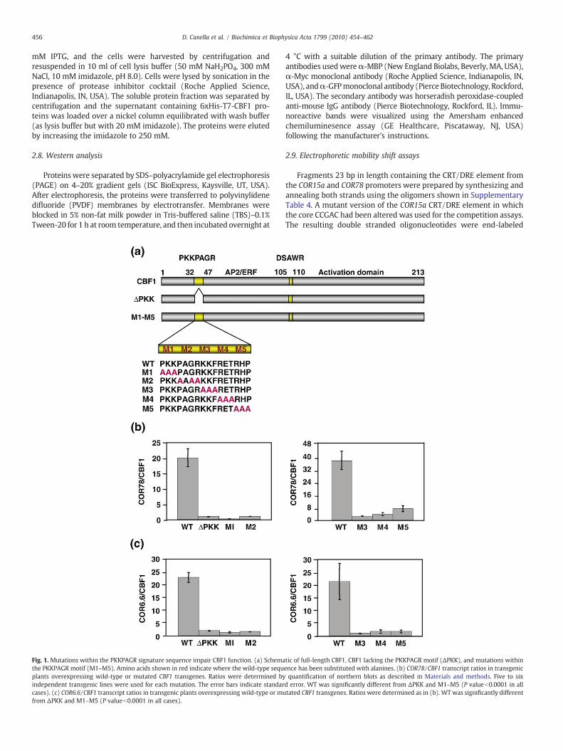
Fig. 1. Mutations within the PKKPAGR signature sequence impair CBF1 function. (a) Schemathe PKKPAGR motif (M1–M5). Amino acids shown in red indicate where the wild-type sequplants overexpressing wild-type or mutated CBF1 transgenes. Ratios were determined byindependent transgenic lines were used for each mutation. The error bars indicate standarcases). (c) COR6.6/CBF1 transcript ratios in transgenic plants overexpressing wild-type or mufrom ΔPKK and M1–M5 (P valueb0.0001 in all cases).
4 °C with a suitable dilution of the primary antibody. The primaryantibodies usedwereα-MBP (New England Biolabs, Beverly, MA, USA),α-Myc monoclonal antibody (Roche Applied Science, Indianapolis, IN,USA), andα-GFPmonoclonal antibody (Pierce Biotechnology, Rockford,IL, USA). The secondary antibody was horseradish peroxidase-coupledanti-mouse IgG antibody (Pierce Biotechnology, Rockford, IL). Immu-noreactive bands were visualized using the Amersham enhancedchemiluminesence assay (GE Healthcare, Piscataway, NJ, USA)following the manufacturer's instructions.
2.9. Electrophoretic mobility shift assays
Fragments 23 bp in length containing the CRT/DRE element fromthe COR15a and COR78 promoters were prepared by synthesizing andannealing both strands using the oligomers shown in SupplementaryTable 4. A mutant version of the COR15a CRT/DRE element in whichthe core CCGAC had been altered was used for the competition assays.The resulting double stranded oligonucleotides were end-labeled
tic of full-length CBF1, CBF1 lacking the PKKPAGR motif (ΔPKK), and mutations withinence has been substituted with alanines. (b) COR78/CBF1 transcript ratios in transgenicquantification of northern blots as described in Materials and methods. Five to six
d error. WT was significantly different from ΔPKK and M1–M5 (P valueb0.0001 in alltated CBF1 transgenes. Ratios were determined as in (b). WT was significantly different

457D. Canella et al. / Biochimica et Biophysica Acta 1799 (2010) 454–462
with 32P and purified through a Sephadex G-50 column. The bindingof recombinant protein to the CRT/DRE-containing DNA probes wastested by incubating 0.5 ng of 32P-probe labeled by end-filling with agradient concentration of each recombinant protein in the presence/absence of 100 ng unlabeled competitor DNA for 20 min at roomtemperature. After incubation, samples were separated on non-denaturing polyacrylamide gels. The gels were dried and exposed toa phosphorimager screen. The screen was scanned and quantifiedusing QuantityOne software (BioRad, Hercules, CA, USA).
2.10. Fluorescence imaging of Arabidopsis root tips overexpressingCBF1:GFP:GUS
Arabidopsis seedlings were grown on vertically oriented plates andallowed to grow for 6–7 days before being imaged. Laser confocalimages were collected using an upright LSM Zeiss 510 META micro-scope (Carl Zeiss, Thornwood, NY, USA) equipped with a 40× oilimmersion objective. To visualize the nuclei, Arabidopsis seedlingswere incubated with a solution of 50 μg/ml propidium iodide (PI) and5 μg/ml RNase in the dark for 15–30 min. Samples were rinsed withdistilled water and mounted in tap water. GFP fluorescence imageswere obtained using Argon ion laser excitation of 488 nm with a 505/530 nm bandpass filter. PI fluorescence images were collected using anexcitation line of 543 nmwith a 560-nm longpass filter. Postacquisitionimage processing was done with the LSM 5 Image Browser and AdobePhotoshop 5.0 software (Adobe Systems, San Jose, CA, USA).
2.11. ANOVA
Statistical difference in COR/CBF1 ratios of transgenic plants over-expressing a wild-type CBF1 transgene or CBF1 transgenes mutated inthe signature sequences was assessed by analysis of variance(ANOVA) using SAS Proc Mixed procedures (SAS Institute, Cary, NC,USA), version 9.1. To test for significant difference between COR/CBF1ratios between transgenic plants overexpressing wild-type CBF1 orthe mutated versions, we estimated least-square means for eachgenotype and compared them to the least-squaremeans of the controlplants overexpressing wild-type CBF1. These estimates were used tocalculate the t-values and the statistical significance at Pb0.0001 forall the transgenic lines.
Fig. 2. Reduced function of CBF1 carrying mutations within the PKKPAGR signature sequencoverexpressing 6xMyc-tagged CBF1 (WT) or 6xMyc-tagged CBF1 with mutations in the PKKPand G26 are two independent lines overexpressing wild-type CBF1 without a Myc-tag. CoWestern blot of transgenic lines overexpressing 6xMyc-tagged CBF1 without (WT) or with (detected using a monoclonal anti-Myc antibody as described in Materials and methods.
3. Results
3.1. The PKKPAGR signature sequence is required for CBF1 to induceexpression of COR genes
The importance of the PKKPAGR motif in CBF1 function wasassessed by determining whether changes in the sequence affectedthe ability of the transcription factor to induce COR gene expression.Two types of modification were made; a complete deletion of thePKKPAGR motif, ΔPKK, and short alanine substitutions, M1–M5,within the motif (Fig. 1a). The mutant versions of CBF1 were placedunder control of the strong constitutive CaMV 35S promoter,transformed into Arabidopsis, and the transcript levels for two CBF1target genes, COR78 and COR6.6, were determined (Fig. 1b and c;Supplementary Figs. 1 and 2). The results indicated that each of themutations greatly impaired CBF function; i.e., the ratios of the COR toCBF1 transcripts were much higher in the lines transformed with thewild-type (WT) CBF1 construct than they were in the transgenic linesexpressing the CBF1 ΔPKK and M1–M5 mutations. In all cases, thedifference in COR78/CBF1 and COR6.6/CBF1 transcript ratios betweenplants overexpressing WT CBF1 and any of the mCBF1 was highlysignificant (Pb0.0001) according to the ANOVA analysis.
3.2. The PKKPAGR sequence is not required for protein stability
The reduced level COR gene expression observed in the PKKPAGRmutant lines could have been due to the mutations resulting inprotein instability; i.e., the mutant versions of the CBF1 protein couldhave been much less stable than the WT protein and thus accumulateto much lower levels than that of the WT protein given a similartranscript level. We were unable to test this possibility in the existingtransgenic lines as we were unsuccessful in developing an antibodyagainst full-length CBF1, or specific peptides of it, that could clearlydetect the CBF1 protein in cold-acclimatedWT Arabidopsis plants or intransgenic plants overexpressing CBF1 under control of the CaMV 35Spromoter. Therefore, we tagged the WT CBF1 protein and the M1–M5mutant CBF1 proteins with c-Myc (see Materials and methods),transformed the constructs into Arabidopsis, and tested severaltransgenic lines for CBF1 and COR gene transcript levels (Fig. 2a)and the accumulation of WT and mutant CBF1 proteins (Fig. 2b).
e is not due to reduced protein levels. (a) Northern blot of transgenic Arabidopsis linesAGR motif (M1–M5 as in Fig. 1). The numbers indicate the different transgenic lines. G6l-0 is a non-transgenic control. 18S ribosomal RNA was used as a loading control. (b)M1–M5) mutations in the PKKPAGR region as detailed in (a). 6xMyc-tagged CBF1 was

458 D. Canella et al. / Biochimica et Biophysica Acta 1799 (2010) 454–462
Transgenic plants overexpressing the c-Myc:WT CBF1 protein (Fig.2a, WT transgenic lines 6, 18, 49), like those expressing the non-tagged WT CBF1 protein (Fig. 2a, lines G6 and G26), were found toaccumulate high levels of COR15a transcripts. In contrast, transgenicplants overexpressing the c-Myc:M1–M5 mutant CBF1 proteinsaccumulated low levels of COR15a transcripts (Fig. 2a, M1–M5transgenic lines). This was despite the fact that the CBF1 transcriptlevels were generally greater in the plants expressing the c-Myc:M1–M5 CBF1 transgenes than they were in the plants expressing thec-Myc:WT CBF1 transgene. Thus, as observed in the experiments
Fig. 3. The PKKPAGR motif is not required for targeting of CBF1 to the nucleus. (a) SchemTranslational fusions to GFP:GUS of each of the CBF1 constructs shown in (a) were genemicroscopy as described in Materials and methods. The panels show images obtained fromfused to GFP:GUS, and GFP:GUS is GFP:GUS alone. GFP fluorescence images were obtained (G(GFP and PI).
using the non-tagged CBF1 proteins (Fig. 1b and c), CBF1 function wasseverely impaired by mutations within the PKKPAGR. Further, theresults indicated that this decrease in activity was not due to proteininstability as the c-Myc:M1–M5 CBF1 proteins accumulated to levelscomparable to those in the transgenic lines expressing the c-Myc:WTCBF1 protein (Fig. 2b). For instance, the two lines (2 and 25) ex-pressing the c-Myc:M2 CBF1 transgene had CBF1 transcript (Fig. 2a)and protein (Fig. 2b) levels that were very similar to those in c-Myc:WT CBF1 line 18. Moreover, the c-Myc:M3 CBF1 lines 1 and 13 as wellas the c-Myc:M5 CBF1 line 14 accumulated considerably greater levels
atic of CBF1:GFP:GUS constructs. AP2/ERF is the AP2/ERF DNA-binding domain. (b)rated and transformed into Arabidopsis plants. Root tips were examined by confocalplants expressing the GFP:GUS constructs indicated. NIa is nuclear inclusion protein aFP), and propidium iodide was used to visualize the nuclei; these images were overlaid

459D. Canella et al. / Biochimica et Biophysica Acta 1799 (2010) 454–462
of CBF1 protein than did the WT c-Myc:WT CBF1 lines 6 and 18(Fig. 2b), yet the COR15a transcript levels in the M3 and M5 lineswere much lower than those in the WT lines (Fig. 2a).
3.3. The PKKPAGR motif is not required for targeting CBF1 to the nucleus
When the Arabidopsis CBF1 protein was first described [32], it wassuggested that the PKKPAGR sequence might be a nuclear localizationsignal (NLS). This was based on sequence similarities between thePKKPAGR motif and known NLS sequences for other plant proteins[33–35]. If the PKKPAGR sequence indeed has this function, then it ispossible that the inability of mutant PKKPAGR CBF1 proteins to induceCOR gene expression might be due to the proteins not being importedinto the nucleus. To test this, Arabidopsis was transformed withconstructs that haddifferent parts of the CBF1 protein fused to theGFP:GUS reporter protein (Fig. 3a), and protein localization was deter-mined using laser confocalmicroscopy (Fig. 3b).Western analysis wasalso performed to verify that fluorescence detected in plantscorresponded to the expression of full-length proteins (Supplemen-tary Fig. 3). The results indicated that the GFP:GUS reporter proteinalone did not accumulate in the nucleus (Fig. 3b, GFPGUS). This wasexpected as the GFP:GUS fusion protein has a mass of approximately100 kDa, which is well above the limit for free diffusion into thenucleus [36,37]. However, fusion of the NIa nuclear localization signal(NIa) [38] to the GFP:GUS reporter (Fig. 3b; NIa) or fusion of the CBF1proteinwithout the PKKPAGR region to the GFP:GUS reporter (Fig. 3b;ΔPKK) resulted in nuclear localization of the fusion proteins. Thus, thePKKPAGR motif was not required for nuclear localization of CBF1,indicating that the greatly reduced ability of the ΔPKK and M1–M5proteins to activate COR gene expressionwas not due to inactivation ofan NLS required for the proteins to be imported into the nucleus.
Fig. 4. Mutations in the PKKPAGR motif impair DNA binding. Electrophoretic mobilityshift assays using the CRT/DRE element from the COR15a promoter as a probe. ΔPKKand M1–M5 mutations of CBF1 are as shown in Fig. 1. Recombinant protein (200 ng)was used in a 12-μl binding reaction with 0.5 ng of radiolabeled probe as described inMaterials and methods. Where indicated 100 ng of unlabeled competitor DNA (theCOR15a CRT/DRE element) was added. W, wild-type competitor DNA; M, mutatedcompetitor DNA in which the core DNA-binding sequence, CCGAC, was altered asdescribed [32].
Additional protein fusions were examined in an attempt to localizethe CBF1 NLS sequence(s). Fusion of the 47 N-terminal residues of CBF1to GFP:GUS did not result in nuclear localization of the protein fusion(Fig. 3b, PKK), indicating that the PKKPAGR motif was not sufficient topromote nuclear localization. Deletion of CBF1 amino acids 1–32(construct 802) or 1–47 (construct 801) did not significantly affectnuclear localization of the reporter fusion, but further deletionsremoving either the AP2/ERF domain (construct 800) or the AP2/ERFdomain and the DSAWR sequence (construct 799) did (Fig. 3b). Theseresults were consistent with the PKKPAGRmotif not being essential forlocalizing CBF1 to the nucleus and indicated that an NLS was locatedwithin the AP2/ERF DNA-binding domain.
3.4. Mutations within the PKKPAGR signature sequence greatlyimpair CBF1 binding to the CRT/DRE DNA regulatory element
Wehypothesized that the PKKPAGRmotif might play a role in DNAbinding based on the proximity of this motif to the AP2/ERF domainand the presence of positively charged residues that could potentiallyprovide favorable electrostatic interactions with the negativelycharged DNA backbone. This hypothesis was tested using EMSA toassess the DNA-binding activity of the WT and PKKPAGR mutantversions of CBF1 described in Fig. 1a. The ΔPKK and M1–M5 proteinswere fused to a histidine tag, expressed in E. coli, purified and assayedfor binding to the CRT/DRE DNA regulatory element (see Materialsand methods). A band shift was observed with the WT CBF1 protein(Fig. 4). The binding was abolished by addition of unlabeled wild-typecompetitor CRT/DRE DNA (Fig. 4, W), but not by addition of a versionof the sequence in which the core CRT/DRE binding sequence, CCGAC,
Fig. 5. The PKKPAGR motif is required for CBF1 to bind to the CRT/DRE element. (a)Schematic of maltose binding protein-CBF1 construct. Amino acids 27 to 112,containing the PKKPAGR motif (PK), the AP2/ERF DNA-binding domain (AP2), andthe DSAWRmotif (DS) were translationally fused tomaltose binding protein (MPB). (b)Electrophoretic mobility shift assays using the CRT/DRE element from the COR15apromoter as a probe. Increasing amounts (0.2, 2.0, 15 μg) of each recombinant proteinwere used in a 25-μl binding reaction in the presence of 0.5 ng of radiolabeled probe asdescribed in Materials and methods. WT, wild-type fusion protein as indicated in (a);M1–M5, fusion proteins with mutations in the PKKPAGR region as detailed in Fig. 1;MBP, MBP alone. (c) Western blot of the same proteins in the same amounts as in (b)probed with anti-MBP antibody.

460 D. Canella et al. / Biochimica et Biophysica Acta 1799 (2010) 454–462
was mutated (Fig. 4, M). These results indicated that the WT CBF1protein bound specifically to the CRT/DRE sequence. In sharp contrast,little if any binding could be detected with the ΔPKK and M1–M5mutant CBF1 variants, indicating that PKKPAGR sequence had a majorrole in DNA binding.
The requirement for the PKKPAGRmotif in binding to the CRT/DREmotif was tested further using a construct that contained the CBF1AP2/ERF domain with surrounding signature sequences fused to themaltose binding protein (Fig. 5a). As with the full-length CBF1 protein,the CBF1 AP2/ERF domain with the surrounding signature sequencesbound to the CRT/DRE element (Fig. 5b). This binding was nearlyabolished by each of the M1–M5 mutations, again indicating animportant role for the PKKPAGR signature sequence in binding to theCRT/DRE element.
3.5. Specific amino acid side chain chemistry within the PKKPAGRsignature sequence is required for effective binding of CBF1 to theCRT/DRE regulatory element
Additional analysis was conducted to determine whether specificamino acid side chain chemistry within the PKKPAGR motif was
Fig. 6. Specific residues within the RKKFRET region of the PKKPAGR are crucial for CBF1binding to the CRT/DRE promoter element. (a) The PKKPAGR sequence from CBF1 withthe shaded boxes indicating amino acids that were mutated. Predicted α-helix (H) andrandom coil (C) are indicated. Conservative (Con) and non-conservative (Non-Con)amino acid changes are shown. (b) Electrophoretic mobility shift assays showingbinding activity of maltose binding protein (MBP):CBF27–112 (see Fig. 5a) withmutations detailed in (a) using the CRT/DRE element from the COR78 promoter as aprobe. DNA-binding reactions were carried out in a 15-μl reaction containing increasingamounts (300 and 600 ng) of each recombinant protein in the presence of 0.5 ng ofradiolabeled probe as described inMaterials andmethods. Non-mutatedMBP:CBF27–112(WT) andmaltose binding protein alone (MBP) were used as controls. (c) Western blotof the same proteins in the same amounts as in (b) probed with anti-MBP antibody. Thearrows indicate MBP:CBF27–112.
important for DNA binding.We focused on the RKKFRET regionwithinthe PKKPAGR motif as it was predicted to form an α-helix (D. Canellaand L. Kuhn, unpublished). Both conservative and non-conservativeamino acid changes were made to preserve or alter, respectively, sidechain chemistry within the RKKFRET sequence (Fig. 6a). Likewise, inall but one case, the changes made were chosen such that they wouldbe compatible with maintaining the predicted α-helical structure ofthe RKKFRET region [26]. The one exception was the Phe40→Promutation, which was designed to test the effect of a helix-breakingresidue on the stability of the protein–DNA complex while maintain-ing a hydrophobic side chain. The effects of each mutation wereassessed by EMSA.
The results indicated that the Arg37 and Phe40 residues werecritical for DNA binding; the Arg37→Lys, Arg37→Ser and Phe40→Alasubstitutions all resulted in greatly impaired binding to the CRT/DRE regulatory element (Fig. 6b). The loss of binding caused bysubstitution of Arg37 with Lys, two amino acids with very similarside-chain length and charge, indicated that preserving the positivecharge was insufficient for binding and that the specific side-chainchemistry was critical. The loss of DNA binding caused by thePhe40→Ala substitution suggested that the aromatic ring in thisresidue was essential for interaction with the CRT/DRE element.Consistent with this suggestion was the finding that the Phe40→Tyrsubstitution preserved, or even enhanced, DNA binding. Finally, thefinding that the Phe40→Pro substitution resulted in a nearcomplete loss of DNA binding. The Phe40→Pro substitution resultedin near-complete loss of DNA binding. Given that Pro disfavorshelicity, this result is consistent with the importance of a helicalbackbone conformation at this position for DNA binding.
4. Discussion
The CBF transcription factors of Arabidopsis have a key role in coldacclimation, controlling the expression of a regulon of more than 100genes that contribute to freezing tolerance. Determining how the CBFproteins regulate the expression of the CBF regulon is basic to an overallunderstanding of the CBF cold response pathway. Here we furtherexplore the structure–function relationships of the CBF proteins. Thehigh degree of conservation of the CBF signature sequences among CBFtranscription factors fromdiverseplant species suggested that theyhavean important functional role. Here we show that this is the case for thePKKPAGR sequence. Deletion of this sequence, or mutations within it,was found to greatly impair the ability of CBF1 to induce expression oftarget COR genes (Fig. 1, Supplementary Figs. 1 and 2). This functionalimpairment was not due to the mutations causing protein instability orloss of protein import into the nucleus. Rather, the PKKPAGRmutationswere found to greatly impair the ability of CBF1 to bind to the CRT/DREDNA recognition sequence.
A role for the PKKPAGR region in DNA binding was notanticipated as the AP2/ERF domain has typically been assigned thefunction of DNA binding for AP/ERF family proteins. Allen et al. [14]determined the 3D solution structure of the 60-amino-acid AP2/EREBP domain of the AtERF1 transcription factor and examined itsinteractions with its DNA-binding sequence, the GCC box. The AP2/ERF domain was found to be composed of a three-stranded anti-parallel structure followed by an α-helix that is packed parallel tothe β-sheet. Arginine and tryptophan residues within the β-sheetwere found to contact eight of nine consecutive nucleotides of thebinding site within the major groove of the DNA. The high degree ofsequence identity between CBF1 residues 46–107 and residues withinthe AtERF1 solution structure (Protein Data Bank entry 1gcc) impliesthe same arrangement of structural elements, with the full PKKP/RAGRxKFxETRHP sequence occurring immediately N-terminal tothis domain. Thus, there was no prior reason to suspect an essentialrole for the PKKPAGR sequence in CBF1 binding to the CRT/DREelement.

461D. Canella et al. / Biochimica et Biophysica Acta 1799 (2010) 454–462
A somewhat different picture was presented by Hao et al. [15].These investigators found that the 10 amino acids immediatelyupstream of the AP2/ERF-binding domain of the EREBP2 transcrip-tion factor, a protein that also binds to the GCC box, were requiredfor effective binding to the GCC box. However, unlike the CBFsignature sequences, these 10 amino acids did not match the aminoacids immediately flanking the AP2/ERF domains of other membersof the EREBP protein family. Thus, the authors suggested that the 10amino acids might not have a direct role in DNA binding, but instead,be required for the AP2/ERF domain to maintain an active con-formation. The study did not include a functional analysis of the 10amino acids, and thus, it was unclear whether specific amino acidswere required, or that simply an N-terminal peptide extension wasrequired to stabilize, in a non-specific manner, the AP2/ERF domainof EREBP2.
Our results show that the PKKPAGR sequence is required for CBF1to effectively bind to the CRT/DRE element. Moreover, they show aneed for specific amino acid side-chain chemistry at certain positions.In particular, the results show that the Arg37 and Phe40 residuesare critical for CBF1 binding to the CRT/DRE regulatory element.Substituting Arg37 with Lys essentially abolished binding eventhough the two amino acids have very similar side-chain length andcharge. In addition, the loss of DNA binding brought about bysubstituting Phe40 with Ala, and the preservation of binding (if notenhanced binding) observed with the Phe40 to Tyr substitution,indicates a requirement for a bulky hydrophobic side chain at thisposition. The challenge now is to determine the specific functions ofthe Arg37 and Phe40 side chains. One possibility is that they interactspecifically with other CBF1 amino acids, stabilize the DNA-bindingdomain, and enable effective protein–DNA interaction. However,another possibility is that these residues interact directly withnucleotides surrounding the CRT/DRE core motif and contribute tobinding site specificity. In this case, the residues would contribute todetermining the composition of the CBF regulon. Future experimentswill be directed at distinguishing between these possibilities.
Acknowledgments
This research was supported in part by grants to MFT from the NSFPlant Genome Project (DBI 0110124 and DBI 0701709), theDepartment of Energy (DE-FG02-91ER20021) and the MichiganAgricultural Experiment Station.
Appendix A. Supplementary data
Supplementary Table 1. Primers used for PKKPAGR mutagenesis.Supplementary Table 2. Primers used in making constructs.Supplementary Table 3. Primers used RKKFRET mutagenesis.Supplementary Table 4. Oligomers used in EMSAs.Supplementary Figure 1. Mutations in the PKKPAGR signature
sequence impair CBF function. Northern blots of Arabidopsis over-expressing CBF1 with mutations in the PKKPAGR region as shown inFig. 1 were performed using CBF1 and COR78 as probes [(b) and (d)].B6 is a transgenic line expressing the vector alone. 18S ribosomal RNAwas used as a loading control. The blots were quantified [(a) and (c)]as described in Materials and methods.
Supplementary Figure 2. Mutations in the PKKPAGR signaturesequence impair CBF function. Northern blots of Arabidopsis over-expressing CBF1 with mutations in the PKKPAGR region as shown inFig. 1 were performed using CBF1 and COR6.6 as probes [(b) and (d)].B6 is a transgenic line expressing the vector alone. 18S ribosomal RNAwas used as a loading control. The blots were quantified [(a) and (c)]as described in Materials and methods.
Supplementary Figure 3. Western blot analysis of protein extractsfrom Arabidopsis overexpressing CBF1-GFP:GUS constructs. Total
protein extracts were made from 12-day-old seedlings, and Westernblots were prepared as described in Materials and methods. Totalprotein (70 μg) was used per lane. Detection was with monoclonalantibody against GFP. Upper and lower panels represent short andlong exposures of the same film, respectively.
Note: The supplementary material accompanying this article isavailable at (doi:10.1016/j.bbagrm.2009.11.017).
References
[1] V. Chinnusamy, J. Zhu, J.K. Zhu, Cold stress regulation of gene expression in plants,Trends Plant Sci. 12 (2007) 444–451.
[2] M.F. Thomashow, Plant cold acclimation: freezing tolerance genes and regulatorymechanisms, Annu. Rev. Plant Physiol. Plant Mol. Biol. 50 (1999) 571–599.
[3] S.J. Gilmour, D.G. Zarka, E.J. Stockinger, M.P. Salazar, J.M. Houghton, M.F.Thomashow, Low temperature regulation of the Arabidopsis CBF family of AP2transcriptional activators as an early step in cold-induced COR gene expression,Plant J. 16 (1998) 433–442.
[4] J. Medina, M. Bargues, J. Terol, M. Perez-Alonso, J. Salinas, The Arabidopsis CBFgene family is composed of three genes encoding AP2 domain-containing proteinswhose expression is regulated by low temperature but not by abscisic acid ordehydration, Plant Physiol. 119 (1999) 463–470.
[5] Q. Liu, M. Kasuga, Y. Sakuma, H. Abe, S. Miura, K. Yamaguchi-Shinozaki, K.Shinozaki, Two transcription factors, DREB1 and DREB2, with an EREBP/AP2 DNAbinding domain separate two cellular signal transduction pathways in drought-and low-temperature-responsive gene expression, respectively, in Arabidopsis,Plant Cell 10 (1998) 1391–1406.
[6] S.S. Baker, K.S. Wilhelm, M.F. Thomashow, The 5′-region of Arabidopsis thalianacor15a has cis-acting elements that confer cold-, drought- and ABA-regulatedgene expression, Plant Mol. Biol. 24 (1994) 701–713.
[7] K. Yamaguchi-Shinozaki, K. Shinozaki, A novel cis-acting element in an Arabi-dopsis gene is involved in responsiveness to drought, low-temperature, or high-salt stress, Plant Cell 6 (1994) 251–264.
[8] K. Maruyama, Y. Sakuma, M. Kasuga, Y. Ito, M. Seki, H. Goda, Y. Shimada, S.Yoshida, K. Shinozaki, K. Yamaguchi-Shinozaki, Identification of cold-inducibledownstream genes of the Arabidopsis DREB1A/CBF3 transcriptional factor usingtwo microarray systems, Plant J. 38 (2004) 982–993.
[9] J.T. Vogel, D.G. Zarka, H.A. Van Buskirk, S.G. Fowler, M.F. Thomashow, Roles of theCBF2 and ZAT12 transcription factors in configuring the low temperaturetranscriptome of Arabidopsis, Plant J. 41 (2005) 195–211.
[10] K.R. Jaglo-Ottosen, S.J. Gilmour, D.G. Zarka, O. Schabenberger, M.F. Thomashow,Arabidopsis CBF1 overexpression induces COR genes and enhances freezingtolerance, Science 280 (1998) 104–106.
[11] J.L. Riechmann, E.M. Meyerowitz, The AP2/EREBP family of plant transcriptionfactors, Biol. Chem. 379 (1998) 633–646.
[12] K.D. Jofuku, B.G. den Boer, M. Van Montagu, J.K. Okamuro, Control of Arabidopsisflower and seed development by the homeotic gene APETALA2, Plant Cell 6(1994) 1211–1225.
[13] M. Ohme-Takagi, H. Shinshi, Ethylene-inducible DNA binding proteins thatinteract with an ethylene-responsive element, Plant Cell 7 (1995) 173–182.
[14] M.D. Allen, K. Yamasaki, M. Ohme-Takagi, M. Tateno, M. Suzuki, A novel modeof DNA recognition by a beta-sheet revealed by the solution structure of theGCC-box binding domain in complex with DNA, EMBO J. 17 (1998)5484–5496.
[15] D. Hao, M. Ohme-Takagi, A. Sarai, Unique mode of GCC box recognition by theDNA-binding domain of ethylene-responsive element-binding factor (ERFdomain) in plant, J. Biol. Chem. 273 (1998) 26857–26861.
[16] Y. Sakuma, Q. Liu, J.G. Dubouzet, H. Abe, K. Shinozaki, K. Yamaguchi-Shinozaki,DNA-binding specificity of the ERF/AP2 domain of Arabidopsis DREBs, transcrip-tion factors involved in dehydration- and cold-inducible gene expression,Biochem. Biophys. Res. Commun. 290 (2002) 998–1009.
[17] K.R. Jaglo, S. Kleff, K.L. Amundsen, X. Zhang, V. Haake, J.Z. Zhang, T. Deits, M.F.Thomashow, Components of the Arabidopsis C-repeat/dehydration-responsiveelement binding factor cold-response pathway are conserved in Brassica napusand other plant species, Plant Physiol. 127 (2001) 910–917.
[18] S.J. Gilmour, S.G. Fowler, M.F. Thomashow, Arabidopsis transcriptional activatorsCBF1, CBF2, and CBF3 have matching functional activities, Plant Mol. Biol. 54(2004) 767–781.
[19] S.J. Clough, A.F. Bent, Floral dip: a simplified method for Agrobacterium-mediatedtransformation of Arabidopsis thaliana, Plant J. 16 (1998) 735–743.
[20] D.G. Zarka, J.T. Vogel, D. Cook, M.F. Thomashow, Cold induction of Arabidopsis CBFgenes involves multiple ICE (inducer of CBF expression) promoter elements and acold-regulatory circuit that is desensitized by low temperature, Plant Physiol. 133(2003) 910–918.
[21] J. Sambrook, D.W. Russell, Molecular Cloning, A laboratory manual, third ed., ColdSpring Harbor Laboratory Press, Cold Spring Harbor, 2001.
[22] G. Church, G. Walter, Genomic sequencing, Proc. Natl. Acad. Sci. U. S. A. 81 (1984)1991–1995.
[23] S. Li, M.F.Wilkinson, Site-directed mutagenesis: a two-stepmethod using PCR andDpnI, Biotechniques 23 (1997) 588–590.
[24] W.Wang, B.A. Malcolm, Two-stage PCR protocol allowing introduction ofmultiplemutations, deletions and insertions using QuikChange Site-Directed Mutagenesis,Biotechniques 26 (1999) 680–682.

462 D. Canella et al. / Biochimica et Biophysica Acta 1799 (2010) 454–462
[25] G.E. An, P.R. Ebert, A. Mitra, S.B. Ha, Binary vectors, Plant Molecular BiologyManual, vol. A3, Kluwer Academic Publishing, Dordrecht, 1988, pp. 1–19.
[26] J. Overington, D. Donnelly, M.S. Johnson, A. Sali, T.L. Blundell, Environment-specific amino acid substitution tables: tertiary templates and prediction ofprotein folds, Protein Sci. 1 (1992) 216–226.
[27] K.E. Vlachonasios,M.F. Thomashow, S.J. Triezenberg, Disruptionmutations ofADA2band GCN5 transcriptional adaptor genes dramatically affect Arabidopsis growth,development, and gene expression, Plant Cell 15 (2003) 626–638.
[28] M. Schena, D. Picard, K. Yamamoto, Vectors for constitutive and inducible geneexpression in yeast, Methods Enzymol. 194 (1991) 389–398.
[29] J. Haseloff, K.R. Siemering, D.C. Prasher, S. Hodge, Removal of a cryptic intron andsubcellular localization of green fluorescent protein are required to marktransgenic Arabidopsis plants brightly, Proc. Natl. Acad. Sci. U. S. A. 94 (1997)2122–2127.
[30] B.P. Cormack, R.H. Valdivia, S. Falkow, FACS-optimized mutants of the greenfluorescent protein (GFP), Gene 173 (1996) 33–38.
[31] T. Kleber-Janke, W.M. Becker, Use of modified BL21(DE3) Escherichia coli cells forhigh-level expression of recombinant peanut allergens affected by poor codonusage, Protein Expr. Purif. 19 (2000) 419–424.
[32] EJ. Stockinger, SJ. Gilmour, MF. Thomashow, Arabidopsis thaliana CBF1 encodes anAP2 domain-containing transcriptional activator that binds to the C-repeat/DRE, acis-acting DNA regulatory element that stimulates transcription in response to lowtemperature and water deficit, Proc. Natl. Acad. Sci. U. S. A. 94 (1997) 1035–1040.
[33] S. Abel, P. Oeller, A. Theologis, Early auxin-induced genes encode short-livednuclear proteins, PNAS 91 (1994) 326–330.
[34] R. Lyck, U. Harmening, I. Hohfeld, E. Treuter, K.D. Scharf, L. Nover, Intracellulardistribution and identification of the nuclear localization signals of two plantheat-stress transcription factors, Planta 202 (1997) 117–125.
[35] A.G. von Arnim, X.W. Deng, Light inactivation of Arabidopsis photomorphogenicrepressor COP1 involves a cell-specific regulation of its nucleocytoplasmicpartitioning, Cell 79 (1994) 1035–1045.
[36] R.J. Grebenok, E. Pierson, G.M. Lambert, F.C. Gong, C.L. Afonso, R. Haldeman-Cahill,J.C. Carrington, D.W. Galbraith, Green-fluorescent protein fusions for efficientcharacterization of nuclear targeting, Plant J. 11 (1997) 573–586.
[37] A. Kaffman, E.K. O'Shea, Regulation of nuclear localization: a key to a door, Annu.Rev. Cell Dev. Biol. 15 (1999) 291–339.
[38] J.C. Carrington, D.D. Freed, A.J. Leinicke, Bipartite signal sequence mediates nucleartranslocation of the plant potyviral NIa protein, Plant Cell 3 (1991) 953–962.