biochemical and biophysical research communications, issue date
TRANSCRIPT
Instructions for use
Title Anti-parallel membrane topology of two components of EbrAB, a multidrug transporter.
Author(s) Kikukawa, Takashi; Miyauchi, Seiji; Araiso, Tsunehisa; Kamo, Naoki; Nara, Toshifumi
Citation Biochemical and Biophysical Research Communications, 358(4): 1071-1075
Issue Date 2007-07-13
Doc URL http://hdl.handle.net/2115/28010
Type article (author version)
File Information BBRC358-4.pdf
Hokkaido University Collection of Scholarly and Academic Papers : HUSCAP
Anti-parallel membrane topology of two components of EbrAB,
a multidrug transporter ‡
Takashi Kikukawaa,*, Seiji Miyauchib, Tsunehisa Araisoa, Naoki Kamoc,
Toshifumi Narab
aLaboratory of Biomolecular Systems, Creative Research Initiative
"Sosei" (CRIS), Hokkaido University, Sapporo 001-0021, Japan
bLaboratory of Biophysical Chemistry, Graduate School of
Pharmaceutical Sciences, Hokkaido University, Sapporo 060-0812,
Japan
cDivision of Molecular Life Science, Faculty of Advanced Life
Science, Hokkaido University, Sapporo 060-0812, Japan
‡ This work was supported in part by a grant-in-aid for scientific
research from the Japanese Ministry of Education, Science,
Technology, Sports and Culture.
* Corresponding author:
Tel: +81-11-706-7199, Fax: +81-11-706-7321
E-mail: [email protected]
1
Abstract
EbrAB is a multidrug-resistance transporter in Bacillus subtilis
that belongs to the small multidrug resistance, and requires two
polypeptides of both EbrA and EbrB, implying that it functions in
the hetero-dimeric state. In this study, we investigated the
transmembrane topologies of EbrA and EbrB. Various single-cysteine
mutants were expressed in E. coli cells, and the efflux activity
was measured. Only mutants having a high activity were used for
the topology experiments. The reactivity of a membrane impermeable
NEM-fluorescein against the single cysteine of these fully
functional mutants was examined when this reactive fluorophore was
applied either from the outside or both sides of the cell membrane
or in the denatured state. The results clearly showed that EbrA
and EbrB have the opposite orientation within the membrane or an
anti-parallel configuration.
Keywords: Efflux transporter; EmrE; Small multidrug resistance
(SMR); Dual topology configuration; Fluorescein-5-maleimide
(NEM-fluorescein); Ethidium efflux
2
Introduction
Small multidrug resistance (SMR) proteins are the smallest
antiporters which expel various toxicants from bacterial cells to
confer the multidrug resistance to the cell [1-3]. Interestingly,
SMR is composed of two or more polypeptides whose lengths are only
100 – 120 amino acids long, and each polypeptide then forms four
α-helical transmembrane helices. This implies an oligomer structure
in the active form. EmrE found in Escherichia coli is a family
archetype and is well documented to function as a homo-dimer [3-6].
EbrA/EbrB and YkkC/YkkD from B. subtilis and Ydge/YdgF from E. coli
are composed of paired components that are encoded in distinct
operons, and the simultaneous expression of both components is
absolutely indispensable for the efflux activity [7-11]. Thus, it
is most likely that the SMR transporter is generally composed of
two components irrespective of the homo- or hetero-dimeric
structures.
The next concern is the topology of the components within
the membrane. Biochemical approaches such as chemical modification
[12] and cross-linking [13,14] suggested the parallel topology of
3
the dimer of EmrE. A parallel topology was also reported for SugE,
another SMR homologue in E. coli [15]. On the other hand, electron
microscope (EM) data and a modeling analysis based on its EM data
[4,16,17] have proposed an anti-parallel topology, which is
sometimes called a “dual topology” configuration.
The anti-parallel configuration is consistent with the
topology analyses of E. coli inner-membrane proteins performed by
von Heijine and colleagues [18,19]. C-terminal tagging with
alkaline phosphatase or green fluorescence protein led to the
suggestion of the anti-parallel configuration of EmrE and SugE.
The same analysis also predicted the anti-parallel orientation of
YdgE/YdgF, one of the hetero-dimeric SMR’s [18]. However, these
analyses focused only on the location of the C-termini of the
components. In addition, it should be noted that, in these
experiments, fusion tags were attached that are much larger than
SMR transporter itself; the large fusion tags might disturb the
valid topology and result in a topology different from the natural
one. Therefore, we may say that the essential structure of the SMR’s
is now under debate.
4
In the present study, we investigated the topology of the
hetero-dimeric SMR, EbrA/EbrB. To avoid the possibility of
disturbing the topology, large reporter tags are not attached.
Instead, a relatively small, membrane-impermeable and SH-reactive
fluorescein-5-maleimide (NEM-fluorescein)[20] is labeled to a
single cysteine residue at various positions. This is one kind of
“cysteine scanning” method [21]. In addition, we examined the
topology of only the mutants having the normal efflux activity,
which assures the valid structure of the examined protein. The
obtained results strongly imply the anti-parallel topology.
5
Materials and Methods
Mutagenesis
To obtain a single cysteine mutant protein, a cysteine-less (CL)
background mutant was first obtained by replacing the 13th position
of EbrA and the 116th position of EbrB with valine and serine residues,
respectively. From the CL-background, various single-cysteine
mutants were constructed using the QuickChange site-directed
mutagenesis kit (Stratagene, La Jolla, CA) and sets of two
overlapping primers. The DNA sequences were confirmed using the
standard procedure (377 DNA sequencer, Applied Biosystems, Foster
City, CA). A single cysteine residue on the CL-background was
introduced at the N-terminus, C-terminus positions and in the loop
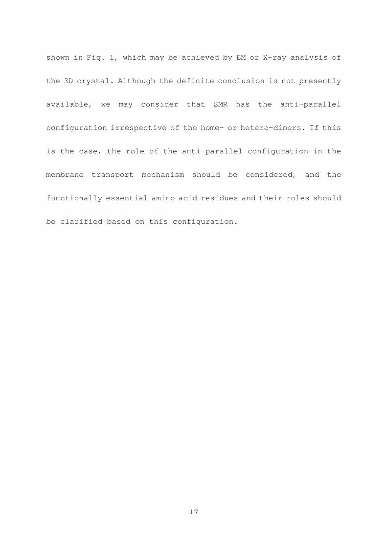
region connecting successive membrane helices as shown in Fig. 1.
The loop regions were predicted by a hydropathy analysis [10] and
the consideration for the amphiphilicity index of polar residues
[22]. Here, the transmembrane regions are assigned so as to end
with amphiphilic residues of lysine, histidine, glutamic acid,
tyrosine or tryptophan.
6
Construction of the expression plasmid has been previously
described [10]. For the simultaneous expression of EbrA and EbrB,
the plasmid was obtained by inserting the gene pair of ebrA and
ebrB downstream of the tac promoter of pFLAG-CTC (Sigma, St. Louis,
MO).
Ethidium efflux assay from E. coli cells
We wanted to perform a topology analysis only for the mutant proteins
whose efflux activity is normal. To estimate the activity, the
efflux of ethidium, one of typical substrates of SMR, was measured,
using a previously described method [10].
Topology studies using NEM-fluorescein labeling
A concentrated solution of NEM-fluorescein (20 mM; Molecular Probes,
Eugene, OR) was freshly prepared in N,N’-dimethylformamide. The
cells were washed twice with a buffer solution containing 400 mM
NaCl and 50 mM sodium phosphate (pH 7.0), and were re-suspended
at 0.5 of OD660. This cell suspension was divided into three aliquots
and saved in three test tubes (1 ml each), followed by the labeling
7
with the membrane-impermeable NEM-fluorescein [20] under three
different conditions. They are:
(1) Labeling from outside of the cell membrane – NEM-fluorescein
was added to the intact cell suspension at a final concentration
of 0.5 mM and incubated for 30 min at room temperature while shaking.
To stop the reaction and to prevent redundant labeling,
β-mercaptoethanol (final concentration of 5%) was added, and the
cells were washed three times with the above-described buffer
supplemented with 5% β-mercaptoethanol, followed by ultrasonic
disruption. The membrane fractions were collected by
ultra-centrifugation at 106,000 x g for 1 h at 4 oC and solubilized
in an SDS-PAGE-loading buffer containing 5% β-mercaptoethanol.
(2) Labeling from both sides of the cell membrane – The intact cells
were disrupted by sonication in the presence of 0.5 mM
NEM-fluorescein, and this suspension was allowed to be shaken for
30 min at room temperature. To stop the reaction, β-mercaptoethanol
was added to a final concentration of 5% v/v. The collection and
solubilization of the membrane fractions were performed in the same
manner as described above.
8

(3) Labeling under the denatured condition – The intact cells were
disrupted by sonication, and the membrane fractions were collected
by ultra-centrifugation. The membrane fractions were then
solubilized in SDS-PAGE-loading buffer containing 0.5 mM
NEM-fluorescein, followed by a 30-min incubation at room
temperature. Finally, β-mercaptoethanol was added at a final
concentration of 5% v/v.
The final volumes of these three samples were the same, giving
approximately equal protein concentrations in each container. The
samples were analyzed by tricine-SDS-PAGE together with
fluorescein-labeled protein standards. Fluorescence images of the
gels were acquired using a Fujifilm FLA-2000 imaging system (Tokyo,
Japan).
9
Results and Discussion
A single cysteine residue was introduced into the CL-background
mutants (C13V of EbrA and C116S of EbrB) at the respective five
regions of EbrA and EbrB: these regions were the N-terminus (1.5C
for EbrA; G3C for EbrB), the first loop connecting the first and
second membrane helices (S25C for EbrA; T28C, for EbrB), the second
loop (S58C for EbrA and EbrB), the third loop (N85C for EbrA; T82C
for EbrB) and the C-terminus (106C for EbrA; A106C for EbrB). Here,
1.5C and 106C for EbrA represent the mutants having cysteine
residues inserted after the 1st methionine and the C-terminal
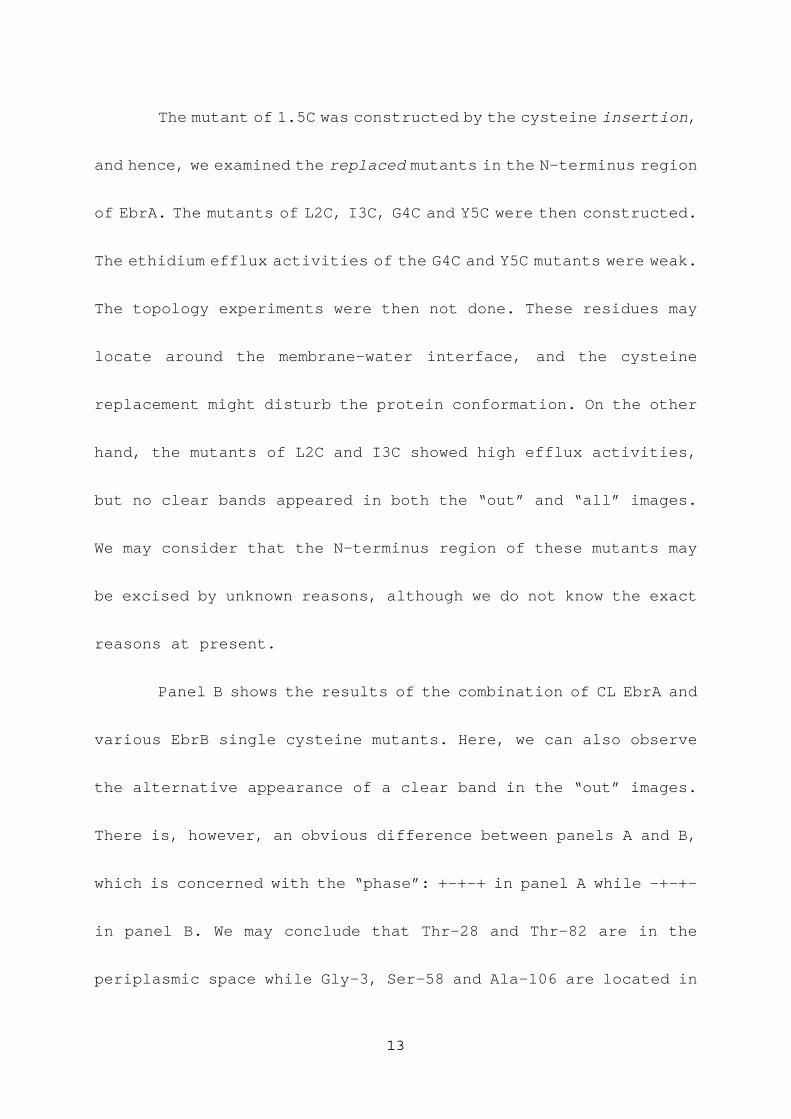
proline at the 105th position, respectively. Figure 1 illustrates
the possible membrane helix and loop regions together with the
positions where the cysteine residues are introduced. Note that
the membrane topologies of EbrA and B are drawn based on results
of the present study.
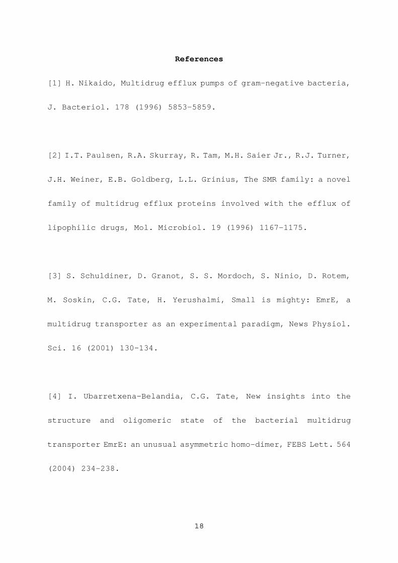
The normal transport activities of these single-cysteine
mutants were confirmed by the ethidium efflux assays. Figure 2 shows
the time-dependent decrease in the ethidium fluorescence that
represents the ethidium efflux, because the fluorescence originates
10
only from ethidium inside the cells [10]. Panel A is for cells
expressing combinations of various single-cysteine EbrA mutants
and the CL EbrB, and panel B is for cells expressing combinations
of the CL EbrA and various single-cysteine EbrB mutants. All mutants
maintained activities as high as that of the wild-type EbrAB (thick
solid lines). The CL-background EbrAB also showed a high activity
(data not shown).
The EbrAB mutants were labeled with an SH-reactive and
membrane-impermeable NEM-fluorescein by three different
procedures. They were labeled from the outside, from both sides
and under the denatured condition, which are hereafter called “out”,
“both” and “all”, respectively. The obtained fluorescence images
are shown in Fig. 3, where “None” means the image from cells
harboring a vacant plasmid. The “out” image of the “None” sample
does not show clear bands, while the “all” image of the “None” shows
several bands, indicating that intrinsic proteins are not labeled
with the NEM-fluorescein from the outside of the intact cells and
also that NEM-fluorescein does not penetrate into the inside of
the cells. The image of “all” of the CL sample is almost the same
11
as that of “all” of the “None” sample, which is reasonable because
“CL” has no cysteine residues. The presence of these intrinsic
proteins fortunately did not interfere with the further analyses,
because the two bands from the transporter components are much more
dense than those of the intrinsic proteins. The molecular masses
calculated from the amino acid compositions are 11.4 kDa for EbrA
(105 A.A. residues) and 12.3 kDa for EbrB (117 A.A. residues). Thus,
the upper and lower bands are from EbrB and EbrA, respectively.
We considered the results of five mutants in panel A, in which
clear bands appear in all the “all” images with almost the same
density. Note that the lanes are arranged in the order from the
N- to C-terminus. When we focus on the “out” images, the bands
alternatively appear from 1.5C to 106C. No bands appear in the “out”
images of S25C and N85C. Therefore, we may conclude that the
N-terminus (1.5C), Ser-58, and the C-terminus (106C) are located
in the periplasmic space, while the membrane-impermeable
NEM-fluorescein cannot access Ser-25 and Asn-85. Combining the
hydropathy analysis, we may conclude the location of these residues
as shown in the left panel of Fig. 1.
12
The mutant of 1.5C was constructed by the cysteine insertion,
and hence, we examined the replaced mutants in the N-terminus region
of EbrA. The mutants of L2C, I3C, G4C and Y5C were then constructed.
The ethidium efflux activities of the G4C and Y5C mutants were weak.
The topology experiments were then not done. These residues may
locate around the membrane-water interface, and the cysteine
replacement might disturb the protein conformation. On the other
hand, the mutants of L2C and I3C showed high efflux activities,
but no clear bands appeared in both the “out” and “all” images.
We may consider that the N-terminus region of these mutants may
be excised by unknown reasons, although we do not know the exact
reasons at present.
Panel B shows the results of the combination of CL EbrA and
various EbrB single cysteine mutants. Here, we can also observe
the alternative appearance of a clear band in the “out” images.
There is, however, an obvious difference between panels A and B,
which is concerned with the “phase”: +-+-+ in panel A while -+-+-
in panel B. We may conclude that Thr-28 and Thr-82 are in the
periplasmic space while Gly-3, Ser-58 and Ala-106 are located in
13
the cytoplasm (the right panel of Fig. 1). It is worthwhile to note
that the topologies are opposite between EbrA and B (see Fig. 1).
Thus, our present results reveal the anti-parallel configuration
of EbrA/B. Figure 1 also indicates the location of the Lys- and
Arg-residues from which we can see that the “positive-inside” rule
holds.
Strictly speaking, the absence of the dense bands in the “out”
images does not necessarily mean that the locations of the
respective residues are in the cytoplasmic space. There is the
possibility that these residues may be located within the membrane
helices, although the hydropathy analysis predicts that these
residues belong to the loop region connecting the two membrane
helices. Therefore, we compared the band densities between the
“both” and “all” images of S25C and N85C of EbrA, and G3C, S58C
and A106C of EbrB (panel C in Fig. 3). The results reveal the almost
equal densities of the same mutants, implying that the hydrophilic
fluorescent probe can react with the cysteine residues of these
mutants from the cytoplasm. In other words, the locations of these
residues are in the cytoplasm as shown in Fig. 1.
14
In panels B and C of Fig. 3, the band positions of the EbrB
mutants slightly vary. The reason for this is not clear at present.
It seems that even in the presence of SDS, the 3D structure may
not be completely destroyed, but it is noted that the band positions
between the “out”, “all” and “both” are the same within one mutant.
These differences in the migration rate then do not interfere these
concluding the anti-parallel configuration of EbrA/B.
Our conclusion is also consistent with previous studies on
the other hetero-dimeric SMR’s of YdgE/F, in which the C-terminus
locations were examined using the fusion of the reporter proteins
[18,19]. YdgE is a shorter member (corresponding to EbrA in this
sense) whose C-terminus is located outside, and the C-terminus of
the longer member, YdgF (corresponding to EbrB), is located inside.
This configuration is similar with EbrA/B as shown in Fig. 1.
The conclusion of anti-parallel configuration of EbrA/B may
be expanded to the membrane topology of EmrE, a homo-dimeric SMR.
Contrary to EbrA/B, EmrE obviously does not have the
“positive-inside” bias [19], and then we loose one of the criteria
of the topology. Therefore, we will consider this from the other
15
aspects; i.e., What happens when the “positive-inside” bias is
removed from EbrA/B? Actually this was previously done [10]. We
prepared mutants of EbrA and EbrB lacking hydrophilic regions by
the replacement of the positively charged lysine residues with
neutral ones, and then the resultant mutants lost the clear
“positive-inside” biases. Interestingly, expressing only one
monomer (either of the EbrA or EbrB mutants) conferred resistance
to the cell. In other words, we succeeded in preparing the homo-dimer
SMR of the Ebr homologues. Although there is no direct evidence
that these two monomers have the anti-parallel configuration as
in the wild-EbrA/B (Fig. 1), there may be a high possibility of
the anti-parallel configuration because the locations of the
hydrophilic amino-acid residues participating in the substrate
transport within the membrane may not be changed between the wild
and the functional homo-dimer mutant. Therefore, we may infer that
EmrE also has an anti-parallel configuration. This is now in
progress in our laboratory.
Further investigation on the configuration of EbrA/B is
needed to reveal more direct and definite evidence on the topology
16
shown in Fig. 1, which may be achieved by EM or X-ray analysis of
the 3D crystal. Although the definite conclusion is not presently
available, we may consider that SMR has the anti-parallel
configuration irrespective of the home- or hetero-dimers. If this
is the case, the role of the anti-parallel configuration in the
membrane transport mechanism should be considered, and the
functionally essential amino acid residues and their roles should
be clarified based on this configuration.
17
References
[1] H. Nikaido, Multidrug efflux pumps of gram-negative bacteria,
J. Bacteriol. 178 (1996) 5853-5859.
[2] I.T. Paulsen, R.A. Skurray, R. Tam, M.H. Saier Jr., R.J. Turner,
J.H. Weiner, E.B. Goldberg, L.L. Grinius, The SMR family: a novel
family of multidrug efflux proteins involved with the efflux of
lipophilic drugs, Mol. Microbiol. 19 (1996) 1167-1175.
[3] S. Schuldiner, D. Granot, S. S. Mordoch, S. Ninio, D. Rotem,
M. Soskin, C.G. Tate, H. Yerushalmi, Small is mighty: EmrE, a
multidrug transporter as an experimental paradigm, News Physiol.
Sci. 16 (2001) 130-134.
[4] I. Ubarretxena-Belandia, C.G. Tate, New insights into the
structure and oligomeric state of the bacterial multidrug
transporter EmrE: an unusual asymmetric homo-dimer, FEBS Lett. 564
(2004) 234-238.
18
[5] Y. Elbaz, S. Steiner-Mordoch, T. Danieli, S. Schuldiner, In
vitro synthesis of fully functional EmrE, a multidrug transporter,
and study of its oligomeric state, Proc. Natl. Acad. Sci. USA 101
(2004) 1519-1524.
[6] P.J.G. Butler, I. Ubarretxena-Belandia, T. Warne, C.G. Tate,
The Escherichia coli multidrug transporter EmrE is a dimer in the
detergent-solubilised state, J. Mol. Biol. 340 (2004) 797-808.
[7] Y. Masaoka, Y. Ueno, Y. Morita, T. Kuroda, T. Mizushima, T.
Tsuchiya, A two-component multidrug efflux pump, EbrAB, in Bacillus
subtilis, J. Bacteriol. 182 (2000) 2307-2310.
[8] D.L. Jack, M.L. Storms, J.H. Tchieu, I.T. Paulsen, M.H. Saier
Jr., A broad-specificity multidrug efflux pump requiring a pair
of homologous SMR-type proteins, J. Bacteriol. 182 (2000)
2311-2313.
19
[9] K. Nishino, A. Yamaguchi, Analysis of a complete library of
putative drug transporter genes in Escherichia coli, J. Bacteriol.
183 (2001) 5803-5812.
[10] T. Kikukawa, T. Nara, T. Araiso, S. Miyauchi, N. Kamo,
Two-component bacterial multidrug transporter, EbrAB: mutations
making each component solely functional, Biochim. Biophys. Acta.
1758 (2006) 673-679.
[11] Z. Zhang, C. Ma, O. Pornillos, X. Xiu, G. Chang, and M.H. Saier
Jr., Functional characterization of the heterooligomeric EbrAB
multidrug efflux transporter of Bacillus subtilis, Biochemistry,
46 (2007) 5218-5225.
[12] S. Ninio, Y. Elbaz, S. Schuldiner, The membrane topology of
EmrE - a small multidrug transporter from Escherichia coli, FEBS
Lett. 562 (2004) 193-196.
20
[13] M. Soskine, S. Steiner-Mordoch, S. Schuldiner, Crosslinking
of membrane-embedded cysteines reveals contact points in the EmrE
oligomer, Proc. Natl. Acad. Sci. USA 99 (2002) 12043-12048.
[14] M. Soskine, S. Mark, N. Tayer, R. Mizrachi, S. Schuldiner,
On parallel and antiparallel topology of a homodimeric multidrug
transporter, J. Biol. Chem. 281 (2006) 36205-36212.
[15] M.S. Son, C. Del Castilho, K.A. Duncalf, D. Carney, J.H. Weiner,
R.J. Turner, Mutagenesis of SugE, a small multidrug resistance
protein, Biochem. Biophys. Res. Commun. 312 (2003) 914-921.
[16] I. Ubarretxena-Belandia, J.M. Baldwin, S. Schuldiner, C.G.
Tate, Three-dimensional structure of the bacterial multidrug
transporter EmrE shows it is an asymmetric homodimer, EMBO J. 22
(2003) 6175-6181.
[17] S.J. Fleishman, S.E. Harrington, A. Enosh, D. Halperin, C.G.
Tate, N. Ben-Tal, Quasi-symmetry in the cryo-EM structure of EmrE
21
provides the key to modeling its transmembrane domain, J. Mol. Biol.
343 (2006) 54-67.
[18] D.O. Daley, M. Rapp, E. Granseth, K. Melén, D. Drew, G. von
Heijne, Global topology analysis of the Escherichia coli inner
membrane proteome, Science 308 (2005) 1321-1323.
[19] M. Rapp, E. Granseth, S. Seppälä, G. von Heijne, Identification
and evolution of dual-topology membrane proteins, Nat. Struct. Mol.
Biol. 13 (2006) 112-116.
[20] I. Sobczak, J.S. Lolkema, Loop VIII/IX of the Na+-citrate
transporter CitS of Klebsiella pneumoniae folds into an amphipathic
surface helix, Biochemistry 44 (2005) 5461-5470.
[21] T. Kimura-Someya, S. Iwaki, S. Konishi, N. Tamura, Y. Kubo,
A. Yamaguchi, Cysteine-scanning mutagenesis around transmembrane
segments 1 and 11 and their flanking loop regions of Tn10-encoded
22
metal-tetracycline/H+ antiporter, J. Biol. Chem. 275 (2000)
18692-19697.
[22] S. Mitaku, T. Hirokawa, T. Tsuji, Amphiphilicity index of polar
amino acids as an aid in the characterization of amino acid
preference at membrane–water interfaces, Bioinformatics 18 (2002)
608-616.
23
Figure Legends
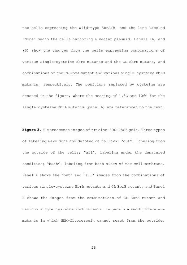
Figure 1. Positions where cysteine residues were introduced on the
CL-background mutants (C13V of EbrA and C116S of EbrB). The regions
of the membrane α-helices (enclosed by gray boxes) and loops outside
of membrane phospholipid core were predicted by the hydropathy
analysis and the consideration for the amphiphilicities of polar
residues. The orientations of EbrA and EbrB are drawn to be opposite,
and this is a conclusion of the present paper. The positively charged
residues, lysine and arginine, are highlighted by circles filled
in black. The membrane topology in this figure obeys the
“positive-inside” rule.
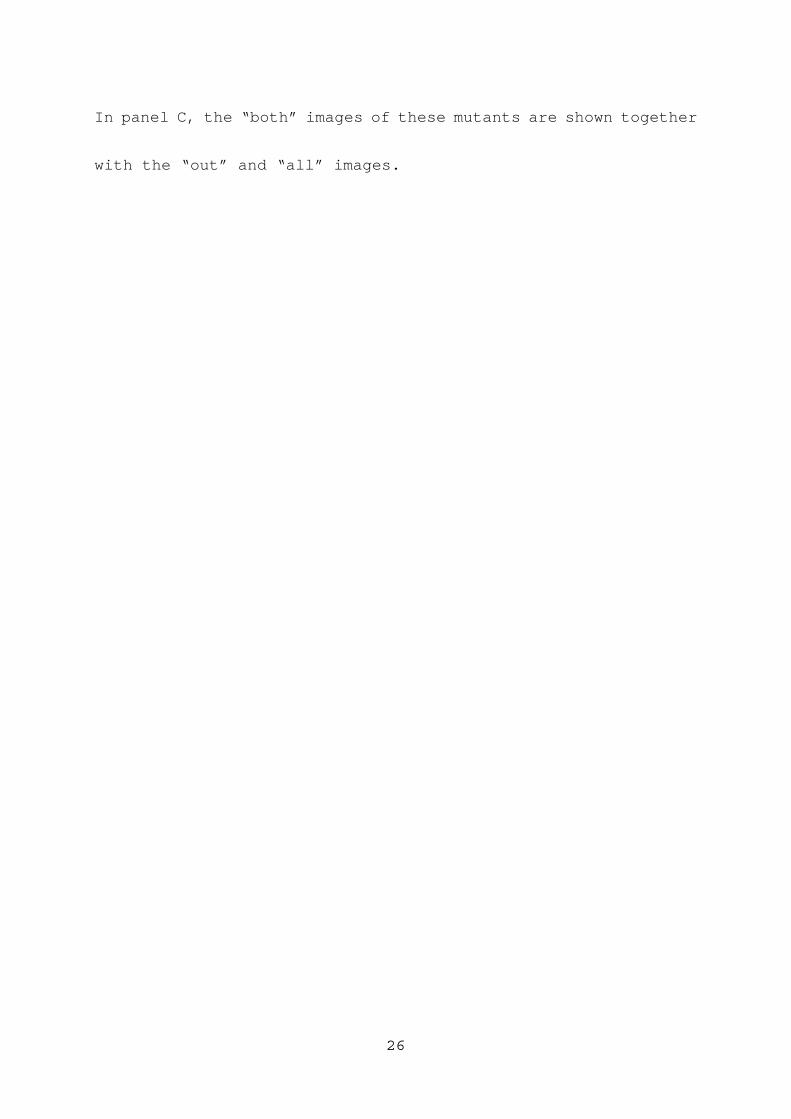
Figure 2. The confirmation that single-cysteine mutants have as
high efflux activity as the wild-type. The ethidium concentrations
remaining inside of the E. coli cells were monitored continuously
by measuring the fluorescence from ethidium. The excitation and
emission wavelengths were 545 nm and 610 nm, respectively. At time
0, glucose was added to energize the cells to start the ethidium
efflux. The thick solid lines are the fluorescence changes from
24
the cells expressing the wild-type EbrA/B, and the line labeled
“None” means the cells harboring a vacant plasmid. Panels (A) and
(B) show the changes from the cells expressing combinations of
various single-cysteine EbrA mutants and the CL EbrB mutant, and
combinations of the CL EbrA mutant and various single-cysteine EbrB
mutants, respectively. The positions replaced by cysteine are
denoted in the figure, where the meaning of 1.5C and 106C for the
single-cysteine EbrA mutants (panel A) are referenced to the text.
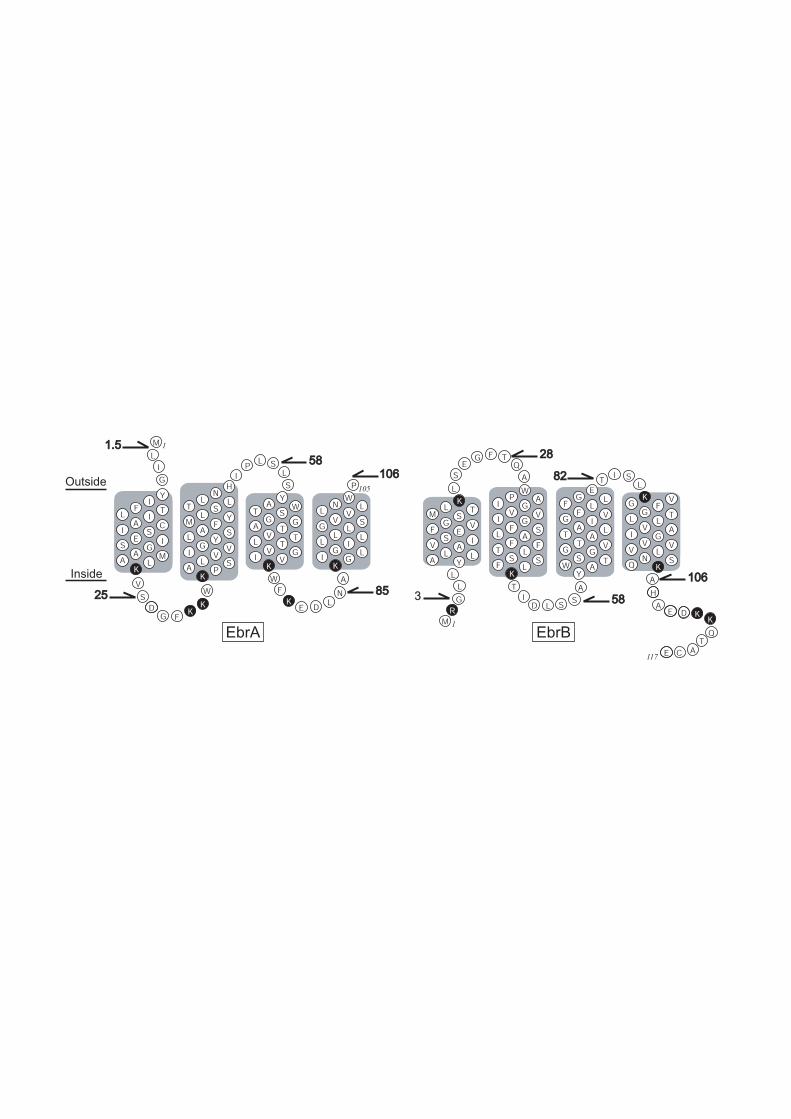
Figure 3. Fluorescence images of tricine-SDS-PAGE gels. Three types
of labeling were done and denoted as follows: “out”, labeling from
the outside of the cells; “all”, labeling under the denatured
condition; “both”, labeling from both sides of the cell membrane.
Panel A shows the “out” and “all” images from the combinations of
various single-cysteine EbrA mutants and CL EbrB mutant, and Panel
B shows the images from the combinations of CL EbrA mutant and
various single-cysteine EbrB mutants. In panels A and B, there are
mutants in which NEM-fluorescein cannot react from the outside.
25
In panel C, the “both” images of these mutants are shown together
with the “out” and “all” images.
26
EbrA EbrB
M
L
G
KG V
FG
L TL
VI
AG
VV
VL
NQ S
K
EG
FL
LF
G VI
AT L
AT
G VG
SW
TA
Y
WP
IA
GV
I VG
FL S
AF
TF
LS
FS
LK
KL
M TS
GF V
ES
V IA
LA
LY
YA
TW
SG
A GT
VL T
TV
IG
VK
WN
L LV
VG S
LL
LL
IG
IL
GK
HN
LT
LS
LM Y
FA
LS
YG
I VV
LA
SP
K
YI
FL T
IA
IC
SE
S IG
AA M
LK
I
V
S
G F
W
PI
L SL
S
FW
L
A
N
P105
1
KK DK EDI
CE A
A
S
T
SS
T
A
KK
I
T
E
G
AS
Q
F TE
DM
G
L
Q
DR
H
L
L
L
L
A
117
1
2828
8282106106
5858
8585
Outside
Inside 106106
58583
1.51.5
2525
0.0
0.2
0.4
0.6
0.8
1.0
0.0
0.2
0.4
0.6
0.8
1.0
0 50
Time [sec]
(A)
(B)
Flu
ore
scence Inte
nsity
100 150
106C
T28C
G3C
T82C A106C
None
None
S58C
N85C
1.5CS25C
S58C

outkDa
14.4
10.0
all out all out all out all out all out allall
WT CL
EbrA(single-cys) + EbrB(cys-less)
1.5C S25C S58C N85C 106CNone
(A) (B)
all kDa
EbrA(cys-less) + EbrB(single-cys)
out all out all out all out all out all out all
14.4
10.0
None G3C T28C S58C T82C A106C
EbrA(single-cys) + EbrB(cys-less)
kDa
(C)
14.4
10.0
out allboth
None
out allboth
S25C
EbrA(cys-less) + EbrB(single-cys)
out allboth
N85C
out allboth
G3C
out allboth
S58C
out allboth
A106C