bio-materials: paper review on bone response to titanium implants
TRANSCRIPT

Michelle Calender
Ngozi Aberdeen
Jurinus Lesporis
Larsson et al.
International Journal of Biomaterials
1994

Background• Surface composition includes attributes
such as roughness and surface oxide
thickness.
• Titanium’s surface properties can be varied
systematically by electropolishing and
anodizing.
• Are implant-tissue interactions influenced
by implant surface composition?

Preparation Methods
Electropolishing
Polishing using
electrical current.
• Titanium oxides
dissolve
• methanol, butanol,
and perchloric acid
• 22.5 V
• -30 °C
Anodizing
Adding a layer of
surface oxide using
electrical current.
• Titanium oxides form
protective layer
• acetic acid
• 10 or 80 V
• Room temperature

Groups
Each rabbit received 4 threaded titanium
implants, two in each tibia:
1. Electropolishing
2. Electropolishing and 10 V anodizing
3. Electropolishing and 80 V anodizing
4. None
4 rabbits were examined after 7 weeks
6 rabbits, after 12 weeks

Analysis Method
Morphometry:
• Implants and surrounding tissue examined
under a microscope connected to a computer
• Measured surface oxide thickness and
smoothness
• Calculated contact between implant and
tissues
• Calculated percentage of different tissues
between threads
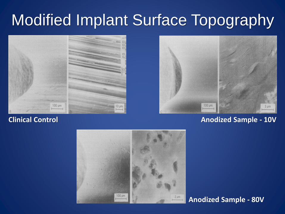
Modified Implant Surface Topography
Clinical Control Anodized Sample - 10V
Anodized Sample - 80V
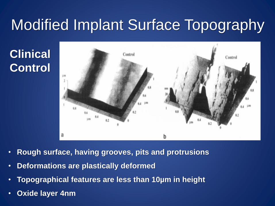
Modified Implant Surface Topography
Clinical
Control
• Rough surface, having grooves, pits and protrusions
• Deformations are plastically deformed
• Topographical features are less than 10µm in height
• Oxide layer 4nm

Modified Implant Surface Topography
Electropolished
• Smooth surface, having small pits
• Topographical features are less than 1µm in height
• Oxide layer 4-5nm
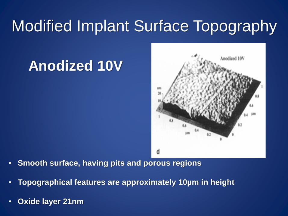
Modified Implant Surface Topography
Anodized 10V
• Smooth surface, having pits and porous regions
• Topographical features are approximately 10µm in height
• Oxide layer 21nm

Modified Implant Surface Topography
Anodized 80 V:
Smooth (left)
Rough (right)
• Heterogeneous surface, having grooves, pits and protrusions
• Topographical features are approximately10µm in height
• Oxide layer 180nm
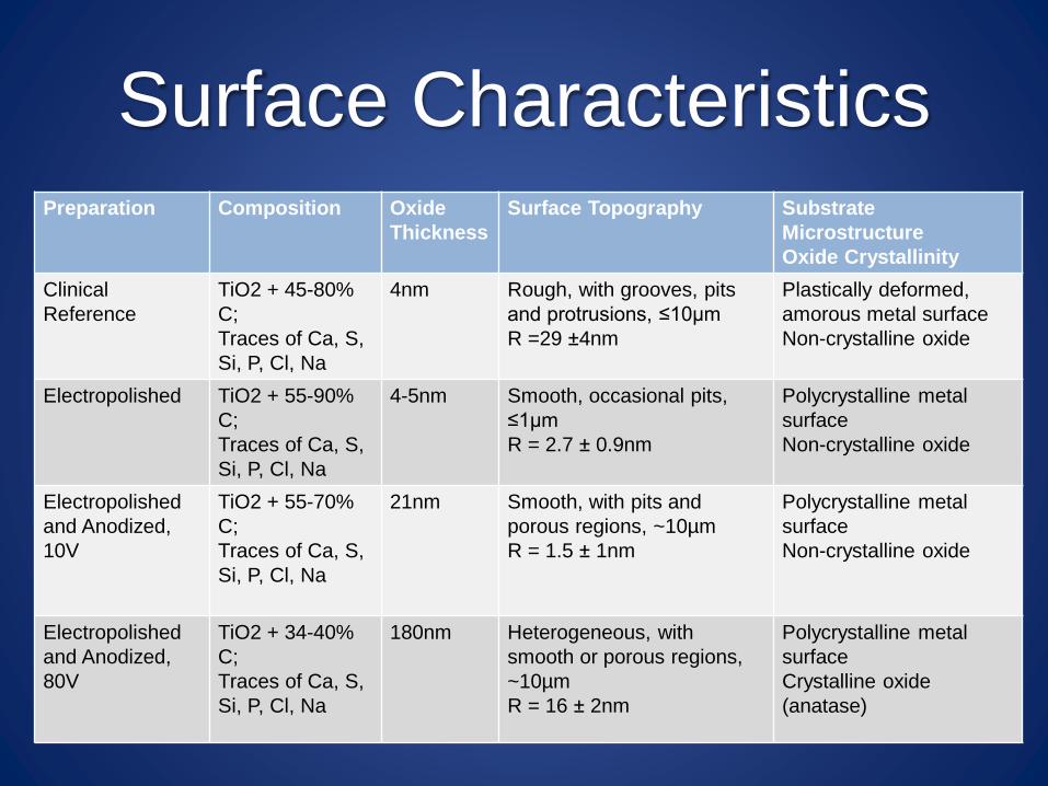
Surface CharacteristicsPreparation Composition Oxide
Thickness
Surface Topography Substrate
Microstructure
Oxide Crystallinity
Clinical
Reference
TiO2 + 45-80%
C;
Traces of Ca, S,
Si, P, Cl, Na
4nm Rough, with grooves, pits
and protrusions, ≤10µm
R =29 ±4nm
Plastically deformed,
amorous metal surface
Non-crystalline oxide
Electropolished TiO2 + 55-90%
C;
Traces of Ca, S,
Si, P, Cl, Na
4-5nm Smooth, occasional pits,
≤1µm
R = 2.7 ± 0.9nm
Polycrystalline metal
surface
Non-crystalline oxide
Electropolished
and Anodized,
10V
TiO2 + 55-70%
C;
Traces of Ca, S,
Si, P, Cl, Na
21nm Smooth, with pits and
porous regions, ~10µm
R = 1.5 ± 1nm
Polycrystalline metal
surface
Non-crystalline oxide
Electropolished
and Anodized,
80V
TiO2 + 34-40%
C;
Traces of Ca, S,
Si, P, Cl, Na
180nm Heterogeneous, with
smooth or porous regions,
~10µm
R = 16 ± 2nm
Polycrystalline metal
surface
Crystalline oxide
(anatase)

Bone Response to Modified Implants After Surgery
(Clinical Control and Electropolished)
7 weeks 12 weeks

Bone Response to Modified Implants After Surgery
(Anodized 10V and 80V)
7 weeks 12 weeks
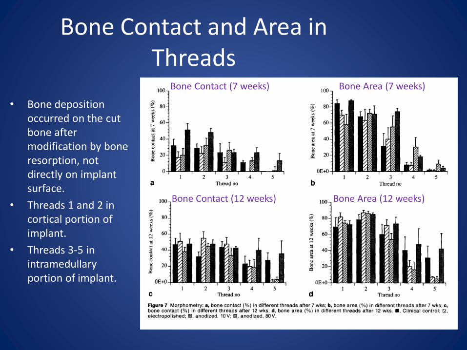
Bone Contact and Area in Threads
• Bone deposition occurred on the cut bone after modification by bone resorption, not directly on implant surface.
• Threads 1 and 2 in cortical portion of implant.
• Threads 3-5 in intramedullary portion of implant.
Bone Contact (7 weeks)
Bone Contact (12 weeks)
Bone Area (7 weeks)
Bone Area (12 weeks)

Bone Contact Results
The data suggests the 80V-anodized sample has the most bone contact and bone area within the threads.
Total Bone Contact Total Bone Area

Discussion: Findings
1. All had a high degree of bone contact, and there was no evidence of soft tissue encapsulation.
Why: for bone apposition the Oxide Layer surface chemical properties is a lot more important than its Thickness & Microstructure.
2. Electropolished implants had the lowest degree of bone contact and intra-thread bone amount.
Why: very smooth surface topography
Discussion: Findings (cntd.)
3. 80V Anodized Implant had faster bone formation
Why: I. Maybe Thicker oxide
II. Surface TopographyBut: Topographical features occurred on 1µm level and cells are only influenced by structures 10 times that.

Discussion: Limitations
• Longer implantation periods are especially
necessary for evaluating the clinical
implications of the results of this study.
• Oxide Microstructure was not looked at in
this study.

Q&A