binocular fixation disparity during the recognition of ... · the tracker’s output was recorded...
TRANSCRIPT

JEP:HPP, in press
Binocular fixation disparity in single word displays
Kevin B. Paterson
Timothy R. Jordan
&
Stoyan Kurtev
School of Psychology
Faculty of Medicine and Biological Sciences
Henry Wellcome Building
University of Leicester
Leicester LE1 9HN
U.K.
Running Head: Binocular fixation disparity in single word displays.
Binocular fixation disparity in single word displays. 2
Abstract
It has been claimed that, when a word is presented in isolation, recognition is affected by the
precise location at which the word is fixated. However, this putative role for fixation location has yet to be
reconciled with the finding from reading research that fixations made by each eye are frequently
misaligned and so more than one location in a word is often fixated simultaneously. The accuracy and
alignment of the two eyes’ fixations during single word processing have never been assessed. Therefore, to
investigate this issue, single words (and nonwords) were presented for lexical decision at various locations
around a central fixation point. Participants viewed each stimulus binocularly and were required to fixate
the fixation point. An eye-tracker recorded the fixation location of each eye. The data revealed that
participants often fixated inaccurately and that fixations were frequently misaligned but that this did not
affect word recognition. These findings indicate that binocular fixation disparity is pervasive even in single
word presentations and a potential source of confound for research investigating the effects of fixation
location on word recognition.
Key words: word recognition, binocular fixation disparity, fixation accuracy; optimal viewing position;
split fovea.
Binocular fixation disparity in single word displays. 3
The role of fixation location in single word recognition has been investigated for many years (e.g.,
O’Regan, Lévy-Schoen, Pynte, & Brugaillère, 1984) and a major motivation for this research is the steep
drop-off in retinal acuity that occurs with increased distance from the centre of the fovea (e.g., Hilz &
Cavonius, 1974). This issue has often been addressed by presenting words at different offsets to the left
and right of a central fixation point so that participants fixate a specific location within each word (e.g.,
O’Regan, 1981; O’Regan & Jacobs, 1992; O’Regan et al., 1984; Stevens & Grainger, 2003). The standard
finding is that words are recognised fastest and with fewest errors when fixated in a region between the
beginning of a word and its center (usually referred to as the optimal viewing position or OVP). The
leftward offset in the location of the OVP from a word’s center has generally been attributed to asymmetry
in the visibility of letters to each side of fixation, asymmetry in the perceptual span in reading, or
informativeness of different parts of words (for discussions, see Stevens & Grainger, 2003).
More recently, the role of fixation location in word recognition has received renewed attention in
theoretical accounts that propose that the fovea of each eye is divided precisely at its vertical meridian (for
reviews, see Brysbaert, 2004; Jordan & Paterson, 2009; Lavidor & Walsh, 2004; Lindell & Nicholls, 2003;
Shillcock, Ellison, & Monaghan, 2000). According to this Split-Fovea Theory of word recognition
(hereafter SFT), when a word is fixated so that its retinal image straddles the vertical midline of each
fovea, the word is effectively split in two such that all letters to the left of fixation project unilaterally to
the right hemisphere (RH) and all letters to the right of fixation project unilaterally to the left hemisphere
(LH). Thus, according to this view, if the stimulus “word” were fixated at the inter-letter space between
“o” and “r”, “wo” would project only to the RH and “rd” would project only to the LH.
SFT research has employed similar approaches to OVP research (e.g., Brysbaert, 1994; Brysbaert,
Vitu, & Schroyens, 1996; Hunter, Brysbaert, & Knecht, 2007; Lavidor, Ellis, Shillcock, & Bland, 2001;
Martin, Thierry, Démonet, Roberts, & Nazir, 2007). Accordingly, the typical approach has been to present
words at offsets to the left or right of a fixation point so that they straddle this point at various locations
Binocular fixation disparity in single word displays. 4
(and in some studies are shown entirely to the left or right of this point in nearby locations). This research
has shown a word recognition advantage when most letters in a word, or words in their entirety, are shown
to the right of the fixation point rather than the left, and these effects are interpreted by advocates of SFT
as evidence for unilateral projections to LH and RH processes on either side of fixation due to split-fovea
processing.
Clearly, it is important for understanding the role of fixation location in single word recognition
that participants accurately fixate the designated fixation location when words are presented in
experiments. However, under these conditions, it is well established that participants have difficulty
monitoring and precisely controlling their fixation locations when attempting to fixate a specified location,
and so often do not fixate accurately (e.g., see Findlay & Kapoula, 1992; Jones & Santi, 1978; Jordan,
Patching, & Milner, 1998, 2000; Jordan, Patching, & Thomas, 2003; Jordan & Paterson, 2009; Jordan,
Paterson, Kurtev, & Xu, 2009a; Jordan, Paterson, & Stachurski, 2009; Terrace, 1959). Nevertheless,
participants in experiments investigating the role of fixation location in word recognition are usually only
instructed to fixate a designated fixation location (sometimes accompanied by a secondary fixation task)
with no external monitoring or control (e.g., using an eye-tracker) to determine which locations are
actually fixated (e.g., Brysbaert, 1994; Brysbaert et al., 1996; Hunter et al., 2007; Lavidor et al., 2001;
Martin et al., 2007; O’Regan, 1981; O’Regan & Jacobs, 1992; O’Regan et al., 1984; Stevens & Grainger,
2003; although see O’Regan et al., 1984, Experiment 2). 1
A further, major problem is the implicit yet fundamental assumption that the points of fixation for
the left and right eyes coincide precisely. Thus, according to this view, when a word is fixated, information
in the left and right hemifield of one eye will match the information in the left and right hemifield of the
other eye, with the result that precisely the same letters each side of fixation will project from both eyes to
the corresponding contralateral hemisphere. However, there is growing evidence that, during normal
(binocular) reading, the two eyes often do not fixate the same locations (Blythe, Liversedge, Joseph,
Binocular fixation disparity in single word displays. 5
White, Findlay, & Rayner, 2006; Heller & Radach, 1999; Juhasz, Liversedge, White, & Rayner, 2006;
Kliegl, Nuthmann, & Engbert, 2006; Liversedge, White, Findlay, & Rayner, 2006; Liversedge, Rayner,
White, Findlay, & McSorley, 2006; for a review, see Kirkby, Webster, Blythe, & Liversedge, 2008).
Accordingly, when fixating a word during reading, two fixation locations will often occur, one for each
eye, and so information on either side of each foveal midline will often differ substantially between the two
eyes. Indeed, when reading, fixation disparities have been found to occur on almost 50% of fixations and
are produced by fixations that land as much as 2 or more characters apart. Indeed, sometimes these
fixations may even be on different words. This situation is complicated further because misaligned
fixations can be either crossed (when the right eye fixates to the left of the left eye’s fixation), or uncrossed
(when the right eye fixates to the right of the left eye’s fixation), and the nature and extent of the disparity
may vary from fixation to fixation.
--------------------Figure 1 here-----------------
The issue of fixation disparity adds to the problem of determining fixation accuracy when assessing
the role of fixation location in single word displays. To date, previous research has examined binocular
fixation disparity only in reading, and the occurrence and influence of fixation disparity during single word
recognition has yet to be determined. Accordingly, the present study assessed the extent and influence of
binocular fixation disparity during single word recognition using the paradigm employed widely in studies
of the effects of fixation location on single word recognition. Specifically, five-letter stimuli were
presented at normal reading size (subtending 1.25°) at one of 6 locations such that a central fixation point
coincided with the space immediately to the left or right of each stimulus, or one of 4 inter-letter spaces
(see Figure 1). In line with previous research, participants were emphatically instructed to fixate the
designated fixation point for each stimulus display. However, unlike previous research, a binocular eye-
tracker recorded the actual fixation locations of both eyes, and so revealed the nature and extent of
binocular fixation accuracy and fixation disparity in single word displays typical in this area of research.
Binocular fixation disparity in single word displays. 6
Experiment
Method
Participants. Twenty native English speakers from the University of Leicester were paid for
participating in a single 30 minute session. Participants had normal visual acuity, determined by a Bailey-
Lovie Eye Chart, and were right-handed, determined by a revised Annett Handedness Questionnaire
(Annett, 1970). Eye dominance was determined individually for each participant using both the Miles test
(Miles, 1930) and the Porta test (Porta, 1593; see also, Roth, Lora, & Heilman, 2002) of ocular dominance.
Stimuli and Design. Stimuli were 78, 5-letter words (mean frequency 171 per million; Baayen,
Piepenbrock, & Gulikers, 1995) and 78, 5-letter pronounceable nonwords generated from existing English
words by substitution of one letter. Thirty-six additional stimuli (18 words and 18 nonwords) served as
practice items at the beginning of the experiment. Stimuli were presented in lowercase Courier font as
black text on a white background and subtended approximately 1.25° (4 letters per degree), which
approximates normal reading size (e.g., Rayner & Pollatsek, 1989). Stimuli were presented in a random
order across each of the 6 screen locations. Each location was selected at random on each trial.
Apparatus. Stimuli were presented on a high-definition display monitor. A Cambridge Research
Systems VSG 2/5 card controlled stimulus presentations and timing. Responses were collected via a
Cambridge Research Systems CT3 button box. The experiment was conducted in a sound-attenuated and
darkened room and displays were observed using a head brace and chinrest to ensure a constant viewing
distance of 60 cm. Stimulus viewing was binocular and the position of each eye was monitored using a
Skalar IRIS eye-tracking system (Cambridge Research Systems). The eye tracker was clamped firmly to
each participant’s head, which in turn was clamped in a rigid head brace (that incorporated the chin-rest)
throughout the experiment to prevent head movements. This arrangement allowed accurate and consistent
measurement of fixation location in the experiment to within 5 minutes of arc (for further details, see
Patching & Jordan, 1998; Jordan & Patching, 2006). The tracker’s output was recorded each ms by the
Binocular fixation disparity in single word displays. 7
Cambridge Research Systems VSG2/5 card, which also controlled the visual display (see Jordan, Patching,
& Milner, 1998, 2000).
Procedure. Before the start of each session, participants were given instructions that included
emphasising the importance of accurate fixation, and the eye-tracker was calibrated for the fixation of each
eye 2. Calibration was checked at the beginning of each trial. Binocular fixations were recorded in two
conditions. In the uncontrolled condition, participants were instructed emphatically to fixate a point at the
center of the screen at the start of each trial, after which this fixation point disappeared and a stimulus was
presented for 150 ms. In the controlled condition, the fixation point was presented at the centre of the
screen and stimulus presentation was withheld until accurate dominant eye fixation of this point actually
occurred for 300 ms. When this criterion was satisfied, the fixation point disappeared and a stimulus was
presented for 150 ms. In the controlled condition, if fixation deviated from the fixation point before
stimulus presentation, no stimulus presentation took place until accurate fixation was re-established (see
Patching & Jordan, 1998, for further details). Participants made a lexical decision for each stimulus using
their right (dominant) hand to press one of two keys on the response box.
Results
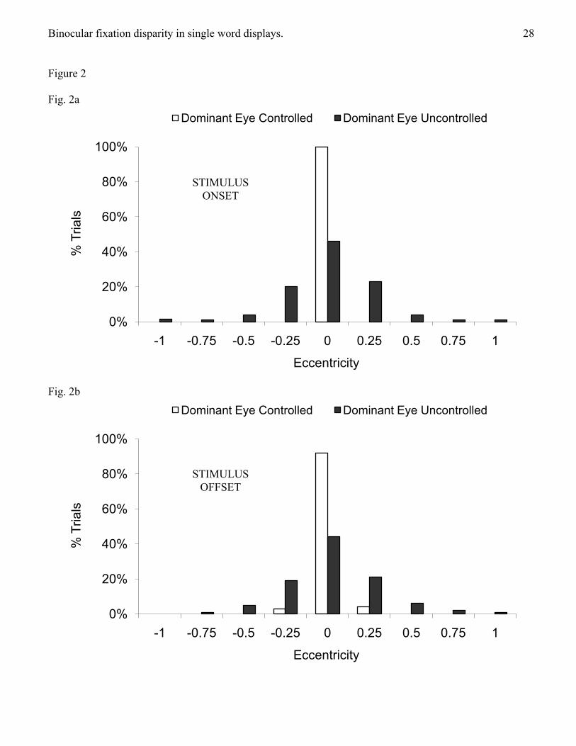
Fixation accuracy in controlled and uncontrolled conditions. Figure 2 shows the location of
dominant eye fixations at stimulus onset (Fig. 2a) and offset (Fig. 2b). In the controlled condition, the
dominant eye fixated accurately on 100% of trials at stimulus onset (as accurate dominant eye fixations
were ensured in this condition). By comparison, accurate dominant eye fixations occurred on only 46% of
trials at stimulus onset in the uncontrolled condition. On 54% of trials in this condition, the dominant eye
fixated between 0.25° (i.e., approximately 1 letter) and 1° (i.e., approximately 4 letters) away from this
location, indicating that the dominant eye did not fixate accurately on the majority of trials. At stimulus
offset, accurate dominant eye fixations fell to 92% in the controlled condition and to 44% in the
uncontrolled condition, although chi-square analyses used to assess separately the change in the proportion
Binocular fixation disparity in single word displays. 8
of accurate dominant eye fixations between stimulus onset and offset in controlled and uncontrolled
fixation conditions indicated that neither reduction was significant (ps>.10). At stimulus offset in the
controlled condition inaccurate dominant eye fixations fell equally to the left and right of center. Chi-
square analyses comparing the proportion of fixations that landed on each side of the central fixation point
revealed that the distribution of inaccurate dominant eye fixations in the uncontrolled condition was
symmetrical about the fixation point at both stimulus onset and stimulus offset (ps>.10).
--------------------Figures 2 & 3 here-----------------
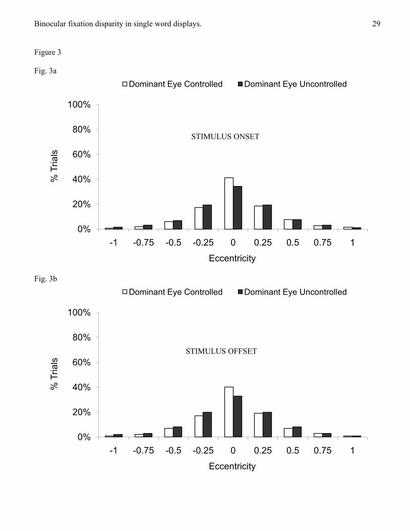
Figure 3 shows the distribution of non-dominant eye fixations at stimulus onset (Fig. 3a) and offset
(Fig. 3b). Note that the location of these fixations was uncontrolled in both fixation conditions. In the
controlled condition (i.e., when all dominant eye fixations were accurate at onset), the non-dominant eye
fixated accurately on only 41% of trials at stimulus onset and inaccurate fixations fell between 0.25° and
1° away from the fixation point on 59% of trials. Thus, even when participants fixated the fixation point
with their dominant eye, the non-dominant eye did not fixate this location on the majority of trials. In the
uncontrolled condition, the non-dominant eye fixated accurately on only 34% of trials at stimulus onset
and inaccurate fixations fell between 0.25° and 1° away from the fixation point on 66% of trials. Thus, the
non-dominant eye did not fixate accurately on the majority of trials in this condition. The change in the
proportion of accurate non-dominant eye fixations between stimulus onset and offset was analysed
separately for the controlled and uncontrolled conditions using Chi-square analyses. This revealed that
there was little change in the accuracy of non-dominant eye fixations between stimulus onset and offset in
either controlled or uncontrolled conditions (ps>.10). In addition, Chi-square analyses that compared the
proportion of fixations on each side of the central fixation point indicated that inaccurate fixations were
symmetrical about the fixation point in both controlled and uncontrolled conditions (ps>.10).
Fixation alignment during word recognition. Following the convention adopted by previous
research (Liversedge, White et al., 2006), fixations were considered aligned if the point of fixation of each
Binocular fixation disparity in single word displays. 9
eye fell within the width of one character space. 3 As 5-letter stimuli subtended 1.25°, fixations were
deemed aligned when the disparity between the two points of fixation subtended a horizontal visual angle
less than or equal to 0.25°. Fixations were crossed when the right eye fixated more than one character
space to the left of the left eye’s fixation, and uncrossed when the right eye fixated more than one character
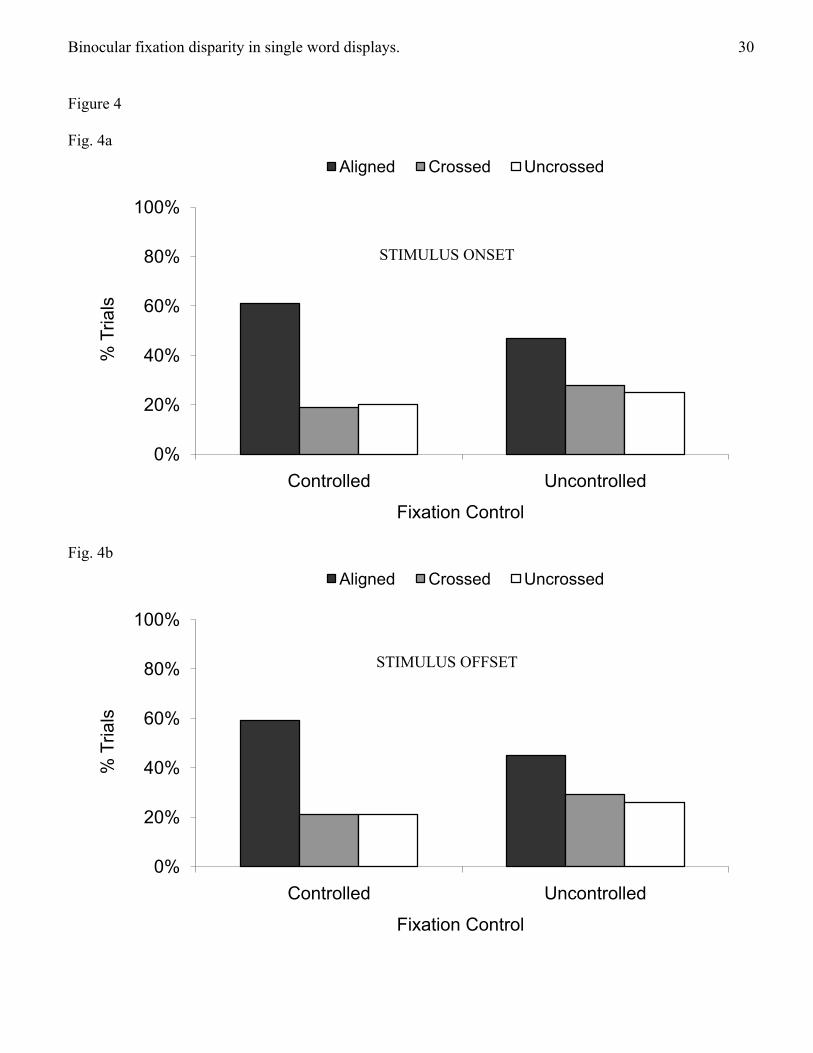
space to the right of the left eye’s fixation. Figure 4 shows the proportion of each type of fixation
alignment at stimulus onset (Fig. 4a) and offset (Fig 4b).
--------------------Figure 4 here-----------------
In the controlled condition, fixations were aligned at stimulus onset on only 61% of trials (59% at
offset) and, in the uncontrolled condition, on only 47% of trials (45% at offset). In the controlled condition
at stimulus onset, 19% of fixations were crossed (21% at offset), and 20% were uncrossed (21% at offset).
In the uncontrolled condition at stimulus onset, 28% of fixations were crossed (29% at offset) and 25%
were uncrossed (26% at offset). Chi-square analyses were used to compare separately the proportion of
aligned, crossed, and uncrossed fixations across controlled and uncontrolled conditions. The analyses
indicated that the proportion of aligned, crossed, and uncrossed fixations at stimulus onset differed
significantly between controlled and uncontrolled conditions, 2(df = 2) = 87.84, p<.001, and an analysis
of residuals (Seigel & Castellan, 1988) confirmed that more fixations were aligned in the controlled
condition (p<.001). Further Chi-square analyses were used to compare separately the change in the
proportion of aligned, crossed, and uncrossed, fixations between stimulus onset and offset. These analyses
confirmed that the slight changes in the proportions of each type of fixation alignment between stimulus
onset and offset were not significant in either controlled or uncontrolled conditions (p>.10).
Across all fixations (aligned and misaligned), fixations were on average 0.32° (1.3 character
spaces) apart at stimulus onset (controlled condition, 0.27°; uncontrolled condition, 0.37°), and 0.32° (1.3
character spaces) apart at stimulus offset (controlled condition, 0.29°; uncontrolled condition, 0.37°).
Repeated measures t-tests revealed that fixation disparity was smaller in the controlled than uncontrolled
Binocular fixation disparity in single word displays. 10
condition at both stimulus onset, t(19) = 2.94, p<.01, and offset, t(19) = 2.65, p<.05. Misaligned fixations
averaged 0.56° (2.2 character spaces) apart at stimulus onset (controlled condition, 0.52°; uncontrolled
condition, 0.59°), and 0.56° (2.2 character spaces) apart at stimulus offset (controlled condition, 0.52°;
uncontrolled condition, 0.57°). Fixation disparity for misaligned fixations did not differ significantly
between controlled and uncontrolled conditions at stimulus onset or offset (t>.10).
The focus of the current study was to determine natural variation in fixation disparity and
participants did not produce equal amounts of aligned, crossed, and uncrossed fixations for each fixation
location and did not fixate accurately with both eyes on the majority of trials, even in the controlled
condition. Consequently, it was not possible to perform statistical analyses that assessed the effects of
fixation alignment on word recognition when words were presented at each of the 6 screen locations in
controlled and uncontrolled conditions by performing analyses based on a traditional analysis of variance
(ANOVA). We therefore computed an analysis using a linear mixed-effect model specifying participants
as a random effect. An advantage of such an analysis is that it results in substantially reduced loss of
statistical power in unbalanced designs than ANOVA (see, e.g., McCulloch & Searle, 2000). Overall error
rates were low (3%) and did not support meaningful analyses so only reaction times (for correct responses)
were analysed. This analysis produced a significant main effect of fixation control, F(1,2666) = 29.01,
p<.001, which revealed that reaction times were longer in the controlled (545 ms) than uncontrolled
conditions (517 ms). There was also a significant main effect of screen location, F(5, 2666) = 9.18, p<.001.
Mean reaction times produced an OVP effect similar to that obtained in other research using binocular
displays (e.g., O’Regan, 1981; O’Regan & Jacobs, 1992; O’Regan et al., 1984; Stevens & Grainger, 2003),
indicating normal processes of word recognition. Specifically, reactions times were shortest at locations 2
and 3, when the fixation point was between the beginning and middle of words (i.e., location 2 = 521 ms,
location 3 = 508 ms), intermediate when the fixation point was at other locations within words (location 4
= 528 ms, location 5 = 530 ms), and longest when words were shown entirely to the right or left of the
Binocular fixation disparity in single word displays. 11
fixation point (location 1 = 534 ms, location 6 = 567 ms). However, it should be noted that because the
present research intentionally did not control the fixation locations of both eyes (and previous research
generally has not controlled the fixation locations of even one eye), neither this experiment nor previous
OVP research provides an accurate indication of the effects of fixating specific locations within words
viewed binocularly (but see Jordan, Paterson, Kurtev, & Xu, 2009b). Crucially, there was no effect of
fixation alignment, F<1, indicating that there was no significant difference in reaction times for words
when fixations were aligned (532 ms), crossed (529 ms), or uncrossed (532 ms). No other effects were
significant, Fs<2.6, indicating that fixation alignment did not affect word recognition performance in
different fixation control conditions or when words were presented at different screen locations.4
A further analysis was conducted to determine if both eyes had to fixate within the region of text
corresponding to the OVP to produce an OVP effect, by examining reaction times for words when
participants fixated within this region with both eyes, one eye, or neither eye. This analysis was
conducted using a traditional ANOVA (because the data produced a balanced design) with variables
fixation control (controlled, uncontrolled) and type of fixation within the OVP (both eyes, one eye,
neither eye) and revealed a main effect of the type of fixation, F(2,76) = 13.11, p<.001, ηp2 =.26, that did
not interact with fixation control, F<1. Tukey tests showed that reaction times were shortest of all when
both eyes fixated within the OVP (519 ms, ps<.001), and that reaction times were shorter when one eye
fixated within the OVP (537 ms) than when neither eye fixated within this region (549 ms, p = .05).
Discussion
This study examined fixation accuracy and alignment during binocular viewing of single words
using a paradigm widely employed to investigate effects of fixation location on single word recognition,
and a binocular eye-tracker to record the actual fixation locations of both eyes.
Fixation accuracy during single word recognition. Previous research using this paradigm generally
has not assessed the actual locations of participants’ fixations (e.g., Brysbaert, 1994; Brysbaert et al., 1996;
Binocular fixation disparity in single word displays. 12
Hunter et al., 2007; Lavidor et al., 2001; Martin et al., 2007; see also O’Regan, 1981; O’Regan & Jacobs,
1992; Stevens & Grainger, 2003). Moreover, even when fixation location has been assessed (e.g., O’Regan
et al., 1984, Experiment 2), the fixations of just one eye (during binocular viewing) have been monitored
and this is uninformative about the location of both eyes’ fixations. The present study revealed that even
when accurate dominant eye fixation was ensured, the non-dominant eye did not fixate the designated
location on the majority of trials. The extent of this inaccuracy was even greater when fixation location
was only monitored (not controlled); in this case neither eye fixated accurately on the majority of trials.
Thus, in both fixation conditions, and on most trials, participants did not fixate stimuli as required.
These findings contrast with findings from research showing that fixations of small isolated
stationary targets, like a fixed central point, are extremely stable over several seconds when the head is
firmly supported, suggesting that the demands of maintaining central fixation under these perceptually
simple conditions are small (for a review, see Kowler, 1990). Indeed, Ratliff and Riggs (1950; also
Putnam, Hofer, Doble, Chen, Carroll, & Williams, 2005; Steinman, 1977; Steinman, Haddad, Skavenski,
& Wyman, 1973) found that the total movement of the eyes over a period of several seconds is usually less
than 10 min of arc, occurring equally often to the left and right of fixation, and Steinman et al. (1973)
observed that maintaining stable fixation of a fixed central point is simple to do and requires no special
training. However, these findings were made using paradigms in which only fixation stimuli were
presented. In contrast, accurately fixating a fixed central point appears to be difficult in studies where the
primary task is to recognise words presented around a central fixation point (e.g., Jordan & Paterson, 2009;
Jordan, Paterson, Kurtev, & Xu, 2009a; Jordan, Paterson, & Stachurski, 2009) or in lateralized displays
(e.g., Findlay & Kapoula, 1992; Jones & Santi, 1978; Jordan, Patching, & Milner, 1998; 2000; Jordan,
Patching, & Thomas, 2003; Terrace, 1959). Indeed, the present results support the view that participants
have considerable difficulty in monitoring and precisely controlling their fixation locations when
attempting to fixate a specified location and that instructions alone do not ensure precise fixation in studies
Binocular fixation disparity in single word displays. 13
of word recognition. The present results also reveal that there is considerable variability in the exact
fixation location of each eye, and further caution that even when participants are known to be fixating a
fixation point with one eye on each trial of an experiment, the other eye is unlikely to be fixating the same
location on the majority of trials. Moreover, our results indicate that these problems occur even when (as in
the experiment reported) participants have ample time and strong encouragement to fixate accurately.
The alignment of binocular fixations during single word recognition. Studies of reading have
revealed frequent and substantial disparities in the location of binocular fixations (Blythe et al., 2006;
Heller & Radach, 1999; Juhasz et al., 2006; Kliegl et al., 2006; Liversedge, White et al, 2006; Liversedge,
Rayner, et al., 2006). The present data show that similar fixation disparities occur in single word
presentations. We found that misaligned fixations were more frequent (and average fixation disparity was
greater) when participants were merely instructed to fixate a designated location and actual fixation
locations were only monitored. Using an eye-tracker to ensure accurate dominant eye fixation increased
the proportion of aligned fixations (and reduced fixation disparity), but fixations were still misaligned on
many trials. Thus, even though procedures that ensure accurate fixation can improve the coordination of
binocular fixations, these procedures do not ensure that fixations are always aligned. It seems inevitable
that a similar lack of fixation alignment will have occurred in research employing the same paradigm as
the present study but without fixation monitoring and control (e.g., Brysbaert, 1994; Brysbaert et al., 1996;
Hunter et al., 2007; Lavidor et al., 2001; Martin et al., 2007; see also, e.g., O’Regan, 1981; O’Regan &
Jacobs, 1992; Stevens & Grainger, 2003). Consequently, this earlier research is unlikely to have provided
an accurate indication of the effects of fixating designated locations within words.
The findings were also informative about the incidence of crossed and uncrossed fixations.
Previous research has revealed considerable variability in the proportion of crossed and uncrossed fixations
in reading. Thus, whereas Liversedge and his colleagues found that most misaligned fixations were
uncrossed (e.g., Liversedge, White et al., 2006), other research reported that most misaligned fixations
Binocular fixation disparity in single word displays. 14
were crossed (Kliegl et al., 2006). The present research revealed no such biases, and showed instead that
misaligned fixations were equally likely to be crossed as uncrossed. Thus, although the reason for
variability in the proportion of crossed and uncrossed fixations in reading (and differences between
findings from different laboratories) remains to be determined, the same biases were not observed in the
present experiment.
Effects of fixation alignment on word recognition. Notwithstanding the variability in fixation
alignment, the present findings indicate that fixation misalignment does not affect single word recognition.
Instead, word recognition was no more difficult when the two eyes fixated locations one or more character
spaces apart than when fixations were aligned to within a single character space. This finding is in line
with other research showing that adult readers can tolerate disparity in the location of the two eye’s
fixations during textual reading without disruption to normal word recognition processes (Heller &
Radach, 1999; Juhasz et al., 2006). Thus, although the frequency of the fixation misalignment is of
considerable importance for theories that emphasize the role of fixation location in word recognition and
reading (as discussed below), the actual practical consequences for skilled reading appear to be less
sensitive, given that word recognition performance did not vary across fixation alignment conditions.
However, this is not to say that fixation alignment is entirely unimportant in reading. For example,
developmental differences may occur in the coordination of saccades during word recognition (e.g., Bucci
& Kapoula, 2006) and in fixation alignment during reading (Blythe et al., 2006) that may be relevant to the
development of reading skills, and other research has attributed reading difficulties in dyslexia to difficulty
in maintaining stable aligned binocular fixation (e.g., Stein & Fowler, 1983; see Kirkby et al., 2008, for a
review).
Although fixation misalignment did not affect word recognition in our experiment, and standard
OVP effects were obtained, word recognition was slower when fixation accuracy of the dominant eye was
ensured than when participants were merely instructed to fixate accurately. This finding is in line with
Binocular fixation disparity in single word displays. 15
other research showing that word recognition is slower, and that variation in word length affects reaction
times, when the accuracy of fixations within words is enforced (Jordan, Paterson, & Stachurski, 2009).
Thus, it appears that participants not only have difficulty in monitoring and precisely controlling their
fixation locations in tasks involving word recognition but that ensuring the precise fixation of a particular
location within (and close to) words can impose additional task demands that impede normal word
recognition processes. This and other aspects of the present findings are problematic for SFT, which
emphasizes the importance of precise fixation location in word recognition and for research in which
participants are instructed emphatically to fixate a particular location on each trial (e.g., Brysbaert, 1994;
Brysbaert et al., 1996; Hunter et al., 2007; Lavidor et al., 2001; Martin et al., 2007; Shillcock, Ellison, &
Monaghan, 2000). Our findings indicate not only that these efforts are unlikely to be successful but that,
even if they were, normal processes of word recognition would be impeded. Thus, in contrast to the
arguments of SFT, fixating precisely-defined locations in foveally-presented words appears to impair word
recognition rather than improve it.
Indeed, the present findings are problematic for SFT’s account of word recognition. According to
this account, when a string of letters is fixated so that its retinal image straddles the vertical midline of each
fovea, all letters to the left of fixation project to the RH and all letters to the right project to the LH (e.g.,
Lavidor & Walsh, 2004; Shillcock et al., 2000). The present findings, and other research into binocular
fixation disparity during textual reading (Blythe et al., 2006; Heller & Radach, 1999; Juhasz et al., 2006;
Kliegl et al., 2006; Liversedge, White et al., 2006; Liversedge, Rayner, et al., 2006), reveal that the two
eyes often fixate different locations in a word during normal word recognition, and so information on
either side of the foveal midline will often differ between the two eyes. In general, SFT has not
accommodated disparities in binocular fixation and has overlooked the influence of these disparities in
experiments reported in support of split-foveal processing. The present findings are important for SFT
because until now the theory has assumed that when participants are instructed to fixate a particular
Binocular fixation disparity in single word displays. 16
location within a word, both eyes will accurately fixate this location, and therefore there is no conflict in
the information provided around the two points of fixation. Our data show that there often is a conflict but
that this does not affect word recognition. Moreover, the data reveal that both the nature and the magnitude
of the disparity in the location of the two eyes fixations is likely to vary from fixation to fixation, thus
changing both the form and the amount of overlap in information projected to each hemisphere from
fixation to fixation in word recognition, and from trial to trial in an experiment. Given this situation, it
seems that the assumptions regarding fixation behavior in many experiments that provide support for SFT,
and the account of word recognition provided by the theory itself, present, at best, an oversimplified
account of the role of fixation location in word recognition.
By comparison, the present findings are consistent with the approach taken in OVP research (e.g.,
O’Regan et al., 1984), which makes rather more lenient demands on fixation location. According to this
account, word recognition operates best when fixations land somewhere between the beginning and middle
of a word (i.e. the OVP) and word recognition is impeded only when fixations are made at locations that
lie outside this region. The present findings show that OVP effects do not depend on accurate fixation of a
designated fixation location, or even that the two eye’s fixations are aligned, which may explain why the
OVP effect has proved to be so robust in research, even when participants are only instructed to fixate a
designated fixation location without external monitoring or control of actual fixation location, and viewing
is binocular (e.g., Brysbaert, 1994; Brysbaert et al., 1996; Hunter et al., 2007; O’Regan, 1981; O’Regan &
Jacobs, 1992; O’Regan et al., 1984; Stevens & Grainger, 2003; see also, Jordan, Paterson, & Stachurski,
2009). Indeed, the present data suggest that it is sufficient that participants fixate within the broad intra-
word region that encompasses the OVP to produce an OVP effect (see also, Jordan, Paterson, Kurtev, &
Xu, 2009b), although our data indicate that the effect is seen most clearly when both eyes fixate within this
region. Thus, while the precise reasons for the OVP have yet to be fully determined (see, e.g., Stevens and
Grainger, 2003), our findings indicate that the effect of the OVP has nothing to do with the fixation
Binocular fixation disparity in single word displays. 17
precision posited by split-foveal accounts of word recognition.
In sum, our findings reveal substantial and frequent inaccuracies and disparities in binocular
fixations when viewing and processing single words. Nevertheless, fixation accuracy and misalignment did
not affect word recognition performance. These findings provide important information concerning the
nature of binocular fixations and their effect on word recognition. They also caution against the common
misapprehension that the points of fixation for the left and right eyes coincide precisely at the designated
fixation point in experiments using single word displays.
Binocular fixation disparity in single word displays. 18
References
Annett, M. (1970). A classification of hand preference by association analysis. British Journal of
Psychology, 61, 303-321.
Baayen, R.H., Piepenbrock, R., & Gulikers, L. (1995). The CELEX Lexical Database (Release 2) [CD-
ROM]. Philadelphia, PA: Linguistic Data Consortium, University of Pennsylvania.
Blythe, H. I., Liversedge, S.P., Joseph, H.S.S.L., White, S.J., Findlay, J.M., & Rayner, K. (2006). The
binocular coordination of eye movements during reading in children and adults. Vision Research,
46, 3898-3908.
Brysbaert, M. (1994). Interhemispheric transfer and the processing of foveally presented stimuli.
Behavioural Brain Research, 64, 151-161.
Brysbaert, M. (2004). The importance of interhemispheric transfer for foveal vision: A factor that has been
overlooked in theories of visual word recognition and object perception. Brain and Language, 88,
259-267.
Brysbaert, M., Vitu, F., & Schroyens, W. (1996). The right visual field advantage and the optimal viewing
position effect: On the relation between foveal and parafoveal word recognition. Neuropsychology,
10, 385-395.
Bucci, M.P. & Kapoula, Z. (2006). Binocular coordination of saccades in 7 years-old children in single
word reading and target fixation. Vision Research, 46, 457-466.
Findlay, J.M., & Kapoula, Z. (1992). Scrutinization, spatial attention, and the spatial programming of
saccadic eye movements. Quarterly Journal of Experimental Psychology, 45A, 633–647.
Heller, D. & Radach, R. (1999). Eye movements in reading. Are two eyes better than one? In E. Becker
(Ed.), Current oculomotor research. Plenum Press: New York.
Hilz, R.L., & Cavonius, C.R., (1974). Functional organisation of the peripheral retina: Sensitivity to
periodic stimuli. Vision Research, 14, 1333-1337.
Binocular fixation disparity in single word displays. 19
Hunter, Z., Brysbaert, M., & Knecht, S. (2007). Foveal word reading requires interhemispheric
communication. Journal of Cognitive Neuroscience, 19, 1373-1387.
Jones, B. & Santi, A. (1978). Lateral asymmetries in visual perception with and without eye movements.
Cortex, 14, 164-168.
Jordan, T.R., & Patching, G.R. (2006). Assessing effects of fixation demands on perception of lateralized
words: A visual window technique for studying hemispheric asymmetry. Neuropsychologia, 44,
686-692.
Jordan, T.R., Patching, G.R., & Milner, A.D. (1998). Central fixations are inadequately controlled by
instructions alone: Implications for studying cerebral asymmetry. Quarterly Journal of
Experimental Psychology, 51A, 371-391.
Jordan, T.R., Patching, G.R., & Milner, A.D. (2000). Lateralized word recognition: Assessing the role of
hemispheric specialization, modes of lexical access and perceptual asymmetry. Journal of
Experimental Psychology: Human Perception and Performance, 26, 1192-1208.
Jordan, T.R., Patching, G.R., & Thomas, S.M. (2003). Assessing the role of hemispheric specialization,
serial-position processing and retinal eccentricity in lateralized word perception. Cognitive
Neuropsychology, 20, 49-71.
Jordan, T.R., & Paterson, K.B. (2009). Re-evaluating split-fovea processing in word recognition: A critical
assessment of recent research. Neuropsychologia, in press.
Jordan, T.R., Paterson, K.B., Kurtev, S., & Xu, M. (2009a). Do fixation cues ensure fixation accuracy in
split-fovea studies of word recognition? Neuropsychologia, in press.
Jordan, T.R., Paterson, K.B., Kurtev, S., & Xu, M. (2009b). Re-evaluating split-fovea processing in word
recognition: Effects of fixation location within words. Cortex, in press.
Jordan, T.R., Paterson, K.B., Kurtev, S., & Xu, M. (2009c). Re-evaluating split-fovea processing in word
recognition: Effects of word length during monocular viewing. Cortex, in press.
Binocular fixation disparity in single word displays. 20
Jordan, T.R., Paterson, K.B., & Stachurski, M. (2008). Re-evaluating split-fovea processing in word
recognition: Effects of retinal eccentricity. Neuropsychology, 22, 738-745.
Jordan, T.R., Paterson, K.B., & Stachurski, M. (2009). Re-evaluating split-fovea processing in word
recognition: Effects of word length. Cortex, 45, 495-505.
Juhasz, B., Liversedge, S.P., White, S.J., & Rayner, K. (2006). Binocular coordination of the eyes during
reading: Word frequency and case alternation affect fixation duration but not fixation disparity.
Quarterly Journal of Experimental Psychology, 59, 1614–1625.
Kirkby, J.A., Webster, L.A.D., Blythe, H.I., & Liversedge, S.P. (2008). Binocular coordination during
reading and non-reading tasks. Psychological Bulletin, 134, 742-763.
Kliegl, R., Nuthmann, A., & Engbert, R. (2006). Tracking the mind during reading: The influence of past,
present, and future words on fixation durations. Journal of Experimental Psychology: General, 135,
12-35.
Kowler, E. (1990). The role of visual and cognitive processes in the control of eye movement. In E.
Kowler (Ed.), Eye movements and their role in visual and cognitive processes (pp. 1–70).
Amsterdam: Elsevier.
Lavidor, M., Ellis, A.W., Shillcock, R., & Bland, T. (2001). Evaluating a split processing model of visual
word recognition: Effects of word length. Cognitive Brain Research, 12, 265-272.
Lavidor, M., & Walsh, V. (2004). The nature of foveal representation. Nature Reviews Neuroscience, 5,
729-735.
Lindell, A.K., & Nicholls, M.E.R. (2003). Cortical representation of the fovea: implications for visual half-
field research. Cortex, 39, 111-117.
Liversedge, S.P., White, S.J., Findlay, J.M., & Rayner, K. (2006). Binocular coordination of eye
movements during reading. Vision Research, 46, 2363-2374.
Liversedge, S. P., Rayner, K., White, S.J., Findlay, J.M., & McSorley, W. (2006). Binocular coordination
Binocular fixation disparity in single word displays. 21
of the eyes during reading. Current Biology, 16, 1726-1729.
Martin, C.D., Thierry, G., Démonet, J.F., Roberts, M., & Nazir, T. (2007). ERP evidence for the split fovea
theory. Brain Research, 1185, 212-220.
McCulloch, C.E., & Searle, S.R. (2000). Generalized, Linear, and Mixed Models. John Wiley and Sons.
Miles, W.R. (1930) Ocular dominance in human adults. Journal of General Psychology, 3, 412-30.
O’Regan, J.K. (1981). The convenient viewing position hypothesis. In D. F. Fisher, R. A. Monty, & J.W.
Senders (Eds.), Eye movements, cognition, and visual perception (pp. 289-298). Hillsdale, NJ:
Erlbaum.
O’Regan, J.K., & Jacobs, A.M. (1992). Optimal viewing position effect in word recognition: A challenge
to current theory. Journal of Experimental Psychology: Human Perception & Performance, 18,
185-197.
O’Regan, J.K., Lévy-Schoen, A., Pynte, J., & Brugaillère, B. (1984). Convenient fixation location within
isolated words of different length and structure. Journal of Experimental Psychology: Human
Perception and Performance, 10, 250-257.
Patching, G.R., & Jordan, T.R. (1998). Increasing the benefits of eye-tracking devices in divided visual
field studies of cerebral asymmetry. Behavior Research Methods, Instruments, & Computers, 30,
643-650.
Porta, G. della. (1593). De refractione. Optices parte: libri novem. Napoli: Ex officina horatii salviani,
apud Jo. Jacobum Carlinum, & Anotnium Pacem.
Putnam, N. M., Hofer, H. J., Doble, N., Chen, L., Carroll, J., & Williams, D. R. (2005). The locus of
fixation and the foveal cone mosaic. Journal of Vision, 5, 632-639
Ratliff, F., & Riggs, L. A. (1950). Involuntary motions of the eye during monocular fixation. Journal of
Experimental Psychology, 40, 687–701.
Rayner, K., & Pollatsek, A. (1989). The Psychology of Reading. Englewood Cliffs, NJ: Prentice-Hall.
Binocular fixation disparity in single word displays. 22
Roth, H.L., Lora, A.N., & Heilman, K.M. (2002). Effects of monocular viewing and eye dominance on
spatial attention. Brain, 125, 2023-2035.
Shillcock, R., Ellison, M. T., Monaghan, P. (2000). Eye-fixation behavior, lexical storage, and visual word
recognition in a split processing model. Psychological Review, 107, 824-851.
Siegel, S., Castellan, N.J. (1988). Nonparametric statistics for the behavioural sciences. McGraw-Hill:
New York.
Stein, J.F. & Fowler, M.S. (1993). Unstable binocular control in children with specific reading retardation.
Journal of Research in Reading, 16, 30–45.
Steinman, R. M. (1977). Role of eye movements in maintaining a phenomenally clear and stable world. In
R. A. Monty & J. W. Senders (Eds.), Eye movements and psychological processes. Hillsdale, NJ:
Lawrence Erlbaum Associates Inc.
Steinman, R. M., Haddad, G. M., Skavenski, A. A., & Wyman, D. (1973). Miniature eye movement.
Science, 181, 810–819.
Stevens, M., & Grainger, J. (2003). Letter visibility and the viewing position effect in visual word
recognition. Perception & Psychophysics, 65, 133-151.
Terrace, H.S. (1959). The effects of retinal locus and attention on the perception of words. Journal of
Experimental Psychology, 58, 382-385.

Binocular fixation disparity in single word displays. 23
Acknowledgements
This research was funded by a grant from the Ulverscroft Foundation. We thank Keith Rayner and two
anonymous reviewers for helpful comments.
Binocular fixation disparity in single word displays. 24
Footnotes
1. In studies that have used a secondary fixation task in an attempt to prevent fixation errors, participants
are required to identify a stimulus (e.g., a digit) presented at the required fixation location. However,
accurate performance on a secondary fixation task does not require accurate fixation, does not ensure
fixation accuracy, and may contaminate performance on the primary task (i.e., word recognition; see e.g.,
Jordan, Patching, & Milner, 1998, for review and discussion). In fact, recent evidence reveals that when a
secondary fixation task is used to control fixation accuracy, accurate fixation occurs on only 25% of trials
and this level of accuracy is no better than when no secondary task is used (Jordan, Paterson, Kurtev, &
Xu, 2009a).
2. The eye-tracker was calibrated to participants’ right and left eyes under normal binocular viewing
conditions. A pilot study established that the disparity in the locations of the two eyes’ fixations was
similar when viewing was binocular and when one eye was occluded during calibration of the other eye.
3. Note that this procedure provides an assessment of fixation alignment but not the accuracy with which
each eye fixates a predetermined location. For example, in the uncontrolled condition, the fixations made
by each individual eye could be accurate irrespective of the alignment of both eyes, and the fixations of
both eyes could be aligned or misaligned irrespective of their individual fixation accuracy. Moreover, in
the controlled condition, a non-dominant eye fixation aligned with the dominant eye (according to this
procedure) could, nevertheless, be approximately one character space to the left or right of the fixation
point.
4. Main effects of fixation control and fixation alignment were also observed in analyses that used
traditional ANOVA techniques to examine separately the effects of fixation alignment and of fixation

Binocular fixation disparity in single word displays. 25
location on word recognition performance in controlled and uncontrolled conditions. Like the linear mixed
model analysis, these analyses also did not reveal any effects of fixation alignment on performance.
Binocular fixation disparity in single word displays. 26
Figure Legends
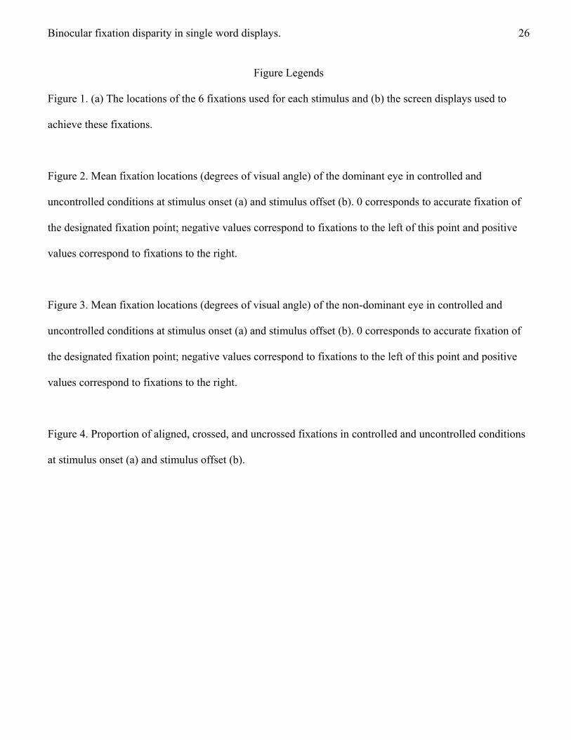
Figure 1. (a) The locations of the 6 fixations used for each stimulus and (b) the screen displays used to
achieve these fixations.
Figure 2. Mean fixation locations (degrees of visual angle) of the dominant eye in controlled and
uncontrolled conditions at stimulus onset (a) and stimulus offset (b). 0 corresponds to accurate fixation of
the designated fixation point; negative values correspond to fixations to the left of this point and positive
values correspond to fixations to the right.
Figure 3. Mean fixation locations (degrees of visual angle) of the non-dominant eye in controlled and
uncontrolled conditions at stimulus onset (a) and stimulus offset (b). 0 corresponds to accurate fixation of
the designated fixation point; negative values correspond to fixations to the left of this point and positive
values correspond to fixations to the right.
Figure 4. Proportion of aligned, crossed, and uncrossed fixations in controlled and uncontrolled conditions
at stimulus onset (a) and stimulus offset (b).

Binocular fixation disparity in single word displays. 27
Figure 1
a
1t2a3b4l5e6
b Fixation point
.
Location 1 table
Location 2 table
Location 3 table
Location 4 table
Location 5 table
Location 6 table
Binocular fixation disparity in single word displays. 28
Figure 2
Fig. 2a
Fig. 2b
0%
20%
40%
60%
80%
100%
-1 -0.75 -0.5 -0.25 0 0.25 0.5 0.75 1
% T
ria
ls
Eccentricity
Dominant Eye Controlled Dominant Eye Uncontrolled
0%
20%
40%
60%
80%
100%
-1 -0.75 -0.5 -0.25 0 0.25 0.5 0.75 1
% T
ria
ls
Eccentricity
Dominant Eye Controlled Dominant Eye Uncontrolled
STIMULUS
ONSET
STIMULUS
OFFSET
Binocular fixation disparity in single word displays. 29
Figure 3
Fig. 3a
Fig. 3b
0%
20%
40%
60%
80%
100%
-1 -0.75 -0.5 -0.25 0 0.25 0.5 0.75 1
% T
ria
ls
Eccentricity
Dominant Eye Controlled Dominant Eye Uncontrolled
0%
20%
40%
60%
80%
100%
-1 -0.75 -0.5 -0.25 0 0.25 0.5 0.75 1
% T
ria
ls
Eccentricity
Dominant Eye Controlled Dominant Eye Uncontrolled
STIMULUS ONSET
STIMULUS OFFSET
Binocular fixation disparity in single word displays. 30
Figure 4
Fig. 4a
Fig. 4b
0%
20%
40%
60%
80%
100%
Controlled Uncontrolled
% T
ria
ls
Fixation Control
Aligned Crossed Uncrossed
0%
20%
40%
60%
80%
100%
Controlled Uncontrolled
% T
ria
ls
Fixation Control
Aligned Crossed Uncrossed
STIMULUS ONSET
STIMULUS OFFSET