binding and internalization of herpes simplex virus-antibody complexes by polymorphonuclear...
DESCRIPTION
immune enhancement, antiviralsTRANSCRIPT

Journal of Medical Virology 20:151-163 (1986)
Binding and Internalization of Herpes Simplex Vi rus-Ant i body Complexes by Polymorphonuclear Leukocytes Jerry W. Smith, James R. Jachimowicz, and Eve L. Bingham
Department of Ophthalmology, University of Michigan, Ann Arbor, Michigan (J. W S., E. L. B.); Difco Laboratories, Detroit, Michigan (J. W S.); Department of Microbiology and Immunology, Louisiana State University, New Orleans, Louisiana (J. R. J.)
We studied the interactions between rabbit polymorphonuclear leukocytes (PMN) and the RE strain of herpes simplex virus type 1 (HSV-1) to determine better the role of inflammatory cells in herpetic stromal keratitis. PMN were found to be nonpermissive for HSV replication and were unable to bind virus in the absence of antibody. However, PMN did bind and internalize HSV-antibody complexes in vitro as was demonstrated visually by electron microscopic studies and quantita- tively by measurement of activity associated with radiolabeled HSV-antibody complexes. Virus used for immune complex formation was labeled with either 12’Iodine or 35S-methionine. In some experiments, anti-HSV IgG used for immune complex formation was labeled with 12’Iodine before incubation with virus. Use of all three radiolabeling approaches resulted in the same general pattern of binding, indicating a requirement for both antibody and virus for interaction with PMN. The activity associated with PMN was increased by preincubation with complement. The results suggest an active role for PMN in controlling HSV infection through their ability to bind and ingest virus-antibody complexes.
Key words: herpes simplex virus type 1, polymorphonuclear leukocyte, antigen-antibody complex
INTRODUCTION
Traditionally, the most important cells involved in antiviral immunity have been thought to be those comprising the lymphocyte-macrophage defense system. Al- though polymorphonuclear leukocytes (PMN) have been shown to function as primary effector cells in acute inflammatory responses to bacterial and mycotic infections, there is increasing evidence to suggest PMN play an important role in antiviral defense.
PMN have been observed in the inflammatory infiltrates in specific infections; for example, the cerebrospinal fluid of children with viral meningitis [Feigin and
Accepted for publication April 21, 1986.
Address reprint requests to Dr. J.W. Smith, Department of Ophthalmology, Kellogg Eye Center, 990 Wall St., Ann Arbor, MI 48105.
0 1986 Alan R. Liss, Inc.

152 Smith, Jachimowicz, and Bingham
Shakelford, 19731 and the cutaneous lesions of varicella zoster infections [Stevens et al, 19751. In herpetic stromal keratitis, a disease characterized by corneal opacity and scarring, an infiltrate consisting mainly of PMN and macrophages persists in the limbus and later the cornea for 4-5 days postinfection [Metcalf and Reichert, 19791.
A possible role for PMN in lower respiratory tract infections caused by respi- ratory syncytial virus (RSV) has been suggested by demonstrating in vitro activation of oxidative and arachidonic acid metabolism of PMN with RSV immune complexes [Kaul et al, 1981; Faden et al, 19831.
Although interactions that occur between host phagocytes and herpes simplex virus (HSV) during infection have not been extensively investigated, it has been assumed that the known mechanisms of antimicrobial immunity mediated by PMN participate in the activation and clearance of virus and virus-antibody complexes [Notkins et al, 1970; Theofilopoulos and Dixon, 1979; Sedlacek, 19801. Previous studies in our laboratory, using chemiluminescence as an indicator of membrane perturbation and/or phagocytosis, suggest that PMN exert anti-HSV activity in the presence of specific antibody, but the virus alone has no effect [Bingham et al, 19851. In an effort to clarify further the interactions between PMN and HSV in ocular infection, we studied the ability of PMN to mediate clearance of virus and virus- antibody complexes. The rabbit was used for our studies because the clinical disease in this model parallels the sequence of disease in human herpes keratitis [Meyers- Elliot et al, 19831. The parameters addressed were: (1) the permissiveness of PMN to HSV replication, (2) the ability of PMN to bind and ingest HSV, and (3) the ability of PMN to bind and phagocytize HSV-antibody complexes.
MATERIALS AND METHODS Preparation of PMN
Blood was collected from adult New Zealand white rabbits into a syringe containing heparin (20 units/ml blood). PMN were separated from whole blood by modification of the method described by Boyum [ 19681. Briefly, 10 ml of heparinized blood was diluted 1 :4 with Hanks balanced salt solution (HBSS), layered on a Ficoll- Hypaque gradient, and centrifuged for 40 min at 400 g. The resulting erythrocyte- PMN pellet was resuspended in 20 ml of autologous plasma and 8 ml of a 4.5% Dextran T-500 solution and incubated for 60 min at 37°C.
Following incubation, the PMN-rich supernatant was collected and spun for 10 min at 300 g. Residual erythrocytes were lysed with ACK buffer (30 mM NH4C1, 2 mM KHC03, 0.02 mM disodium EDTA). PMN purity was greater than 95% as assessed by Wrights stain and PMN viability exceeded 90% as determined by trypan blue exclusion.
Preparation of Virus
The RE strain of HSV-1 was used. This strain was originally isolated from a human corneal lesion and has been used in studies of herpetic stromal disease in rabbits without a high incidence of mortality from encephalitis. It produces a spectrum of disease in rabbits comparable to that seen in humans [Irvine and Kimura, 1967; Metcalf et al, 1976; Wander et al, 19801.
The RE strain was grown in SIRC (Staaten Serum Institut Rabbit Cornea) monolayers with minimal essential medium (MEM) and 5 % fetal calf serum (FCS) at

Binding of HSV-Ab Complexes by HSV 153
37°C. When cytopathological changes involved 100% of the monolayer, culture fluid from infected cells was clarified by two cycles of low speed centrifugation (500 g for 15 min). The resulting supernatant was precipitated with polyethylene glycol (PEG) as described by Powell and Watson [I9751 to obtain an enriched population of enveloped virus.
After an overnight incubation, the HSV precipitate was layered over a continu- ous 20-60% sucrose gradient and centrifuged at 85,000 g for 60 min. The virus band was collected, repelleted, rebanded on a sucrose gradient, and resuspended in MEM supplemented with 2% FCS and stored at -70°C. The PEG-purified HSV prepara- tion had a titer of 8 X lo7 plaque forming units (PFU)/ml in Vero cells.
Virus Replication in PMN
Peripheral blood PMN from normal rabbits were suspended in RPMI-1640 medium with 20% rabbit serum and seeded into 24-well tissue culture plates (5 x lo5 PMN/well). After 60 min at 37"C, PMN cultures were incubated with HSV until harvest or exposed to virus 2 hr, washed, and overlaid with fresh medium. Culture supernatants were harvested at various time intervals up to 96 hr. Fluids were clarified by centrifugation (500 g for 10 min) and quantitated for plaque forming units on Vero cell monolayers using 1 % agarose in MEM as the overlay medium.
Virus Adsorption to PMN
Both rabbit peripheral blood PMN and Vero cells were adjusted to a concentra- tion of 2 X lo6 cells/ml in RPMI-1640 supplemented with 10% rabbit serum. We added 0.5 ml of cells or medium alone to wells of a 24-well plastic tissue culture plate and mixed with 0.1 ml of HSV at 22°C. Virus was diluted to achieve input multiplicities of infection (MOI) of 10 and 1. At designated times after addition of virus (5, 15, 30, and 60 min) culture fluid was harvested, clarified by centrifugation (500 g for 10 min), and quantitated by plaque assay in Vero cells.
Purification of IgG
Antiserum to the RE strain of HSV-1 was produced in New Zealand white rabbits weighing 2-3 kg. Each eye was injected intrastromally with 0.02 ml of virus preparation containing lo6 PFU.
At 28 days postinfection, animals were exsanguinated and the blood allowed to clot at room temperature. The serum was separated by one cycle of centrifugation and then heated for 30 min at 56°C to inactivate complement.
IgG was precipitated from immune serum with a neutralization titer of 1024 by the addition of ammonium sulfate and then separated from other charged serum proteins by ion exchange chromatography on DEAE cellulose [Garvey et al, 19771. Peak protein fractions from the column were pooled and concentrated on a PM-10 membrane (Amicon Corp., Lexington, MA). The protein content of purified IgG was determined by absorbance at 280 nm and purity analysis done by Ouchterlony immunodiffusion.
Radiolabeling of Purified HSV and IgG
Purified HSV was radiolabeled with '2510dine (New England Nuclear, Boston, MA) either by the lactoperoxidase method described by Fenger et a1 [ 19781 or through

154 Smith, Jachimowicz, and Bingham
metabolic incorporation of the amino acid 35S-methionine [Eberle and Courtney, 19801.
Purified IgG was radiolabeled by the chloramine-T method of McConahey and Dixon [ 19661 and by the lactoperoxidase technique of Marchalonis [ 19691, modified by Kozel and McGaw [ 19791.
Preparation of Virus-Antibody Complexes
Purified radioiodinated HSV (1.3 m g / d protein) was combined in equal parts with either a 1 : 10 dilution of rabbit antiserum to HSV or commercial rabbit serum that showed no antibody to HSV. Following a 30-min incubation at 37"C, virus- antibody complexes and free virus were pelleted by centrifugation (85,000 g for 60 min), washed twice with MEM plus 2% FCS, and stored at -70°C. Immune complexes containing 3'S-HSV were made, washed, and stored in a similar manner.
Radioiodinated IgG (1 mg/ml) was prepared from rabbit antiserum to HSV and combined in equal volumes with purified virus. The mixture was incubated for 30 min at 37°C in a shalung waterbath and then centrifuged at 1000 g for 30 min to sediment the insoluble complexes. The complexes were washed twice, resuspended in MEM plus 2% FCS, and stored at -70°C.
Immune Complex Binding Assay
The uptake of radiolabeled complexes by rabbit PMN was measured by a modification of the coverslip assay of Smith and Rommel [ 19771. In this assay system, 12 mm round #1 thickness coverslips were cleaned with acetone and placed in 16 mm wells of a 24-well tissue culture plate. PMN in RPMI-1640 supplemented with 20% normal rabbit serum were seeded onto the coverslip (5 X 10' PMN/coverslip) and allowed to adhere for 1 hr at 37°C in a 5% C02 atmosphere. Control (blank) coverslips were treated with medium plus rabbit serum. Following incubation, each coverslip was washed with medium to elute nonadherent cells and serum components.
We added 50 pl of radiolabeled complexes to each coverslip. After a 10-min incubation at 37"C, the coverslips were removed from wells, washed four times with medium, drained dry with a tissue, and inserted into a bio-vial (Beckman). Iodinated samples were counted on a Biogamma counter (Beckman) to quantitate coverslip associated radioactivity. Quantitation of 35S-radioactivity was accomplished by tissue solubilization and liquid scintillation spectrometry as described by Smith et a1 [ 19791.
Electron Microscopy
Complexes formed by combining PEG concentrated HSV and ammonium sul- fate precipitated IgG were incubated with rabbit PMN for 5 min at 37°C. In preparation for thin sectioning, PMN were washed twice with Tyrode's solution (pH 7.2), fixed in 1% glutaraldehyde, postfixed in osmium tetroxide, dehydrated in ethanol, and embedded in epoxy resin (Araldite). Thin sections were stained with uranyl acetate and lead citrate (pH 7.0) and examined in a Phillips 201 electron microscope.
RESULTS Permissiveness of Rabbit Peripheral Blood PMN to HSV Replication
Studies were done to determine if the interaction between HSV and PMN results in viral replication. The findings in Figure 1 indicate that no detectable replication of

Binding of HSV-Ab Complexes by HSV 155
"1
96 12 2 24 48
HOURS POST A D D I T I O N OF VIRUS
Fig. 1. Replication of HSV in PMN. PMN from peripheral blood of normal rabbits were allowed to adhere to bottoms of 24-well plastic tissue culture plates (5 x lo5 cells/well) before exposure to infectious virus (MOI of 100). Tissue culture fluid was harvested at various times after addition of virus. The titer of infectious HSV was determined by plaque assay under agarose on Vero cell monolayers. Results are expressed as the mean of triplicate cultures. Symbols: A medium + HSV; 0 PMN + HSV incubated for 2 hr at 37°C and washed to remove HSV inoculum; 0 PMN + HSV incubated together at 37°C.
HSV occurs in rabbit PMN. Cultures exposed to infectious virus for 2 hr or longer showed no increase in virus yield. Slight decreases in virus titer were observed in all samples examined beyond 2 hr. Both PMN exposed to virus and that incubated with medium alone exhibited similar patterns of loss of infectivity. Electron microscopy revealed no evidence of permissive or abortive infection in PMN examined 24-48 hr after addition of HSV (data not shown).
Binding of HSV to Rabbit Peripheral Blood PMN To determine if rabbit PMN bound or absorbed HSV, experiments were per-
formed in which infectious virus was exposed to PMN in culture. Supernatant fluids were examined for virus at various time intervals. Results shown in Figure 2 indicate no adsorption or binding of HSV to PMN. Supernatant fluids contained the same amount of infectious virus before and after incubation with PMN for both MOI's examined, 10 and 1. Vero cell monolayers served as positive binding controls. Decreases in virus titer were observed in supernatants from Vero cells as early as 5 min after exposure to HSV.
Binding of HSV-Antibody Complexes to PMN Using Radiolabeled Virus Preparations
In an effort to determine if virus-antibody complexes interact with PMN, experiments were performed in which enveloped HSV, either externally labeled with '''Iodine or metabolically labeled with 35S-methionine, was exposed to immune serum and the resulting complexes were assayed for binding. Results in Table I show that enveloped '251-HSV incubated with normal rabbit serum was bound to PMN at

156 Smith, Jachimowicz, and Bingham
Lz
E
4 5s I I
' I I I I 5 1 5 M 60
MINUTES POST ADDITION OF V IRUS
Fig. 2. Binding of HSV to PMN. Infectious HSV was added to suspensions of either 2 X lo6 rabbit PMN or 2 x lo6 Vero cells at MOI's of 10 and 1. The cultures were continuously agitated and at designated time intervals aliquots were collected. Culture fluids were clarified of cells by centrifugation at 500 fig and screened for reduction in HSV titer by plaque assay under agarose on Vero cells. Results are expressed as mean values of triplicate samples. MOI of 10: A medium + HSV; 0 PMN + HSV;
Vero + HSV. MOI of 1: A medium + HSV; 0 PMN + HSV; 0 Vero + HSV.
TABLE I. Binding of Virus-Antibody Complexes to PMN Using Radiolabeled HSV Preparations
Reaction mixture Activity Cells ComDlex (CPMY
None Iz5I-HSV + NRSb 980 + 82 None Iz5I-HSV + Ab 1446 k 162 PMN IZ5I-HSV + NRS 4591 + 171 PMN Iz5I-HSV + Ab 14942 k 1001 None 35S-HSV + NRS 105 k 10 None 35S-HSV + Ab 602 + 54 PMN 35S-HSV + NRS 2750 244 PMN 35S-HSV + Ab 6516 f 595
aResults represent the mean of sextuplicate coverslips + SEM. bNRS, normal rabbit serum.
greater than fourfold the amount of activity associated with control coverslips. Addition of complexes of '251-HSV and HSV specific antibody produced activity tenfold greater than blank coverslips and threefold greater than that associated with PMN and virus exposed to nonimmune serum.
The same pattern of response was seen when 35S-HSV was substituted for 1251- HSV in immune complexes. Approximately the same ratios of activity were observed. Immune complexes formed using 35S-HSV antibody demonstrated the highest activity associated with PMN.
Binding of HSV-Ab Complexes by HSV 157
Binding of Virus-Antibody Complexes to PMN Using Radiolabeled Anti-HSV IgG
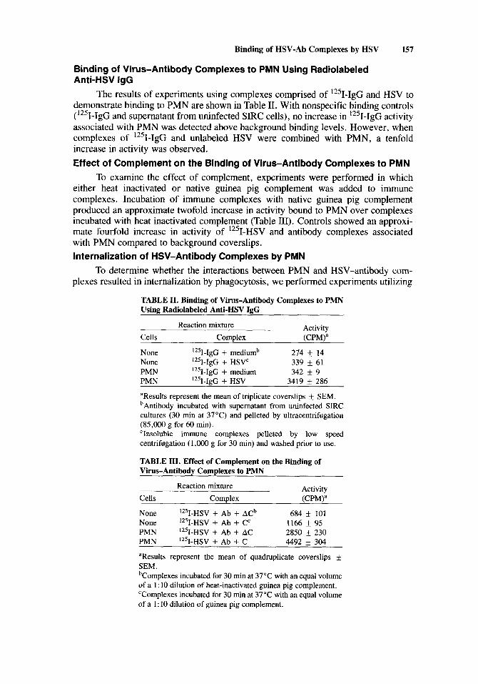
The results of experiments using complexes comprised of '251-IgG and HSV to demonstrate binding to PMN are shown in Table 11. With nonspecific binding controls ( '251-IgG and supernatant from uninfected SIRC cells), no increase in '251-IgG activity associated with PMN was detected above background binding levels. However, when complexes of '251-IgG and unlabeled HSV were combined with PMN, a tenfold increase in activity was observed. Effect of Complement on the Binding of Virus-Antibody Complexes to PMN
To examine the effect of complement, experiments were performed in which either heat inactivated or native guinea pig complement was added to immune complexes. Incubation of immune complexes with native guinea pig complement produced an approximate twofold increase in activity bound to PMN over complexes incubated with heat inactivated complement (Table 111). Controls showed an approxi- mate fourfold increase in activity of 1251-HSV and antibody complexes associated with PMN compared to background coverslips. Internalization of HSV-Antibody Complexes by PMN
To determine whether the interactions between PMN and HSV-antibody com- plexes resulted in internalization by phagocytosis, we performed experiments utilizing
TABLE 11. Binding of Virus-Antibody Complexes to PMN Using Radiolabeled Anti-HSV IgG
Reaction mixture Activity Cells Comolex (CPM)a
None 1251-IgG + mediumb 274 k 14 None "'I-IgG + HSVC 339 +_ 61 PMN Iz5I-IgG + medium 342 k 9 PMN Iz5I-IgG + HSV 3419 f 286
aResults represent the mean of triplicate coverslips k SEM. bAntibody incubated with supernatant from uninfected SIRC cultures (30 min at 37°C) and pelleted by ultracentrifugation (85,000 g for 60 min). 'Insoluble immune complexes pelleted by low speed centrifugation (1,OOO g for 30 min) and washed prior to use.
TABLE 111. Effect of Complement on the Binding of Virus-Antibodv Comolexes to PMN
Activity Cells Comulex (CPM\a
Reaction mixture
None 1251-HSV + Ab + ACb 684 f 101
PMN I2%HSV + Ab + AC 2850 k 230 PMN IZ5I-HSV + Ab + C 4492 k 304
aResults represent the mean of quadruplicate coverslips & SEM. bComplexes incubated for 30 min at 37°C with an equal volume of a 1 : 10 dilution of heat-inactivated guinea pig complement. 'Complexes incubated for 30 min at 37°C with an equal volume of a 1 : 10 dilution of guinea pig complement.
None 1251-HSV + Ab + Cc 1166 +_ 95

158 Smith, Jachimowicz, and Bingham
electron microscopy. Figure 3 shows the results of incubating virus-antibody com- plexes with PMN. Enveloped HSV can clearly be seen within phagocytic vacuoles and attached to the PMN cytoplasmic membrane. Experiments using HSV incubated either alone or after exposure to nonimmune serum resulted in no visible internaliza- tion of virions (data not shown).
Fig. 3. Internalization of HSV-antibody complexes by PMN. The arrows denote complete enveloped HSV particles that are attached to the cytoplasmic membrane and internalized within the phagocytic vacuoles of rabbit PMN. Photograph was taken 5 min after addition of virus-antibody complexes to PMN. Magnification, X36,OOO.

Binding of HSV-Ab Complexes by HSV 159
DISCUSSION
Herpetic stromal keratitis causes corneal damage that may result in blindness. At present, it is not clear whether PMN induce, mediate, or contribute to the pathogenesis of the disease in vivo.
Enhancement of HSV virulence may result from the ability of the virus to infect, replicate, and persist in phagocytic cells. Peritoneal exudate macrophages from strains of mice susceptible to HSV infection in vivo have been shown to support the replication of HSV [Brucher et al, 19841. It has also been shown that HSV [Daniels et al, 19781, rubella [Van Deg Logt, 19801, and dengue-2 [Brandt et al, 19791 viruses are capable of replicating in human monocytes and macrophages. Our laboratory has reported HSV permissiveness following mitogenic stimulation of human lymphocytes [Plaeger-Marshall and Smith, 19781. In addition, we have demonstrated limited permissiveness of rabbit macrophages to HSV infection [Plaeger-Marshall et al, 19811. Rabbit monocytes, on the other hand, showed no detectable interaction with virus. In addition to in vitro studies, fluorescent antibody probes have suggested the presence of HSV or HSV antigens within PMN from infiltrates associated with herpetic keratitis [Meyers and Pettit, 1973; Meyers-Elliot et al, 1980; Meyers-Elliot and Chitjian, 19811.
In the present study of HSV-PMN interactions, no replication or adsorption of HSV to PMN was observed. Regarding replication, no increase in virus titer was seen in PMN exposed to HSV. Samples of PMN taken 2 4 4 8 hr after exposure to HSV and examined by electron microscopy showed no evidence of complete virions or abortive infection. Binding experiments using PMN mixed with HSV at two MOI’s revealed no measurable adsorption, whereas Vero cell controls gave positive binding results under the same conditions. The data presented suggest that rabbit PMN do not recognize HSV by itself, supporting results from studies in this laboratory using chemiluminescence as an assay system [Bingham et al, 19851. Explanations for PMN nonpermissiveness could be the phagocyte’s lack of receptors, short life span, and limited capacity for protein synthesis, all of which my reflect differences in degrees of cellular maturation and differentiation.
It has been well established that PMN ingest and kill bacteria and fungi [Hilger and Danley, 1980; Klebanoff, 1975; Schuit, 19791. In antiviral defense, PMN are presumed to be active in the clearance of virus-antibody complexes through phago- cytosis following recognition by receptors that bind to antibody and complement [Allison, 19741. Opsonins, such as IgG, interact with the material to be phagocytized, adding a host protein coating that facilitates internalization via the PMN Fc receptor [Eden et al, 1973; Phillips-Quagliata et al, 1969; Theofilopoulos et al, 1974; Wiggins and Cochrane, 19811.
A variety of techniques have been employed to quantitate phagocytosis by PMN. Some methods determine the number of particles removed from the culture medium; others count the number of particles that appear within phagocytes. The uptake of radioactive protein from the suspending medium is another approach to measuring phagocytosis [Chang, 19691. Using this last approach, Ward and Zvaifler [ 19731 described a method for the quantitative measurement of phagocytosis by neutrophils using radiolabeled immune complexes. Because of particle size, virus uptake by cells cannot be visually assessed by light microscopy. Therefore, the ability to assay phagocytosis with labeled immune complexes becomes a particularly useful tool when studying the interactions between PMN and viruses.

160 Smith, Jachimowicz, and Bingham
In our current study using radiolabeled complexes, we have demonstrated that HSV-antibody complexes bind to PMN. We employed complexes containing either '251-HSV, 35S-HSV, or 1251-IgG and obtained similar patterns of response, thus minimizing the chance that the results observed were an artifact of assay conditions. Increased binding occurred using HSV labeled externally with 12510dine or metaboli- cally with 35S-methionine for the formation of antigen-antibody complexes. When radiolabeled antibody (1251-IgG anti-HSV) was combined with virus in immune complexes, there was a dramatic increase in the binding activity to PMN in contrast to the low activity produced by unaggregated 12510dine labeled HSV-IgG controls. In an unrelated study, using similar procedures, Shinomaya and Koyama [ 19761 found that '251-IgG alone bound minimally to guinea pig macrophages whereas '251-IgG combined with hapten was rapidly bound and internalized.
The addition of complement to virus-antibody complexes is presumed to en- hance neutralization and immune clearance through enlargement of viral aggregates. Studies have shown that immune complexes containing complement bind to phago- cytes with greater affinity than those without complement [Kijlstra et al, 1979; Mantovani et al, 1972; Van Snick and Masson, 19781. Our data support the concept that complement increases binding of immune complexes to PMN. The addition of active complement to radiolabeled complexes was found to increase activity associ- ated with PMN by as much as 25 % .
The data presented clearly demonstrate that PMN bind HSV-antibody com- plexes and that the binding activity increases with the addition of complement. To determine whether ingestion as well as adsorption of immune complexes occurs, we employed electron microscopy. This approach resulted in direct visualization of internalized HSV-antibody complexes. Enveloped virions were observed both within phagocytic vacuoles and adherent to the PMN cytoplasmic membrane.
In the initial stages of acute inflammation large numbers of PMN are recruited to destroy and remove the inflammatory stimuli via phagocytosis [Issekutz et al, 19811. The presence of PMN in the corneal stroma during HSV keratitis can be explained by the liberation of proteases from damaged cells, the activation of the complement cascade by antigen-antibody complexes, the release of leukotactic factors by virus-sensitized lymphocytes, and the chemotactic properties of collagenase in the cornea [Meyers and Pettit, 19741.
Gradually, macrophages accumulate at the same locations for further removal of inflammatory stimuli. Macrophages also initiate tissue repair and immunological responses such as antigen presentation to T lymphocytes. Phagocytic cells at the sites of inflammation bind or ingest immune complexes formed during the course of the disease. Occasionally the removal of such complexes is not efficiently achieved resulting in the subsequent synthesis and release of more inflammatory mediators by PMN and macrophages [Davies and Bonney, 19811.
The pathogenesis of herpetic stromal keratitis remains unclear. It is not fully understood how much of the disease process is due to viral replication and how much is due to the immune response to viral antigens. Our observations that PMN are capable of binding and ingesting HSV-antibody complexes suggest that PMN play a beneficial role in the clearance of virus during herpetic keratitis.
Previous reports [Dawson and Togni, 1976; Pettit and Meyers-Elliot, 19731 have suggested a major role for specifically sensitized lymphocytes in chronic recur- rent corneal inflammation. Our data do not exclude such a possibility. It is clear that

Binding of HSV-Ab Complexes by HSV 161
a combination of inflammatory cells are active during the course of herpetic keratitis and that a complete understanding of their interrelationships and mechanisms involved in immune destruction will require further investigation.
ACKNOWLEDGMENTS
This work was supported by Public Health Service grant EY05105 from the National Eye Institute.
REFERENCES
Allison AC (1974): Interaction of antibody, complement, and various cell types in immunity against viruses and pyogenic bacteria. Transplantation Review 19:3-55.
Bingham EL, Fenger TW, Sugar A, Smith JW (1985): Dependence on antibody for induction of chemiluminescence in polymorphonuclear leukocytes by herpes simplex virus. Investigative Ophthalmology and Visual Science 26: 1236-1243.
Boyum A (1968): Isolation of mononuclear cells and granulocytes from human blood. Scandinavian Journal of Clinical Laboratory Investigation 21 :77-89.
Brandt WE, McCown JM, Top FH, Bancroft WH, Russell PK (1979): Effect of passage history on dengue-2 virus replication in subpopulations of human leukocytes. Infection and Immunity
Brucher J, Domke I, Schroder CH, Kirchner H (1984): Experimental infection of inbred mice with herpes simplex virus. Archives of Virology 82:83-93.
Chang Y (1969): Studies on phagocytosis. I. Uptake of radioiodinated (I3’I) human serum albumin as a measure of the degree of phagocytosis in vitro. Experimental Cell Research 54:42-48.
Daniels CA, Kleinerman ES, Snyderman R (1978): Abortive and productive infections of human mononuclear phagocytes by type 1 herpes simplex virus. American Journal of Pathology 91: 119- 136.
Davies P, Bonney RJ (1981): Some basic mechanisms of inflammatory responses. In Suran A, Gery I, Nussenblatt RB (eds): “Immunology of the Eye; Workshop 111. ” Arlington, VA: Information Retrieval Inc., pp 273-290.
Dawson CR. Togni B (1976): Herpes simplex eye infections: Clinical manifestations, pathogenesis and management. Survey of Ophthalmology 21: 121-135.
Eberle R, Courtney RJ (1980): Preparation and characterization of specific antisera to individual antigens comprising the major glycoprotein region of herpes simplex virus type 1. Journal of Virology
Eden A, Bianco C, Nussenzweig V (1973): Mechanisms of binding soluble immune complexes to lymphocytes. Cellular Immunology 7:459-473.
Faden H, Hong JJ, Ogra PL (1983): Activation of oxidative and arachidonic acid metabolism in neutrophils by respiratory syncytial virus antibody complexes: Possible role in disease. Journal of Infectious Disease 148: 110-116.
Feigin RD, Shakelford PG (1973): Value of repeat lumbar puncture in differential diagnosis of menin- gitis. New England Journal of Medicine 289
Fenger TW, Smith JW, Howe C (1978): Analysis of ecipitated surface glycoproteins in measles virions and in membranes of infected cells. Journal of Virology 28:292-299.
Garvey JS, Cremer NS, Sussdorf DH (eds) (1977): Ammonium sulfate precipitation. In “Methods in Immunology.” Reading, MA: WA Benjamin Inc., pp 218-219.
Garvey IS, Cremer NS, Sussdorf DH (eds) (1977): DEAE cellulose chromatography. In “Methods in Immunology.” Reading, MA: WA Benjamin Inc., pp 222-226.
Hilger AE, Danley DL (1980): Alteration of polymorphonuclear leukocyte activity by viable Candida albicans. Infection and Immunity 27:714-720.
Irvinc AR, Kimura SJ (1967): Experimental stromal herpes simplex keratitis in rabbits. Archives of Ophthalmology 78:654-663.
Issekutz TB, Issekutz AC, Movat HZ (1981): The in-vitro quantitation and kinetics of monocyte migration into acute inflammatory tissue. American Journal of Pathology 103:47-55.
26:534-541.
35:902-9 17.

162 Smith, Jachimowicz, and Bingham
Kaul TN, Faden H, Ogra PL (1981): Effect of respiratory syncytial virus and virus-antibody complexes on the oxidative metabolism of human neutrophils. Infection and Immunity 32:649-654.
Kijlstra A, Vanes LA, Daha MR (1979): The role of complement in the binding and degradation of immunoglobulin aggregates by macrophages. Journal of Immunology 123:2488-2493.
Klebanoff SJ (1975): Antimicrobial systems of the polymorphonuclear leukocyte. In Dayton DH (ed): “The Phagocytic Cell in Host Resistance.” New York: Raven Press, pp 45-59.
Kozel TR, McGaw TG (1979): Opsonization of Cryptococcus neoformans by human IgG: Role of IgG in phagocytosis by macrophages. Infection and Immunity 25:255-261.
Mantovani B, Rabinovitch M, Nussenzweig V (1972): Phagocytosis on immune complexes by macro- phages, different roles for the macrophage receptor sites for complement and IgG. Journal of Experimental Medicine 135:780-792.
Marchalonis JJ (1969): An enzymatic method for trace iodination of immunoglobulins and other proteins. Biochemistry Journal 113:299-305.
McConahey PJ, Dixon FJ (1966): A method of trace iodination of proteins for immunologic studies. International Archives of Allergy 24: 185-189.
Metcalf JF, McNeill JI, Kaufman HE (1976): Experimental disciform edema and necrotizing keratitis in the rabbit. Investigative Ophthalmology 15:979-985.
Metcalf JF, Reichert RW (1979): Histological and electron microscopic studies of experimental herpetic keratitis in the rabbit. Investigative Ophthalmology 18: 1123-1138.
Meyers RL, Pettit TH (1973): The pathogenesis of corneal inflammation due to herpes simplex virus. I. Corneal hypersensitivity in the rabbit. Journal of Immunology 111: 1031-1042.
Meyers RL, Pettit TH (1974): Chemotaxis of polymorphonuclear leukocytes in corneal inflammation: Tissue injury in herpes virus infection. Investigative Ophthalmology 13: 187-197.
Meyers-Elliot RH, Pettit TH, Maxwell WA (1980): Viral antigens in the immune ring of herpes simplex stromal keratitis. American Journal of Ophthalmology 89: 897-904.
Meyers-Elliot RH, Chitjian PA (1981): Immunopathogenesis of corneal inflammation in herpes simplex stromal keratitis: Role of the polymorphonuclear leukocyte. Investigative Ophthalmology and Visual Science 20:784-798.
Meyers-Elliot RH, Chitjian PA, Dethlefs BA (1983): Experimental herpes virus keratitis in the rabbit: Topical versus intrastromal infection routes. Ophthalmic Research 15:240-256.
Notkins AL, Mergenhagen S, Howard R (1970): Effects of virus infections on the functions of the immune response. Annual Review of Microbiology 24:525-538.
Phillips-Quagliata JM, Levine BB, Uhr JW (1969): Studies on the mechanisms of binding immune complexes to phagocytes. Nature 222: 1290-1291.
Plaeger-Marshall S, Smith JW ( 1978): Experimental infection of subpopulations of human peripheral blood leuckocytes by herpes simplex virus. Proceedings of the Society for Experimental Biology and Medicine 158:263-268.
Plaeger-Marshall S, Wilson LA, Smith JW (1981): Permissiveness of rabbit monocytes and macrophages for herpes simplex virus type 1. Infection and Immunity 35:151-156.
Powell KL, Watson DH (1975): Some structural antigens of herpes simplex virus. Journal of General Virology 29: 167-178.
Schuit K (1979): Phagocytosis and intracellular killing of pathogenic yeasts by human monocytes and neutrophils. Infection and Immunity 24:932-938.
Sedlacek HH (1980): Pathophysiological aspects of immune complex disease. Klinische Wochenschrift
Shinomiya T, Koyama J (1976): In vitro uptake and digestion of immune complexes containing guinea pig IgGl and IgG2 antibodies by macrophages. Immunology 30:267-275.
Smith DL, Rommel F (1977): A rapid micro method for the simultaneous determination of phagocyte- microbicidal activity of human peripheral blood leukocytes in vitro. Journal of Immunological Methods 17:241-247.
Smith JW, Torres JE, Holmquist ND (1979): Association of herpes simplex virus (HSV) with cervical cancer by lymphocyte reactivity with HSV-1 and HSV-2 antigens. American Journal of Epide- miology 110: 141-147.
Stevens DA, Ferrington RA, Jordan GW, Merigan TC (1975): Cellular events in zoster vesicles: Relation to clinical course and immune parameters. Journal of Infectious Diseases 131 :509-514.
Theofilopoulos AN, Dixon FJ, Boksich VA (1974): Binding of soluble immune complexes to human lymphoblastoid cells. I. Characterization of the receptors for IgG, Fc, and complement and description of the binding mechanisms. Journal of Experimental Medicine 140:877-894.
58593-605.

Binding of HSV-Ab Complexes by HSV 163
Theofilopoulos AN, Dixon FJ (1979): The biology and detection of immune complexes. Advances in Immunology 28: 89-220.
Van Deg Logt JTM, Van Loon AM, Van Der Veen J (1980): Replication of rubella virus in human mononuclear blood cells. Infection and Immunity 27:309-3 14.
Van Snick J, Masson PL (1978): The effect of complement on the ingestion of soluble antigen-antibody complexes and IgM aggregates by mouse peritoneal macrophages. Journal of Experimental Medicine 148:903-914.
Wander AH, Centifanto YM, Kaufman HE (1980): Strain specificity of clinical isolates of herpes simplex virus. Archives of Ophthalmology 98: 1458-1461.
Ward PA, Zvaifler NJ (1973): Quantitative phagocytosis by neutrophils. I. A new method with immune complexes. Journal of Immunology 111:1771-1776.
Wiggins RC, Cochrane GC (1981) Immune complex mediated biologic effects. New England Journal of Medicine 304518-520.