bilateral stress fracture of the tibia diagnosed by ultrasound. a case report
TRANSCRIPT
Journal of Ultrasound (2012) 15, 130e134
Available online at www.sciencedirect.com
journal homepage: www.elsevier .com/locate/ jus
Bilateral stress fracture of the tibia diagnosed byultrasound. A case report
V. Khy a,b,*, B. Wyssa c, S. Bianchi b
aHotel Dieu d’Amos, Amos, Quebec, CanadabCIM - Cabinet Imagerie Medicale, Geneve, SwitzerlandcChemin de Beau-Soleil, Geneve, Switzerland
KEYWORDSFatigue fracture;Sonography;Ultrasound;Stress fracture.
* Corresponding author. Hotel Dieu dAmos, Quebec J9t 2S2, Canada.
E-mail address: [email protected] (V.
1971-3495/$ - see front matter ª 201doi:10.1016/j.jus.2011.09.002
Abstract We report the case of a 35 years old patient presenting with bilateral pain in themedial aspect of both knees. Ultrasound examination revealed hyperechoic appearance ofthe subcutaneous tissue and periarticular soft tissue bilateral. Color Doppler studies showedsignificant vascular signals at the surface of the tibial plateaux. US appearance, together withclinical findings, suggested a diagnosis of simultaneous bilateral fatigue fracture. An MRIconfirmed the diagnosis and the patient’s symptoms resolved with rest. US may be a usefulimaging tool in the diagnosis of stress fracture.
Sommario Presentiamo il caso di un paziente di 35 anni con dolore bilaterale nella partemediale delle ginocchia. L’ecografia evidenziava ipoecogenicita del tessuto sottocutaneo edelle parti molli periarticolari bilateralmente. L’esame con color Doppler mostrava significativisegnali vascolari in corrispondenza del piatto tibiale. L’aspetto ecografico, associato ai segniclinici, suggeriva la diagnosi di simultanea frattura da fatica, bilaterale. La risonanza magne-tica confermava la diagnosi e i sintomi del paziente regredivano con il riposo. L’ecografia puoessere un’utile metodica nella diagnosi frattura da stress.ª 2011 Elsevier Srl. All rights reserved.
Introduction
Stress fractures (SF), bone failures secondary to submax-imal chronic overloads, can be divided depending on theirpathogenesis in fatigue (FF) and insufficiency fractures (IF).
’Amos, 622 4ieme rue ouest,
Khy).
1 Elsevier Srl. All rights reserved.
FF result from repeated abnormal stresses on a normalbone and are commonly seen in both amateur and high-level athletes. Their incidence is estimated as high as 61%in certain group of athletes [1e5]. IF are seen when normalstresses are applied to bones weakened by a variety ofsystemic disorders such as osteoporosis or rheumatoidarthritis.
Although they can affect the upper extremity, FF mostlyinvolve the lower extremity with prevalence for the tibia,the neck of the femur, the metatarsal bones and thecalcaneus [1,2,6,7]. Patients present with local pain
Bilateral stress fracture of the tibia diagnosed by ultrasound 131
typically accentuated by activity and diminished by rest.Physical examination can only show pain at palpation, whenthe fracture involves deep-seated bones, or also reveallocal swelling and redness if a superficial bone is fractured.FF can be suspected clinically but an imaging technique isusually required to confirm the suspicion and assess theseverity of the fracture. Imaging studies allow earlydetection of the fracture, instauration of an adequatetreatment thus reducing morbidity.
Conventional radiographs are not enough sensitive sincepresence of cortical discontinuity or formation of a bonecallus can take weeks to be evident [8e10]. The sensitivityof bone scan has been demonstrated in various studies butits specificity is low [4,9e11]. Computed tomography (CT)scan can show FF, but due to utilisation of radiation it is lessfavoured than the magnetic resonance imaging (MRI). MRI isthe examination of choice in the early detection of SF butits use is limited by lack of access and associated cost [3]. Inthe past few years, various authors have reported ultra-sound (US) as an inexpensive, non-invasive and readymodality with high sensitivity for the investigation of SF ofthe metatarsal [10,11]. Reports of SF affecting other bonesthen metatarsals diagnosed by US are rare [6]. NeverthelessUS can be useful in diagnosing SF if the examination isaccurately performed by an expenciered sonologist witha high degree of clinical suspicion. We report the case ofa young patient with a bilateral simultaneous FF of thetibial plateau diagnosed by US and confirmed by MRI.
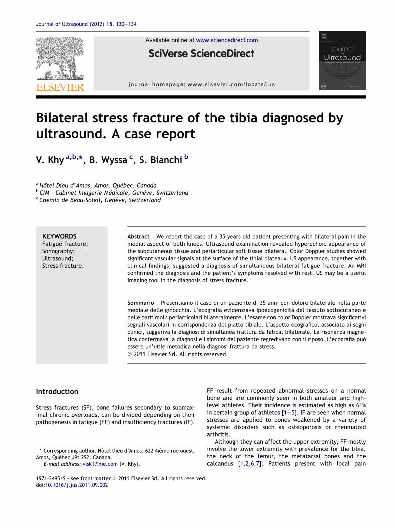
Figure 1 Coronal (top) and coronal colour Doppler (bottom)sonograms obtained over the medial aspect of the left knee.The grey-scale image shows thickening of the soft tissuelocated between the tibial cortex (white arrowhead) and theinferior part of the medial collateral ligament (black arrow-heads). Colour Doppler shows flow signals due to local hyper-vascular changes.
Case report
A 35-year-old Caucasian man (172 cm/90 kg, body massindex Z 30,4) presented with pain in the medial aspect ofboth knees. The patient had a sedentary job and regularlyengaged in swimming and hiking. History did not showedany systemic disorders/treatment or risk factors predis-posing to osteoporosis. Three weeks earlier, he starteda jogging program (one-half hour/day, seven days a week)in an attempt to loose weight. After 10 days he reportedbilateral medial knee pain first noted during running andthen persisting at rest. Clinical examination revealedlocalized bilateral pain and tenderness on the lower medialaspect of the knees, more evident at the insertion site ofthe hamstrings. The range of motion of both knees wasnormal (135� of flexion, 0�of extension) and there was nosign of joint or periarticular inflammation. There wasa varus of 10� for both knees. Previous blood tests werereferred as normal. Based on clinical findings an initialdiagnosis of hamstring tendinopathy was made anda conservative treatment with non-steroidal anti-inflam-matory drugs was prescribed and US was obtained toconfirm the clinical diagnosis.
A musculoskeletal radiologist with 25 years of experi-ence in US performed the examination using a commerciallyavailable equipment with a linear broadband transducerworking at 5e12 MHz. Axial, longitudinal and oblique grey-scale and colour Doppler images were obtained on themedial aspect of both knees and proximal tibiae in thesupine patient with the knees slightly flexed. A largeamount of gel was deployed. US revealed the presence ofbilateral thickening and hyperechogenicity of the
subcutaneous tissue, paraarticular soft tissues adjacent tothe bone cortex and periosteum (Fig. 1). The hyperechoiccortex of both tibias showed mild irregularity without signof discontinuity. Colour Doppler showed significant vascularsignals of the surface of the tibial plateaux. The pesanserinus region and the medial collateral ligament werenormal on both sides. Examination of the knee joints wasnormal. US appearance, together with clinical findings,suggested a diagnosis of simultaneous bilateral FF of theproximal tibiae. An MRI was then obtained to confirm the USfindings. MRI was obtained, a week after the US examina-tion, on both knees using a 1,5 T equipment and an 8-channels knee dedicated coil. The examination protocolincluded coronal (FOV 16 cm, 18 slices thickness 3 mm TR3000, TE 30) and axial (FOV 15 cm, 18 slices thickness4 mm TR 2990, TE 15) proton density fat suppressed images,sagittal proton density images with (FOV 17 cm, 24 slicesthickness 3.5 mm TR 2685, TE 15) and without (FOV 17 cm,24 slices thickness 3.5 mm TR 5000, TE 30) fat saturationand sagittal proton density images (FOV 15 cm, 18 slicesthickness 4 mm TR 2990, TE 15). On both proximal epiphysisof the tibia MRI showed an irregular hypo intense lineroughly parallel to the joint surface. The lines were evidentin all sequences and were surrounded by an area of bonemarrow oedema appearing hyperintense in PD fat saturatedsequences and slightly hypo intense in PD images (Fig. 2).The appearance was typical of an FF. The rest of theexamination was normal.
The patient was treated by rest (crutches) and analgesictreatment upon pain. Physical examination obtained after 1month showed complete relief of symptoms and signs.Bilateral AeP and lateral radiographs of the knees obtained

Figure 2 Coronal PD fat sat MRI images obtained on both knees. MRI depicts the stress fracture (white arrows) as a low intensity,serpiginous, line surrounded by an ill-defined hyperintense area related to marrow oedema (black arrows). Note also hyperintensity(withe arrowheads) of soft tissues located between the medial collateral ligaments (black arrowheads) and the bone cortex relatedto local inflammation.
132 V. Khy et al.
at this time (Fig. 3) showed a sclerotic subchondral linelocated at the medial aspect of the proximal metaphysis ofboth tibiae. The appearance was typical of FF.
Informed consent was obtained from the patient for thepublication of this case report.
Discussion
Sports injuries have significantly increased in frequency inthe past few years, especially among individuals whoselevel of physical fitness is ill suited to the intensity or thevery nature of the activity undertaken. Although data onthe prevalence of sports injuries seem to be difficult tocollect, it is estimated that approximately 6% of those whoengage in sports require medical care for their injuries[13,14]. Sports injuries can be acute or derive from
Figure 3 A-P standard radiographs of both knee show two rougsurface. This aspect is typical of stress fracture.
excessive (overuse injuries) or not well equilibrated (disuseinjuries) activity such as excessive training in an unpre-pared organism. Overuse-disuse injures affect primarilytendons and muscles but depending on the activity andstress applied can also affect bone resulting in FF.
The main site for SF the sacrum (29.6%), the tibia(16,5%), the femur (9.9%), the metatarsus and the calca-neus [2,8]. In most patients, FF cannot be diagnosed only onthe basis of clinical findings although a detailed history andcareful clinical examination can orient to the diagnosis.Usually an increase in the intensity or duration of physicalactivity previous to the appearance of pain can beretraced. The main clinical finding is localized pain aggra-vated from activity and decreased by rest. An imagingmodality is required to confirm the clinical suspicion and tostart adequate treatment. Conventional radiographs arenot sensitive in diagnosing SF since they are normal in the
hly symmetric sclerotic lines (arrows) parallel to the articular

Bilateral stress fracture of the tibia diagnosed by ultrasound 133
first weeks. The radiographic appearance of SF depends onthe bone affected. In tubular bones, although corticaldiscontinuity is the hallmark of SF, the earliest diagnosticsign is a thin periosteal calcific reaction that can be easilymissed if the diagnosis is not suspected. Later on, with boneremodelling, a bone callus becomes evident. In cancellousbones the first radiographic sign is a sclerotic band of thetrabecular bone due to bone apposition from theendosteum.
A bone scan is a sensitive method for early diagnosis ofa fatigue fracture. The radioactive tracer is incorporatedinto the cells responsible for bone remodelling within 24 hfollowing the fracture. However, this technique is notsufficiently specific because bone turnover can beincreased in a wide variety of others conditions includingtumours, infections or inflammations [4,9,14].
At CT-Scan the bone repair reaction appears as bonysclerosis surrounding the line of fracture. In some studies,this technic is even more sensitive and specific than MRI[11,15].
Due to its mutiplanar capabilities and high tissuecontrast, MRI is the imaging modality of choice for diag-nosing an SF. MRI is sensitive and specific, revealing intra-medullary oedema, the periosteal reaction and the fractureline.
Nevertheless it is expensive and still today barelyaccessible in some countries.
Recent technological advancements have increased thediagnostic possibilities of US in the assessment of muscu-loskeletal system thus widely increasing its utilisation.Wide accessibility, low cost, as well as absence of sideeffects, has increased the popularity of this technic.Although its chief application in musculoskeletal disordersis the assessment of soft tissues, US can help in detectingSF. Banal et al. reported 70% sensitivity for US compared toan MRI for the examination of fractures of the secondmetatarsal [12]. Several studies have described ultrasono-graphic findings of SF (11,14). Five US hallmarks have beendescribed in SF: [1] cortical disruption which is a rare andlate finding [2] posterior shadowing related to periostealthickening. This sign can be seen only in small bone such asthe metatarsals [3] thickening of the periosteum whichappears as a hypoechoic band overlying the hyperechoicbone cortex [4] Increased periosteal colour Doppler flowrelated to local hyperaemia [5] hyperechogenicity of thesurrounding soft tissue which indicates soft tissue oedemaand inflammatory reaction. Doppler abnormalities andhyperechogenicity of the local soft tissue are most impor-tant for establishing an early diagnosis [6,12,15].
Most articles discussing the US appearance of SF havebeen focused on metatarsals [12,16]. Reports on SFaffecting other bones are scanty [6].
The differential diagnosis of periarticular pain of themedial aspect of the knee in sports enthusiasts includestendinopathy, bursitis and sprain of the medial collateralligament. In pur case, these diagnosis were excluded by USthat allowed demonstration of thickening, oedema andhypervascular changes in the periosteum and adjacent softtissues associated with irregularities of the bone cortex.These findings were noted at the level of maximal tender-ness. The US appearance in our patient was not specificbut, considering the clinical findings and the normal
appearance of the paraarticular tendons, was highly sus-pected of an FF. MRI was necessary to establish a definitediagnosis.
Our case shows the importance for the sonologist toperform a comprehensive US examination targeted to allstructures from skin to the bone cortex and the necessity tocorrelate the US findings to clinical appearance. The USfindings of local oedema, thickening and hyperaemia of theperiosteum could be easily overlooked without consideringthe clinical appearance.
In case of suspicion of an FF, when evaluating the bonecortex, the sonologist must be careful in considering smallcortical discontinuities as fractures since these can corre-spond to the entries of feeding arteries as it can bedemonstrated by colour Doppler. At the proximal medialtibia, we’ve found this physiological discontinuity in 15healthy subjects aged from 30 to 50 years 80% (unpublisheddata). Nevertheless the Doppler signal among thesesubjects was not as intense as in our patient, was closelylocalized at the discontinuity and and did not affect theperiosteum and periarticular tissues.
In conclusion we present a case in which US showedbilateral thickening of the periosteum and periarticulartissues of the medial tibia associated with increased localvascularity at Color Doppler and ruled out the clinicalsuspicion of hamstrings tendinitis. The US appearance,togheter with the clinical setting, were high presumptive ofa bilateral FF that was later confirmed by MRI. US whenperformed with an accurate technique of examination canstrongly indicate a diagnosis of FF in the tibia. MRI is thepreferred method of choice to confirm the diagnosis.
Conflict of interest statement
The authors have no conflict of interest to disclose.
Appendix. Supplementary material
Supplementary material associated with this article canbe found, in the online version, at doi:10.1016/j.jus.2011.09.002.
References
[1] Albisetti W, Erugina D, De Bartolomeo O, Tagliabue L,Camerucci E, Calori MG. Stress fractures of the base of themetatarsal bones in young trainee ballet dancers. Int Orthop2010;34:51e5.
[2] Pohl M, Mullineaux D, Milner C, Hamill J, Davis I. Biochemicalpredictors of retrospective tibial stress fractures in runners. JBiomech 2008;41:1160e5.
[3] Raasch WG, Hergan DJ. Treatment of stress fractures: thefundamentals. Clin Sports Med 2006;25:29e36.
[4] Sofka C. Imaging of stress fractures. Clin Sports Med 2006;25:53e62.
[5] Warden S, Burr D, Brukner P. Stress fractures: pathophysi-ology, epidemiology and risk factors. Curr Osteoporos Rep2006;4(3):103e9.
[6] Arni D, Lambert V, Delmi M, Bianchi S. Insufficiency fracture ofthe calcaneum: sonographic findings. J Clin Ultrasound 2009;37:424e7.
134 V. Khy et al.
[7] Matheson GO, Clement B, McKenzie DC, Taunton JE, Lloyd-Smith DR, Macintyre JG. Stress fractures in athletes. A studyof 320 cases. Am J Sports Med 1987;15:46e58.
[8] Krestan C, Hojreh A. Imaging of insufficiency fractures. Eur JRadiol 2009;71:398e405.
[9] Soubrier M, Dubost JJ, Boisgard S, Sauvezie B, Gaillard P,Michel J, et al. Insufficiency fracture. A survey of 60 cases andreview of the literature. Jt Bone Spine 2003;70:209e18.
[10] Patel DS, Roth M, Kapil N. Stress fracture: diagnosis,treatment and prevention. Am Fam Physician 2011;83(1):39e46.
[11] Gaeta M, Minutoli F, Scribano E, Ascenti G, Vinci S,Bruschetta D, et al. CT and MR imaging findings in athleteswith early tibial stress injuries: comparison with bone scin-tigraphy findings and emphasis on cortical abnormalities.Radiology 2005;235:553e61.
[12] Banal F, Grandjbakh F, Foltz V, Goldcher A. Sensitivity andspecificity of ultrasonography in early diagnosis of metatarsalbone stress fractures: a pilot study of 37 patients. J Rheu-matol 2009;36:1715e9.
[13] Dreinhofer KE, Reichel H, Kafer W. Strategies for preventionand management of musculoskeletal conditions. Lower limbpain. Best Pract Res Clin Rheumatol 2007;21:135e52.
[14] Peris P. Stress fractures. Best Pract Res Clin Rheumatol 2003;17(6):1043e61.
[15] Ganiyusufoglu AK, Onat L, Karatoprak O, Enercan M. Ham-zaoglu. Diagnostic accuracy of magnetic resonance imagingversus computed tomography in stress fractures of the lumbarspine. Clin Radiol 2010;65:902e7.
[16] Bodner G, Stockl B, Fierlinger A, Schocke M, Bernathova M.Sonographic findings in stress fractures of the lower limb:preliminary findings. Eur Radiol 2005;15:356e9.