bilateral high division of the brachial artery in one human male
TRANSCRIPT
J. Morphol. Sci., 2011, vol. 28, no. 3, p. 204-207204
Case report
Bilateral high division of the brachial artery in one human male cadaver: case report
Rossi Junior, WC.*, Esteves, A., Simões, JS. and Fernandes, GJM.
Institute of Biomedic Sciences, Federal University of Alfenas – UNIFAL Rua Gabriel Monteiro da Silva, 700, CEP 37130-000, Alfenas, MG, Brazil
*E-mail: [email protected]
Abstract
In general, the brachial artery divides into the radial and ulnar arteries at the level of the cubital fossa (elbow), just proximal to or right over the cubital anastomosis and these arteries are the main responsible for the irrigation of the forearm. After revising an extensive bibliographical literature, we found out that only few authors admit that divisions can appear below the cubital fossa (low division) and at the upper, middle or lower third of the arm (high divisions), but these are considered rare events. While dissecting one male cadaver on an ordinary practical class in the Laboratory of Gross Anatomy, we observed a high division of the brachial artery, located at the level of the superior part of the upper third in both arms, close to the axilla. We decided to report this anatomical variation because a bilateral event such as that is rarer yet.
Keywords: anatomy, cadaver, brachial artery.
1 Introduction
Arteries are muscular and/or elastic cylindrical tubes that conduct blood away from the heart. According to their internal diameter, they can be classified as large-diameter artery (some centimeters to 7 mm); medium-diameter artery (from 7 mm to 2.5 mm); small-diameter artery (from 2.5 mm to 0.5 mm); and arterioles (less than 0.5 mm) (ROUVIÈRE, 1956; DÂNGELO and FATTINI, 2007).
In respect to their elasticity, arteries can enlarge either in length or in width in order to retain the greatest amount of blood volume possible and to keep the blood flow steady (PETROIANU, 1999).
Arteries emit terminal and collateral branches. Terminal branches appear when the main artery divides (bifurcation or trifurcation) and no longer exists, for instance, as the brachial artery divides into the radial and ulnar arteries. Collateral branches occur when the main artery gives off other vessels but still continues ahead as the same vessel (SNELL, 1999).
The brachial artery is the principal artery of the arm, following the axillary artery. Its proximal boundary is at the level of the inferior margin of the teres major muscle and its distal boundary is frequently located at the level of the elbow. Passing beneath the bicipital aponeurosis, the brachial artery divides into the radial and ulnar arteries. The brachial artery is quite superficial, being palpable in almost all its way in a groove between the brachialis and triceps brachii muscles. Proximally, it courses medially to the shaft of the humerus but distally it turns forward to become anterior to it. In its downward course, the brachial artery follows closely the median nerve, which eventually crosses the vessel anteriorly, and emits muscular branches and the nutrient artery for the humerus from its lateral side. The main branches of the brachial artery, the deep artery of the arm (profunda brachii artery) and the superior and inferior ulnar collateral arteries arise from the medial side (MOORE and DALLEY, 2007).
The brachial artery is almost straight and oblique downward and outward (lateral). Its course can be
represented by a line from the axilla’s apex to the middle of the elbow (cubital fossa) (ROUVIÈRE, 1956).Being not so rare, the bifurcation of the brachial artery far above the level of the elbow (cubital fossa) has been reported at every eighth or tenth individual. This high division is seen more at the upper third of the arm than at the medium and lower thirds (TESTUT, 1954).
The objective of this report is to describe a rarer case of a bilateral high division of the brachial artery, over the brachial plexus, found on one male cadaver during a routine dissection class at the Laboratory of Gross Anatomy of the Federal University of Alfenas (UNIFAL-MG), and to correlate this kind of division to clinical and/or surgical aspects.
2 Case report
This study was performed at the Laboratory of Gross Anatomy of the Federal University of Alfenas (UNIFAL-MG) and was approved by the Human Research Ethics Committee. We dissected 56 cadavers, registering and storing photographically the data concerning the brachial artery in both arms of each specimen (Figure 1a,b).
The dissection began by the removal of the skin and subcutaneous tissue using a scalpel directed at a right angle from the corium. The muscles were dissected by removing adipose tissue and fasciae around and over them in order to show their origin, insertion, vasculature and innervation. Blood vessels and nerves were dissected by withdrawing the adjacent tissues (MIZERES and GARDNER, 1963; WEBER, 2001).The brachial artery and its branches were exposed after classical dissection and separation of the principal muscles of the arm and forearm related to this artery and the excision of the medial and lateral intermuscular septum of the arm, the brachial and the antebrachial fasciae.
Bilateral high division of the brachial artery
J. Morphol. Sci., 2011, vol. 28, no. 3, p. 204-207 205
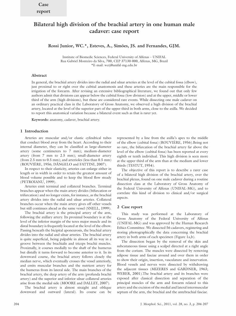
Figure 1. a,b) The brachial artery (blue arrow) bifurcates in the cubital fossa into the radial (red arrow) and the ulnar (yellow arrow) arteries in the utmost sample of cadavers presenting a division of the brachial artery at the elbow (a more frequent presentation).
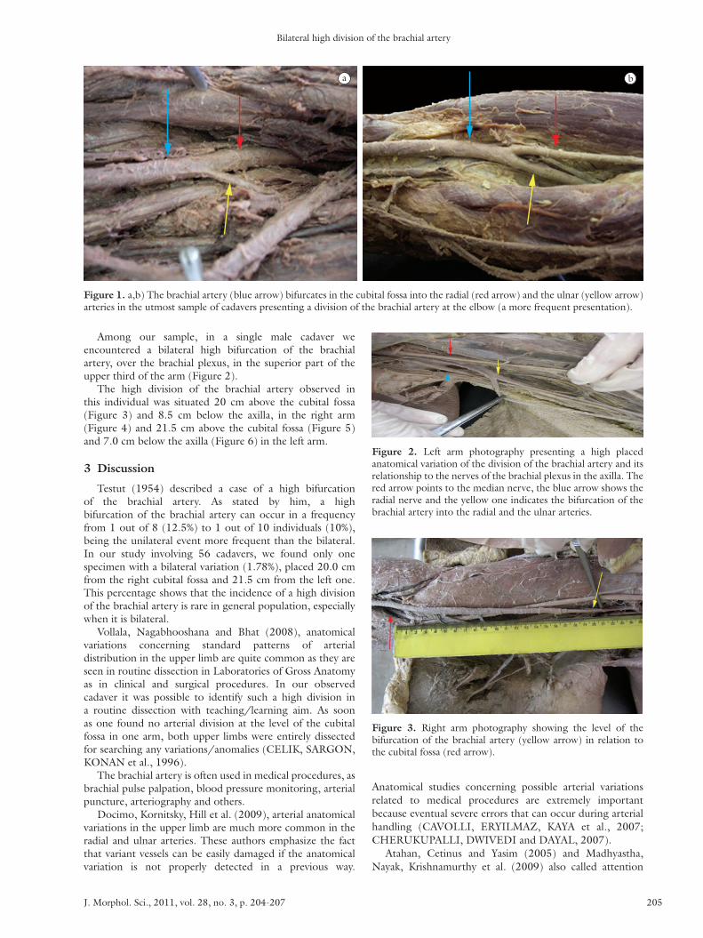
Figure 2. Left arm photography presenting a high placed anatomical variation of the division of the brachial artery and its relationship to the nerves of the brachial plexus in the axilla. The red arrow points to the median nerve, the blue arrow shows the radial nerve and the yellow one indicates the bifurcation of the brachial artery into the radial and the ulnar arteries.
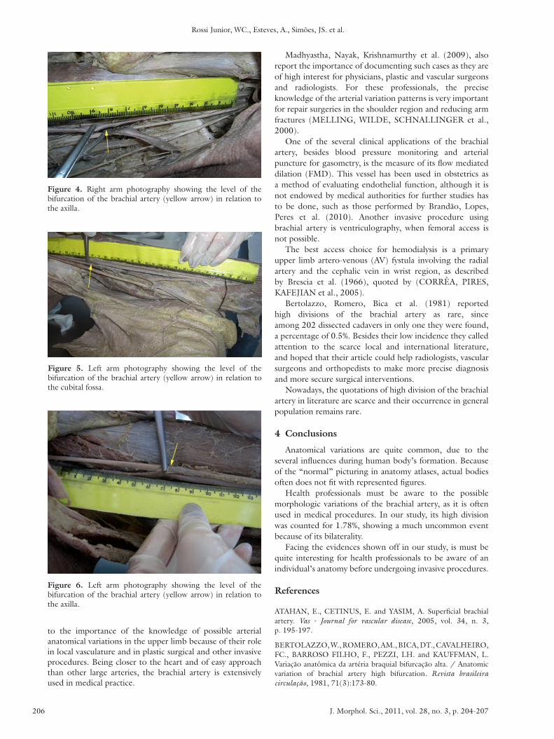
Figure 3. Right arm photography showing the level of the bifurcation of the brachial artery (yellow arrow) in relation to the cubital fossa (red arrow).
Among our sample, in a single male cadaver we encountered a bilateral high bifurcation of the brachial artery, over the brachial plexus, in the superior part of the upper third of the arm (Figure 2).
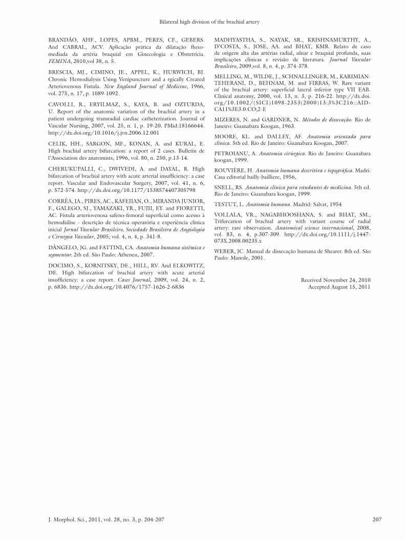
The high division of the brachial artery observed in this individual was situated 20 cm above the cubital fossa (Figure 3) and 8.5 cm below the axilla, in the right arm (Figure 4) and 21.5 cm above the cubital fossa (Figure 5) and 7.0 cm below the axilla (Figure 6) in the left arm.
3 Discussion
Testut (1954) described a case of a high bifurcation of the brachial artery. As stated by him, a high bifurcation of the brachial artery can occur in a frequency from 1 out of 8 (12.5%) to 1 out of 10 individuals (10%), being the unilateral event more frequent than the bilateral. In our study involving 56 cadavers, we found only one specimen with a bilateral variation (1.78%), placed 20.0 cm from the right cubital fossa and 21.5 cm from the left one. This percentage shows that the incidence of a high division of the brachial artery is rare in general population, especially when it is bilateral.
Vollala, Nagabhooshana and Bhat (2008), anatomical variations concerning standard patterns of arterial distribution in the upper limb are quite common as they are seen in routine dissection in Laboratories of Gross Anatomy as in clinical and surgical procedures. In our observed cadaver it was possible to identify such a high division in a routine dissection with teaching/learning aim. As soon as one found no arterial division at the level of the cubital fossa in one arm, both upper limbs were entirely dissected for searching any variations/anomalies (CELIK, SARGON, KONAN et al., 1996).
The brachial artery is often used in medical procedures, as brachial pulse palpation, blood pressure monitoring, arterial puncture, arteriography and others.
Docimo, Kornitsky, Hill et al. (2009), arterial anatomical variations in the upper limb are much more common in the radial and ulnar arteries. These authors emphasize the fact that variant vessels can be easily damaged if the anatomical variation is not properly detected in a previous way.
Anatomical studies concerning possible arterial variations related to medical procedures are extremely important because eventual severe errors that can occur during arterial handling (CAVOLLI, ERYILMAZ, KAYA et al., 2007; CHERUKUPALLI, DWIVEDI and DAYAL, 2007).
Atahan, Cetinus and Yasim (2005) and Madhyastha, Nayak, Krishnamurthy et al. (2009) also called attention
Rossi Junior, WC., Esteves, A., Simões, JS. et al.
J. Morphol. Sci., 2011, vol. 28, no. 3, p. 204-207206
Madhyastha, Nayak, Krishnamurthy et al. (2009), also report the importance of documenting such cases as they are of high interest for physicians, plastic and vascular surgeons and radiologists. For these professionals, the precise knowledge of the arterial variation patterns is very important for repair surgeries in the shoulder region and reducing arm fractures (MELLING, WILDE, SCHNALLINGER et al., 2000).
One of the several clinical applications of the brachial artery, besides blood pressure monitoring and arterial puncture for gasometry, is the measure of its flow mediated dilation (FMD). This vessel has been used in obstetrics as a method of evaluating endothelial function, although it is not endowed by medical authorities for further studies has to be done, such as those performed by Brandão, Lopes, Peres et al. (2010). Another invasive procedure using brachial artery is ventriculography, when femoral access is not possible.
The best access choice for hemodialysis is a primary upper limb artero-venous (AV) fystula involving the radial artery and the cephalic vein in wrist region, as described by Brescia et al. (1966), quoted by (CORRÊA, PIRES, KAFEJIAN et al., 2005).
Bertolazzo, Romero, Bica et al. (1981) reported high divisions of the brachial artery as rare, since among 202 dissected cadavers in only one they were found, a percentage of 0.5%. Besides their low incidence they called attention to the scarce local and international literature, and hoped that their article could help radiologists, vascular surgeons and orthopedists to make more precise diagnosis and more secure surgical interventions.
Nowadays, the quotations of high division of the brachial artery in literature are scarce and their occurrence in general population remains rare.
4 Conclusions
Anatomical variations are quite common, due to the several influences during human body’s formation. Because of the “normal” picturing in anatomy atlases, actual bodies often does not fit with represented figures.
Health professionals must be aware to the possible morphologic variations of the brachial artery, as it is often used in medical procedures. In our study, its high division was counted for 1.78%, showing a much uncommon event because of its bilaterality.
Facing the evidences shown off in our study, is must be quite interesting for health professionals to be aware of an individual’s anatomy before undergoing invasive procedures.
References
ATAHAN, E., CETINUS, E. and YASIM, A. Superficial brachial artery. Vas - Journal for vascular disease, 2005, vol. 34, n. 3, p. 195-197.
BERTOLAZZO, W., ROMERO, AM., BICA, DT., CAVALHEIRO, FC., BARROSO FILHO, F., PEZZI, LH. and KAUFFMAN, L. Variação anatômica da artéria braquial bifurcação alta. / Anatomic variation of brachial artery high bifurcation. Revista brasileira circulação, 1981, 71(3):173-80.
Figure 4. Right arm photography showing the level of the bifurcation of the brachial artery (yellow arrow) in relation to the axilla.
Figure 5. Left arm photography showing the level of the bifurcation of the brachial artery (yellow arrow) in relation to the cubital fossa.
Figure 6. Left arm photography showing the level of the bifurcation of the brachial artery (yellow arrow) in relation to the axilla.
to the importance of the knowledge of possible arterial anatomical variations in the upper limb because of their role in local vasculature and in plastic surgical and other invasive procedures. Being closer to the heart and of easy approach than other large arteries, the brachial artery is extensively used in medical practice.
Bilateral high division of the brachial artery
J. Morphol. Sci., 2011, vol. 28, no. 3, p. 204-207 207
MADHYASTHA, S., NAYAK, SR., KRISHNAMURTHY, A., D’COSTA, S., JOSE, AA. and BHAT, KMR. Relato de caso de origem alta das artérias radial, ulnar e braquial profunda, suas implicações clínicas e revisão de literatura. Journal Vascular Brasileiro, 2009,vol. 8, n. 4, p. 374-378.
MELLING, M., WILDE, J., SCHNALLINGER, M., KARIMIAN-TEHERANI, D., BEHNAM, M. and FIRBAS, W. Rare variant of the brachial artery: superficial lateral inferior type VII EAB. Clinical anatomy, 2000, vol. 13, n. 3, p. 216-22. http://dx.doi.org/10.1002/(SICI)1098-2353(2000)13:3%3C216::AID-CA11%3E3.0.CO;2-E
MIZERES, N. and GARDNER, N. Métodos de dissecação. Rio de Janeiro: Guanabara Koogan, 1963.
MOORE, KL. and DALLEY, AF. Anatomia orientada para clínica. 5th ed. Rio de Janeiro: Guanabara Koogan, 2007.
PETROIANU, A. Anatomia cirúrgica. Rio de Janeiro: Guanabara koogan, 1999.
ROUVIÈRE, H. Anatomia humana descritiva e topográfica. Madri: Casa editorial bailly-bailliere, 1956,
SNELL, RS. Anatomia clínica para estudantes de medicina. 5th ed. Rio de Janeiro: Guanabara koogan, 1999.
TESTUT, L. Anatomia humana. Madrid: Salvat, 1954
VOLLALA, VR., NAGABHOOSHANA, S. and BHAT, SM., Trifurcation of brachial artery with variant course of radial artery: rare observation. Anatomical science internacional, 2008, vol. 83, n. 4, p.307-309. http://dx.doi.org/10.1111/j.1447-073X.2008.00235.x
WEBER, JC. Manual de dissecação humana de Shearer. 8th ed. São Paulo: Manole, 2001.
Received November 24, 2010 Accepted August 15, 2011
BRANDÃO, AHF., LOPES, APBM., PERES, CF., GEBERS. And CABRAL, ACV. Aplicação prática da dilatação fluxo-mediada da artéria braquial em Ginecologia e Obstetrícia. FEMINA, 2010,vol 38, n. 5.
BRESCIA, MJ., CIMINO, JE., APPEL, K., HURWICH, BJ. Chronic Hemodialysis Using Venipuncture and a rgically Created Arteriovenous Fistula. New England Journal of Medicine, 1966, vol. 275, n. 17, p. 1089-1092.
CAVOLLI, R., ERYILMAZ, S., KAYA, B. and OZYURDA, U. Report of the anatomic variation of the brachial artery in a patient undergoing transradial cardiac catheterization. Journal of Vascular Nursing, 2007, vol. 25, n. 1, p. 19-20. PMid:18166644. http://dx.doi.org/10.1016/j.jvn.2006.12.001
CELIK, HH., SARGON, MF., KONAN, A. and KURAL, E. High brachial artery bifurcation: a report of 2 cases. Bulletin de l’Association des anatomists, 1996, vol. 80, n. 250, p.13-14.
CHERUKUPALLI, C., DWIVEDI, A. and DAYAL, R. High bifurcation of brachial artery with acute arterial insufficiency: a case report. Vascular and Endovascular Surgery, 2007, vol. 41, n. 6, p. 572-574. http://dx.doi.org/10.1177/1538574407305798
CORRÊA, JA., PIRES, AC., KAFEJIAN, O., MIRANDA JUNIOR, F., GALEGO, SJ., YAMAZAKI, YR., FUJII, EY. and FIORETTI, AC. Fístula arteriovenosa safeno-femoral superficial como acesso à hemodiálise - descrição de técnica operatória e experiência clínica inicial Jornal Vascular Brasileiro, Sociedade Brasileira de Angiologia e Cirurgia Vascular, 2005; vol. 4, n. 4, p. 341-8.
DÂNGELO, JG. and FATTINI, CA. Anatomia humana sistêmica e segmentar. 2th ed. São Paulo: Atheneu, 2007.
DOCIMO, S., KORNITSKY, DE., HILL, RV. And ELKOWITZ, DE. High bifurcation of brachial artery with acute arterial insufficiency: a case report. Cases Journal, 2009, vol. 24, n. 2, p. 6836. http://dx.doi.org/10.4076/1757-1626-2-6836