beyond the c/d ratio: evaluating a glaucomatous … the c/d ratio: evaluating a glaucomatous optic...
TRANSCRIPT

Beyond the C/D Ratio: Evaluating a Glaucomatous
Optic Nerve
Marcus Gonzales, OD, FAAO Cedar Springs Eye Clinic COPE ID#: 27809-GL

Points to Remember
• Glaucoma affects the ONH in characteristic patterns
• Typically affects the eyes and rims asymmetrically
• Damage usually occurs at the ONH before corresponding VF loss
• Monitoring for change is the key

Points to Remember
• More comprehensive documentation of the optic nerve at baseline helps better determine this change over time
• Scanning laser instrumentation and disc photography can be a huge aid
• With all the technology out there, don’t forget that you’re the best instrument in determining risk

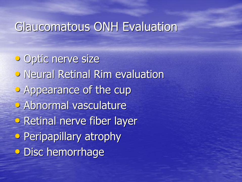
Glaucomatous ONH Evaluation
• Optic nerve size
• Neural Retinal Rim evaluation
• Appearance of the cup
• Abnormal vasculature
• Retinal nerve fiber layer
• Peripapillary atrophy
• Disc hemorrhage

Why is a C/D ratio not enough?
• Doesn’t take into account nerve size
– Small, Average, Large
• Doesn’t take into account rim configuration
– ISNT rule
• Doesn’t tell us how C/D was determined
– Color or contour
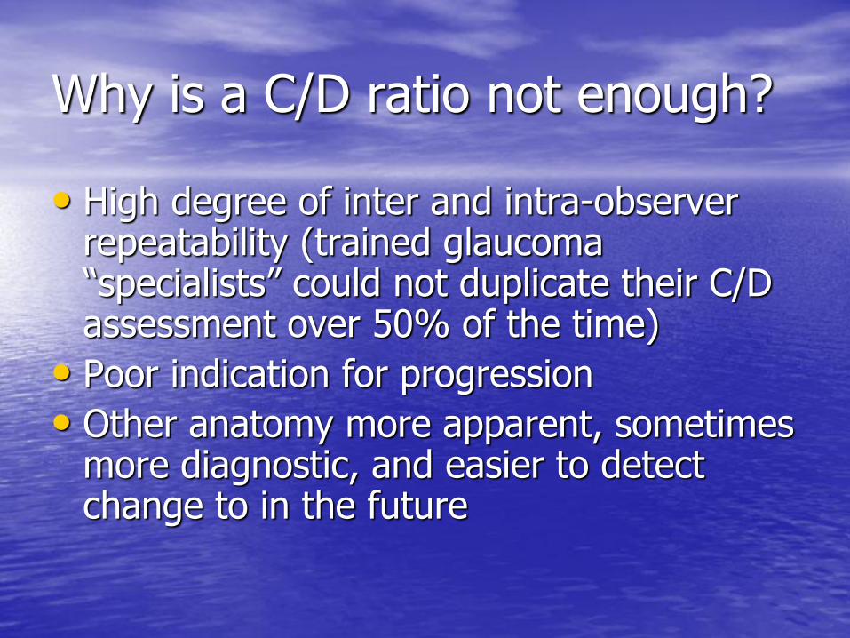
Why is a C/D ratio not enough?
• High degree of inter and intra-observer repeatability (trained glaucoma “specialists” could not duplicate their C/D assessment over 50% of the time)
• Poor indication for progression
• Other anatomy more apparent, sometimes more diagnostic, and easier to detect change to in the future
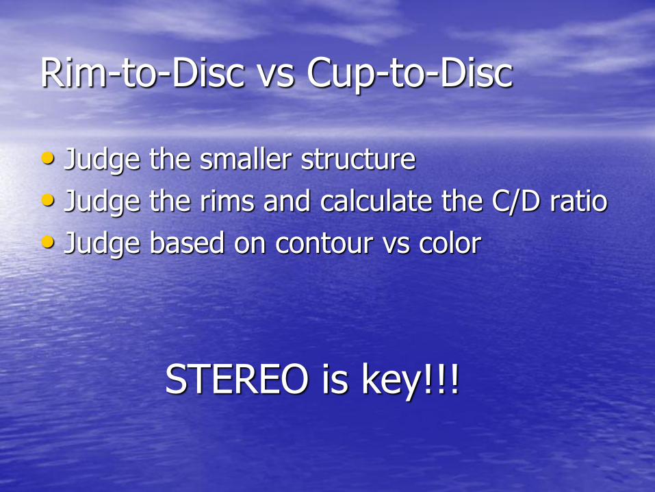
Rim-to-Disc vs Cup-to-Disc
• Judge the smaller structure
• Judge the rims and calculate the C/D ratio
• Judge based on contour vs color
STEREO is key!!!

Rim-to-Disc vs Cup-to-Disc
Glaucomatous ONH Evaluation
• Optic nerve size
• Neural Retinal Rim evaluation
• Appearance of the cup
• Abnormal vasculature
• Retinal nerve fiber layer
• Peripapillary atrophy
• Disc hemorrhage

Optic Nerve Size
• Small <1.5mm
• “Normal” 1.5-2.2mm
• Large >2.4mm

ON Size Affects C/D Estimation
C/D: .3/.3 C/D: .65/.65
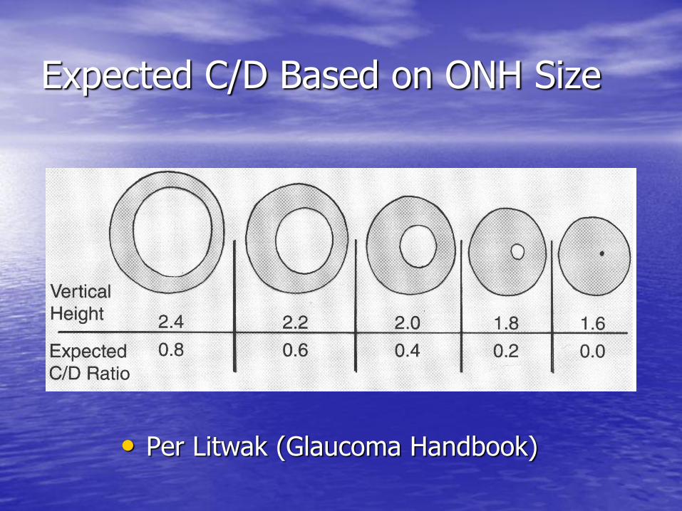
Expected C/D Based on ONH Size
• Per Litwak (Glaucoma Handbook)

• Per Litwak (Glaucoma Handbook)
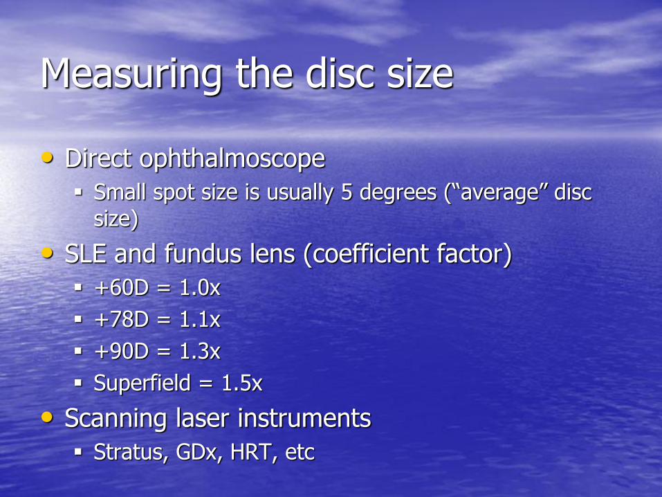
Measuring the disc size
• Direct ophthalmoscope
Small spot size is usually 5 degrees (“average” disc size)
• SLE and fundus lens (coefficient factor)
+60D = 1.0x
+78D = 1.1x
+90D = 1.3x
Superfield = 1.5x
• Scanning laser instruments
Stratus, GDx, HRT, etc

Factors that affect disc size
• Refractive error
Hyperope – smaller discs » smaller C/Ds
Myope – larger discs » larger C/Ds
• Race
Black – larger disc
White – smaller disc
Hispanic – somewhere in the middle
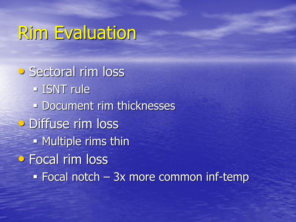
Rim Evaluation
• Sectoral rim loss
ISNT rule
Document rim thicknesses
• Diffuse rim loss
Multiple rims thin
• Focal rim loss
Focal notch – 3x more common inf-temp
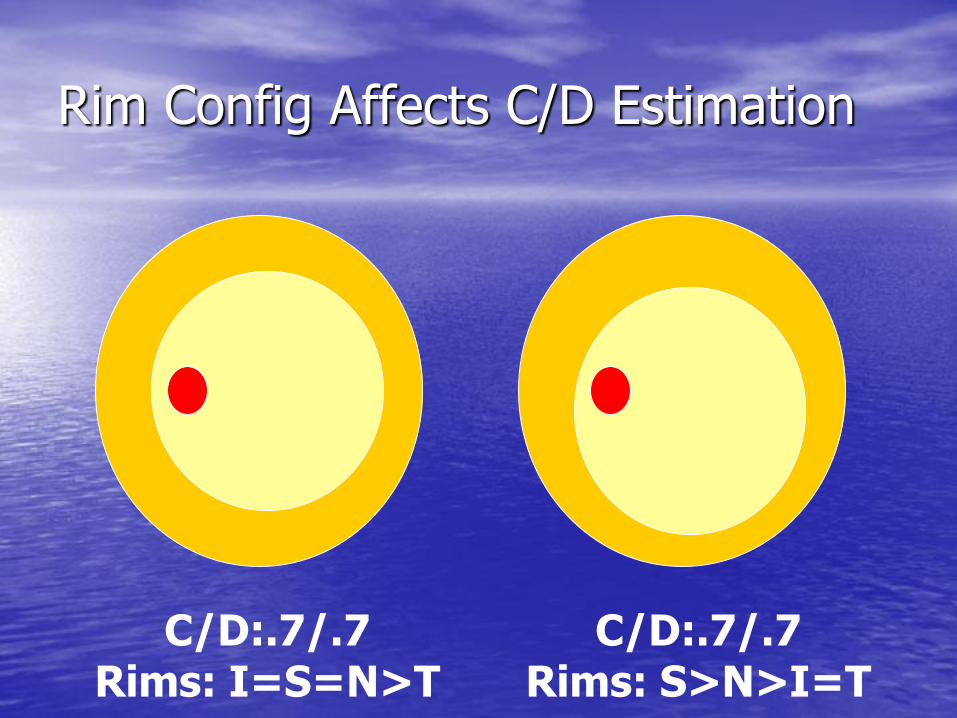
Rim Config Affects C/D Estimation
C/D:.7/.7 Rims: I=S=N>T
C/D:.7/.7 Rims: S>N>I=T
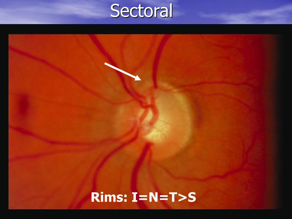
Sectoral
Rims: I=N=T>S

Sectoral
Rims: N>I>S=T Rims: I=N>S>T

Diffuse
Rims: I=S=N=T

Diffuse
Rims: I=N>S=T
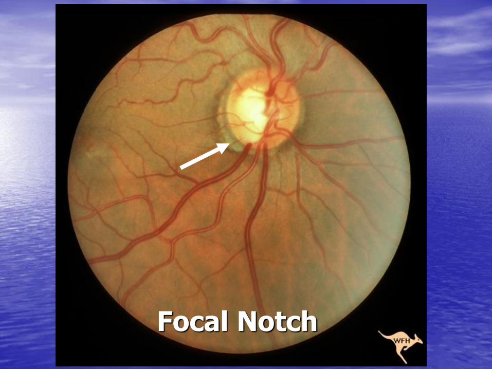
Focal Notch
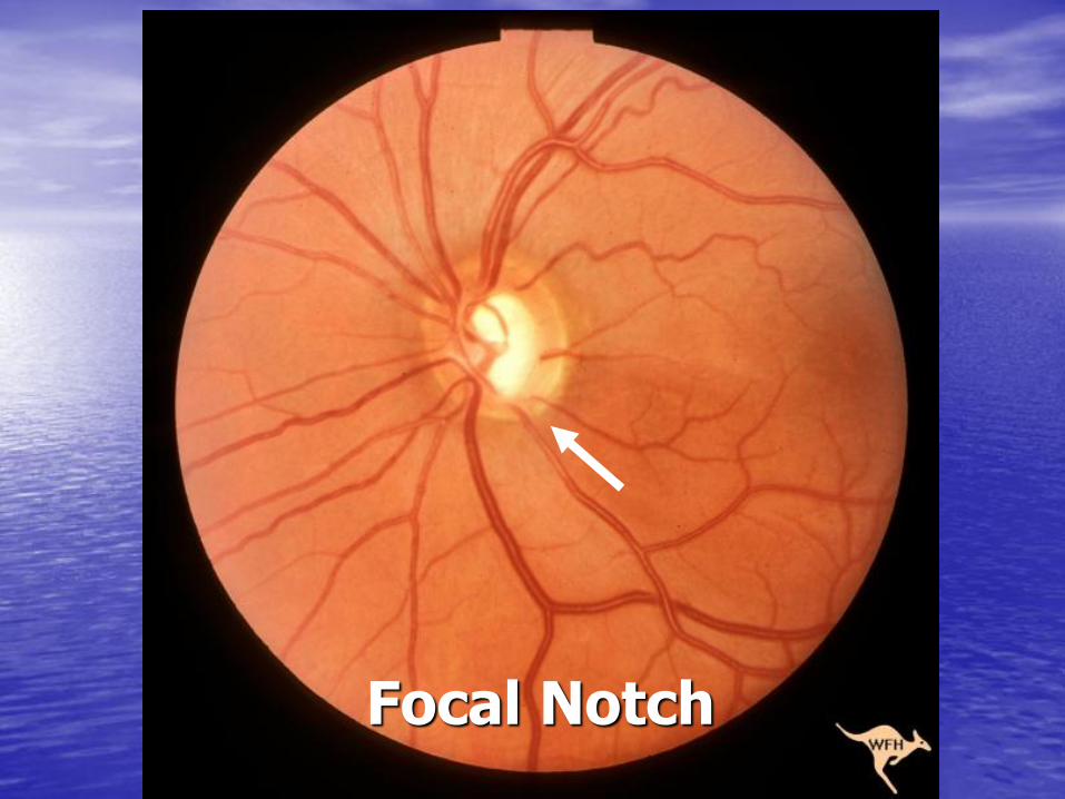
Focal Notch

Focal Notch
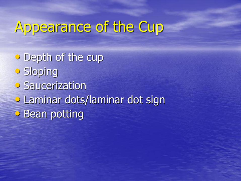
Appearance of the Cup
• Depth of the cup
• Sloping
• Saucerization
• Laminar dots/laminar dot sign
• Bean potting
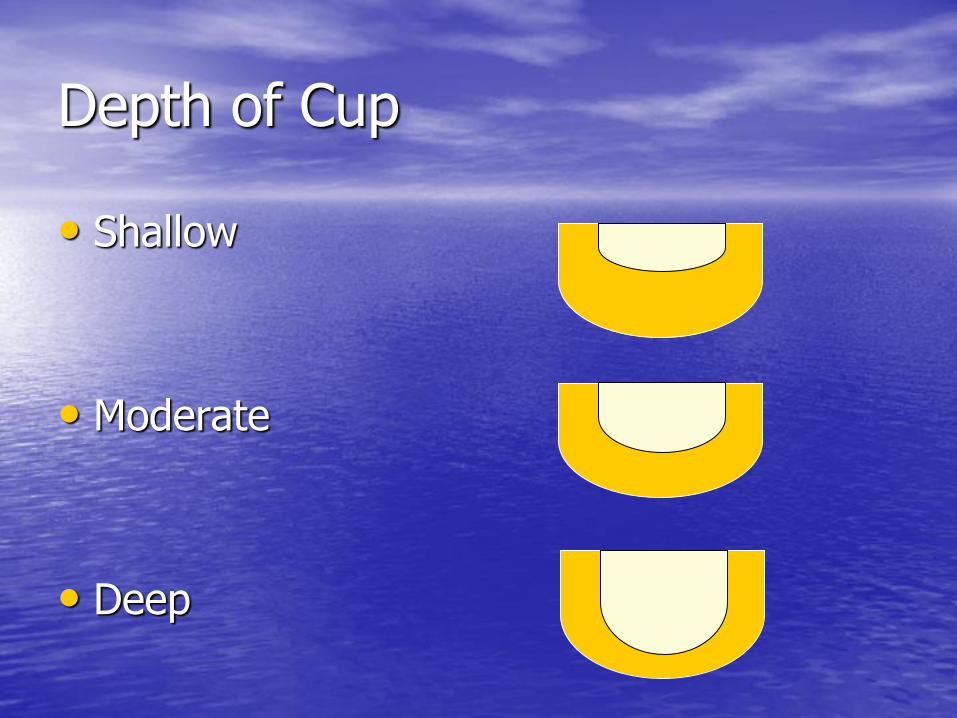
Depth of Cup
• Shallow
• Moderate
• Deep
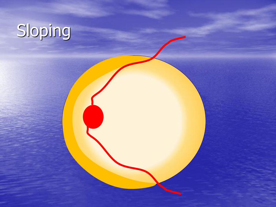
Sloping

Sloping

Sloping

Sloping
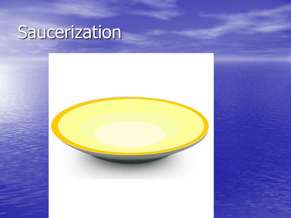
Saucerization

Saucerization
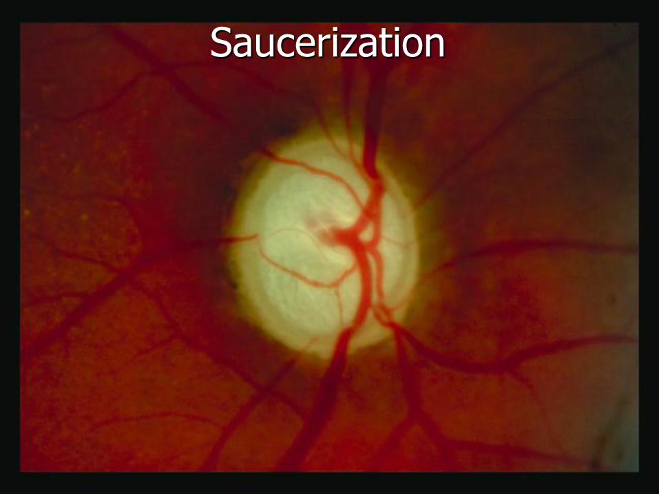
Saucerization
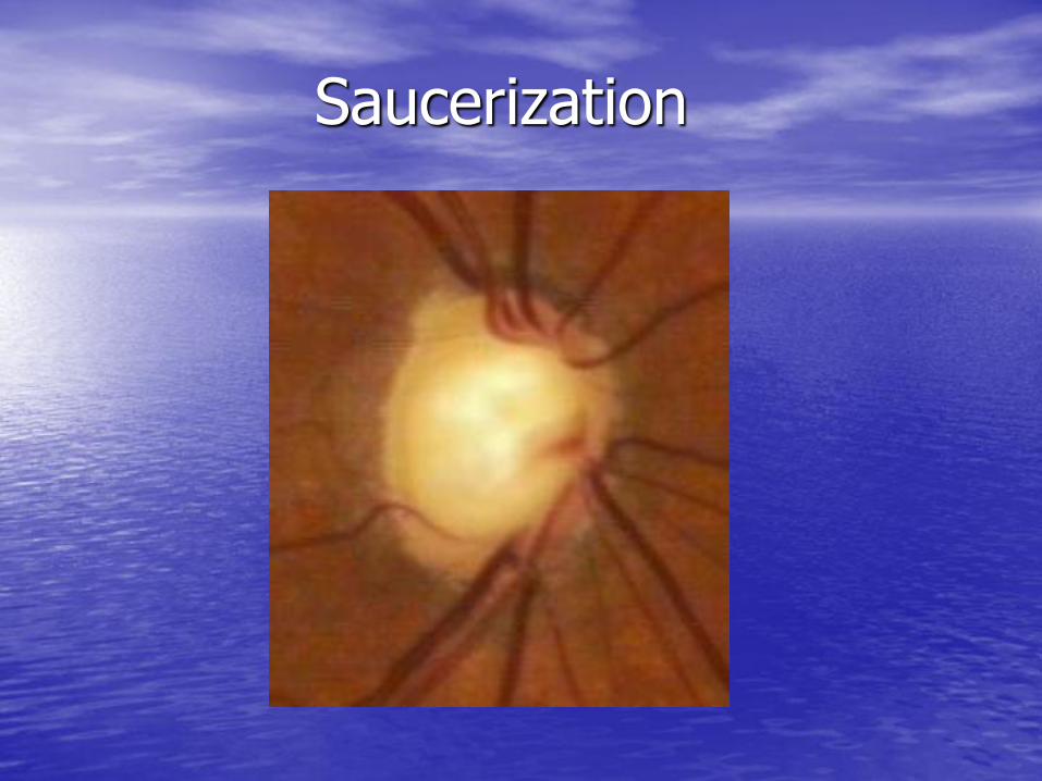
Saucerization

Laminar Dots
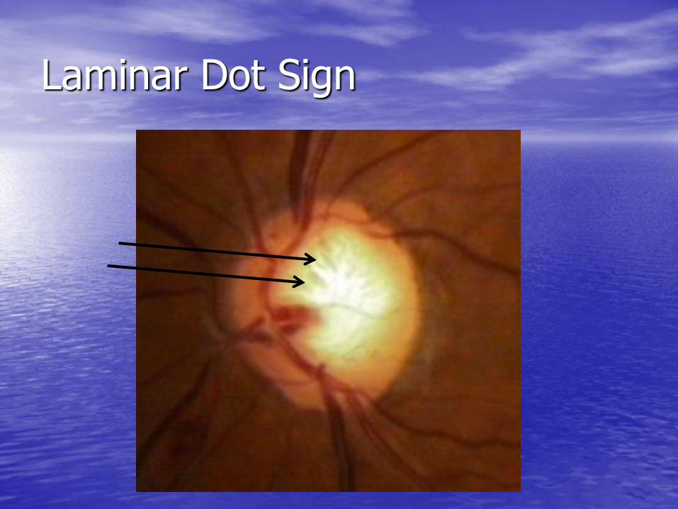
Laminar Dot Sign
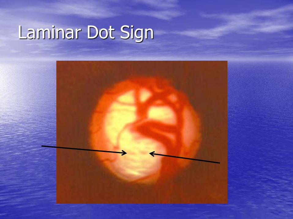
Laminar Dot Sign
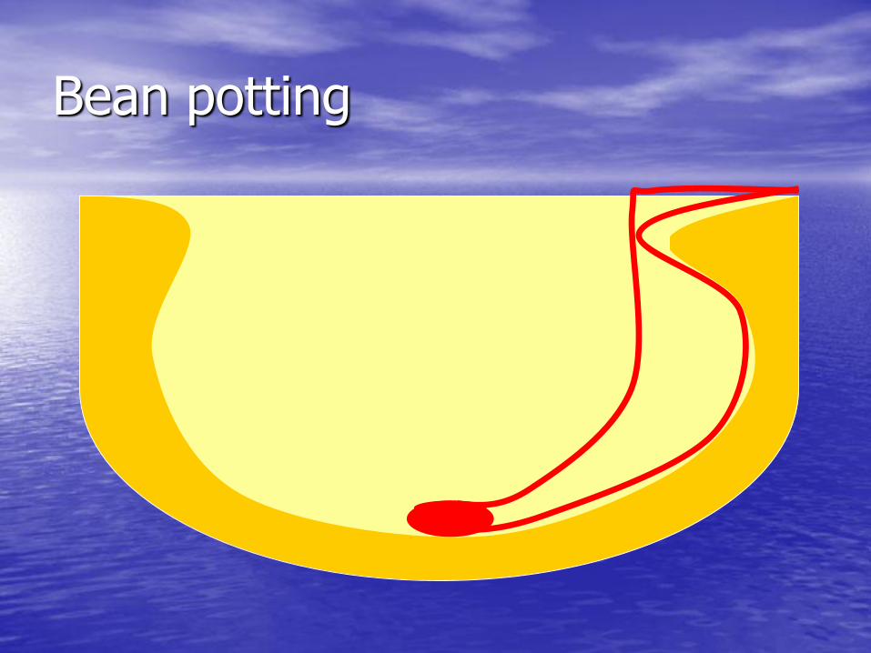
Bean potting

Bean potting

Bean potting
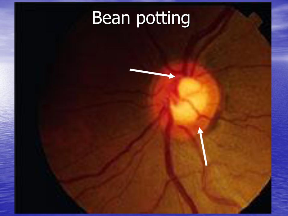
Bean potting

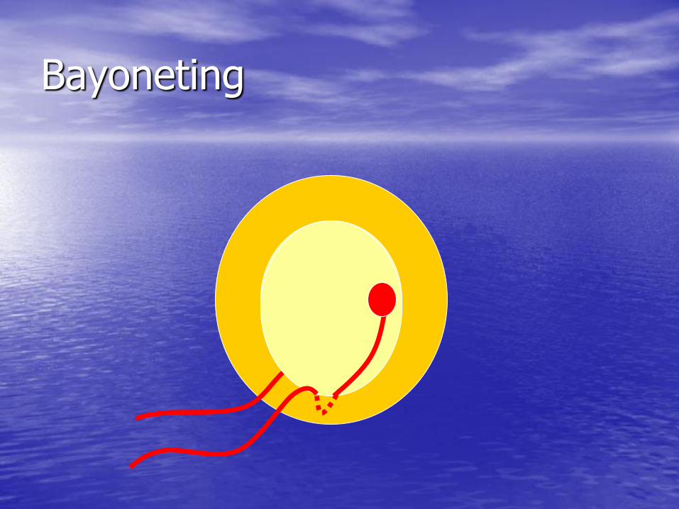
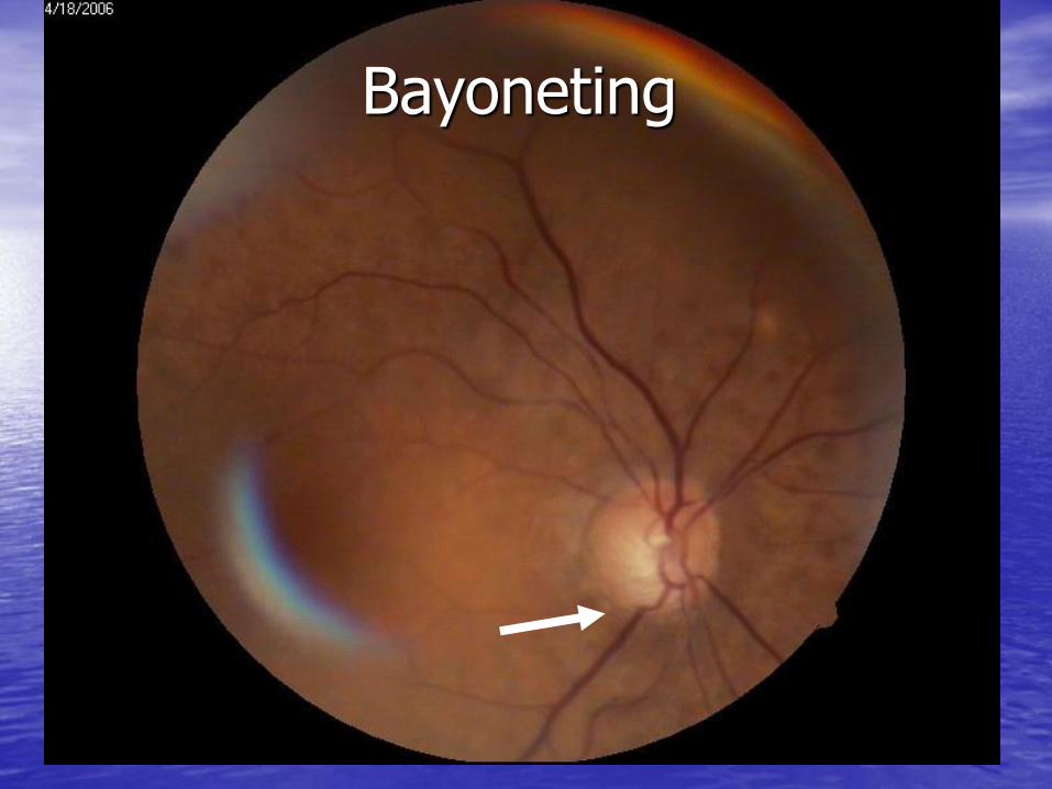
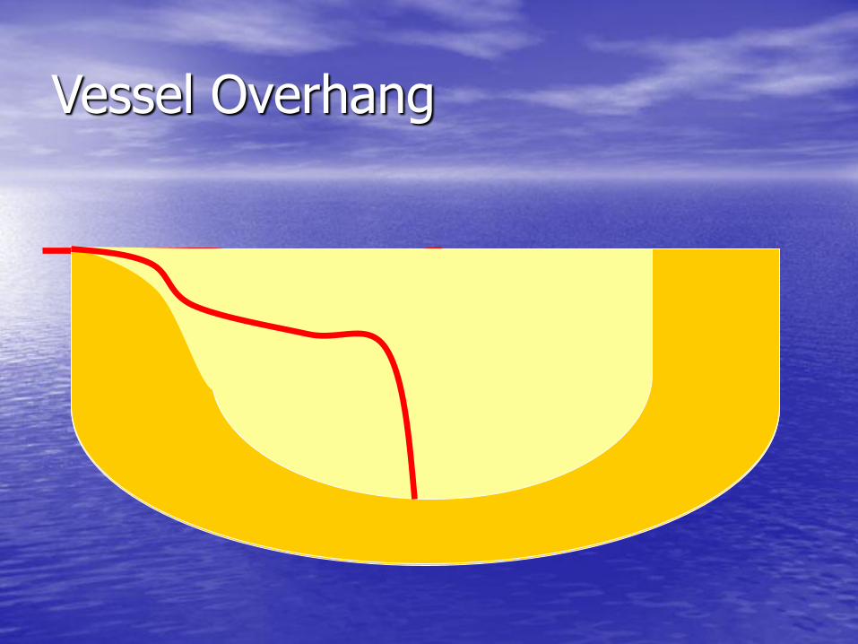
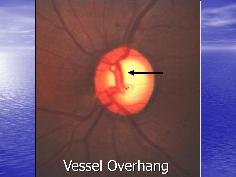
Abnormal Vasculature
• Bayoneting
• Vessel Overpass/Overhang or Baring of circumlinear vessels
• Nasalization

Bayoneting
Bayoneting
Bayoneting

Bayoneting

Bayoneting
Vessel Overhang
Vessel Overhang
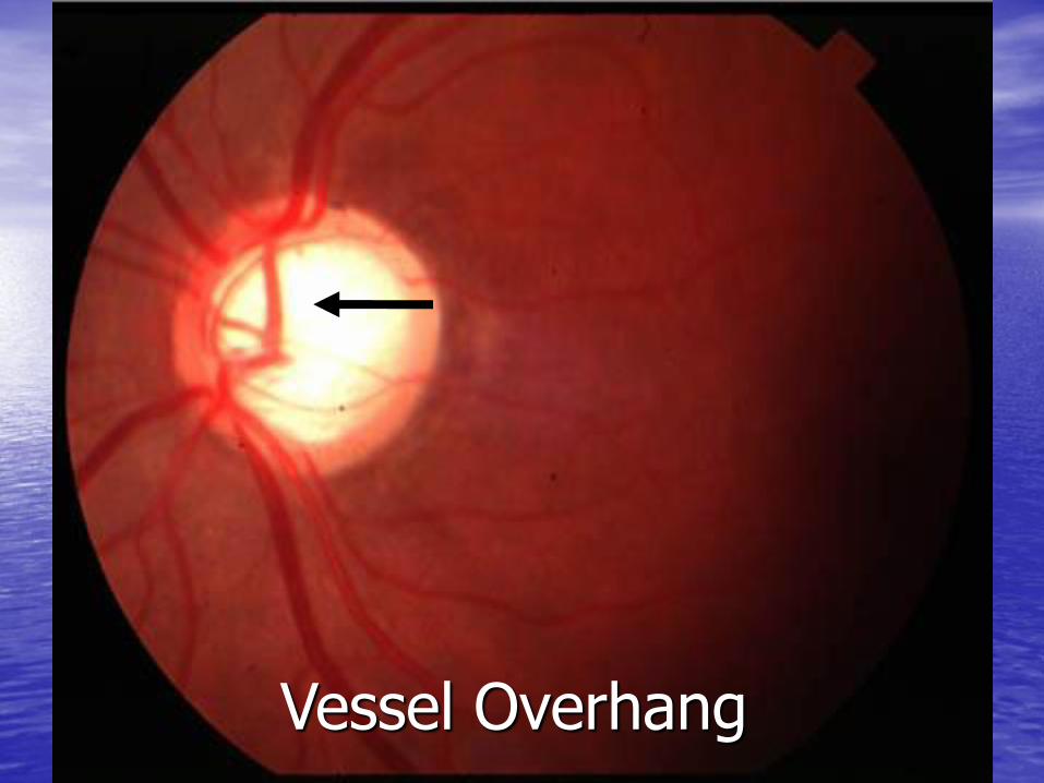
Vessel Overhang
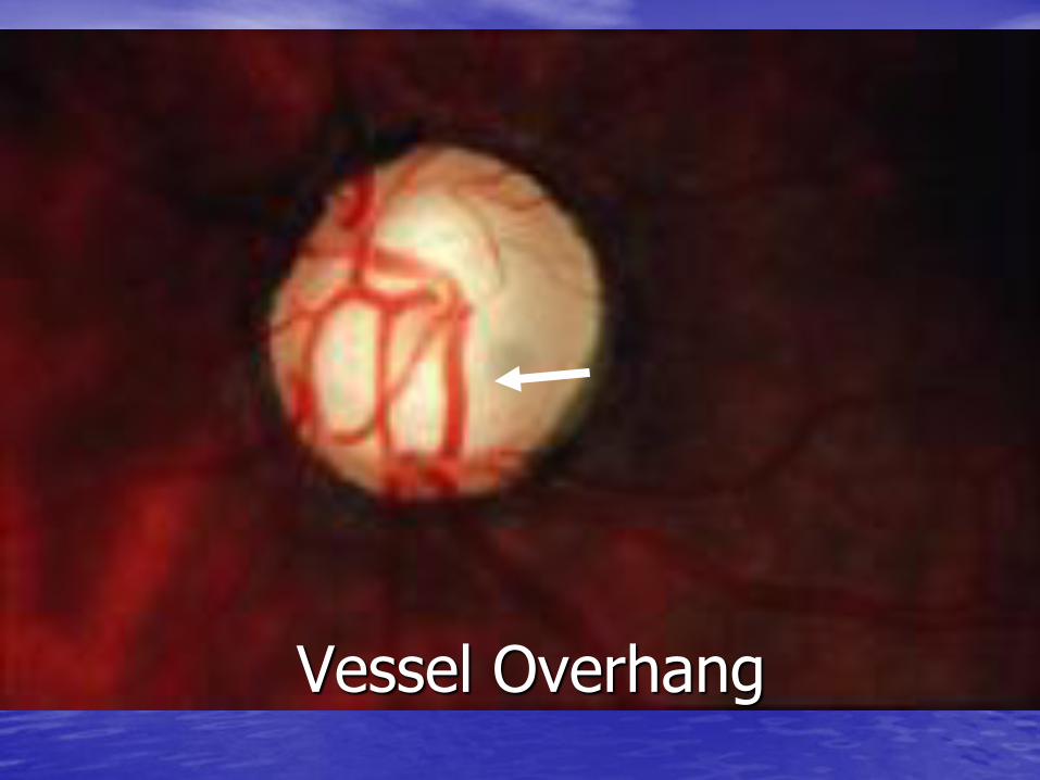
Vessel Overhang

Nasalization
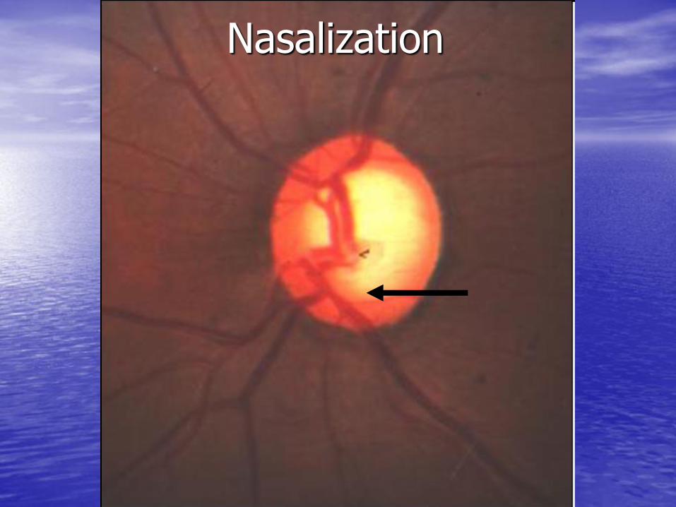
Nasalization

Nasalization
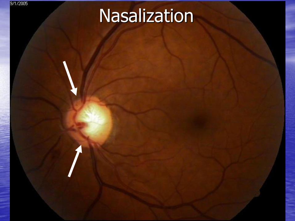
Nasalization

Retinal Nerve Fiber Layer
• Bright/Dim/Bright reflection
• Brighter reflections blur underlying blood vessels
• Best visualized with brighter illumination and with red-free (green) filter
• Don’t overlook photography as an aid
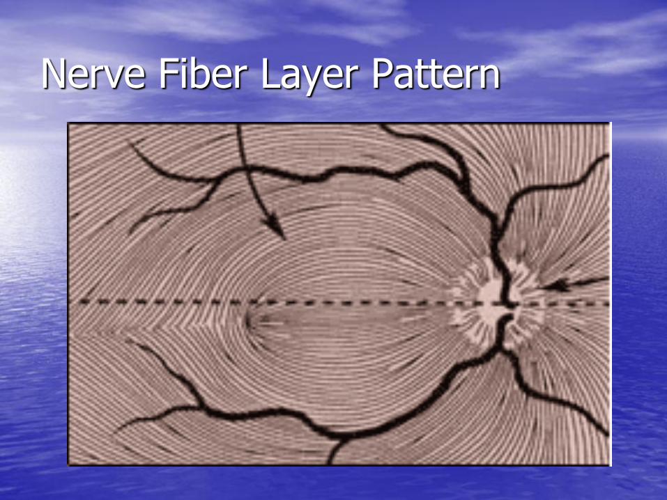
Nerve Fiber Layer Pattern
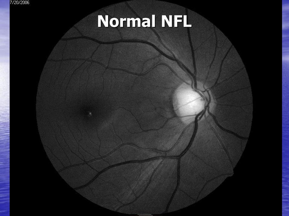
Normal NFL Photography Normal NFL

Retinal Nerve Fiber Layer
• Wedge defect Triangular-shaped dark area extending from disc
• Diffuse loss Entire quadrant of disc with less reflectance
• Slit defect Dark area at least an arteriole in width extending
from disc
• Pseudo-slit defect May be physiologic or could progress to a slit defect

Wedge Defect
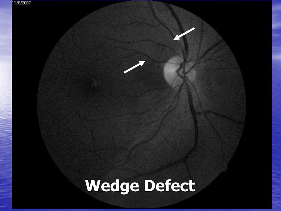
Wedge Defect
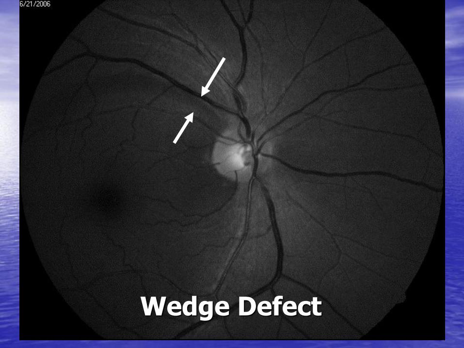
Wedge Defect

Diffuse NFL Loss

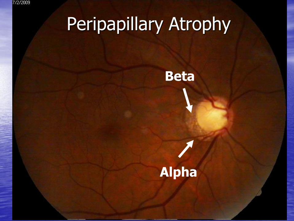
Peripapillary Atrophy
• Atrophy of tissue surrounding ONH
• Pathogenesis: Ischemia of peripapillary choroidal circulation and/or vascular deficiency in the ONH
• Correlation of size and location of PPA to the extent of damage to ONH
• Correlation to changes in PPA associated with progression of VF loss

Peripapillary Atrophy
• Alpha (α) zone changes Irregular hyper/hypopigmentation of the RPE
Normal variation with age
• Beta (β) zone changes Atrophy of the RPE and choriocapillaris making large
choriodal vessels and sclera apparent
More common and extensive in glaucomatous nerves
May precede notch, disc heme, NFL defect
If both changes are present, alpha changes occur outside the beta zone changes
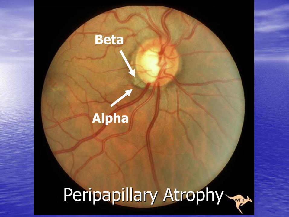
Beta
Alpha
Peripapillary Atrophy
Peripapillary Atrophy
Beta
Alpha

Alpha
Beta
Peripapillary Atrophy
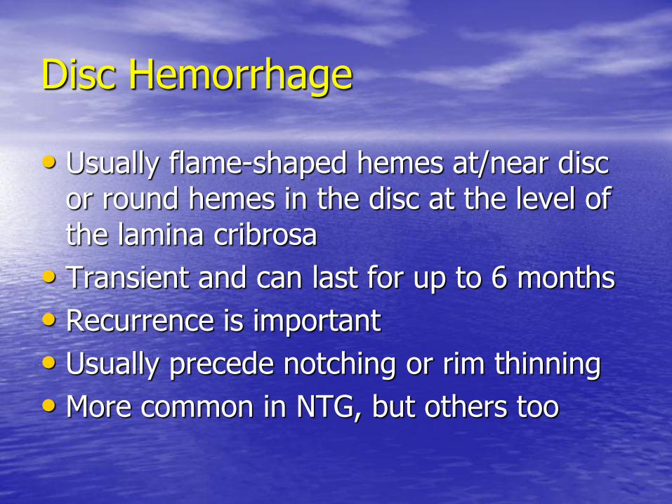
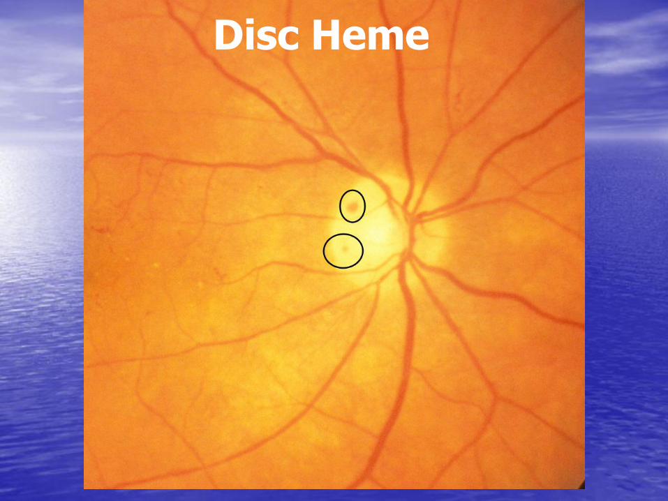
Disc Hemorrhage
• Usually flame-shaped hemes at/near disc or round hemes in the disc at the level of the lamina cribrosa
• Transient and can last for up to 6 months
• Recurrence is important
• Usually precede notching or rim thinning
• More common in NTG, but others too

Disc Heme

Disc Heme
Disc Heme

Disc Heme

Glaucomatous ONH Evaluation
• Optic nerve size
• Neural Retinal Rim evaluation
• Appearance of the cup
• Abnormal vasculature
• Retinal nerve fiber layer
• Peripapillary atrophy
• Disc hemorrhage

Thank you for your attention
Questions?