berberine reverses epithelial-to-mesenchymal transition and...
TRANSCRIPT

MOL #94037
1
Berberine reverses epithelial-to-mesenchymal transition and inhibits metastasis
and tumor-induced angiogenesis in human cervical cancer cells
Shu-Chen Chu, Cheng-Chia Yu, Li-Sung Hsu, Kuo-Shuen Chen, Mei-Yu Su,
Pei-Ni Chen
Institute and Department of Food Science, Central Taiwan University of Science and
Technology, Taiwan (S.-C. C.)
Institute of Oral Science, School of Dentistry, Chung Shan Medical University,
Taiwan (C.-C. Y.)
Institute of Biochemistry and Biotechnology, Chung Shan Medical University, Taiwan
(P.-N. C., M.-Y. S., L.-S. H.)
Institute of Medicine, Chung Shan Medical University, Taiwan (K.-S. C.)
Clinical Laboratory, Chung Shan Medical University Hospital, Taiwan (P.-N. C., M.-Y.
S.)
Department of Dentistry, Chung Shan Medical University Hospital, Taiwan (C.-C. Y.)
Department of Internal Medicine, Chung Shan Medical University Hospital, Taiwan
(K.-S. C.)
This article has not been copyedited and formatted. The final version may differ from this version.Molecular Pharmacology Fast Forward. Published on September 12, 2014 as DOI: 10.1124/mol.114.094037
at ASPE
T Journals on M
ay 28, 2019m
olpharm.aspetjournals.org
Dow
nloaded from

MOL #94037
2
Running title: Berberine inhibits metastasis, EMT and angionenesis
Corresponding author:
Pei-Ni Chen, P.h.D., Institute of Biochemistry and Biotechnology, Chung Shan
Medical University, No 110, Section 1, Jianguo N. Road, Taichung, Taiwan. E-mail:
[email protected], Phone: +886-4-2473-0022 ext. 11685, Fax: +886-4-2324-8195
9 Figures
0 Tables
47 text pages
41 References
223 words in the Abstract
734 words in Introduction
1362 words in Discussion
Abbreviations: EMT, epithelial–mesenchymal transition; MMP, matrix
metalloproteinase; u-PA, urokinasetype plasminogen activator; VEGF, vascular
endothelial growth factor; TGF-β1, transforming growth factor-β1
This article has not been copyedited and formatted. The final version may differ from this version.Molecular Pharmacology Fast Forward. Published on September 12, 2014 as DOI: 10.1124/mol.114.094037
at ASPE
T Journals on M
ay 28, 2019m
olpharm.aspetjournals.org
Dow
nloaded from

MOL #94037
3
Abstract
Metastasis is the most common cause of cancer-related death in patients, and
epithelial to mesenchymal transition (EMT) is essential for cancer metastasis, which
is a multistep complicated process that includes local invasion, intravasation,
extravasation, and proliferation at distant sites. When cancer cells metastasize,
angiogenesis is also required for metastatic dissemination, given that an increase in
vascular density will allow easier access of tumor cells to circulation and represents a
rational target for therapeutic intervention. Berberine has several anti-inflammation
and anticancer biological effects. In this study, we provided molecular evidence that is
associated with the anti-metastatic effect of berberine by showing a nearly complete
inhibition on invasion (P < 0.001) of highly metastatic SiHa cells via reduced
transcriptionally activities of matrix metalloproteinase-2 and urokinasetype
plasminogen activator. Berberine reversed transforming growth factor-β1-induced
EMT and caused upregulation of epithelial markers such as E-cadherin and inhibited
mesenchymal markers such as N-cadherin and snail-1. Selective snail-1 inhibition by
snail-1-specific-siRNA also showed increased E-cadherin expression in SiHa cells.
Berberine also reduced tumor-induced angiogenesis in vitro and in vivo. Importantly,
in vivo BALB/c nude mice xenograft model and tail vein injection model showed that
berberine treatment reduced tumor growth and lung metastasis by oral gavage,
respectively. Taken together, these findings suggested that berberine could reduce
metastasis and angiogenesis of cervical cancer cells, thereby constituting an adjuvant
treatment for metastasis control.
This article has not been copyedited and formatted. The final version may differ from this version.Molecular Pharmacology Fast Forward. Published on September 12, 2014 as DOI: 10.1124/mol.114.094037
at ASPE
T Journals on M
ay 28, 2019m
olpharm.aspetjournals.org
Dow
nloaded from
MOL #94037
4
Introduction
Cervical cancer is the second most common female cancer worldwide, with an
estimated 530,000 new cases ever year and the third greatest cause of death from
cancer in women. Although cervical cytology screening has helped reduce mortality
rates, managing pre-invasive and invasive cervical lesions remains a challenge (Smith
et al., 2013). Cancer metastasis and resistance to treatment are two major causes for
poor survival and prognosis of cervical cancer patients. Most patient deaths from
cervical cancer are related to metastasis, which is a complicated and currently
uncontrolled process. Therefore, reducing the metastasis of cervical tumor cells is one
of the most important research areas in medicine.
Tumor malignancy consists of a series of complicated processes, including invasion,
migration, adhesion, angiogenesis, and proliferation. During tumor progression, tumor
cells acquire expression of mesenchymal markers, such as vimentin, N-cadherin, and
fibronectin, as well as loss of epithelial markers, such as E-cadherin and α-catenin, to
result in epithelial–mesenchymal transition (EMT), subsequent tumor metastasis, and
proliferation at distant sites. Suppressing E-cadherin expression by its transcriptional
suppressor, snail-1, is a key process in EMT (de Herreros et al., 2010). During cancer
metastasis, secreting extracellular proteases is significant in cancer invasion (Rao,
2003). Among these proteases, matrix metalloproteinase-2 (MMP-2, also known as
This article has not been copyedited and formatted. The final version may differ from this version.Molecular Pharmacology Fast Forward. Published on September 12, 2014 as DOI: 10.1124/mol.114.094037
at ASPE
T Journals on M
ay 28, 2019m
olpharm.aspetjournals.org
Dow
nloaded from

MOL #94037
5
gelatinase A), which belongs to a family of structurally related zinc-dependent
extracellular matrix (ECM)-degrading enzymes, is a proteolytic enzyme that is
capable of degrading the structural support network for normal and malignant cells,
which could serve a crucial role in invasion and angiogenesis of tumor cells (Roomi et
al., 2012). In addition to matrix metalloproteinases, serine protease urokinasetype
plasminogen activator (u-PA), which is secreted in cervical cancer, is a key factor to
initiate a cascade of proteolytic steps that accumulates in degrading the ECM (Roomi
et al., 2012; Tee et al., 2012).
Angiogenesis is a physiologic process that generates new blood vessels from
pre-existing capillaries and is critical to growth and metastasis of solid tumors. In
normal tissues, the vascular system is regulated by a balance of anti-angiogenic and
pro-angiogenic molecules, which ensures that an efficient and orderly network of
blood vessels is maintained to meet the metabolic demands of the tissue. Unlike
normal tissue vasculature, tumor vessels are generally long and highly chaotic, with
numerous abnormalities that result in poor blood flow and high vascular permeability,
which may lead to reduced efficiency of chemotherapy in cancer patients and elevated
potential for metastasizing to distant organs (Vaupel, 2004). A fundamental step in the
transition of tumors from dormant to malignant state is followed by the tumor
potential for metastasis. Inhibiting the angiogenesis and metastasis of human cervical
This article has not been copyedited and formatted. The final version may differ from this version.Molecular Pharmacology Fast Forward. Published on September 12, 2014 as DOI: 10.1124/mol.114.094037
at ASPE
T Journals on M
ay 28, 2019m
olpharm.aspetjournals.org
Dow
nloaded from
MOL #94037
6
cancer cells could serve as an effective strategy against cervical cancer progression
(Xie et al., 2013).
Chemopreventive approach using non-toxic botanicals could be one of the
strategies for cancer management. These natural substances are of interest, as they are
potential sources of anticancer compounds with minimal debilitating toxicity and side
effects. Berberine, which is a naturally occurring isoquinoline alkaloid, is an active
component in the roots, rhizomes, and stem barks of many medicinal plants, including
Berberis vulgaris (barberry), Berberis aristata (tree turmeric), Berberis aquifolium
(Oregon grape), and Coptis chinensis (Chinese goldthread). Berberine was initially
used as an antibiotic because of its potent antimicrobial activity against many
organisms including bacteria, fungi, protozoans, viruses, chlamydia, and helminthes
(Yu et al., 2005). Berberine has a wide range of pharmacological and biochemical
effects for various clinical conditions, such as diarrhea, hypertension, arrhythmias,
and inflammation (Rabbani et al., 1987). Recently, berberine has been demonstrated
to possess anticancer activities including DNA-modified electrodes (Tian et al., 2008),
reduction of AP-1 activity to induce growth arrest and apoptosis (Mahata et al., 2011),
and induction of caspase-dependent apoptosis in cervical cancer (Mantena et al.,
2006a, b). Berberine inhibited invasion of human lung carcinoma A549 cells,
increased expression of E-cadherin, and repressed expression of vimentin during
This article has not been copyedited and formatted. The final version may differ from this version.Molecular Pharmacology Fast Forward. Published on September 12, 2014 as DOI: 10.1124/mol.114.094037
at ASPE
T Journals on M
ay 28, 2019m
olpharm.aspetjournals.org
Dow
nloaded from

MOL #94037
7
initiation of transforming growth factor (TGF)-β1-induced EMT. Berberine inhibited
the capacity of hepatocellular carcinoma to stimulate human umbilical vein
endothelial cell (HUVEC) proliferation, migration, and endothelial tube formation
(Jie et al., 2011). Berberine also inhibited HIF-1α expression via enhanced proteolysis
in gastric adenocarcinoma cell line SC-M1 (Lin et al., 2004). However, the effects of
berberine on cancer invasion, angiogenesis, and EMT of human cervical carcinoma
and the underlying mechanisms of such effects remain unclear. In the current study,
we tested the hypothesis that berberine has .the anti-metastatic and reverse EMT
potential for human cervical cells.
This article has not been copyedited and formatted. The final version may differ from this version.Molecular Pharmacology Fast Forward. Published on September 12, 2014 as DOI: 10.1124/mol.114.094037
at ASPE
T Journals on M
ay 28, 2019m
olpharm.aspetjournals.org
Dow
nloaded from

MOL #94037
8
Materials and Methods
Materials and Chemicals.
Berberine, heparin, Giemsa, gelatin, 4'-6-Diamidino-2-phenylindole, dimethyl
sulfoxide (DMSO), 3-(4,5-dimethylthiazol-2-y1)-2,5-diphenyltetrazolium bromide
(MTT), Coomassie brilliant blue R-250 and crystal violet were obtained from Sigma
Chemical Co. (St. Louis, MO, USA). Matrigel was purchased from BD Biosciences
(Bedford, MA, USA). The Immobilon Western Chemiluminescent HRP substrate kit
was obtained from Millipore (Burlington, MA). Dulbecco's modified Eagle medium
(DMEM), medium 199, penicillin, streptomycin and trypsin-EDTA were obtained
from Gibco Invitrogen Corparation (Barcelona, Spain).
Cell culture
SiHa cells were obtained from the American Type Culture Collection (Manassas, VA),
whereas HeLa and CaSki were obtained from the Bioresource Collection and
Research Center (BCRC, Hsinchu, Taiwan) and were cultured in DMEM with 10%
fetal bovine serum (FBS), 2 mM glutamine, 100 U/mL of penicillin, and 100 μg/mL
of streptomycin. HUVECs were obtained from the BCRC and cultured on
gelatin-coated culture dishes in medium 199 with 10% FBS, 25 U/ml of heparin, 30
µg/ml of endothelial cell growth supplement (ECGS, Sigma, St. Louis, MO), 100
This article has not been copyedited and formatted. The final version may differ from this version.Molecular Pharmacology Fast Forward. Published on September 12, 2014 as DOI: 10.1124/mol.114.094037
at ASPE
T Journals on M
ay 28, 2019m
olpharm.aspetjournals.org
Dow
nloaded from

MOL #94037
9
U/ml of penicillin, and 0.1 mg/ml of streptomycin. Subcultures were performed with
trypsin-EDTA. Cells from passages 5–10 were used. Media were refreshed every
other day. All cell cultures were maintained at 37 °C in a humidified atmosphere of
5% CO2.
Determination of cell viability (MTT assay)
Cells were incubated with 0.5 mg/mL of MTT in culture medium for an additional 4 h;
the blue formazan crystals of viable cells were dissolved and measured
spectrophotometrically at 570 nm (Chen et al., 2005).
Boyden chamber cell invasion and motility assays
After pre-treatment with berberine for 24 h, cells were harvested and seeded to
Boyden chamber (Neuro Probe, Cabin John, MD) at 1.5 × 10⁴ cells/well in serum free
medium and then incubated for another 24 h at 37 °C. For the invasion assay, 10 µL of
Matrigel (0.5 mg/1 mL) was applied to 8 µm pore-sized polycarbonate membrane
filters, in which the bottom chamber of the apparatus contained standard medium. The
invaded cells were fixed with methanol and stained with Giemsa. Cell numbers were
counted under a light microscope, whereas motility assay was carried out as described
for the invasion assay without coating of Matrigel (Chen et al., 2011).
Wound healing migration assay
This article has not been copyedited and formatted. The final version may differ from this version.Molecular Pharmacology Fast Forward. Published on September 12, 2014 as DOI: 10.1124/mol.114.094037
at ASPE
T Journals on M
ay 28, 2019m
olpharm.aspetjournals.org
Dow
nloaded from

MOL #94037
10
Cells were seeded into a 12-well culture dish, and then wounds were introduced to the
confluent monolayer of cells with a sterile 200 µL plastic pipette tip to create a
denuded area. Cell movement into the wound area was photographed at 0 and 24 h
under a microscope (Ho et al., 2011).
Cell matrix adhesion assay
After 24 h treatment with berberine, cells were placed on 24-well dishes that were
coated with type I collagen (10 µg/mL). Non-adherent cells were removed by PBS
washes. After staining with 0.1% crystal violet, fixed cells were lysed in 0.2% Triton
X-100, and the absorbance was measured at 550 nm (Ho et al., 2011).
Determination of MMPs and u-PA by zymography
In gelatin zymography, collected media were subjected to 0.1% gelatin–8% SDS
polyacrylamide gel electrophoresis to determine the MMPs. After electrophoresis,
gels were washed with 2.5% Triton X-100 and then incubated in reaction buffer. Gel
was then stained with Coomassie brilliant blue R-250. The u-PA activity was
visualized by casein zymography (Chen et al., 2011).
Measurement of MMP-2 and u-PA promoter activity
A 460 bp (−218 to +243) segment from the 5’-promoter region of the MMP-2 gene
and a 644 bp (−562 to +83) segment from the 5’-promoter region of the u-PA gene
This article has not been copyedited and formatted. The final version may differ from this version.Molecular Pharmacology Fast Forward. Published on September 12, 2014 as DOI: 10.1124/mol.114.094037
at ASPE
T Journals on M
ay 28, 2019m
olpharm.aspetjournals.org
Dow
nloaded from

MOL #94037
11
were cloned. The pGL3-MMP-2 and pGL3-u-PA plasmids were transfected into SiHa
cells using PolyJetTM reagent (SignaGen Laboratories, Gaithersburg, MD) according
to the manufacturer’s instructions. After incubation with berberine, cells were
collected and disrupted by Luciferase Assay System (Promega, San Diego, CA).
Firefly luciferase activities were standardized for β-galactosidase activity (Lin et al.,
2010).
NF-κB binding assay
Binding of NF-κB in nuclear extracts was assessed by electrophoretic mobility shift
assay (EMSA) with biotin-labeled double-stranded NF-κB oligonucleotides. EMSA
was carried out with Lightshift kit. Specific binding was confirmed with a 200-fold
excess of unlabeled probe as specific competitor. Gel shifts were visualized with a
streptavidin-horseradish peroxidase (HRP) followed by chemiluminescent detection
(Lin et al., 2010).
Immunofluorescence staining
Cells were cultured on sterile glass coverslips in six-well plates. Slides were
incubated overnight at 4 °C with Texas-568 phalloidin (invitrogen, Carlsbad, CA).
The slides were counterstained with 4'-6-Diamidino-2-phenylindole and analyzed by
confocal microscopy.
This article has not been copyedited and formatted. The final version may differ from this version.Molecular Pharmacology Fast Forward. Published on September 12, 2014 as DOI: 10.1124/mol.114.094037
at ASPE
T Journals on M
ay 28, 2019m
olpharm.aspetjournals.org
Dow
nloaded from

MOL #94037
12
Western blot
Samples of cell lysates were separated in 10% polyacrylamide gel and transferred
onto a nitrocellulose membrane as previously described. The blot was subsequently
operated with standard procedures and probed with primary and secondary antibodies.
Protein expression was detected by chemiluminescence using Immoblon Western
Chemiluminescent HRP Substrate kit (Ho et al., 2010).
Snail-1 siRNA
The 1:1:1 mixture of Snail-1-siRNA #5, Snail-1-siRNA #6, and Snail-1-siRNA #7
were obtained from Invitrogen. Forward transfections were performed with
Lipofectamine RNAiMax (Invitrogen, Carlsbad, CA) following the guidelines
according to the manufacturer (Hsieh et al., 2013). After treatment, the cell lysates
and unclear extracts were extracted and analyzed by Western blot.
Chicken chorioallantoic membrane assay
Fertilized chicken eggs were transferred into an egg incubator maintained at 37 °C
and 50% humidity and allowed to grow for 9 d. For separation of chicken
chorioallantoic membrane (CAM) from the shell membrane, small holes were drilled
in the shell, one at the broad end of the egg where the air sac is located and the other
at a position 90° halfway down the length of the egg. Gentle suction was applied at
This article has not been copyedited and formatted. The final version may differ from this version.Molecular Pharmacology Fast Forward. Published on September 12, 2014 as DOI: 10.1124/mol.114.094037
at ASPE
T Journals on M
ay 28, 2019m
olpharm.aspetjournals.org
Dow
nloaded from

MOL #94037
13
the hole at the broad end of the egg to create a false air sac directly over the CAM,
and a 1 cm2 window was removed from the eggshell immediately over the second
hole. DMSO (control group) and berberine (10 µg) were placed on the CAM, and the
embryos were further incubated for 48 h. Neovascular zones under the disks were
photographed.
Zebrafish angiogenesis model
Transgenic Tg [fli-1:enhanced green fluorescent protein (EGFP)] zebrafish embryos,
in which EGFP is expressed in all endothelial cells of the vasculature, were used to
monitor the effects of berberine on embryonic angiogenesis (Lawson and Weinstein,
2002). Zebrafish embryos were generated by natural pairwise mating and raised at 28
°C in embryo water (0.2 g/l of Instant Ocean Salt in distilled water). Approximately
10 healthy embryos were placed in 6 cm dishes, and berberine was added into embryo
water at 6 h post fertilization. The embryo water with 40 µM berberine was replaced
daily. At 48 h post fertilization, the embryos were anesthetized using 0.05%
2-phenoxyethanol in the embryo water. The embryos were further observed for blood
vessel development, particularly in dorsal longitudinal anastomotic vessels (DLAVs)
and intersegmental arteries (ISAs) under confocal microscopy (630×).
Preparation of conditioned medium (CM)
This article has not been copyedited and formatted. The final version may differ from this version.Molecular Pharmacology Fast Forward. Published on September 12, 2014 as DOI: 10.1124/mol.114.094037
at ASPE
T Journals on M
ay 28, 2019m
olpharm.aspetjournals.org
Dow
nloaded from

MOL #94037
14
SiHa cells were cultured in DMEM + 10% FBS until confluence for 48 h. CM was
collected and centrifuged at 1000 rpm for 5 min.
Reverse Transcription- Polymerase Chain Reaction (RT-PCR)
For reverse transcription, 2 μg of total RNA were used as templates in a 20 μl reaction
with 4 μl of dNTPs (2.5 mM), 2.5 μl of Oligo dT (10 pmole/μL), and 200 U of RTase.
PCR was performed using Platinum Taq polymerase (Invitrogen) as follows: 25
cycles at 94 °C for 1 min, 55 °C (u-PA and PAI-1) or 63 °C (MMP-2, TIMP-2, and
GAPDH) for 1 min, 72 °C for 2 min followed by 10 min at 72 °C.
Matrigel tube formation assay
The 96-well plates were coated with 50 µl of Matrigel (10 mg/ml) (BD Bioscience
Pharmingen) by incubation at 37 °C for 1 h. HUVECs were suspended in M199 with
10% FBS and ECGS, and then plated onto a layer of Matrigel at a density of 2.5 × 10⁴
cells/well with or without CMs of SiHa cells. The plates were then incubated for 8 h
at 37 °C, and capillary-like tube formation was observed under microscope.
Tumor growth and lung metastasis
All procedures that involved animals were in accordance with the Institutional Animal
Care and Use Committee (IACUC) of the institutional animal welfare guidelines of
the Chung Shan Medical University (IACUC Approval Number: 1217). For nude
This article has not been copyedited and formatted. The final version may differ from this version.Molecular Pharmacology Fast Forward. Published on September 12, 2014 as DOI: 10.1124/mol.114.094037
at ASPE
T Journals on M
ay 28, 2019m
olpharm.aspetjournals.org
Dow
nloaded from
MOL #94037
15
mice xenograft model, 5 to 6 weeks old immunodeficient nude mice (BALB/c
AnN.CgFoxn nu/Crl Narl mice) weighing 17 g to 19 g were used. The mice were
housed with a regular 12 h light/12 h dark cycle and ad libitum access to standard
rodent chow diet (Laboratory Rodent Diet 5001, LabDiet, St. Louis, MO) and were
kept in a pathogen-free environment at the Laboratory Animal Unit. SiHa cells (4 ×
106 cells/0.1 mL/mouse) were injected subcutaneously into the right front axilla. Eight
days post implantation, the mice were randomly divided into three groups (n = 5 for
each group) and fed by oral gavage with placebo (control) and berberine (10 and 20
mg/day/kg). Bioluminescence imaging was performed using IVIS50 animal imaging
system (Xenogen Corp., Alameda, CA) (Hu et al., 2012). Tumor growth was
monitored by luciferase activity in SiHa cells, and the emitted photons from the target
site penetrated through the mammalian tissue and could be externally detected and
quantified using a sensitive light imaging system. For lung metastasis assay, SiHa
cells (1.5 × 10⁶ cells) that were suspended in 0.1 ml of PBS were injected into the tail
vein of BALB/c nude mice. On the following day (day 1), mice were randomly
divided into three groups (n = 5 for each group) to be fed by oral gavage with placebo
(control) or berberine (20 mg/kg of body weight, daily). Five untreated mice were
used as wild type control. After 21 d, animals were euthanized with CO2. Lungs were
isolated and weighed, and metastatic nodules on the surface of the lungs were counted
This article has not been copyedited and formatted. The final version may differ from this version.Molecular Pharmacology Fast Forward. Published on September 12, 2014 as DOI: 10.1124/mol.114.094037
at ASPE
T Journals on M
ay 28, 2019m
olpharm.aspetjournals.org
Dow
nloaded from
MOL #94037
16
under a microscope. Lungs were fixed in neutral buffered 5% formalin, and sections
were collected and stained with hematoxyline and eosine for morphological studies
(Kim et al., 2009).
Immunohistochemistry analysis
Paraffin-embedded slides were deparaffinized, and antigen unmasking was carried out
by microwave heating in citrate buffer for 20 min. Slides were incubated with primary
anti-Ki67, anti-VEGF, and anti-CD31 antibodies, and biotinylated secondary
anti-mouse antibodies were also added.
Statistical analysis
Statistical significances were analyzed by one-way analysis of variance (ANOVA)
with post hoc Dunnett’s test. P value < 0.05 was considered statistically significant
(Sigma-Stat 2.0, Jandel Scientific, San Rafael, CA).
This article has not been copyedited and formatted. The final version may differ from this version.Molecular Pharmacology Fast Forward. Published on September 12, 2014 as DOI: 10.1124/mol.114.094037
at ASPE
T Journals on M
ay 28, 2019m
olpharm.aspetjournals.org
Dow
nloaded from

MOL #94037
17
Results
Berberine exerts strong anti-invasion and anti-migration efficacy against SiHa
cells
MTT assay results showed that berberine only slightly reduced the viability of HeLa,
SiHa, and CaSki cervical cancer cell lines (Figure 1A). The 20 µM berberine
treatment inhibited the invasion potential of CsSki, SiHa, and HeLa cells by 37%,
65%, and 22%, respectively (Figure 1B). Among the three cervical cancer cell lines,
SiHa cells were affected at the highest extent. Berberine exhibited the strongest
reduction effect on invasion and motility of SiHa cells in a dose-dependent manner
(Figure 1C). Quantification analysis indicated that the invasiveness of SiHa cells
were reduced by 69 % (P < 0.01) when cells were treated with 20 µM of berberine.
Berberine also significantly reduced the motility (P < 0.001) of SiHa cells in a
concentration-dependent manner (Figure 1D). Wound healing migration assay was
performed to assess if berberine affects cell migration. Incubating SiHa with 1% FBS
produced a marked cell migration in the wound area. Berberine was able to
significantly inhibit SiHa cell migration by 87.7% in wound healing assays performed
over a period of 24 h (Figure 1E). Berberine was then tested to determine its effects
on cell–matrix adhesion. The results showed that berberine significantly reduced the
cell–collagen interactions of SiHa cells (Figure 1F). To screen for preventive
This article has not been copyedited and formatted. The final version may differ from this version.Molecular Pharmacology Fast Forward. Published on September 12, 2014 as DOI: 10.1124/mol.114.094037
at ASPE
T Journals on M
ay 28, 2019m
olpharm.aspetjournals.org
Dow
nloaded from
MOL #94037
18
effectors against migration of other cervical cancer cells, the inhibitory effect of
berberine on migration of HeLa and CaSki cells were examined by wound healing
assay. The result showed that berberine significantly reduced the cell migration in
both HeLa and CaSki cells (data not shown).
Berberine exerts an inhibitory effect on MMP-2 and u-PA, and has an increment
effect on TIMP-2 and PAI-1
Given that the expression and activity of u-PA and MMP-2 are critical to cell invasion,
the expression and activity of u-PA and MMP-2 of SiHa cells that were treated with
different concentrations of berberine were examined by casein zymography and
gelatin zymography, respectively. Berberine reduced the activities of both u-PA
(Figure 2A) and MMP-2 (Figure 2B). Western blot showed that MMP-2 and u-PA
protein expression were significantly decreased along with the concentration of
berberine (Figure 2C). Physiological activity of MMP-2 is closely related to that of
the specific endogenous inhibitor TIMP-2. Therefore, Western blot was performed to
determine the effects of berberine on TIMP-2 expression. The results showed that
TIMP-2 protein levels were gradually increased along with the concentration of
berberine in SiHa (Figure 2C). To evaluate the effects of berberine on MMP-2 and
u-PA promoters, transient transfection was performed with pGL3-u-PA (Figure 2D)
and pGL3-MMP-2 (Figure 2E) promoters. Luciferase activities of the
This article has not been copyedited and formatted. The final version may differ from this version.Molecular Pharmacology Fast Forward. Published on September 12, 2014 as DOI: 10.1124/mol.114.094037
at ASPE
T Journals on M
ay 28, 2019m
olpharm.aspetjournals.org
Dow
nloaded from

MOL #94037
19
berberine-treated transfectants were reduced in a dose-dependent manner. To examine
whether the inhibitory effect of berberine on MMP-2 expression was linked to NF-κB
activities, nuclear extract was analyzed by EMSA for NF-κB DNA binding activity.
The result showed that a pre-treatment with berberine suppressed NF-κB binding
activity (Figure 2F). Subsequently, Western blot was performed to further confirm
these results. The findings indicated that berberine suppressed the nuclear levels of
NF-κB with C23 as the internal control. Berberine also significantly inhibited the
phosphorylation of NF-κB in the cytoplasm (Figure 2G). To further delineate
whether the inhibition of MMP-2 secretions by berberine were mainly affected by
inhibition of NF-κB signaling pathway, effects of the NF-κB inhibitor on SiHa cells
were investigated. The results showed that sole treatment with the 10μM NF-κB
inhibitor led to inhibition of MMP-2 activity similar to that of 10μM berberine. The
combined treatment of inhibitor with 10μM berberine could also decrease the MMP-2
activity (Figure 2H).
Berberine targets signaling molecules that regulate EMT in SiHa cells
We examined the berberine effect on major regulators and markers of EMT.
Berberine significantly elicited the upregulation of epithelial markers, such as
E-cadherin. Berberine treatment slightly increased ZO-1 expression but did not affect
Claudin-1 expression. Berberine also significantly decreased occludin expression, as
This article has not been copyedited and formatted. The final version may differ from this version.Molecular Pharmacology Fast Forward. Published on September 12, 2014 as DOI: 10.1124/mol.114.094037
at ASPE
T Journals on M
ay 28, 2019m
olpharm.aspetjournals.org
Dow
nloaded from
MOL #94037
20
well as mesenchymal markers, such as N-cadherin. Berberine treatment slightly
decreased fibronectin and vimentin expressions (Figure 3A). Focal adhesion kinase
(FAK) and paxillin are protein tyrosine kinases that are linked to signaling events
between cells and the ECM. To understand the possible mechanism underlying
berberine anti-migratory and anti-invasive efficacy on focal adhesion, we examined
the effects of berberine on p-FAK, total-FAK, p-paxillin, and total-paxillin
expressions. Our results showed that upon treatment of SiHa cells with berberine,
phosphorylation of FAK, paxillin and Src were noticeably reduced (Figure 3B).
Berberine significantly decreased the transcription factor nuclear protein expression
of snail-1 with C23 as the internal control sample in SiHa cells (Figure 3C).
Cadherin-bound β-catenin is required for cell adhesion. Upon activation of wet
signaling, β-catenin translocated to nucleus to induce the expression EMT-related
genes. Western blot analyses clearly revealed elevated β-catenin protein levels in the
cytoplasm, whereas nuclear β-catenin was decreased by berberine treatment (Figure
3D). Quantification of E-cadherin, Occludin, ZO-1, p-FAK, p-paxillin, and p-Src was
shown in Supplemental Figure 1.
We used snail-1 siRNA to examine further the role of snail-1 in regulating
E-cadherin expression. Snail -1 siRNA strongly decreased the level of snail-1 after
48 h of treatment (Figure 3E). Knockdown of snail-1 expression by snail-1 siRNA
This article has not been copyedited and formatted. The final version may differ from this version.Molecular Pharmacology Fast Forward. Published on September 12, 2014 as DOI: 10.1124/mol.114.094037
at ASPE
T Journals on M
ay 28, 2019m
olpharm.aspetjournals.org
Dow
nloaded from
MOL #94037
21
was accompanied by an increase in E-cadherin level in SiHa cells. We silenced the
snail-1 expression by snail-1 siRNA and berberine treatment to determine if berberine
increases the E-cadherin level by other pathways independent of snail-1. Western blot
results showed that the combined treatment of berberine slightly increased the
E-cadherin level when the snail-1 level is selectively inhibited, which suggests that
part of the function of snail-1 in berberine inhibition increased the E-cadherin level
(Figure 3E).
To clarify whether or not berberine could inhibit the expression of MAPK and
PI3K/Akt pathways, Western blot analysis was conducted. Berberine significantly
inhibited the phosphorylation of p38 after a treatment of 40 µM berberine, whereas it
did not affect on p-ERK1/2 and p-Akt expression. Berberine treatment slightly
decreased p-JNK1/2 expression. Moreover, no significant change in the total amount
of ERK1/2, p38, JNK1/2, PI3K and Akt proteins were observed (Figure 3F and 3G).
Berberine reduced ability of TGF-β1-induced EMT, cell invasion, and MMP-2
expression
TGF-β1-mediated EMT of human cervical cancer cells may contribute to cervical
cancer metastasis. To determine whether berberine could affect the TGF-β-induced
scattering, SiHa cells were pretreated with berberine prior to stimulation with TGF-β1.
This article has not been copyedited and formatted. The final version may differ from this version.Molecular Pharmacology Fast Forward. Published on September 12, 2014 as DOI: 10.1124/mol.114.094037
at ASPE
T Journals on M
ay 28, 2019m
olpharm.aspetjournals.org
Dow
nloaded from

MOL #94037
22
SiHa cells were pretreated with berberine for 2 h (10 and 20 μM) prior to stimulation
with TGF-β1 (5 ng/mL) for 24 h. After treatment with TGF-β1, the cells adopted a
more fibroblast-like morphology and reduced their cell−cell contact. Berberine
blocked TGF-β1-induced scattering in a dose-dependent manner (Figure 4A). To
confirm the morphology change in Fig. 4a, immunofluorescence was performed to
examine the actin profile. The SiHa cells underwent morphological and compositional
changes consistent with EMT following treatment with TGF-β1. These changes
included loss of apical polarity with the acquisition of a more fibroblast-like spindle
shape and cytoskeletal remodeling with the appearance of actin stress fibers.
Berberine reversed TGF-β1-induced morphological changes (Figure 4B). We further
examined whether berberine also affected TGF-β1-induced cell invasion. Quantitative
analyses by cell invasion assay showed that the invasion of SiHa cells was increased
by ~3.0-fold upon TGF-β1 treatment, and TGF-β1-induced invasion was reduced by
berberine treatment in a dose-dependent manner (Figure 4C). Berberine also reduced
TGF-β1-induced MMP-2 activity of SiHa cells in a dose-dependent manner (Figure
4D). To ensure that berberine-blocked TGF-β1-induced scattering was not caused by
cell death or inhibition of proliferation, MTT assay revealed that berberine had no
effect on cell viability (Figure 4E).
Anti-angiogenic effects of berberine on HUVECs in vitro and on treated chicken
This article has not been copyedited and formatted. The final version may differ from this version.Molecular Pharmacology Fast Forward. Published on September 12, 2014 as DOI: 10.1124/mol.114.094037
at ASPE
T Journals on M
ay 28, 2019m
olpharm.aspetjournals.org
Dow
nloaded from
MOL #94037
23
CAM and transgenic zebrafish embryos in vivo
MMP-2 and u-PA are key factors in degrading the ECM by invading and proliferating
endothelial cells with subsequent invasion of the underlying stroma. Preventing ECM
degradation by inhibiting MMP and u-PA activities could be a potential therapeutic
approach to block the invasion that occurs during angiogenesis. The present study
aimed to confirm whether berberine has anti-angiogenic effects on HUVECs. The
HUVEC cells were treated with berberine at 0, 10, 20, 30, and 40 µM for proliferation,
invasion, and expression of MMP-2 and u-PA. HUVEC cell proliferation was
evaluated by MTT assay. The results showed that berberine slightly reduced the
viability of HUVEC cells with 87.6% remaining after a treatment of 40 µM berberine
(Figure 5A). Boyden chamber Matrigel invasion assay was performed to assess
whether berberine affects HUVEC migration. The result showed that berberine
significantly reduced the invasion (P < 0.001) (Figure 5B). Zymography showed a
dose-dependent inhibition of MMP-2 and u-PA expression with virtual total inhibition
at 10 µM concentration (Figure 5C). Western blot was performed to determine the
effects of berberine on MMP-2, u-PA, TIMP-2, and PAI-1 expression, and the results
showed that MMP-2 and u-PA protein expressions were gradually decreased, whereas
TIMP-2 and PAI-1 protein levels were gradually increased along with the
concentration of berberine in HUVECs (Figure 5D). Regulatory effects of berberine
This article has not been copyedited and formatted. The final version may differ from this version.Molecular Pharmacology Fast Forward. Published on September 12, 2014 as DOI: 10.1124/mol.114.094037
at ASPE
T Journals on M
ay 28, 2019m
olpharm.aspetjournals.org
Dow
nloaded from
MOL #94037
24
on proteases and their endogenous inhibitors on mRNA levels were also validated by
semi-quantitative RT-PCR analysis. With glyceraldehyde-3-phosphate dehydrogenase
(GAPDH) as an internal control, mRNA levels of MMP-2 and u-PA were
significantly reduced, whereas mRNA levels of TIMP-2 and PAI-1 were slightly
increased in HUVEC cells (Figure 5E). CAM assay is an important in vivo model of
microvessel formation. Anti-angiogenic activities of berberine analogs were
investigated using CAM assay. A marked inhibition of angiogenesis was seen upon
examination 2 d after berberine (10 µg) was placed at the vascular membrane when
compared to DMSO-treated controls (Figure 5F). We then investigated the influence
of berberine on vascular development in transgenic Tg (flil-1:EGFP) zebrafish.
Berberine significantly decreased fluorescent intensities of DLAVs and ISAs in
zebrafish (Figure 5G). For tube formation, HUVECs were cultured in previously
polymerized Matrigel. Berberine inhibited tube formation of HUVECs (Figure 5H).
These results together with our earlier findings suggest that berberine has
anti-angiogenic effects, such as inhibiting vascular tube formation and endothelial cell
invasion and protease expression.
Berberine inhibits angiogenic potential of SiHa cells by VEGF downregulation
We examined whether berberine was capable of inhibiting tumor-induced tube
formation, invasion, and proliferation on HUVECs. HUVECs that were cultured with
This article has not been copyedited and formatted. The final version may differ from this version.Molecular Pharmacology Fast Forward. Published on September 12, 2014 as DOI: 10.1124/mol.114.094037
at ASPE
T Journals on M
ay 28, 2019m
olpharm.aspetjournals.org
Dow
nloaded from
MOL #94037
25
conditioned media from SiHa cells appeared in a tube-like structure or tube network
form. HUVECs that were cultured with conditioned media from SiHa cells with 10
and 20 μM of berberine led to decreases in tube network compared with the control
(Figure 6A). SiHa-induced proliferation (Figure 6B) and invasion (Figure 6C) were
reduced by berberine treatment in a dose-dependent manner. Vascular endothelial
growth factor (VEGF) is the most potent angiogenic factor and is associated with
tumor-induced angiogenesis. To determine the effects of berberine on VEGF
secretion, VEGF protein level in the conditioned medium was measured by ELISA.
Berberine reduced the VEGF secretion in the culture media in a dose-dependent
manner (Figure 6D). Western blot was performed to determine the effects of
berberine on VEGF expression. The results showed that the VEGF protein expression
was significantly decreased along with the concentration of berberine in SiHa cells
(Figure 6E). Considering that hypoxia-inducible factor-1α (HIF-1α), which is a
transcription factor, has a primary role in mediating hypoxia-induced VEGF
transcription, we hypothesized that berberine might downregulate the expression of
HIF-1α. The results showed that HIF-1α expression was significantly decreased in
SiHa cells (Figure 6F).
Antitumor effects of berberine in vivo
SiHa-bearing nude mice were treated with placebo or berberine to verify the in vivo
This article has not been copyedited and formatted. The final version may differ from this version.Molecular Pharmacology Fast Forward. Published on September 12, 2014 as DOI: 10.1124/mol.114.094037
at ASPE
T Journals on M
ay 28, 2019m
olpharm.aspetjournals.org
Dow
nloaded from
MOL #94037
26
antitumor effects of berberine. A 3.8-fold reduction in the berberine-treated (20 mg/kg)
animals were observed on day 32 compared with that of the control animals (Figure
7A). Berberine (20 mg/kg) feeding also induced a 4.1-fold reduction in tumor weight
by day 33 (Figure 7B) without any apparent signs of toxicity as proven by the body
weight monitoring (Figure 7C) throughout the experiment. Consistent with the
profound effect on tumor size, a significant increase in proliferation was determined
by Ki-67 stain in tumors (Figure 7D). Histochemical analysis of the pathologic
sections of these tumors showed that berberine-treated tumors had low levels of CD31
(endothelial surface marker) (Figure 7E) and VEGF (Figure 7F) compared with
control SiHa tumors. These data suggested that berberine treatment reduced
angiogenesis and tumor growth properties in vivo.
Inhibition of lung colonization of SiHa by berberine treatment
Nude mice were injected via the tail vein with SiHa cells, and administration of
berberine reduced pulmonary metastasis formation of SiHa cells. Within 21 d of
injection, the control mice were visibly riddled with metastatic tumor nodules
compared with the lungs of SiHa-treated mice (Figure 8A). Mean lung weights for
animals that received 20 mg/kg/day berberine (195 ± 41.3 mg; P < 0.001) were
significantly lower than those from control animals (677 ± 119.1 mg; Figure 8B).
Vehicle-treated control animals had massive tumor growth and were given an
This article has not been copyedited and formatted. The final version may differ from this version.Molecular Pharmacology Fast Forward. Published on September 12, 2014 as DOI: 10.1124/mol.114.094037
at ASPE
T Journals on M
ay 28, 2019m
olpharm.aspetjournals.org
Dow
nloaded from

MOL #94037
27
arbitrary-maximum countable number of about 259 ± 55.6. The number was reduced
to 50 ± 67.0 (20 mg/kg/day; P < 0.001) countable colonies by berberine treatment
(Figure 8C). The average body weight of berberine-treated mice and control group
were not significantly affected (Figure 8D). Histopathology of the lungs also showed
marked reduction in tumor mass in the lungs of berberine-treated animals (Figure
8E).
Taken together, these findings suggested that berberine is able to transcriptionally
regulate MMP-2 expression via the down-regulation of NF-κB pathway, reverse EMT
by modulating E-cadherin and snail-1 expression and inhibit angiogenesis, lung
metastasis and tumor growth (Figure 9).
This article has not been copyedited and formatted. The final version may differ from this version.Molecular Pharmacology Fast Forward. Published on September 12, 2014 as DOI: 10.1124/mol.114.094037
at ASPE
T Journals on M
ay 28, 2019m
olpharm.aspetjournals.org
Dow
nloaded from
MOL #94037
28
Discussion
Metastasis is the spread of cancer cells from the primary site to other parts of the body.
This condition is a major cause of cancer-related death and is a multiple and intricate
process that may complicate clinical management and may lead to poor prognosis for
cancer patients, which has tremendous economical or physical effects to patients. At
the advanced stage, tumors also express high levels of proteases, such as MMPs and
u-PAs that degrade tissue ECM and facilitate tumor invasion and metastasis
(Pulyaeva et al., 1997). NF-κB is an important transcription factor involved in the
regulation of immune responses as well as in cell proliferation, survival and
metastasis (Park et al., 2007). A recent study has identified that the inhibitor of
nuclear factor kappa B alpha (IκB α)/NF-κB signaling pathway was involved in the
mRNA and protein expression of MMP-2 in human ciliary muscle cells (Lan et al.,
2009). The down-regulation of IκBα or up-regulation of p-NF-κB could induce
NF-κB nuclear translocation and consequently promote the increase of MMPs in
several types of human cells (Javelaud et al., 2002; Park et al., 2007; Zhao et al.,
2013). Furthermore, NF-κB regulates the expression of u-PA and its receptor (u-PAR),
and expression of both u-PA and u-PAR correlates with invasive cancer cell
phenotype and poor prognosis in highly invasive breast cancer cells (Sliva,
2004). Since earlier reports have indicated that NF-κB play significant roles in the
This article has not been copyedited and formatted. The final version may differ from this version.Molecular Pharmacology Fast Forward. Published on September 12, 2014 as DOI: 10.1124/mol.114.094037
at ASPE
T Journals on M
ay 28, 2019m
olpharm.aspetjournals.org
Dow
nloaded from
MOL #94037
29
expression of MMP-2 (Lan et al., 2009) and u-PA (Sliva et al., 2002), the impact of
berberine on NF-κB activity was examined and results showed that berberine could
reduce the binding of NF-κB to DNA in their DNA-binding domains, which was
accompanied by the inhibition of nuclear expression of this factor.
EMT induction in tumor cells has resulted in the acquisition of invasive and
metastatic properties. The inhibition of E-cadherin expression is positively correlated
with the tumor stage and grade (Tseng et al., 2010). Epithelial molecule E-cadherin
connects adjacent cells by homophilic interactions and is linked to the cytoskeleton by
multi-catenin complex attached to their cytoplasmic tails (Inge et al., 2008). In this
complex, p120 and β-catenin are directly associated with E-cadherin, whereas
α-catenin is the link between β-catenin and actin microfilament network of the
cytoskeleton (Inge et al., 2008). β-catenin functions in a dual manner in epithelial
cells, depending on its intracellular localization, when β-catenin translocates to
nucleus and may lead to induce the expression EMT-related genes. In the current
study, berberine increased the expression of cytosolic β-catenin and inhibited the
expression of nuclear β-catenin in SiHa cells. A recent study showed that the absence
of E-cadherin led to an accumulation of free β-catenin and its association with LEF-1,
thereby mimicking Wnt signaling. β-catenin/LEF-1-mediated transactivation in these
cells was antagonized by transient expression of wild-type E-cadherin, but not of
This article has not been copyedited and formatted. The final version may differ from this version.Molecular Pharmacology Fast Forward. Published on September 12, 2014 as DOI: 10.1124/mol.114.094037
at ASPE
T Journals on M
ay 28, 2019m
olpharm.aspetjournals.org
Dow
nloaded from
MOL #94037
30
E-cadherin lacking the β-catenin binding site. The binding with E-cadherin could
recruit β-catenin to the cell membrane and prevent its nuclear localization and
transactivation was also demonstrated in SW480 colon carcinoma cells (Orsulic et al.,
1999). Other reports in epidermoid cancer cells have indicated that phosphorylation of
β-catenin by Akt increases its transcriptional activity and promotes cancer cell
invasion and development (Fang et al., 2007). In the present, berberine significantly
increased the expression of E-cadherin in SiHa cells in a dose-dependent manner,
whereas it has no significant effect on p-Akt and GSk3β (data not shown) expression.
Therefore, the inhibition of β-catenin from cytosol to nuclear may result in increased
expression of E-cadherin, which binding with β-catenin and recruits β-catenin to the
cell membrane and prevents its nuclear localization.
FAK is a focal adhesion-associated protein tyrosine kinase which participates in
many principal cellular processes, including increase of cell migration, formation of
focal adhesion, and cell spreading (Chen et al., 2010). Paxillin is one of the major
binding partners of FAK, and the paxillin signaling pathway is a principal component
of adhesion changes that are associated with EMT in various human carcinomas
(Shah et al., 2012). Paxillin is a focal adhesion-associated adaptor protein known to
participate in cell migration and adhesion (Chiu et al., 2012). Results from the present
study showed that berberine could decrease the phosphorylation of both FAK and
This article has not been copyedited and formatted. The final version may differ from this version.Molecular Pharmacology Fast Forward. Published on September 12, 2014 as DOI: 10.1124/mol.114.094037
at ASPE
T Journals on M
ay 28, 2019m
olpharm.aspetjournals.org
Dow
nloaded from

MOL #94037
31
paxillin. Further studies are needed to understand whether the effects of berberine on
FAK activation is direct or by targeting the molecules that are upstream of FAK, such
as integrins or EGFR, which are also activated in response to E-cadherin loss
(Garouniatis et al., 2013).
TGF-β-1 is a multifunctional cytokine that is involved in the regulation of many
biological processes. In epithelial cells, TGF-β-1 is a potent growth inhibitor, and this
function contributes to its role in tumor suppression (Roberts and Wakefield, 2003).
Paradoxically, TGF-β-1 is overexpressed in many malignant human carcinomas
including cervical cancer cells. Recent studies have shown that overexpression of
TGF-β-1 at early stages of tumorigenesis provided tumor-suppressive effects
primarily via growth inhibition, whereas overexpression of TGF-β-1 at late stages
promoted tumor progression, metastasis, and EMT (Roberts and Wakefield, 2003).
The present study showed that SiHa cells underwent morphological and
compositional changes that are consistent with EMT following treatment with
TGF-β1. These changes included cytoskeletal remodeling with the appearance of
actin stress fibers and the loss of apical polarity with the acquisition of a more
fibroblast-like spindle shape. Berberine also reversed TGF-β-induced-EMT, cell
invasion, and MMP-2 expression in SiHa cells.
This article has not been copyedited and formatted. The final version may differ from this version.Molecular Pharmacology Fast Forward. Published on September 12, 2014 as DOI: 10.1124/mol.114.094037
at ASPE
T Journals on M
ay 28, 2019m
olpharm.aspetjournals.org
Dow
nloaded from

MOL #94037
32
Angiogenesis is a fundamental step in the transition of tumors from dormant to
malignant state, being considered one of the hallmarks of cancer, and having a critical
role in tumor progression, invasion, and metastasis; therefore, angiogenesis represents
a rational target for therapeutic intervention (Hanahan and Weinberg, 2000). Different
strategies for angiogenesis intervention are based on modulating any of the key steps
of the angiogenic process, including endothelial cell proliferation, protease secretion,
cell matrix adhesion, migration, and invasion. The present study demonstrated that
berberine inhibited the capacity of SiHa to induce proliferation, invasion, and
endothelial tube formation of HUVECs, suggesting that berberine could influence the
crosstalk between the vascular endothelial cells and SiHa cancer cells. Subsequent
analyses revealed that berberine prevented secretion of VEGF from SiHa cells and
downregulated HIF-1α expression. Daily oral gavage administration of berberine at
doses of 20 mg/kg in mice resulted in a potent inhibition of tumor-induced
angiogenesis of SiHa cervical cancer cells. In conclusion, berberine effectively
inhibited tumor growth of SiHa cells partly by the anti-angiogenic properties of
berberine. These findings suggested that MMP-2 inhibition correlated well with the
anti-angiogenic and anti-metastatic efficacy, and berberine has the therapeutic
potential to inhibit angiogenesis and metastasis in vivo and in vitro.
The major disadvantages of many effective cancer chemotherapeutic agents are
This article has not been copyedited and formatted. The final version may differ from this version.Molecular Pharmacology Fast Forward. Published on September 12, 2014 as DOI: 10.1124/mol.114.094037
at ASPE
T Journals on M
ay 28, 2019m
olpharm.aspetjournals.org
Dow
nloaded from

MOL #94037
33
drug resistance and systemic toxicity. To overcome such problems, studies of
combination chemotherapy have been focused to find nature compounds with a
known action mechanism that could elevate the therapeutic index of clinical
anticancer drugs (Chen et al., 2005). One recent study indicated a significant benefit
for the use of adjuvant chemotherapy following chemoradiation. Paclitaxel (also
known as taxol) and doxorubicin are also some of clinical chemotherapeutic agents
with broad therapeutic activity against various cancers including cervical cancer.
Phytotherapeutic agents with high anticancer activity and less toxicity to normal
tissues have been suggested as possible candidates for their capability to augment the
effectiveness of clinical anticancer drugs. In this study, accumulating evidence
indicated that treatment with a low concentration of 5 µM berberine did not affect the
viability of SiHa cells, but improved the efficacy of taxol and doxorubicin
(Supplemental Figure 2) and possessed a strong anticancer activity on cervical cancer
cells. The analysis of cell growth showed that berberine could improve the inhibitory
effects of doxorubicin and taxol on cell growth.
In conclusion, this study was the first to show that berberine could inhibit cervical
cancer cell invasion and could reverse EMT, not only by E-cadherin induction, but
also by suppressing MMP-2 and u-PA transcription and enzyme activities. Berberine
also inhibited angiogenic potential of SiHa cells by VEGF downregulation. This study
This article has not been copyedited and formatted. The final version may differ from this version.Molecular Pharmacology Fast Forward. Published on September 12, 2014 as DOI: 10.1124/mol.114.094037
at ASPE
T Journals on M
ay 28, 2019m
olpharm.aspetjournals.org
Dow
nloaded from
MOL #94037
34
further suggests that berberine may be a template for new anticancer drug
developments that can reverse EMT. These results demonstrate anti-angiogenic
properties of berberine and its clinical potential as an inhibitor of tumor angiogenesis
and cancer metastasis in cervical cancer cells.
This article has not been copyedited and formatted. The final version may differ from this version.Molecular Pharmacology Fast Forward. Published on September 12, 2014 as DOI: 10.1124/mol.114.094037
at ASPE
T Journals on M
ay 28, 2019m
olpharm.aspetjournals.org
Dow
nloaded from
MOL #94037
35
Acknowledgements
IVIS, HPLC and confocal microscopy were performed in the Instrument Center of
Chung Shan Medical University, which is supported by National Science Council,
Ministry of Education and Chung Shan Medical University. The authors thank the
Zebrafish Core in Academia Sinica (ZCAS) in Institute of Cellular and Organismic
Biology (ICOB), and the Taiwan Zebrafish Core Facility (TZCF) in National Health
Research Institute (NHRI) for providing the zebrafish AB strain and Tg (fli-1:gfp).
ZCAS and TZCF are supported by the Ministry of Science and Technology (MOST).
This article has not been copyedited and formatted. The final version may differ from this version.Molecular Pharmacology Fast Forward. Published on September 12, 2014 as DOI: 10.1124/mol.114.094037
at ASPE
T Journals on M
ay 28, 2019m
olpharm.aspetjournals.org
Dow
nloaded from
MOL #94037
36
Authorship Contribution
Participated in research design: Pei-Ni Chen
Conducted experiments: Pei-Ni Chen and Shu-Chen Chu
Performed data analysis: Cheng-Chia Yu, Kuo-Shuen Chen and Mei-Yu Su
Contributed new reagents or analytic tools: Cheng-Chia Yu
Wrote or contributed to the writing of the manuscript: Pei-Ni Chen and Li-Sung Hsu
This article has not been copyedited and formatted. The final version may differ from this version.Molecular Pharmacology Fast Forward. Published on September 12, 2014 as DOI: 10.1124/mol.114.094037
at ASPE
T Journals on M
ay 28, 2019m
olpharm.aspetjournals.org
Dow
nloaded from

MOL #94037
37
References
Chen JS, Huang, XH, Wang, Q, Chen, XL, Fu, XH, Tan, HX, Zhang, LJ, Li, W, and
Bi, J (2010) FAK is involved in invasion and metastasis of hepatocellular
carcinoma. Clin Exp Metastasis 27: 71-82.
Chen PN, Chu, SC, Chiou, HL, Chiang, CL, Yang, SF, and Hsieh, YS (2005)
Cyanidin 3-glucoside and peonidin 3-glucoside inhibit tumor cell growth and
induce apoptosis in vitro and suppress tumor growth in vivo. Nutr Cancer 53:
232-243.
Chen PN, Chu, SC, Kuo, WH, Chou, MY, Lin, JK, and Hsieh, YS (2011)
Epigallocatechin-3 gallate inhibits invasion, epithelial-mesenchymal transition,
and tumor growth in oral cancer cells. J Agric Food Chem 59: 3836-3844.
Chiu HY, Sun, KH, Chen, SY, Wang, HH, Lee, MY, Tsou, YC, Jwo, SC, Sun, GH,
and Tang, SJ (2012) Autocrine CCL2 promotes cell migration and invasion
via PKC activation and tyrosine phosphorylation of paxillin in bladder cancer
cells. Cytokine 59: 423-432.
de Herreros AG, Peiro, S, Nassour, M, and Savagner, P (2010) Snail family regulation
and epithelial mesenchymal transitions in breast cancer progression. J
Mammary Gland Biol Neoplasia 15: 135-147.
Fang D, Hawke, D, Zheng, Y, Xia, Y, Meisenhelder, J, Nika, H, Mills, GB,
Kobayashi, R, Hunter, T, and Lu, Z (2007) Phosphorylation of beta-catenin by
AKT promotes beta-catenin transcriptional activity. J Biol Chem 282:
11221-11229.
Garouniatis A, Zizi-Sermpetzoglou, A, Rizos, S, Kostakis, A, Nikiteas, N, and
Papavassiliou, AG (2013) FAK, CD44v6, c-Met and EGFR in colorectal
cancer parameters: tumour progression, metastasis, patient survival and
receptor crosstalk. Int J Colorectal Dis 28: 9-18.
Hanahan D, and Weinberg, RA (2000) The hallmarks of cancer. Cell 100: 57-70.
Ho ML, Chen, PN, Chu, SC, Kuo, DY, Kuo, WH, Chen, JY, and Hsieh, YS (2010)
Peonidin 3-glucoside inhibits lung cancer metastasis by downregulation of
proteinases activities and MAPK pathway. Nutr Cancer 62: 505-516.
Ho ML, Hsieh, YS, Chen, JY, Chen, KS, Chen, JJ, Kuo, WH, Lin, SJ, and Chen, PN
(2011) Antimetastatic Potentials of Dioscorea nipponica on Melanoma In
Vitro and In Vivo. Evid Based Complement Alternat Med 2011: 507920.
Hsieh YS, Chu, SC, Hsu, LS, Chen, KS, Lai, MT, Yeh, CH, and Chen, PN (2013)
Rubus idaeus L. reverses epithelial-to-mesenchymal transition and suppresses
cell invasion and protease activities by targeting ERK1/2 and FAK pathways
in human lung cancer cells. Food Chem Toxicol 62: 908-918.
This article has not been copyedited and formatted. The final version may differ from this version.Molecular Pharmacology Fast Forward. Published on September 12, 2014 as DOI: 10.1124/mol.114.094037
at ASPE
T Journals on M
ay 28, 2019m
olpharm.aspetjournals.org
Dow
nloaded from
MOL #94037
38
Hu FW, Tsai, LL, Yu, CH, Chen, PN, Chou, MY, and Yu, CC (2012) Impairment of
tumor-initiating stem-like property and reversal of epithelial-mesenchymal
transdifferentiation in head and neck cancer by resveratrol treatment. Mol Nutr
Food Res 56: 1247-1258.
Inge LJ, Rajasekaran, SA, Wolle, D, Barwe, SP, Ryazantsev, S, Ewing, CM, Isaacs,
WB, and Rajasekaran, AK (2008) alpha-Catenin overrides Src-dependent
activation of beta-catenin oncogenic signaling. Mol Cancer Ther 7:
1386-1397.
Javelaud D, Poupon, MF, Wietzerbin, J, and Besancon, F (2002) Inhibition of
constitutive NF-kappa B activity suppresses tumorigenicity of Ewing sarcoma
EW7 cells. Int J Cancer 98: 193-198.
Jie S, Li, H, Tian, Y, Guo, D, Zhu, J, Gao, S, and Jiang, L (2011) Berberine inhibits
angiogenic potential of Hep G2 cell line through VEGF down-regulation in
vitro. J Gastroenterol Hepatol 26: 179-185.
Kim HJ, Kim, YM, Lim, S, Nam, YK, Jeong, J, and Lee, KJ (2009) Ubiquitin
C-terminal hydrolase-L1 is a key regulator of tumor cell invasion and
metastasis. Oncogene 28: 117-127.
Lan YQ, Zhang, C, Xiao, JH, Zhuo, YH, Guo, H, Peng, W, and Ge, J (2009)
Suppression of IkappaBalpha increases the expression of matrix
metalloproteinase-2 in human ciliary muscle cells. Mol Vis 15: 1977-1987.
Lawson ND, and Weinstein, BM (2002) In vivo imaging of embryonic vascular
development using transgenic zebrafish. Dev Biol 248: 307-318.
Lin CH, Hsiao, YM, Ou, CC, Lin, YW, Chiu, YL, Lue, KH, Chang, JG, and Ko, JL
(2010) GMI, a Ganoderma immunomodulatory protein, down-regulates tumor
necrosis factor alpha-induced expression of matrix metalloproteinase 9 via
NF-kappaB pathway in human alveolar epithelial A549 cells. J Agric Food
Chem 58: 12014-12021.
Lin S, Tsai, SC, Lee, CC, Wang, BW, Liou, JY, and Shyu, KG (2004) Berberine
inhibits HIF-1alpha expression via enhanced proteolysis. Mol Pharmacol 66:
612-619.
Mahata S, Bharti, AC, Shukla, S, Tyagi, A, Husain, SA, and Das, BC (2011)
Berberine modulates AP-1 activity to suppress HPV transcription and
downstream signaling to induce growth arrest and apoptosis in cervical cancer
cells. Mol Cancer 10: 39.
Mantena SK, Sharma, SD, and Katiyar, SK (2006a) Berberine inhibits growth,
induces G1 arrest and apoptosis in human epidermoid carcinoma A431 cells
by regulating Cdki-Cdk-cyclin cascade, disruption of mitochondrial membrane
potential and cleavage of caspase 3 and PARP. Carcinogenesis 27: 2018-2027.
This article has not been copyedited and formatted. The final version may differ from this version.Molecular Pharmacology Fast Forward. Published on September 12, 2014 as DOI: 10.1124/mol.114.094037
at ASPE
T Journals on M
ay 28, 2019m
olpharm.aspetjournals.org
Dow
nloaded from

MOL #94037
39
Mantena SK, Sharma, SD, and Katiyar, SK (2006b) Berberine, a natural product,
induces G1-phase cell cycle arrest and caspase-3-dependent apoptosis in
human prostate carcinoma cells. Mol Cancer Ther 5: 296-308.
Orsulic S, Huber, O, Aberle, H, Arnold, S, and Kemler, R (1999) E-cadherin binding
prevents beta-catenin nuclear localization and beta-catenin/LEF-1-mediated
transactivation. J Cell Sci 112 ( Pt 8): 1237-1245.
Park JM, Kim, A, Oh, JH, and Chung, AS (2007) Methylseleninic acid inhibits
PMA-stimulated pro-MMP-2 activation mediated by MT1-MMP expression
and further tumor invasion through suppression of NF-kappaB activation.
Carcinogenesis 28: 837-847.
Pulyaeva H, Bueno, J, Polette, M, Birembaut, P, Sato, H, Seiki, M, and Thompson,
EW (1997) MT1-MMP correlates with MMP-2 activation potential seen after
epithelial to mesenchymal transition in human breast carcinoma cells. Clin
Exp Metastasis 15: 111-120.
Rabbani GH, Butler, T, Knight, J, Sanyal, SC, and Alam, K (1987) Randomized
controlled trial of berberine sulfate therapy for diarrhea due to enterotoxigenic
Escherichia coli and Vibrio cholerae. J Infect Dis 155: 979-984.
Rao JS (2003) Molecular mechanisms of glioma invasiveness: the role of proteases.
Nat Rev Cancer 3: 489-501.
Roberts AB, and Wakefield, LM (2003) The two faces of transforming growth factor
beta in carcinogenesis. Proc Natl Acad Sci U S A 100: 8621-8623.
Roomi MW, Kalinovsky, T, Rath, M, and Niedzwiecki, A (2012) Modulation of u-PA,
MMPs and their inhibitors by a novel nutrient mixture in human female cancer
cell lines. Oncol Rep 28: 768-776.
Shah PP, Fong, MY, and Kakar, SS (2012) PTTG induces EMT through integrin
alphaVbeta3-focal adhesion kinase signaling in lung cancer cells. Oncogene
31: 3124-3135.
Sliva D (2004) Signaling pathways responsible for cancer cell invasion as targets for
cancer therapy. Curr Cancer Drug Targets 4: 327-336.
Sliva D, English, D, Lyons, D, and Lloyd, FP, Jr. (2002) Protein kinase C induces
motility of breast cancers by upregulating secretion of urokinase-type
plasminogen activator through activation of AP-1 and NF-kappaB. Biochem
Biophys Res Commun 290: 552-557.
Smith RA, Brooks, D, Cokkinides, V, Saslow, D, and Brawley, OW (2013) Cancer
screening in the United States, 2013: a review of current American Cancer
Society guidelines, current issues in cancer screening, and new guidance on
cervical cancer screening and lung cancer screening. CA Cancer J Clin 63:
88-105.
This article has not been copyedited and formatted. The final version may differ from this version.Molecular Pharmacology Fast Forward. Published on September 12, 2014 as DOI: 10.1124/mol.114.094037
at ASPE
T Journals on M
ay 28, 2019m
olpharm.aspetjournals.org
Dow
nloaded from

MOL #94037
40
Tee YT, Wang, PH, Tsai, HT, Lin, LY, Lin, HT, Yang, SF, Hsieh, YH, and Ying, TH
(2012) Genetic polymorphism of urokinase-type plasminogen activator is
interacting with plasminogen activator inhibitor-1 to raise risk of cervical
neoplasia. J Surg Oncol 106: 204-208.
Tian X, Song, Y, Dong, H, and Ye, B (2008) Interaction of anticancer herbal drug
berberine with DNA immobilized on the glassy carbon electrode.
Bioelectrochemistry 73: 18-22.
Tseng RC, Lee, SH, Hsu, HS, Chen, BH, Tsai, WC, Tzao, C, and Wang, YC (2010)
SLIT2 attenuation during lung cancer progression deregulates beta-catenin and
E-cadherin and associates with poor prognosis. Cancer Res 70: 543-551.
Vaupel P (2004) Tumor microenvironmental physiology and its implications for
radiation oncology. Semin Radiat Oncol 14: 198-206.
Xie F, Meng, YH, Liu, LB, Chang, KK, Li, H, Li, MQ, and Li, DJ (2013) Cervical
carcinoma cells stimulate the angiogenesis through TSLP promoting growth
and activation of vascular endothelial cells. Am J Reprod Immunol 70: 69-79.
Yu HH, Kim, KJ, Cha, JD, Kim, HK, Lee, YE, Choi, NY, and You, YO (2005)
Antimicrobial activity of berberine alone and in combination with ampicillin
or oxacillin against methicillin-resistant Staphylococcus aureus. J Med Food 8:
454-461.
Zhao M, Gao, Y, Wang, L, Liu, S, Han, B, Ma, L, Ling, Y, Mao, S, and Wang, X
(2013) Overexpression of integrin-linked kinase promotes lung cancer cell
migration and invasion via NF-kappaB-mediated upregulation of matrix
metalloproteinase-9. Int J Med Sci 10: 995-1002.
This article has not been copyedited and formatted. The final version may differ from this version.Molecular Pharmacology Fast Forward. Published on September 12, 2014 as DOI: 10.1124/mol.114.094037
at ASPE
T Journals on M
ay 28, 2019m
olpharm.aspetjournals.org
Dow
nloaded from

MOL #94037
41
Footnotes
This study was supported by grants of National Science Council, Republic of China
[NSC102-2320-B040-006-MY3].
This article has not been copyedited and formatted. The final version may differ from this version.Molecular Pharmacology Fast Forward. Published on September 12, 2014 as DOI: 10.1124/mol.114.094037
at ASPE
T Journals on M
ay 28, 2019m
olpharm.aspetjournals.org
Dow
nloaded from

MOL #94037
42
Legends for Figures
Figure 1 The effects of berberine on cell viability, invasion, migration and adhesion
of human cervical cancer cell lines. (A) SiHa, CaSki and HeLa cells were treated with
berberine for 24 h by MTT assay. (B) SiHa, CaSki and HeLa cells were treated with
berberine by invasion assay. (C) SiHa cells were treated with various concentration of
Ber (berberine) for 24 h by invasion and motility assay. (D) Quantification of invasion
and motility ability from C. (E) SiHa cells were subjected to analyze for cell
migration by wound healing assay. (F) SiHa cells were treated with berberine for 24 h, and
then subjected to analyze for cell-matrix adhesion. Results were statistically evaluated by
using one-way ANOVA with post hoc Dunnett's test (*, P<0.05; **, P <0.01; ***,
P<0.001). Results from 3 repeated and separated experiments were similar.
Figure 2 Inhibitory effects of berberine on the proteinase and transcription activities
of MMP-2 and u-PA. SiHa cells were treated with berberine for 24 h, and then
subjected to casein zymography and gelatin zymography to analyze the activities of
(A) u-PA and (B) MMP-2 respectively as described in Materials and Methods. (C)
Western blot analysis of MMP-2, u-PA and TIMP-2 with β-actin being an internal
control. Luciferase activity was measured in transiently transfected SiHa cells using
(D) pGL3-u-PA and (E) pGL3-MMP-2. (F) Nuclear extracts were analysed for DNA
This article has not been copyedited and formatted. The final version may differ from this version.Molecular Pharmacology Fast Forward. Published on September 12, 2014 as DOI: 10.1124/mol.114.094037
at ASPE
T Journals on M
ay 28, 2019m
olpharm.aspetjournals.org
Dow
nloaded from

MOL #94037
43
binding activity of NF-κB using biotin labeled NF-κB specific oligonucleotide in
EMSA. The last lane represented nuclear extracts incubated with unlabeled
oligonucleotide (Comp) to confirm the specificity of binding. (G) Nuclear and cytosol
extracts were subjected to SDS-PAGE followed by western blotting with anti-NF-κB
and anti-p-NF-κB antibodies, respectively. Signals of proteins were visualized with an
ECL detection system. (H) Cells were treated with NF-κB inhibitor and then
incubated in the presence or absence of berberine for 24 h. Condition media were
subjected to gelatin zymography to analyze the activities of MMP-2. Data represented
mean ± SD with that of control being 100%, and the statistical significance of results
was analyzed by using one-way ANOVA with post hoc Dunnett’s test (*, P <0.05; **,
P <0.01; ***, P <0.001).
Figure 3 The effects of berberine on the cytoskeleton related protein. (A&B) Western
blot analysis of cytoskeleton related protein with β-actin being an internal control in
SiHa cells after 24 h of treatment with berberine. (C) Nuclear extracts were subjected
to SDS-PAGE followed by Western blotting with anti-snail-1 antibodies with anti-C23
being an internal control. (D) Cytosol ectracts and nuclear extracts were subjected to
SDS-PAGE followed by Western blotting with anti-β-actenin antibodies. (E) The cells
were treated with snail-1 siRNA and/or 5 μM berberine. The cell lysates were then
This article has not been copyedited and formatted. The final version may differ from this version.Molecular Pharmacology Fast Forward. Published on September 12, 2014 as DOI: 10.1124/mol.114.094037
at ASPE
T Journals on M
ay 28, 2019m
olpharm.aspetjournals.org
Dow
nloaded from

MOL #94037
44
subjected to Western blot with anti-snail-1 and anti-E-cadherin antibodies. (F&G)
Western blot analysis of MAPK and PI3K/Akt pathways with β-actin being an
internal control. Similar results were obtained from three repeated and independent
experiments.
Figure 4 The inhibitory effect of berberine on TGF-β-induced EMT, MMP-2 and cell
invasion. SiHa cells were pre-treated with berberine for 1 h and then cultured in the
presence of 5 ng/mL TGF-β for 24h or 48h. (A) Phase image of cells. (B)
Immunofluorescence staining of SiHa cells with Texas-568 phalloidin to visualize the
actin cytoskeleton. (C) Cells were then subjected to analyses for cell invasion. (D)
Condition media were collected for analysis of MMP-2 by gelatin zymography. (E)
Cell viability by MTT assay. The quantitative data were presented as means ± SD of
three independent experiments (#, P<0.001 compared with control. *, P<0.05; **,
P<0.01; ***, P<0.001 compared with TGF-β1-treated group).
Figure 5 In situ inhibition of angiogenesis in the chicken chorioallantoic membrane
(CAM) and zebrafish embryos. HUVECs were treated with berberine at the indicated
concentration. (A) HUVECs cell viability and (B) cell invasion. (C) Conditioned
media from HUVECs were run on gelatin zymography and casein zymography, and
This article has not been copyedited and formatted. The final version may differ from this version.Molecular Pharmacology Fast Forward. Published on September 12, 2014 as DOI: 10.1124/mol.114.094037
at ASPE
T Journals on M
ay 28, 2019m
olpharm.aspetjournals.org
Dow
nloaded from

MOL #94037
45
the areas of protease activities of MMP-2 and u-PA appeared as clear bands. (D)
HUVECs were treated berberine for 24 h, and then subjected to western blotting to
analyze the expression of MMP-2, u-PA, TIMP-2, and PAI-1 with β-actin being an
internal control. (E) For mRNA levels, HUVECs total RNAs were extracted and
subjected to a semi-quantitative RT-PCR for MMP-2, u-PA, TIMP-2, and PAI-1 with
GAPDH being an internal control. (F) The chicken chorioallantoic membranes were
treated with vehicle (control) and 10 μM berberine. (G) Lateral image of TG
(flil:EGFP) zebrafish embryos 48 h (DLAV, dorsal longitudinal anastomotic vessel;
ISA, intersegmental arteries; DA, dorsal aorta; PCV, posterior cardinal vein). (H)
Inhibitory effects of berberine on VEGF induced-tube formation of HUVEC cells.
Data represented the mean ± SD of at least 3 independent experiments. Results were
statistically evaluated by using one-way ANOVA with post hoc Dunnett's test (*,
P<0.05; **, P<0.01; ***, P<0.001).
Figure 6 Inhibitory effects of berberine on SiHa induced-tube formation capacity of
HUVEC cells and the secretion of VEGF by SiHa cells. HUVECs were cultured in
conditioned media from SiHa cells, treated with berberine at the indicated
concentration. (A) Phase contrast micrographs illustrating the arrangement of
HUVECs into a rich meshwork of capillary-like tubular structures when cultured on
This article has not been copyedited and formatted. The final version may differ from this version.Molecular Pharmacology Fast Forward. Published on September 12, 2014 as DOI: 10.1124/mol.114.094037
at ASPE
T Journals on M
ay 28, 2019m
olpharm.aspetjournals.org
Dow
nloaded from
MOL #94037
46
Matrigel for 8 h. (B) Cell viability of HUVECs by MTT assay. (C) Cell invasion of
HUVECs by invasion assay. (#, P<0.01 compared with control. *, P<0.05; **, P<0.01;
***, P<0.001 compared with CM-treated group) (D) SiHa cells were treated with
various concentrations of berberine for 24 h, and then condition media were subjected
to ELISA. SiHa cells were treated with various concentrations of berberine for 24 h,
and then cell lysates and nuclear extracts were subjected to SDS-PAGE followed by
Western blotting with (E) anti-VEGF and (F) anti-HIF1α antibodies, respectively.
Data represented the mean ± SD of at least 3 independent experiments. Results were
statistically evaluated by using one-way ANOVA with post hoc Dunnett's test (*,
P<0.05; **, P<0.01; ***, P<0.001).
Figure 7 The in vivo anti-tumor effects of berberine. After subcutaneous implantation
of SiHa cells, BALB/c nude mice (N = 5 for each group) were treated with placebo or
berberine and then analyzed for the growth of tumor. (A) Bioluminescence over time
after s.c. inoculation of SiHa cells. (B) Average tumor weight. (C) Average mice body
weight. Immunohistochemistry for (D) Ki-67 (cell proliferation marker; 200×), (E)
CD31 (the endothelial surface marker; 200×), (F) VEGF in SiHa tumors (200×).
Comparisons were performed by using one-way ANOVA with post hoc Dunnett's test
(*, P<0.05; **, P<0.01; ***, P<0.001).
This article has not been copyedited and formatted. The final version may differ from this version.Molecular Pharmacology Fast Forward. Published on September 12, 2014 as DOI: 10.1124/mol.114.094037
at ASPE
T Journals on M
ay 28, 2019m
olpharm.aspetjournals.org
Dow
nloaded from

MOL #94037
47
Figure 8 Suppression of lung metastasis of SiHa cells by berberine. SiHa cells were
injected into the tail veins of 6-week-old female BALB/c nude mice. After injection
of cells, berberine (20 mg/kg/day) and vehicle alone were administered oral gavage
for 21 days. Mice were sacrificed and then analyzed for representative photographs of
(A) lungs, (B) the weight of lung, (C) the number of lung metastasis and (D) the body
weight of mice. (E) Histopathology of lung of metastatic tumor bearing animals.
Lungs of the metastasis- induced animals were fixed in neutral buffered formalin, and
stained with hematoxyline and eosine. Arrows showed areas of metastatic nodules
(tumor). Results were statistically evaluated by using one-way ANOVA with
post-hock Dunnett's test (***, P<0.001).
Figure 9. Proposed molecular targets in anti-invasiveness, EMT and angiogenesis
efficacy of berberine.
This article has not been copyedited and formatted. The final version may differ from this version.Molecular Pharmacology Fast Forward. Published on September 12, 2014 as DOI: 10.1124/mol.114.094037
at ASPE
T Journals on M
ay 28, 2019m
olpharm.aspetjournals.org
Dow
nloaded from
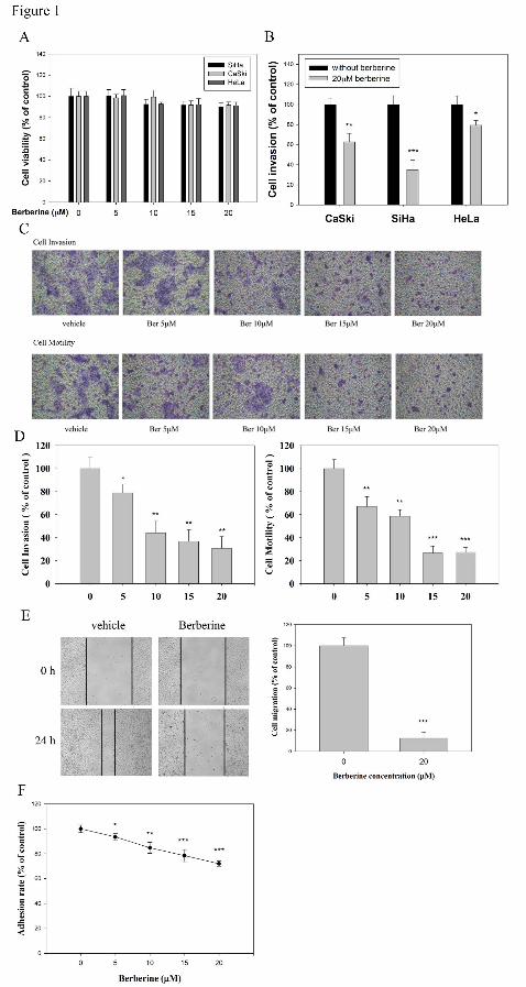
This article has not been copyedited and formatted. The final version may differ from this version.Molecular Pharmacology Fast Forward. Published on September 12, 2014 as DOI: 10.1124/mol.114.094037
at ASPE
T Journals on M
ay 28, 2019m
olpharm.aspetjournals.org
Dow
nloaded from

This article has not been copyedited and formatted. The final version may differ from this version.Molecular Pharmacology Fast Forward. Published on September 12, 2014 as DOI: 10.1124/mol.114.094037
at ASPE
T Journals on M
ay 28, 2019m
olpharm.aspetjournals.org
Dow
nloaded from

This article has not been copyedited and formatted. The final version may differ from this version.Molecular Pharmacology Fast Forward. Published on September 12, 2014 as DOI: 10.1124/mol.114.094037
at ASPE
T Journals on M
ay 28, 2019m
olpharm.aspetjournals.org
Dow
nloaded from

This article has not been copyedited and formatted. The final version may differ from this version.Molecular Pharmacology Fast Forward. Published on September 12, 2014 as DOI: 10.1124/mol.114.094037
at ASPE
T Journals on M
ay 28, 2019m
olpharm.aspetjournals.org
Dow
nloaded from

This article has not been copyedited and formatted. The final version may differ from this version.Molecular Pharmacology Fast Forward. Published on September 12, 2014 as DOI: 10.1124/mol.114.094037
at ASPE
T Journals on M
ay 28, 2019m
olpharm.aspetjournals.org
Dow
nloaded from

This article has not been copyedited and formatted. The final version may differ from this version.Molecular Pharmacology Fast Forward. Published on September 12, 2014 as DOI: 10.1124/mol.114.094037
at ASPE
T Journals on M
ay 28, 2019m
olpharm.aspetjournals.org
Dow
nloaded from
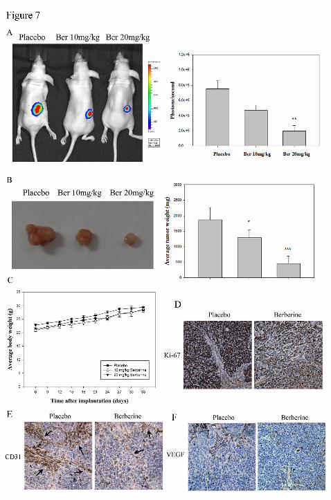
This article has not been copyedited and formatted. The final version may differ from this version.Molecular Pharmacology Fast Forward. Published on September 12, 2014 as DOI: 10.1124/mol.114.094037
at ASPE
T Journals on M
ay 28, 2019m
olpharm.aspetjournals.org
Dow
nloaded from
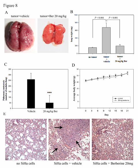
This article has not been copyedited and formatted. The final version may differ from this version.Molecular Pharmacology Fast Forward. Published on September 12, 2014 as DOI: 10.1124/mol.114.094037
at ASPE
T Journals on M
ay 28, 2019m
olpharm.aspetjournals.org
Dow
nloaded from

This article has not been copyedited and formatted. The final version may differ from this version.Molecular Pharmacology Fast Forward. Published on September 12, 2014 as DOI: 10.1124/mol.114.094037
at ASPE
T Journals on M
ay 28, 2019m
olpharm.aspetjournals.org
Dow
nloaded from