benign paroxysmal positional vertigo and treatment in
TRANSCRIPT

Camilla Martens
Benign paroxysmal positionalvertigo and treatment in biaxialrotational chair
2021
Thesis for the degree of Philosophiae Doctor (PhD)University of Bergen, Norway

at the University of Bergen
Avhandling for graden philosophiae doctor (ph.d )
ved Universitetet i Bergen
.
2017
Dato for disputas: 1111
Camilla Martens
Benign paroxysmal positional vertigoand treatment in biaxial rotational chair
Thesis for the degree of Philosophiae Doctor (PhD)
Date of defense: 23.04.2021

The material in this publication is covered by the provisions of the Copyright Act.
Print: Skipnes Kommunikasjon / University of Bergen
© Copyright Camilla Martens
Name: Camilla Martens
Title: Benign paroxysmal positional vertigo and treatment in biaxial rotational chair
Year: 2021

Scientific Environment
The studies that are included in this thesis emerged from National Advisory Unit onVestibular Disorders at the Department of Otolaryngology, Head and Neck Surgeryin collaboration with The Department of Clinical Medicine, University of Bergenand Dr. Karl Fredrik Nordfalk at the Department of Otorhinolaryngology and HeadNeck Surgery, Oslo University Hospital.The principal supervisor was Professor Stein Helge Glad Nordahl and the co-supervisorwas Dr. Frederik Kragerud Goplen. The Norwegian National Advisory Unit onVestibular Disorders, have funded this PhD and made the present study possible
ii

Acknowledgements
Upon accomplishing this thesis, a flow of grateful thoughts comes to mind. I feel
deeply thankful and humble to a lot of people, not only for their addition to scientific
knowledge, but also for my personal experience from the privilege of meeting and
working alongside some of them. It is undeniable that this dissertation, would not
have been possible without its many contributors. The mentorship, scientific envi-
ronment and guidance that I have been offered have been of the best material from
which my graduate education could be made, likewise the support and knowledge
that I have received from my colleagues have been fundamental for this work.
This PhD project has opened a new world of science and knowledge to me. I was first
introduced to vestibular diseases completing an observer-fellowship in Otolaryngol-
ogy Head and Neck surgery/Skull base surgery at St. Vincent Private Hospital in
Sydney in 2011, which was supervised by the brilliant surgeon Professor Paul Fagan
who greatly inspired my interest in scientific work. [1] Our article was rewarded
with Best Paper Prize with a public price reward received in London 2014. He also
introduced me to the world-renowned Professor Gabor Michael Halmagyi at Royal
Prince Alfred Hospital, one of the inventors of the head impulse test. When I was
offered a position as a PhD student at the Norwegian National Advisory Unit on
Vestibular Disorders, I felt very lucky to be offered the opportunity to learn from
two of the national specialists in vestibular diseases in Norway, Professor Stein Helge
Glad Nordahl and Dr. Frederik Kragerud Goplen.
I would like to give my deepest appreciation to my main supervisor Stein Helge
Glad Nordahl the brain, dynamo and initiator behind this project. He is a coura-
geous man with immense energy and brilliant ideas. He explained how to write a
paper and how to present my work effectively. He has the personality to find possi-
bilities and fresh new perspectives in any situation, and is able to pull through when
things seem difficult. You have been a great support through the whole process and
have always been there to help me. I would like to thank you for introducing me to
iii

research and all the opportunities that you have given me by presenting me to your
vast scientific network. You have given me great encouragement throughout the
whole process. I would also like to thank my co-supervisor Frederik Kragerud Go-
plen, an academic analysing and resourceful wise man who has helped me with all of
the articles––thank you for your constructive suggestions and valuable supervision!
He has given me the essential tools and guidance to choose the right direction and
successfully complete my dissertation. He always found a way through my chaos!
I could never have done this without my supervisors, who complement each other
very well.
I would also like to thank my highly skilled, positive and enthusiastic colleague
and inventor: Torbjøn Aasen, who is a true bon vivant, both great with numbers and
essential questions. He makes research exiting by bringing in new ideas, constructing
the most complicated analysing tools and simplifying complicated reasoning. Work-
ing closely with you all of you has opened a whole new world in the scientific field.
Through you I have got to know and been fortunate to visit brilliant minds such
as Professor Halmagyi and Ian Curthoys in Sydney, Dr. Hain in Chicago, Professor
Shepard at the Mayo Clinic in Rochester, Professor Herman Kingma in Maastricht,
Dr. Dai and Dr. Yakushino in New York and also to participate in the expert panel
in Copenhagens international TRV seminar. This medical research-journey has re-
ally been inspiring! Also, my thanks go to The Norwegian Medical Association for
awarding me with travel grants to support some of these travels.
My thanks also go to the staff at the Department of Otorhinolaryngology, Head
and Neck Surgery, Haukeland for making room for my research. I would like to ex-
press my sincere gratitude to all the patients who participated in this study. Special
thanks to Sofia Ekstom, Linda Karin Fauske, Anna Molbo, who have helped me
with the vestibular testing and the logistics. I am also grateful for insightful exper-
tise and discussions with the big expert on Dizziness Handicap Inventory, Kjersti
iv
Thulin Wilhelmsen. My thanks also go to Vegard Løvby Solhaug for his sparkling
interest and expertise in vestibular rehabilitation and his positive energy. I thank
Mari Kalland Knapstad for her great personality and useful discussions. I would
also like to thank Thor Andre Ellingsen for his expertise in photography and for his
help with the photos that I used in our articles. I would like to express my special
thanks to my highly appreciated colleague Elisabeth Thorkildsen, for her invaluable
help in organising and collecting information from the research participants. With-
out her I would be lost.
My thanks go to Rolf Gjestad for his expertise in statistical matters and his great
contribution in the helping me with scrutiny of data. I also thank to my co-PhD
colleagues Kathrin Nilsen and Jan Erik Berge for their interest in vestibular research
and important discussions. My thanks go to my loving parents who are the founda-
tions of everything that I am and have achieved. Extra thanks to my mother Else
Kobbe and my stepfather Harald Kobbe for guiding and for cheering me through
the whole prosess. Ekstra thanks also go to my father Johan Martens and my step-
mother Brigitte Krohn for always being there to support me and being an endless
source of positive energy. Thanks to Petter Gjersvik for your bright spark in struc-
ture and sharp linguistic guidance. And finally, thanks to Consultant Stein Lybak
for allowing me extra time and finish my PhD studies and to professor Hans Jørgen
Aarstad for his broad and deep competence in research and for his useful advice
both during the study and during the work finalising the Thesis.
This work is dedicated to my beloved husband, Thomas who has supported me
throughout the whole prosess, my mum, my dad, Sophie, Lars, Per and all members
of my family.Your love is the joy in my life and you mean the world to me.
Strong people don’t put others down, they lift them up.
– Michael P Watson
v

Abbreviations
Alexander’s law: Nystagmus of peripheral vestibular origin that is enhanced with
gaze toward the fast phase.
Ampullofugal flow: Flow of endolymph away from the ampulla.
Ampullopetal flow: Flow of endolymph toward the ampulla.
Apogeotropic nystagmus: Nystagmus with movement directed away from the
earth.
Canalithiasis: Subtype of BPPV where otoconia are free floating within the semi-
circular canal.
Cupulolithiasis: Subtype of BPPV where otoconia are attached to cupula.
Ewald’s Laws
1st Law: The axis of nystagmus shall match the anatomic axis of the semicircular
canal that generated it.
2nd Law: In the horizontal canal an ampullopetal flow of endolymph produces a
stronger response; whereas in the anterior and posterior canal, an ampullofugal flow
produces a stronger response.
Geotropic nystagmus: Nystagmus movement directed toward the earth.
HSB: High-speed barbecue manoeuvre.
ML: Modified Lempert manoeuvre.
Oscillopsia: Blurring or jumping disturbance of the visual field––the objects oscil-
late.
PN: Positional nystagmus.
PPPD: Persistant Postural Perceptual Dizziness
Semicircular canals: Three circular ducts filled with fluid located in the inner ear
responsible for the detection of angular accelerations.
AC-BPPV: Anterior semicircular canal BPPV.
HC-BPPV: Horizontal semicircular canal BPPV.
PC-BPPV: Posterior canal BPPV.
SPV: Slow-phase Velocity.
vi

SM: Sham maneouvre.
TRV: Mechanical chair for treatment of BPPV named after the inventor Thomas
Richard-Vitton.
VNG: Videonystagmography.
vii

Papers included in the present thesis
Paper I
Martens C, Goplen FK, Nordfalk KF, Aasen T, Nordahl SHG.
Prevalence and Characteristics of Positional Nystagmus in Normal Subjects
Otolaryngol Head Neck Surg. 2016 May;154(5):861-7. doi: 10.1177/0194599816629640.
Epub 2016 Feb 23.
Paper II
Martens C, Goplen FK, Aasen T, Nordfalk KF, Nordahl SHG.
Dizziness handicap and clinical characteristics of posterior and lateral canal BPPV
Eur Arch Otorhinolaryngol. 2019 Aug;276(8):2181-2189. doi: 10.1007/s00405-019-
05459-9. Epub 2019 May 14.
Paper III
Martens C, Goplen FK, Aasen T, Nordfalk KF, Gjestad R, Nordahl SHG.
Treatment of horizontal canal BPPV-a randomized sham-controlled trial comparing
two therapeutic maneuvers of different speeds
Laryngoscope Investig Otolaryngol. 2020 Jun 29;5(4):750-757. doi: 10.1002/lio2.420.
eCollection 2020 Aug.
Reprints of the papers were made with permission from the publishers
viii

Contents
Scientific Environment ii
Acknowledgements vi
Abbreviations vii
List of Publications ix
List of Figures xii
1 ABSTRACT 1
2 INTRODUCTION 4
2.1 History . . . . . . . . . . . . . . . . . . . . . . . . . . . . . . . . . . . 5
2.2 Epidemiology . . . . . . . . . . . . . . . . . . . . . . . . . . . . . . . 7
2.3 Etiology . . . . . . . . . . . . . . . . . . . . . . . . . . . . . . . . . . 7
2.4 Pathogenesis and pathophysiology . . . . . . . . . . . . . . . . . . . . 8
2.5 Otoconia . . . . . . . . . . . . . . . . . . . . . . . . . . . . . . . . . . 9
2.6 Clinical presentation . . . . . . . . . . . . . . . . . . . . . . . . . . . 10
2.7 Subtypes and examination . . . . . . . . . . . . . . . . . . . . . . . . 11
2.7.1 Canalolithiasis of the posterior canal . . . . . . . . . . . . . . 11
2.7.2 Canalolithiasis and cupulolithiasis of the horizontal canal . . . 12
2.7.3 Anterior canal BPPV . . . . . . . . . . . . . . . . . . . . . . . 13
2.8 Therapy . . . . . . . . . . . . . . . . . . . . . . . . . . . . . . . . . . 16
2.8.1 Side effects of treatment . . . . . . . . . . . . . . . . . . . . . 19
2.8.2 Relapses . . . . . . . . . . . . . . . . . . . . . . . . . . . . . . 19
2.9 Treatment chairs . . . . . . . . . . . . . . . . . . . . . . . . . . . . . 19
2.9.1 Potential risk factors . . . . . . . . . . . . . . . . . . . . . . . 21
2.10 Differential diagnosis . . . . . . . . . . . . . . . . . . . . . . . . . . . 21
2.11 Present challenges . . . . . . . . . . . . . . . . . . . . . . . . . . . . . 22
ix
3 AIMS OF THIS THESIS 24
4 MATERIALS AND METHODS 26
4.1 Ethics . . . . . . . . . . . . . . . . . . . . . . . . . . . . . . . . . . . 26
4.2 Design . . . . . . . . . . . . . . . . . . . . . . . . . . . . . . . . . . . 26
4.3 Subjects . . . . . . . . . . . . . . . . . . . . . . . . . . . . . . . . . . 26
4.4 Clinical examination . . . . . . . . . . . . . . . . . . . . . . . . . . . 28
4.5 Biaxial rotational chair . . . . . . . . . . . . . . . . . . . . . . . . . . 29
4.6 Electronystagmography and nystagmus . . . . . . . . . . . . . . . . . 30
4.7 Questionnaires . . . . . . . . . . . . . . . . . . . . . . . . . . . . . . 31
4.8 Statistical methods . . . . . . . . . . . . . . . . . . . . . . . . . . . . 32
5 SUMMARY OF THE MAIN RESULTS 33
5.1 Summary of the results of Paper I . . . . . . . . . . . . . . . . . . . . 33
5.2 Summary of the results of Paper II . . . . . . . . . . . . . . . . . . . 33
5.3 Summary of the results of Paper III . . . . . . . . . . . . . . . . . . . 34
5.4 Adverse events . . . . . . . . . . . . . . . . . . . . . . . . . . . . . . 35
6 DISCUSSION 36
6.1 Discussion of the main results . . . . . . . . . . . . . . . . . . . . . . 36
6.1.1 Prevalence and clinical characteristics of positional nystagmus
in normal subjects . . . . . . . . . . . . . . . . . . . . . . . . 36
6.1.2 Dizziness handicap and clinical characteristics of posterior and
lateral canal BPPV . . . . . . . . . . . . . . . . . . . . . . . . 37
6.1.3 Serum 25-hydroxy vitamin D and its relevance in BPPV . . . 38
6.1.4 Variations of duration in posterior and lateral canal BPPV . . 39
6.1.5 Effect of adding kinetic energy . . . . . . . . . . . . . . . . . . 40
6.2 Internal validity . . . . . . . . . . . . . . . . . . . . . . . . . . . . . . 40
6.3 External validity . . . . . . . . . . . . . . . . . . . . . . . . . . . . . 41
7 CONCLUSION AND FUTURE PROSPECTS 43
x

7.1 Nystagmus . . . . . . . . . . . . . . . . . . . . . . . . . . . . . . . . 43
7.2 Symptoms . . . . . . . . . . . . . . . . . . . . . . . . . . . . . . . . 43
7.3 25 hydroxy vitamin D . . . . . . . . . . . . . . . . . . . . . . . . . . 43
7.4 Manoeuvres of different speed . . . . . . . . . . . . . . . . . . . . . . 44
7.5 Future prospects . . . . . . . . . . . . . . . . . . . . . . . . . . . . . 44
References 64
A Appendices: Questionnaires 65
B Appendices: Article I-III 65

List of Figures
1 A Dix-Hallpike test in a TRV chair . . . . . . . . . . . . . . . . . . . 11
2 Stepwise Epley for the left posterior canal . . . . . . . . . . . . . . . 17
3 Stepwise barbecue for the left horizontal canal . . . . . . . . . . . . . 18
4 Positional tests in a biaxial chair . . . . . . . . . . . . . . . . . . . . 29
xii

1 ABSTRACT
The overall aim of this thesis was to examine a new standardised intervention-tool to
provide the best possible treatment for patients with benign paroxysmal positional
vertigo (BPPV). BPPV is defined as short attacks of vertigo and nystagmus, which
is elicited by certain provocative head positions. In particular, this work consists
primarily of three multicentre studies.
The most significant factor in diagnosing BPPV is the examination and eval-
uation of the characteristics of the nystagmus that is triggered by the diagnostic
positional manoeuvres. The character and direction of the nystagmus are specific
to the semicircular canal that is affected and the subtype of BPPV. It is important
to consider the clinical relevance of nystagmus. For this evaluation, it is crucial to
acquire knowledge about the prevalence and quality of positional nystagmus in the
normal population. However, there is ass yet no consensus on the interpretation
and standardisation of positional nystagmus. Paper I aimed to investigate clinical
characteristics and prevalence of nystagmus in normal subjects. To our knowledge,
this is the first published study to explore positional nystagmus in healthy subjects
in standardised test positions in a particle repositioning chair.
We included 75 adult subjects without a history of vertigo or balance disorder. The
healthy subjects underwent six standardised positional tests, which are identical to
those used to diagnose BPPV patients in repositioning chair. We used videonys-
tagmography to record the eye movements in every test position. We included and
analysed 1329 files of the 1350 videos recorded. Our results showed positional nys-
tagmus in 88% of the healthy subjects in one or more of the test positions, and
in 55% of the subjects in Dix-Hallpike position. The 95th percentile of the max-
imum slow-phase velocity was 5.1 degrees pr. second in the horizontal plane and
6.5 degrees pr. second in the vertical plane. Our study has shown that positional
nystagmus, especially in the Dix-Hallpike position, which is a part of the test for
BPPV, is a common finding in healthy subjects. However, the nystagmus charac-
1

teristics were not similar to the positional nystagmus that is seen in BPPV patients
because it was non-paroxysmal and of slow velocity.
BPPV is diagnosed and divided into the following subtypes: anterior, posterior
and horizontal canal BPPV. In Paper II, we investigated whether the degree of pa-
tient reported symptoms were related to posterior or horizontal canal affection and
canal specific nystagmus intensity because this could have clinical and therapeutic
consequences.
We analysed 132 consecutive BPPV patients with confirmed BPPV by standardised
procedures under diagnostic manoeuvres in a biaxial rotational chair with videonys-
tagmography control. We found that higher DHI scores were associated with hori-
zontal canal BPPV and female gender. Horizontal canal BPPV was associated with
longer symptom duration and lower 25-hydroxyvitamin D levels. We investigated
the association with D-vitamin levels because imbalance in the metabolism of cal-
cium carbonate crystals is associated with BPPV and a shortcoming of D-vtiamin
levels. Furthermore, we found no correlation between DHI scores and nystagmus in-
tensity. This study suggests that patients with horizontal and posterior canal BPPV
are clinically distinct. Patients with horizontal canal BPPV differ with respect to
symptom severity, vitamin D levels and duration. Patient disability was not related
to positional nystagmus slow-phase velocity.
Recently, there has been an extended use of particle repositioning chairs in the
treatment of BPPV. However, randomised studies on the treatment of lateral canal
BPPV in particle repositioning chairs are lacking and the important question of
whether or not to add kinetic energy to the treatment of HC-BPPV has remained
unanswered. In Paper III, we performed a randomised single blinded placebo-
controlled trial that included 57 patients with horizontal canal BPPV. In this study,
we wanted to analyse whether adding kinetic energy through acceleration and de-
celeration in the treatment of horizontal canal BPPV in a biaxial chair had an ef-
2

fect. We evaluated the therapeutic efficacy of treatment for horizontal canal benign
positional vertigo with acceleration and deceleration (high-speed barbecue) HSB,
without acceleration (modified Lempert manoeuvre) ML and sham (sham manoeu-
vre) SM. Patients were analysed after two weeks and after three months. HSB gave
a faster initial recovery. However, the cumulative response rate after three months
did not show any significant differences between the treatment groups.
In summary, our results highlights the risk of misdiagnosing BPPV patients, es-
pecially in patients with low frequency non-paroxysmal positional nystagmus. We
also found that the affected canal seems to predict symptom severity, while nystag-
mus intensity during positional testing does not. The RCT showed a better initial
recovery in patients treated with high-speed manoeuvre in horizontal canal BPPV
in a TRV chair.
3
2 INTRODUCTION
The last few decades have witnessed advances within the field of vestibular research,
and a scientific basis now exists to understand the physiology and pathophysiology
of the vestibular system. The most common peripheral vestibular disorder is Benign
Paroxysmal Positional Vertigo (BPPV). [2] If not treated properly, this disease can
develop into a more severe debilitating condition, such as PPPD. This thesis aimed
to investigate the treatment of BPPV with a new mechanical device ––the TRV
chair. The TRV chair is developed to bypass limitations in conventional reposition-
ing treatment and it offers new analytical possibilities. However, the lack of research
on the TRV chair together with more sensitive techniques for detecting nystagmus
can lead to misinterpretation, which needs further clinical surveillance and research.
Balance is important, and the capacity to maintain posture and orientation is
a fundamental skill. Our balance is regulated by the vestibule, somatic sensation
and vision. If there is a malfunction in any of these systems, then it will cause an
imbalance that will affect everyday life. Being “dizzy” or “out of balance” refers to
the old English word -dysig which meant “foolish, stupid”. From c. 1400 it was re-
ferred to as having a “whirling sensation” and in c .1500 it was described as “giddy,
thoughtless, headless”. Nystagmus comes from the Greek word nystagmos, which
means “nodding, drowsiness” and refers to the brisk movement the head makes when
falling asleep sitting up, nodding, followed by a corrective movement. All of these
words and their associated meanings describe some of the feelings and disabilities
that patients may experience when they acquire BPPV with positional dizziness
and positional nystagmus. An overwhelming whirling sensation that arrives in at-
tacks, which often leaves the patient drained of energy, giddy and with a low level of
concentration. BPPV is characterised by short episodes of a sensation of spinning
that is combined with nystagmus, which are provoked by head movements relative
to gravity. However, patients often feel off-balance, even when avoiding brisk head
movements. Vertigo is a medical expression for imbalance and dizziness, which is
4

defined as an erroneous sense of unsteadiness and motion. [3]
The quality of life scores in patients with BPPV have been found to be on
a level with patients with macular degeneration, hepatitis B and HIV/AIDS [4].
The disease adversely affects functional capacity, balance and quality of life. [5–8].
BPPV is the only vestibular disease that in most cases can be treated efficaciously
by simple intervention. However, there is often a delay in diagnosis and treatment.
This can have a tremendous cost and quality of life implications for the patients,
which leads to a great direct and indirect cost to the health care system. [5, 9] The
benefit of giving these patients an early and correct diagnosis by simple intervention
by performing particle repositioning is enormous. [10] Nevertheless, this has have
proven to be challenging because patients with BPPV who seek medical care are
more likely to undergo brain imaging than to be cured with repositioning. [11]
2.1 History
One of the earliest references to BPPV may by found in Shakespeare’s “Romeo and
Juliet” In Act I, Scene II [enter Romeo and Benvolio] Benvolio says:
“Tut man, one fire burns out another’s burning. One pain is lessened by another’s
anguish; turn giddy, and be holp by backwards turning. . . .” [12]
The first description of positional vertigo in the medical literature was by Adler
[13] and Barany [14]. Robert Barany received the Nobel Prize in 1914 in physiol-
ogy/medicine for his work on the pathology and physiology of the vestibular appa-
ratus. He was the first to describe BPPV in detail in 1921. In 1952 Margaret Dix
and Charles Hallpike [15] presented the currently used provocative positioning tech-
nique. They also defined the clinical manifestations of posterior BPPV, as elicited
by the Dix-Hallpike test.
The pathophysiology of BPPV was not understood until some time later. Pro-
5

fessor of Physiology, Julius Ewald (1855–1921) stated three important laws with
respect to the vestibular system after his studies in pigeons. In his first law, he
declared that the direction of nystagmus should match the anatomic axis of the
semicircular canal that is stimulated. In his second law, he stated that an ampul-
lopetal (towards the vestibule) movement of fluid in the horizontal canal causes the
strongest response, and in the posterior and anterior canals an ampullofugal (away
from the vestibule) fluid movement causes the strongest response. These statements
led to further understanding of the disease.
Harlod Schuknecht (1917-1996), who had a passionate interest of the temporal bone,
proposed that utricular otoconia acting upon the cupula could cause BPPV. He
stated that ” to study the temporal bone on a Sunday morning is closer to religion
than visiting at church services” [16]. Thanks to his insight, the pathophysiology of
BPPV was elucidated. In 1969, Schuknecht presented a theory of “cupulolihiasis”
which was explained by otolitic debris attached to the cupula. Hall et al. proposed
the concept of canalolithiasis with free-floating particles within the semicircular
canals in 1979 and emphasised the importance of the fatigability of nystagmus. [17]
Nevertheless, it was not until the early-1990s that the technique of canalith repo-
sition procedures became well-established and physicians to a wider extent became
aware of the treatment potential of the disease.
In 1992, Epley published his first report on the Epley manoeuvre, thereby intro-
ducing a new form of treatment for BPPV. This may be considered a major break-
through because the disease could now be treated effectively with simple reposi-
tioning manoeuvres. Parnes and McClure made a modification to the concept of
BPPV by proposing that BPPV was not a result of cupulolithiasis but was caused
by a hydrodynamic gravitational pull exerted by debris on the endolymph. [18] In
a recent article, Kao et al. [19] used scanning electron microscope to confirm the
presence of otolitic membrane with embedded free-floating particles in patients with
BPPV.
6

2.2 Epidemiology
A recent epidemiological survey of 70 million individuals found that BPPV with a
prevalence of 0.5 percent was the most common vestibular disease. [2] The cumula-
tive incidence of BPPV is around 10 percent by the age of 80. [20] The prevalence
of unrecognised BPPV has been found to be 9% in both a prospective study of 198
young adults aged from 18–34 years and in a geriatric clinic. [6,21] BPPV can occur
throughout the lifespan but the incidence increases with age, and is most common
between the fifth and seventh decades of life. It is also more prevalent among fe-
males. [5]. A familial tendency has been suggested because patients with BPPV
have been found to be five times more likely to have relatives with BPPV affection
compared to other patients with dizziness. [22]
2.3 Etiology
The accepted cause of BPPV are ectopic gravity-sensitive calcium carbonate parti-
cles (otoconia) that migrate from the utricle and inadvertently drift into the lumen of
the semicircular canal (canalolithiasis). The otoconia can also attach to the cupula
(cupulolithiasis) [18,23]. This generates attacks of positional nystagmus and vertigo.
It is not the debris or otoliths but the hydrodynamic effect that disturbs the normal
endolymphatic flow and bending of the cupula that provokes vertigo and creates
the vestibular disturbance. The ampulla has become a gravitational sensor rather
than a rotation sensor in cupulolithiasis. In cupulolithiasis vertigo is provoked by
rotation. A degenerating otolith apparatus is presumed to be an overall component
for increased risk of BPPV. [24] Over half of all BPPV episodes are primary BPPV
(idiopathic origin). [25, 26]
Secondary BPPV has been reported to be linked with a with several underlying
conditions, including migraine, [27] dental treatment, [28] ear surgery, [29] head
trauma, [30] or other inner ear problems. [31] Interestingly, patients with Meniere’s
disease who develop secondary BPPV are more likely to develop lateral canal in-
volvement, [32] and patients with traumatic BPPV are more prone to get a bilateral
7

involvement [33] and more positioning manoeuvres are often necessary to obtain
relief. [34]
Recent studies that have focused on the risk factors for BPPV have found that low
25-hydroxy vitamin D levels and osteoporosis can be risk factors for both the de-
velopment and recurrence of BPPV. [35–37] A study from Parnes et al. also found
levels of bone turnover to correlate with BPPV. [38] Changes in female sex hormone
levels have been shown to associated with BPPV, and a recent study showed that
low estradiol levels may lead to increased risk of BPPV in postmenopausal female
patients. [39] Another study found that age related increases in BPPV were reversed
in women taking oestrogen replacement therapy. [40] Uric acid levels and lipid pro-
files have been found to be higher in patients with BPPV when compared to controls.
Interestingly, uric acid levels have been shown to decrease one month after a BPPV
attack. [41] Several recent studies have indicated that the calcium metabolism’s rela-
tionship with oxidative stress might play a role in the development of BPPV. [42,43]
Sleep disorders, prolonged bedrest and diabetes may also be potential risk factors
for BPPV. [44, 45] Ototoxic medications like Gentamycin can increase the risk of
BPPV. [46]A study that analysed the comorbidities in 1000 BPPV patients found
that patients with a high number of comorbidities had a higher risk of recurrences
in BPPV. [47]
2.4 Pathogenesis and pathophysiology
The inner ear contributes to both balance and hearing. The vestibular system rises
from an elegant network that is designed to perceive the movement and orientation
of the head with unique precision, thus guiding our movement and stabilising our
visual environment relative to gravity.
The peripheral vestibular system works together with our proprioception, vision and
brain to help us maintain our balance.The vestibular complex is an important end
organ for balance and it contains receptors for our sense of equilibrium. It consist of
five end organs: the sacculus, the utriculus and the three semicircular canals. The
8

three semicircular canals are organised orthogonally to each other, which enables us
to detect the direction and amplitude of the head’s movement in three spatial planes.
The two vertical semicircular canals are oriented at 45 degrees in the saggital plane
and the horizontal canals are tilted upward by approximately 30 degrees. Movement
of the semicircular canals will activate the vestibular system and will generate the
vestibulo-ocular reflex (VOR) with adjusted eye movements to preserve gaze stabil-
ity, because the head’s movements are followed by compensatory eye movements.
This helps us to stabilise vision and muscle tone during movement. The vestibular
complex receives a constant flow of information, including when standing and sit-
ting. A change in nerve firing conducts information on how the head is turned with
regard to rotation and acceleration.
2.5 Otoconia
A certain mass of otoconia is needed to trigger symptoms. [48] House et al’s math-
ematical model [49] estimated that 62 otoconia of approximately 10 micrometers
must be involved to produce symptoms. Movement of the otoconia causes an en-
dolymphatic fluid motion which stimulates the ampullary receptors and thereby
elicits vertigo, which again explains the symptoms and nystagmus findings. As the
crystals move, the brain gets false messages that the individual is spinning. The
characteristics of nystagmus (i.e. latency time, fatiguability, direction and tran-
siency) differ between canalo- and cupulolithiasis, and depends on which vestibular
canal is affected.
Otoconia weigh 2.95 grams pr. cubic cm, which is three times the density of wa-
ter and is the reason they sink in the endolymph. Otoconia are most often hexagonal
in form. They are normally between 3-30 micrometers long. Because of their mass
they sink in the endolymph. A recent study has examined particulate matter in-
traoperatively from two patients undergoing posterior canal occlusion surgery, and
9

has shown otoconia measuring roughly 2-8 micrometers. [19] Otoconia are com-
posed of organic (mainly otoconin 90) and inorganic components, which facilitate
mineralisation of calcium carbonate around the inorganic matrix to form calcite
crystals. Otoconia are anchored together by otolinbased-fibrils. The otolinbased-
fibrils are affected by ageing and various disease processes (e.g endolymph change
in pH). This includes conditions were otoconia can fragment and separate from the
otoconial membrane. Calcium concentration in the endolymph appears to have a
considarable influence on the rate of resorption of otoconia and may affect the dura-
tion in a BPPV episode. [50] The involvement of particulate matter has been found
intraoperatively in patients with BPPV. [18, 19] However, this particulate matter
was not found in patients without BPPV. [51] Studies in animals [52–56] and math-
ematical models [57] support the pathological findings in BPPV. Squires et al. [48]
found in their model that an average-sized otoconia requires around 5 seconds to
exit the ampulla, which can explain the latency in positional nystagmus in BPPV
patients. The latency was predicted to vary depending on the localisation of the
otoconia.
2.6 Clinical presentation
Patients typically present with vertigo of short duration elicited by certain head
movements such as when lying down or turning over in bed. The onset is often
after waking up in the morning. The history can sometimes predict which side
is affected because the position that initially caused the vertigo is reluctant to be
the involved side. [58] Circular acceleration triggers symptoms in canalolithiasis,
whereas position change triggers symptoms in cupulolithiasis. Common symptoms
with clinical presentation are rotational vertigo (86%), imbalance (49%), nausea
(33%), oscillopsia (31%), vomiting (14%), fear of falling (36%) and falls (1%). [5]
Interestingly, older patients (i.e. patients older than 70) in 31% of the cases report
symptoms that are described by unsteadiness and imbalance rather than vertigo.
[59,60]
10

2.7 Subtypes and examination
2.7.1 Canalolithiasis of the posterior canal
Dix-Hallpike:
The most common form of BPPV is canalolithiasis of the posterior canal which ac-
counts for 85 % of the cases. [61,62] The essential clinical manifestations of posterior
canal BPPV are elicited by the Dix-Hallpike test, which is performed by rotating
the patient’s head 45 degrees to the side that is examined. This is done to make the
posterior semicircular canal parallel to the saggital plane of the body in the seated
position. The patient is thereafter moved from the seated to the supine position
with the test-ear down and the neck extended approximately 20 degrees. Patients
are observed for 30 seconds. In a positive test, after a short latency there will be a
vertical up-beating nystagmus in combination with the upper pole of the eye beating
toward the affected ear. Commonly a crescendo-decrescendo pattern is detected.
Figure 1: A Dix-Hallpike test in a TRV chair
Geotropic torsional-up beating nystagmus observed during the Dix-Hallpike test
indicates that the otoconial debris is descending from the posterior canal ampulla
11

of the lowermost ear. The vertigo typically lasts less than 30 seconds in PC-BPPV.
2.7.2 Canalolithiasis and cupulolithiasis of the horizontal canal
Horizontal canal BPPV (HC-BPPV) is triggered in the the supine position where
the head is turned 90 degrees towards each side and it is diagnosed by the presence
of direction-changing horizontal paroxysmal positional nystagmus. The horizontal
canal BPPV is the second-most frequently affected semicircular canal, with an inci-
dence of around 10-15%. [63–65] The geotropic type (nystagmus beating toward the
lower ear) of horizontal nystagmus indicates canalolithiasis of the lateral canal on
the side with the more intense nystagmus, whereas the apogeotropic apogeotropic
(beating toward the upper ear) variety is indicative of cupulolithiasis (or canaloli-
hiasis of the short arm of the lateral canal) with affection on the side with the less
intense nystagmus. The nystagmus is more intense towards the affected side ac-
cording to Ewalds 2nd law, which tells us that ampullopetal endolymphatic flow
(displacement of otoconia toward the ampullated end) produces a stronger response
than ampullofugal flow (displacement of otoconia away from the ampullated end) in
the horizontal canal. In HC-BPPV vertigo usually lasts longer (about 90 seconds),
is more intense and has a shorter latency. HC-BPPV can be difficult to treat, and
persistence of symptoms from HC-BPPV varies. [66–72] A recent study found that
in a group of intractable BPPV patients, 61% of the patients had horizontal canal
affection. [73] Additionally, studies show a lower recovery rate for apogeotropic HC-
BPPV. [67, 74] It can, however, sometimes be difficult to determine the affected
side based on nystagmus type and subjective symptoms. In the literature different
signs for locating the affected side in complicated cases have been investigated and
described. These phenomena have been referred to as “secondary signs of later-
alisation” by Califano et al. [75] Help in defining the involved side may be found
in “pseudo-spontaneous nystagmus” (PS), [76]” neutral position test,” (NP), [77]
”bowing and leaning nystagmus” [78]and look for the direction of nystagmus in the
sitting to supine position test (STS). [67] The bow and lean nystagmus are based
12

on Ewald’s second law. The patient sits on the exam table facing the clinician.
The patient is than asked to bow their head 90 degrees forward and the direction,
amplitude, and duration of any nystagmus is recorded. The patient thereafter tilts
their head backward 45 degrees and again the characteristics of the nystagmus are
documented. The affected ear in geotropic nystagmus is the ear where the nys-
tagmus is beating towards in the bowing position. The affected ear in apogeotropic
nystagmus is the ear where the nystagmus is beating towards in the leaning position.
Canalolithiasis of the horizontal canal) Lateral canal canalolithiasis produces a
paroxysmal geotropic positional nystagmus after short or no latency. The nystag-
mus has a shorter onset latency then with posterior canal BPPV and crescendo-
decresendo pattern is normally of longer duration than posterior canal BPPV, but
under one minute. [79]
Cupulolithiasis of the horizontal canal) Lateral canal cupulolithiasis produces a
paroxysmal geotropic positional nystagmus after short or no latency, and is char-
acterised by persistent apogeotropic positional nystagmus normally lasting longer
than one minute and typically builds up slowly over 30 seconds and then gradually
declines. [79]
2.7.3 Anterior canal BPPV
Dix-Hallpike:
The prevalence of anterior canal BPPV (AC-BPPV) is probably very low because
of its anatomical location. There is still some discussion around the diagnosis and
treatment of AC-BPPV, and one should always keep central pathology in mind. A
review article by Anagnostou et al. found that anterior canal BPPV only comprises
about 3% of all BPPV cases. [80] This type of BPPV is characterised by nystagmus
that is predominantly down beating in Dix-Hallpike position, as shown in Figure 1
Atypical presentations:
13
Apogeotropic posterior canal BPPV (Posterior cupulolithiasis)
Some patients may present with a torsional down beating in the Dix-Hallpike
position. This may represent otoconia in the distal arm of the posterior canal.
When the patients are positioned in Dix-Hallpike for testing, the otoconia move
from the area of the common crus towards the ampulla and produces an inhibitory
nystagmus that is torsional down beating. There is normally no latency, but has
time course with crescendo –decrescendo pattern and nystagmus is not completely
exhaustible. When the patient is raised to the upright position this does not reverse
the nystagmus direction, and the nystagmus does normally not fatigue on repeated
positional testing. [81]
14

Table 1. BPPV diagnostic criteria according to the Consensus document of the Committee for the Classification of Vestibular Disorders of the Bárány Society (von Brevern et al 2015)
Posterior canalithiasis
Lateral canalithiasis
Lateral cupulolithiasis
Anterior canalithiasis
Lithiasis of multiple canals
Recurrent attacks of positional vertigo or
positional dizziness provoked by lying down or
turning over in the supine position
Required
Required
Required
Required
Required
Duration of vertigo attacks
< 1 min
< 1 min
not specified
< 1 min
< 1 min
Diagnostic maneuver
Dix-Hallpike or Semont
diagnostic maneuver
supine roll test
supine roll test
Dix-Hallpike or supine
straight head-hanging
position
Dix-Hallpike or supine roll test
Latency of positional nystagmus
one or few seconds
brief or none
brief or none
one or few seconds
not specified
Direction of positional nystagmus
torsional upbeat
geotropic direction
changing
apogeotropic direction
changing
predominantly downbeat
compatible with canalithiasis of
more than one canal
Duration of positional nystagmus
typically < 1 min
< 1 min
> 1 min
> 1 min
not specified
Not attributable to another disorder
Required
Required
Required
Required
Required
15

2.8 Therapy
Due to the geometry of the semicircular canals and the placement of the cupula,
there is only one exit path for the particles. Treatment is done by seeking to move
the particles out of the semicircular canals and back to utricle. Otoconial debris
seems to be dissolved by the dark cells in the labyrinth. [82] These dark cells are
found adjacent to the utricle and the crista ampullaris.
Successful treatment of BPPV requires the involved canal to be placed such
that gravity can move the particles thorough the exit path. As the mechanisms of
BPPV have become more widely accepted, treatment with repositioning has become
a better alternative than conservative management with restraint from provocative
head movements. The symptoms are resolved using either the Epley or Semont
manoeuvre in posterior canalolithiasis and cupulolithiasis. There is level 1 evidence
for the efficacy of the Epley, Semont and Gufoni manoeuvre. [83,84]
16

Figure 2: Stepwise Epley for the left posterior canal
17

Figure 3: Stepwise barbecue for the left horizontal canal
18

2.8.1 Side effects of treatment
Repositioning maneuvers: Nausea and vertigo during the procedure are common,
as is light headedness and instability around 48 hours after the procedure. In a ret-
rospective study of 1900 BPPV patients who all underwent repeated repositioning,
complications included canal conversion (3.1%), nausea (46.4%), vomiting (4.9%),
head heaviness 50.8%), imbalance (31.9%) and hypotension or palpitations (8.3%)
during or after the procedure. [85].
Surgery: Patients undergoing canal plugging often experience postoperative im-
balance, but spares hearing.
2.8.2 Relapses
BPPV typically tends to recur. The estimated 5 year recurrence risk is 30-50% . [86]
However, the risk of recurrence between the different semicircular canals is debated.
In some studies no differences has been found between the three semicircular canals
. [26, 87] In other long-term studies, there is found to be a higher recurrence rate
of horizontal canal BPPV. [88] Physical activity is considered protective for BPPV,
theoretically due to prevention of otoconial cluster-creation. [89] The presence of
abnormal caloric test in patients with horizontal canal BPPV predicts higher risk
of recurrence. [90]
2.9 Treatment chairs
Since 2005 there has been an extended use of particle repositioning chairs in the
treatment of BPPV. Mechanical assistance chairs have been designed to facilitate
and optimise the diagnostic and therapeutic manoeuvres and to increase the achiev-
ability of treatment in patients with movement restrictions. [91] Examples of cases
where adequate positioning of the patients can become a challenge by reason of
comorbidities such as mobility restrictions could include: reduced cervical mobility,
bodily injures, obesity or lower back pain. All forms of BPPV can be diagnosed and
attempted to be treated with mechanical chairs. [92] However, a total of 10-20%
19

of BPPV patients cannot be diagnosed or treated accurately with ordinary manual
manoeuvres. [93] Some authors have argued that a chair also seems to be more ef-
fective than conventional reposition. [94, 95]
The TRV chair (Interacoustics, model TRV, France), which is named after its in-
ventor (Thomas Richard-Vitton) is currently the most commonly used biaxial chair
for diagnosis and treatment of BPPV. It became commercially available in 2005. [96]
As of January 2020, the TRV chair was used by 87 centres in 26 countries. Our cen-
tre was the first place in North–Europe to acquire this chair and it has been in
use since December 2009. The TRV chair is hand-operated and can swing around
two axes, which may be locked in preset positions so that one axis is at the same
time earth-horizontal and in line with the approximate axis of any one of the six
semicircular canals. The velocity of rotation is also controlled manually. During di-
agnostics and treatment, the patient is fixed in the chair with a 4-point harness, and
is further secured with a headband and straps for the legs. The patient also wears
infrared video goggles for visualisation and quantification of positional nystagmus.
The setup in the TRV chair provides positioning manoeuvres that are exact and
reproducible for diagnosis of each semicircular canal, and further eliminates move-
ment of the spinal column that may interfere proprioceptive signals.
The use of rotational chairs for management of BPPV was pioneered by Dr. John
M. Epley (1930-2019), who developed the Omniax System. This chair is a comput-
erised positioning device with similar diagnostic precision. [97] However, this chair
is no longer being manufactured.
Furthermore, the Rotundum positioning chair (Prolim engineering GmbH) from
Switzerland, has been developed for research and treatment of BPPV.
20

2.9.1 Potential risk factors
The chair should not be used in patients above 150kg. Dumas et al. [98] gave a list of
patients who should be cautious using vibration in treatment, and the same could
be considered to be applied to treatment in the TRV chair. Recent ear surgery,
retinal detachment, history of recent cerebral hematoma and poorly controlled an-
ticoagulant therapy should all be taken into consideration before applying the chair
in treatment.
2.10 Differential diagnosis
Although BPPV is often easy to diagnose and treat, there are some considerations
and differential diagnosis that one needs to bear in mind and the differentiation
between peripheral and central positional nystagmus may be challenging. Menieres
disease and paroxysmal vertigo may also present as recurrent episodes of vertigo with
positional nystagmus. It is also well-known that central disorders may present as
BPPV-like symptoms with positional nystagmus and positional vertigo(e.g. cerebel-
lar strokes, small cerebellar haemorrhage, especially around the vermis, intracranial
tumours etc.) [99].
Vestibular migraine is a prevalent diagnosis that also is also important to con-
sider. It is essential to analyse positional nystagmus and to follow diagnostic cri-
teria for BPPV [100]. Von Brevern et al. wrote a thorough consensus document
for the Committee for the Classification of Vestibular Disorders of the Barany Soci-
ety [79]. Deviations from diagnostic criteria should always warrant special attention
and differential diagnosis should be considered. A recent study compared 27 patients
with apogeotropic type of central positional nystagmus with 20 patients with apo-
geotropic lateral canal BPPV. The authors found that spontaneous horizontal nys-
tagmus differentiated little between the sitting and supine position in CPN, whereas
the augmentation of spontaneous nystagmus while supine favours the diagnosis of
apogeotropic horizontal canal BPPV. [101]
21

2.11 Present challenges
With a demographic trend towards an ageing population we can expect an increase
in patients with BPPV and also an increase in patients with concomitant immobil-
ity that for health reasons are unfit for traditional manual treatment. Therefore, an
effort should be made to identify factors to improve treatment for these challenged
patients. Chronic dizziness is common in elderly patients and persistent or recurrent
BPPV may be an important cause of these symptoms. [102]
The high prevalence of BPPV stresses the need to educate clinicians both for
early detection and treatment in primary care and for treatment of the rarer, more
complicated cases in specialised care. Finally, we need to spread knowledge about
treatment of BPPV as one of most effective and straightforward interventions in all
of clinical medicine. Particle repositioning for BPPV is largely under-utilised. [103]
The severity and nature of presenting complaints vary considerably, and this may
lead to a low recognition rate of BPPV in health care settings. In typical cases,
diagnostic imaging and vestibular laboratory testing is unnecessary. [100]
Diagnostic measurements and ineffective therapy are common. [9]
BPPV has a considerable negative impact from a humanistic viewpoint, but it is
also important to mention the noteworthy impact on work capacity and healthcare
resource use.
A study from a registry that included 13 countries with data collected from
4294 patients showed that one-third of the vertigo patients consulting health ser-
vices were diagnosed with BPPV. Another study from the same material found that
BPPV had a considerable impact on work load and healthcare resource use. Among
the persons still employed, 70% had reduced their workload, 63% had lost working
days and 6% had quit their jobs. In the three months preceding a visit, the use
of emergency services, primary care consultations and specialist consultations were
extensive [104] The failure in diagnosing BPPV may be due to poor referral patterns
22

and the low fraction of patients who receive appropriate treatment and symptom
relief from the available treatment. Older patients with imbalance and dizziness also
wait longer to seek help, and quite often do not present with the classical complains
of spinning sensation with positional changes. [59] Patients with BPPV are known
to limit their activity level, have a greater incidence of falls, impairments of their
daily activities and increased risk of depression. This is of particular concern in the
elderly population because of the increased morbidity and mortality from falls. Un-
recognised BPPV and associated morbidity is common among the elderly. A study
by Oghalai et al. uncovered unrecognised BPPV in 9% of the older population. [6]
BPPV has adverse psychosocial consequences, including severe subjective impair-
ment and avoidance behaviour in 70%. [105] BPPV sufferers also report reduced
health related quality of life, [7] and are more likely to have depression and reduced
daily activities. [6]
23
3 AIMS OF THIS THESIS
The main objective behind these studies was to gain knowledge about BPPV and
to explore a new standardised biaxial chair using consistent positioning manoeu-
vres. Mechanical chairs are currently widely used internationally in the treatment
of BPPV. A mechanical device gives us good conditions for research because it gives
us an improved analytical feasibility and repeatable test conditions. However, con-
trolled data from the normal population in the chair are missing and RCTs are
limited. Consequently, we wanted to conduct these studies where the overall aim
was to achieve better treatment for BPPV patients.
There has been a limited amount of research on the findings of positional SPV of
nystagmus in normal subjects and cut-point levels of underlying pathology in conju-
gation with VNG in specific head positions. The widespread use of VNG to detect
positional nystagmus in BPPV patients is helpful but can also be challenging be-
cause it is very sensitive to detection of nystagmus and may lead to over-diagnosis of
the condition. To analyse when to consider nystagmus to be of clinical relevance, it is
important to know the prevalence and characteristics of positional nystagmus in the
normal population, which was analysed in Paper I. Another interesting concept that
we wished to explore was whether the most frequent subtypes of BPPV (posterior
and horizontal canal BPPV) entail any divergence in patient perceived handicap be-
cause this could have clinical and therapeutic consequences which was analysed this
in Paper II. There has been an increase in the number of studies using mechanical
devices in the treatment of BPPV. These studies state that there is an increased ef-
fect in treatment efficacy compared to conventional manual treatment, [97,106–112].
However, to the best of our knowledge, no randomised placebo-controlled trials have
examined the difference in speed during treatment. [113, 114] The important ques-
tion of whether adding kinetic energy to the treatment of HC- BPPV, which is
the type of BPPV that is most persistent to treatment, [73] by using acceleration
and deceleration has until now remained unanswered. Consequently, a randomised
24

placebo-controlled trial on a biaxial chair with three months follow-up was done in
Paper III.
25

4 MATERIALS AND METHODS
4.1 Ethics
The regional committees for medical and health research ethics of western Norway
approved the protocol and consent forms in advance. All of the subjects were fully
informed about the aim of the study and the test procedures. In addition, all of
the studies were conducted in accordance with the principles of the Declaration of
Helsinki. [115] Participation was based on written informed consent. The studies
were registered at Clinicaltrials.gov. Identifier: NCT01905800
4.2 Design
Studies I and II (Papers I and II, respectively) were prospective observational multi-
centre studies. Study III (Paper III) was a multi-centre randomised controlled, single
blinded clinical trial with three months follow-up.
4.3 Subjects
The first study (Paper I) included 75 healthy subjects aged 21–87 (mean 57, SD 13)
years without any previous history of vertigo or balance disorder, and was conducted
from 2013–2015. In total 39% of the participants were male and 61% were female.
The subjects were collected among hospital staff and voluntaries who met after
having seen the advertisement for the research. The second study (Paper II) included
132 consecutive patients aged 27–90 (mean 57, SD 13) years referred with a BPPV
and confirmed with active BPPV according to international diagnostic criteria, this
study was conducted from June 2013 to June 2016. In the third study (Paper III).
647 patients with positional vertigo referred with BPPV were screened. In total,
57 patients with confirmed active lateral canal BPPV according to international
diagnostic criteria from from August 2013 to August 2017 were randomised.
26

647 Patients assessed for eligibility
589 did not meet the inclusion criteria for lateral BPPV or met the exclusion criteria
1 refused to participate
18 Randomized to high-speed barbecue
21 randomized to modified Lempert manoeuvre
18 randomized to sham manoeuvres
1 withdrawn by principle
investigator(Received a diagnosis of
Migraine)
1 withdrawn by principal
investigator(Uncovered
meningeoma)1 lost to follow
up
1 withdrawn by principal
investigator(Had taken vestibular
suppressants)
53 Completed study through day 90
57 Randomized, allocated (1:1:1)
27

4.4 Clinical examination
All three studies were performed at two university hospitals in Norway (i.e. Bergen
and Oslo). Two of the authors performed the examinations, and the same setup
and equipment were used. All of the participants underwent a clinical examination,
including an ear, nose and throat examination, head impulse testing, static postur-
ography (Synapsys, Marseille, France) and and their serum 25-hydroxy vitamin D
levels were measured. Patients in the second and third study (Papers II and III,
respectively) also underwent complete otoneurological examination with videonys-
tagmography (VNG) (oculomotor testing, spontaneous eye movements, dynamic
positioning and caloric testing (Interacoustics, Denmark). Pure tone audiometry
was performed by trained audiologists and positional tests were conducted in a bi-
axial chair by two trained medical doctors. The same setup and equipment were
used at both participating departments. [116] Mental alerting was used throughout
the tests.
28

4.5 Biaxial rotational chair
The subjects underwent a standardised examination for positional nystagmus per-
formed with video recording in a biaxial chair. Recording was performed sitting up,
in Supine test, in Dix- Hallpike and in Supine roll test /See Figure 2). In Study I,
the test pattern was random, in Studies II and III the testing started towards the
most symptomatic side, because it is suggested to be the affected side. [117]
Figure 4: Positional tests in a biaxial chair
The Dix-Hallpike test was performed in the TRV chair by turning the chair 45
degrees towards the right in the earth-horizontal axis, the chair was locked in a
preset lockable position and the patient was tilted back until the head was placed 45
29

degrees below the horizontal plane. The inverse procedure was performed for Dix-
Hallpike left. When examining the horizontal canal, the patient was positioned in
the supine position and slowly rotated 90 degrees towards the left and the right side
aiming to perform the movement with the same acceleration. The head and trunk
were stabilised with padded plates on each side to ensure that no active movement
in the cervical spine on the part of the patient would occur during testing.
Throughout positional testing, patients were fitted with a standard VNG mask
prohibiting visual fixation. The VNG mask holds an infrared camera that records
the movements of the eyes in three planes using a dark pupil tracking system with a
sampling rate of 25 Hz (Synapsys, Marseille, France). Eye shifts were also analysed
in real time visually on a wall-mounted monitor. The recordings were continued
for 30 seconds after each diagnostic manoeuvre. The changeover time of movement
from one position to another was approximately three to five seconds. All of the
subjects were primed with information to keep their eyes open and to blink as little as
possible while looking straight ahead during the examination. All nystagmography
recordings were reviewed to unmask discrepancies due to flutter, unsatisfactory pupil
detection or other types of artefacts. To keep the subjects mentally alert, they were
asked various questions during the test.
4.6 Electronystagmography and nystagmus
Nystagmus intensity was defined as the maximum slow-phase velocity (SPVmax),
which was measured in degrees per second after each diagnostic manoeuvre. Nys-
tagmus seen during the transition time of movement from one position to another
was ignored. Nystagmus was considered to be present when at least five or more
consecutive nystagmus beats with slow and fast components were identified in each
30-second VNG sequence. Geotropic and apogeotropic nystagmus was defined as
nystagmus beating towards the lower and uppermost ear in one or both side-lying
position, respectively. Six nystagmus time series —one in each head and body posi-
30

tion, were recorded for each subject in Studies I to III. The VNG-files were analysed
in a LabVIEW program that was developed for this study, and two of the authors
did a blind evaluation of the VNG-signals, selecting and measuring the area with
highest slow-phase velocity. In case of discrepancies, two of the authors indepen-
dently reviewed the recordings. The reviewers also checked for cross-talk, to see if
any of the horizontal eye movements generated software responses in the vertical
channel or opposite, caused by a tilt of goggles or the camera. When analysing SPV
as a factor we used horizontal slow-phase velocity in patients with horizontal canal
BPPV and vertical slow-phase velocity for patients with posterior canal BPPV. Due
to limitations in the VNG technology, the slow phase of torsional nystagmus could
not be measured but was quantified visually by the authors as the mean of three
independent observations on a visual analogue scale (VAS), anchored at both ends
with words descriptive of the maximum and minimum extremes of nystagmus.
4.7 Questionnaires
DHI The Dizziness Handicap Inventory (DHI) is a self-assessment inventory that
is used to assess the degree of self-perceived disability. The questionnaire was de-
veloped by Jacobson and Newman [118] and converted to Norwegian by Tamber et
al. [119] Reliability has been found to be high and sustained when translated into
Norwegian. The questionnaire contains 25 questions, where an increased disability
obtains an increased score. A value of 4, 2, or 0 points is attributed to each answer
(Yes- 4 points, Sometimes- 2 points, No- 0 points). The score gives values be-
tween 0–100 and indicates mild handicap for scores up to 30, moderate handicap for
scores between 31– 60 points, and severe handicap for scores above 60 points. The
maximum total score is 100 points. Whitney et al. [120] found that self-perceived
disability is related to functional achievement in persons with vestibular dysfunction.
A copy of the DHI questionnaire is available in the Appendix.
31

4.8 Statistical methods
In Study I, we used descriptive statistics to provide summaries about the observa-
tions.
In Study II, we used multivariable linear regression and binomial regression mod-
els to calculate OR and 95%CI to identify factors associated with dizziness-related
quality of life. Backward stepwise elimination of the least significant factor was
performed until only significant factors remained. We performed a binomial logistic
regression analysis to identify the predictors of lateral vs. posterior canal BPPV.
In Study III, we used Chi-square tests with 3x2 tables and Fishers exact test to
compare groups for primary and secondary outcome. We used non-parametric tests
because of non-normal distribution of data and also due to the small sample size.
Multiple exposure levels were used to study odds ratios. We used a linear regres-
sion model to analyse factors associated with change in dizziness-related quality of
life using DHI score as dependent variable (continuous, ranging from 0–100) and
treatment group and baseline DHI as factors. The significance level p = 0.05 was
corrected for multiple comparisons by the Bonferroni correction, (0.05/2), giving
a value of p=0.03 for significant results. Random multiple imputation(MAR) was
used to correct missing data.
Power analysis were performed in Studies II and III. SPSS 22.0 (IBM, Chicago,
Illinois), Mplus (version 8.0) and/or STATA SE 15.1 (Texas, USA) were used for
statistical evaluation in all three studies.
32

5 SUMMARY OF THE MAIN RESULTS
5.1 Summary of the results of Paper I
A total of 75 healthy subjects without a history of vertigo or balance disorders were
included in Study I.
Nystagmus prevalence: Positional nystagmus was detected in 88% of the sub-
jects in a healthy population. The most common finding was nystagmus in the
Dix-Hallpike position, which occurred in 55% of the subjects.
Nystagmus characteristics: The 95th percentile of the maximum slow-phase ve-
locity for each subject was found to be 5.1 degrees per second (n =54) in the horizon-
tal plane and 6.5 degrees per second (n = 48) in the vertical plane. The positional
nystagmus was found to be of low velocity, persistent and not paroxystic. This is
important to bear in mind when diagnosing patients with BPPV. Nystagmus that is
of low velocity, without latency and that is not paroxystic may be a normal finding.
5.2 Summary of the results of Paper II
A total of 132 patients aged 27–90 years (mean 57 years, SD 13 years) were included
in Study II. The main part of the patients (n = 103) were female. Higher DHI scores
were associated with HC- BPPV 95%CI (1.59–13.95), p=0.01 and female gender
95%CI (0.74–15.52), p=0.03. HC-BPPV was associated with longer symptom dura-
tion OR 1.10, 95% CI (1.03-1.17) p=0.01, lower 25-hydroxyvitamin D levels OR 0.80,
95%CI (0.67,0.95), p=0.03 and higher DHI scores OR 2.08, 95%(1.08,4.02),p=0.03.
The most common BPPV subtype was PC-BPPV (n=87) followed by HC-BPPV
(n=45). Episodes of previous BPPV that had resolved spontaneously or after ther-
apy were reported by 34 (26%) of the patients. Earlier head trauma within three
months prior to BPPV onset was reported by 31 (23%) of the patients. The right-
33

hand side was affected more often (61%) than the left-hand side (39%). Stress was
a provoking factor for dizziness spells in 49 (37%) of the patients. BPPV was most
common in the fifth (n=32, 24%) and sixth decades of life (n=41, 31%).
5.3 Summary of the results of Paper III
The inclusion criteria were met in 57 patients with horizontal canal BPPV who
were randomised (HSB=18; ML=21; SM=18) aged 27–78. The mean age of the pa-
tients were 57 (SD 12 years), and 68% (38) of the patients were female. The mean
duration of symptoms was 56 weeks (SD129). The duration of the disease ranged
from one day to 15 years. A total of 60% (34) of the patients had apogeotropic
nystagmus. The right-hand side was involved in 53% (30) cases. Primary outcome
was analysed in 54 patients (17 in the HSB group, 20 in the ML group and 17 in
the SM group) after two weeks because three of the patients were withdrawn by the
principle investigator due to late findings uncovering exclusion criteria. A total of
14 of 17 in the HSB group (82%), 11 of 20 in the ML group (55%), and 4 of 17
(24%) in the SM group, with significantly better recovery in HSB OR 15.17, 95%CI
(1.85,124.63), p=0.001using sham as base level. The cumulative therapeutic effect
after three months and further treatments (max 3) showed that 40 of 54 cases (75%)
had recovered. The cumulative effect was 15 of 17 (88%) in the HSB group and 15
of 19 (80%) in the ML group. There was no significant difference in cure rate after
three months between the two treatment groups 95%CI (0.30,13.14), p=0.46.
In the group of no recovery, 10 out of 14 (77%) had cupulolithiasis. Eight pa-
tients had short recurrences of BPPV during the study: two patients in the HSB
group and six patients in the ML group.
The mean DHI score before treatment was 46.1 ( SD 22.1, range: 0–96). After
3 months, the DHI score in the HSB group was 22.6 (SD 23.3, range: 0–62), in the
ML group was 22.5 (SD 23.3, range: 0–62.5) and in the SM group 33.1 (SD 28.2,
34

range: 0–90). There were no significant correlations between change in DHI and
treatment group 95% CI ( -16.56,15,02), p=0.92.
At two-weeks port treatment evaluation, complete recovery was seen in 29 of
the patients (54%) secondary outcome, recovery rate after three months were 15/17
(82%) in the HSB group and 15/19 (55%) in the ML group. Cumulative recovery
rate showed no significant differences between the two treatment groups 95% CI
(0.30,13.14), p=0.46 in cure rate or DHI 95% CI (–16.56,15.02), p=0.92. The pe-
riod for recruitment started August 2013 and ended August 2017.
5.4 Adverse events
We noticed that the patients in the HSB group showed a higher amount of vomiting
and complaints of imbalance and nausea straight after treatment than in the ML
group. Among our patients, one withdrew in the ML group because of anxiety
related to treatment in the biaxial chair. Besides vomiting and dizziness, one patient
noticed tinnitus after the Epley manoeuvre. No other side effects were reported.
35

6 DISCUSSION
Our research explored the treatment of BPPV patients in a biaxial rotatory chair,
which includes a study on positional nystagmus in the normal population Paper
I, a descriptive study of the observations of clinical characteristics of lateral and
posterior BPPV Paper II, and an RCT- exploring treatments for lateral canal
BPPV with and without adding kinetic energy to the treatment Paper III.
6.1 Discussion of the main results
6.1.1 Prevalence and clinical characteristics of positional nystagmus in
normal subjects
Paper I studied nystagmus in the healthy population. Nystagmus evaluation is
considered to be an essential element in diagnosing patients with both central and
peripheral vestibular disease. In BPPV, the quality of positional nystagmus is the
backbone of diagnosing and differentiating the affected semicircular canal. Never-
theless, positional nystagmus is common, and interpretations must be done with
care and as a part of the clinical picture. VNG is in widespread use and represents a
considerable diagnostic advancement. However, the evaluation of eye movements in
general, and nystagmus in particular, is a highly specialised clinical skill. It is also
an ongoing field of research. It is not always trivial to differentiate nystagmus in
BPPV patients from nystagmus in the normal population or from central positional
nystagmus. In a recent review, atypical nystagmus in Dix-Hallpike position was
reported in 97.5% of the patients with central positional nystagmus. [121] Several
studies on positional nystagmus in healthy populations have been performed. The
prevalence of positional nystagmus in asymptomatic human subjects has ranged
from 50-88% [122–127]
No earlier studies have been performed on healthy subjects in a biaxial chair. To
achieve normative data for our further studies we examined positional nystagmus
36

in healthy subjects in the same positions as standardised for patients investigated
for BPPV in a TRV chair. In our first study we found that 55% of the population
had nystagmus in Dix-Hallpike position, however the nystagmus was persistent and
of low amplitude. [116] Downbeat positional nystagmus has earlier been listed as a
nystagmus that is central in origin by Brandt, [128] in our study and in a study by
Levo et al. [123], downbeat was also found in healthy subjects. There is a consensus
that there should be a threshold for the SPV of nystagmus in healthy subjects,
although the thresholds found in the different normative studies vary slightly. We
found a 95th percentile to be 5 degrees per second horizontal plane, which is in line
with the recommendations from Barin et al. [127, 129, 130] ; and 6.5 degrees per
second in the vertical plane, which is similar to a recent study by Jeffery et al. [124]
6.1.2 Dizziness handicap and clinical characteristics of posterior and
lateral canal BPPV
The physical imbalance caused by BPPV is known to disable the patient’s daily life,
due to anxiety, fear and depression. [131, 132] BPPV most commonly affects the
posterior or horizontal canal. The differences in clinical characteristics were studied
in Paper II. This exploration is important due to treatment strategies and possible
priorities within this group. We conducted a study examining the patients under
standardised conditions combined with the use of international diagnostic criteria.
The two groups can be distinguished by the DHI. Horizontal canal BPPV had
a trend to give patients a higher score, indicating increased disability. Although
there have been expert opinions that lateral canal has an increased symptom score
in patients with horizontal canal BPPV, [133–135] but there has been limited re-
search. For example, Kim et al. [136] found a link between horizontal canal BPPV
and increased symptoms in terms of functional, emotional and physical limitations.
However, their study was based on a very limited number of patients with horizon-
tal canal BPPV(n=18), and the number of patients in the respective groups with
37

canalo- and cupulolithiasis of the lateral canal varied from section to section in their
paper. Furthermore, the criteria for inclusion were not set to following accordance
with international guidelines.
A recent study found that the DHI does not correspond very well with vestibular
function tests and is influenced by (for example coping) mechanisms and socio-
cultural background. [137] Even though DHI might not be a structural indicator
for vestibular deficit, it rates the patients subjective disability. Furthermore, BPPV
patients with lateral canal affection seem to have an increased perceived handicap
compared to patients experiencing posterior canal affection. This could be caused
by the fact that they suffer from more symptoms during the day time because
their symptoms can be triggered in situations such as sitting, walking, lying and
standing due to the anatomical orientation of the canals. The female patients also
experienced higher DHI scores, indicating that women experienced a higher handicap
than men. The same associations between sex and DHI have been found in other
studies on vestibular diseases by Kim et al. [136] There have been speculations that
the gender differences, where higher scores in female patients found in Menieres
and vestibular neuritis, may be caused by higher daily inconvenience and emotional
concerns. [136] The same might apply to BPPV because important behavioural and
biological differences are connected to gender. Gender differences in the quality of
life have been found in earlier reports, with lower scores in females after adjusting
for sociodemographic and chronic disease conditions. [138] Gender differences may
be important for clinical decision making. Disease management and clinical decision
making should also be considered to be adjusted to gender. [139]
6.1.3 Serum 25-hydroxy vitamin D and its relevance in BPPV
Low levels of serum 25- hydroxyvitamin D have been found in some studies to in-
crease the risk and recurrence rate in BPPV [140–142] through its role in calcium
metabolism. In a study by Talaat et al. [143], the authors found a significantly
38

decrease in both vitamin D concentrations and bone mineral density in BPPV pa-
tients compared to the control group. Studies in mice and rats have found that
the size and density of otoconia are increased in mice with osteoporosis. [144–146]
In our study, we found that serum 25- hydroxyvitamin D levels were lower in pa-
tients with lateral canal affection when compared to posterior canal BPPV. We have
found a handful of studies examining the difference in vitamin D levels of cupulo and
canalolithiasis, [147] but none have examined the difference in Vitamin D between
the subgroups. Consequently, further studies are required to support these findings.
6.1.4 Variations of duration in posterior and lateral canal BPPV
The longer symptom duration in patients with BPPV of the horizontal canal (75
weeks) than that of the posterior canal (23 weeks) is in contrast to previous findings.
[148, 149] However, the latter studies evaluated only the most recent episode in
patients with acute BPPV, while in our research, we asked the patients to report
the first onset of dizziness symptoms. In our study, we did not select only those
patients with acute vertigo. The natural course of BPPV includes improvements,
which may be spontaneous or due to treatment and also relapses. Our findings
imply that the total time course of vertigo symptoms is longer in patients with
lateral canal BPPV. The reason for longer symptom duration in lateral canal BPPV
could be anatomical. In many cases, horizontal canalolithiasis would be expected to
resolve spontaneously simply by lying on the healthy side in bed. However, it might
recur more easily when turning to the diseased side. Thus, a large accumulation of
debris in the utricle could easily cause prolonged symptoms by entering and exiting
the lateral canal over a long period of time before dissolving in the endolymph.
A study by Sakaida et al. showed a higher risk of recurrence of horizontal canal
BPPV (50%) compared to posterior canal BPPV (26%) [88] and the authors also
developed a theory about a different consistency of debris in the horizontal canal
that increased the risk of recurrence or made it more difficult to treat. In contrast,
recurrence of posterior canalolithiasis might be less likely because the ostium of the
39

common crus is located superiorly in the utricle. In addition, clinicians may be
more familiar with diagnosing and treating posterior canal BPPV than lateral canal
BPPV. Furthermore, patients who do not recover spontaneously go untreated over
longer periods; for example BPPV, persist in 30-33% of untreated patients. [73,150]
6.1.5 Effect of adding kinetic energy
Theoretically, accelerations in the direction of the affected semicircular canal could
induce an intracanalicular force, which aids the process of forcing otoconia-debris
back into the utricle. Accelerations in a chair may alter the effect of treatment
for BPPV because it can move the otoliths in a specific speed with rotation in the
right plane. Treatment with acceleration accumulates energy in the otoliths, which
might help to move them back to the utricle and theoretically increase the effect of
treatment compared to conventional treatment. To our knowledge, this is the first
randomised placebo-controlled study on lateral canal BPPV in a particle reposition-
ing chair. The effects of acceleration-deceleration during treatment for HC-BPPV
were analysed and the initial treatment result showed superior recovery in patients
treated with acceleration and deceleration. However, the cumulative treatment ef-
fect did not differ significantly. The efficacy of treatment in a repositioning chair
versus manual treatment seems to be in favour of repositioning chairs. [94, 151]
6.2 Internal validity
We analysed positional nystagmus in the healthy population (Paper I) aiming to
match the BPPV population. The mean age (mean 57, SD 13) was identical in the
normal population and in the BPPV population in Study II, which analysed BPPV
subtypes and clinical characteristics. With regards to selection bias the healthy pop-
ulation had a higher percentage of health workers than in the general population in
Study I, although we cannot rule out some dissimilarities due to differential partici-
pation. In Studies II and III, patients who were referred to tertiary care with BPPV
were included, in addition to patients who referred directly from general practition-
40

ers with BPPV symptoms. Even though they were all included due to international
diagnostic criteria, the fraction of patients included after having been referred to
tertiary care may not necessarily represent patients seen in general practice or in
emergency departments.
In Paper III, we had three treatment arms, where one group received a sham
treatment. Due to ethical reasons, the portion of patients without treatment effect
in this group were allocated to an active treatment group. Bias due to treatment
in the chair cannot be excluded. We aimed to omit confounding bias by making a
thoroughly designed randomised study in Paper III. This was a small study group
and imbalances in prognostic factors may occur despite randomisation.
6.3 External validity
The three studies were all multi-centre studies and were performed at two university
hospitals in Norway, which made it possible to include a wider range of population
groups and increase the generalisability of the study. Both hospitals have specialised
units for investigation of vestibular disorders and they receive referrals from all over
the country, which facilitates a geographically dispersed study, even though the main
part of the participants came from areas near the research hospitals.The equipment
and examination protocols were identical at the two locations.VNGs were stan-
dardised and signals interpreted through the same LabView program in all studies.
Therefore, we consider the reproducibility and generalisability of the results to be
high even though one should consider the following limitations:
The data from Paper I were collected from hospital staff and persons interested
in participating after having seen the announcement for the clinical trial, which
could represent a potential limitation. However, the aim of the study was to assess
the prevalence of positional nystagmus and not describe the population. All of the
participants represent healthy subjects without any known risk for a different nys-
41

tagmus profile. Nevertheless, a homogenous population sample may present better
control for confounders because they permit more exact theoretical predictions than
may be possible with a heterogeneous group. [152] We believe that estimates from
this study have implications in the general population.
The population investigated in Papers II and III were highly selected subjects
who fit the international diagnostic criteria for BPPV. Therefore one can expect
that they are representative of patients with BPPV. However in Paper III the mean
duration of symptoms was 56 weeks and the duration of the disease ranged from 1
day to 15 years. This symptom duration is somewhat higher than BPPV patients
seen in general practice or in emergency departments.
42
7 CONCLUSION AND FUTURE PROSPECTS
BPPV is a common condition, with diversity within its semicircular presentations.
It is a disease with clinical characteristics that at times might not easily be differ-
entiated from other types of positional nystagmus.
7.1 Nystagmus
Clinicians should be aware that positional nystagmus without related vertigo is com-
mon in the normal population. [153] Knowledge about the frequency and quality of
positional nystagmus in the normal population is necessary to avoid over-diagnosis.
7.2 Symptoms
Depending on the involved canal: we found that patients with lateral canal BPPV
had increased symptom score and longer durations of symptoms compared to poste-
rior canal BPPV. Further studies are needed to determine whether there are substan-
tial differences between the BPPV groups that would affect the care and vestibular
rehabilitation plan for these patients.
7.3 25 hydroxy vitamin D
A lower value of 25-hydroxy Vitamin D was found in patients with horizontal
canal affection. A recent article by Han investigating serum 25-hydroxy vitamin
D in subtypes of BPPV also found a lower level of 25-hydroxy vitamin D in pa-
tients with cupulolithiasis of the horizontal canal, but no significant differences were
found. [154]. Because low Vitamin D levels have been found to be associated with
increased risk and recurrence rate of BPPV, our study indicates the clinicians should
have this in mind when treating patients with this subtype of BPPV.
43
7.4 Manoeuvres of different speed
Our study shows that adding velocity to treatment of the horizontal canal gives a
higher success rate. Adding kinetic energy to the otoliths may make it easier to
reposition the debris.
7.5 Future prospects
In Paper III we looked at the effect of horizontal canal BPPV randomly assigned
to high-speed barbecue (HSB), modified Lempert manoeuvre (ML) or sham ma-
noeuvre (SM). We have also done a randomised controlled trial of 87 patients with
posterior canal BPPV randomised to treatment with and without simultaneously
treatment of the horizontal canal, based on the theory that the horizontal canal is
often affected on the same side. However, the results have not yet been finalised.
We have also investigated long-term outcome after high-speed barbecue manoeuvre
compared to the modified Lempert manoeuvre and sham in patients with benign
paroxysmal vertigo of the horizontal canal. We have started a retrospective analysis
of follow-up data for these patients.
We have also performed a static posturography test consisting of five different com-
ponents measured before and after treatment for BPPV, the results have yet to be
analysed. An objective evaluation of balance before and after treatment of BPPV
would be of clinical interest, but the results are yet not finalised.
Vestibular rehabilitation may often be necessary after long-lasting BPPV. [155]
It has previously been shown that residual dizziness after successful repositioning
was observed in two-thirds of patients with BPPV but that it often disappeared
after three months. [156] A recent study found that experiencing a feeling of being
thrown to the ground when assuming the sitting position after the Epley manoeuvre
might be linked to treatment success. [157] We have also observed this in some cases.
Further studies are needed to confirm these results.
In our this study we found that acceleration during treatment for horisontal canal
44

BPPV can be helpful in treatment. It would be interesting to investigate if ”small
shocks” given to enhance the clearance of displaced otoconia from the semicircular
canals increases the effect of treatment. It is likely that we will have an increase in
BPPV cases in the coming years given that BPPV is more prevalent with age and
the aged population is increasing globally. Mobility restrictions increases with age
and devices such as the TRV chair appear to be helpful in this population. It is our
hope that all patients with BPPV will be effectively diagnosed and treated in the
future.
45

References
[1] C. Martens, A. Csillag, M. Davies, and P. Fagan, “Cochlear implantation
after selective vestibular nerve section,” J Laryngol Otol, vol. 127, no. 3, pp.
311–313, Mar 2013.
[2] R. Hulse, A. Biesdorf, K. Hormann, and et al., “Peripheral vestibular disor-
ders: An epidemiologic survey in 70 million individuals.” Otol Neurotol, Oct
2018.
[3] T. Brandt, A. Zwergal, and M. Strupp, “Medical treatment of vestibular dis-
ordes,” Expert Opin. Pharmacother, vol. 10, pp. 1537–1548, 2009.
[4] R. Roberts, H. Abrams, M. Sembach, J. Lister, R. Gans, and T. Chisolm,
“Utility measures of health-related quality of life in patients treated for benign
paroxysmal positional vertigo,” Ear Hear, vol. 30, no. 3, pp. 369–376., Jun
2009.
[5] M. vonBrevern, A. Radtke, F. Lezius, and et al., “Epidemiology of benign
paroxysmal positional vertigo: a population based study.” J Neurol Neurosurg
Psychiatry., vol. 78, no. 7, pp. 710–715, Jul 2007.
[6] J. Oghalai, S. Manolidis, J. Barth, M. Stewart, and H. Jenkins, “Unrecognized
benign paroxysmal positional vertigo in elderly patients.” Otolaryngol Head
Neck Surg., vol. 122, no. 5, pp. 630–634, May 2000.
[7] J. Lopez-Escamez, M. Gamiz, A. Fernandez-Perez, and M. Gomez-Finana,
“Long-term outcome and health-related quality of life in benign paroxysmal
positional vertigo.” Eur Arch Otorhinolaryngol., vol. 262, no. 6, pp. 507–511,
Jun 2005.
[8] F. Gananca, J. Gazzola, C. Gananca, H. Caovilla, M. Gananca, and O. Cruz,
“Elderly falls associated with benign paroxysmal positional vertigo,” Braz J
Otorhinolaryngol., vol. 76, no. 1, pp. 113–120, 2010.
46
[9] J. Li, C. Li, J. Epley, and L. Weinberg, “Cost-effective management of benign
positional vertigo using canalith repositioning.” Otolaryngol Head Neck Surg,
vol. 122, no. 3, pp. 334–339, Mar 2000.
[10] M. Arshad, S. Abbas, and I. Qureshi, “Delay in diagnosis and treatment of
benign paroxysmal positional vertigo in current practice.” J Ayub Med Coll
Abbottabad, vol. 25, no. 1-2, pp. 93–95, jan-jun 2013.
[11] J. Hotson and R. Baloh, “Acute vestibular syndrome.” N Engl J Med., vol.
339, no. 10, pp. 680–685, Sep 1998.
[12] J. Hornibrook, “Benign paroxysmal positional vertigo bppv history, patho-
physiology, office treatment and future directions.” Int J Otolaryngol., vol.
2011, no. 835671, pp. 1–14, May 2011.
[13] A. Adler, “Ueber den einseitigen drehschwindel,” Dtsch Z Nervenheilk, vol. 11,
pp. 358–375, 1897.
[14] R. Barany, “Diagnose von krankheitserschernungen in bereiche des
otolithenapparates.” Acta Otolaryngol (stockh)., no. 2, pp. 434–437, 1921.
[15] M. Dix and C. Hallpike, “The pathology, symptomatology and diagnosis of
certain common disorders of the vestibular system,” Ann Otol Rhinol Laryn-
gol., vol. 61, no. 4, pp. 987–1016, Dec 1952.
[16] R. Gacek, “In memoriam: Harold f. schuknecht, md.” Am J Otol., no. 18, pp.
136–136, 1997.
[17] S. Hall, R. Ruby, and J. McClure, “The mechanics of benign paroxysmal
vertigo.” J Otolaryngol., vol. 8, no. 2, pp. 151–158, April 1979.
[18] L. Parnes and J. McClure, “Free-floating endolymph particles: a new operative
finding during posterior semicircular canal occlusion.” Laryngoscope, vol. 102,
no. 9, pp. 988–992, Sep 1992.
47

[19] W. Kao, L. Parnes, and R. Chole, “Otoconia and otolithic membrane frag-
ments within the posterior semicircular canal in benign paroxysmal positional
vertigo.” Laryngoscope, vol. 127, no. 3, pp. 709–714, Mar 2017.
[20] H. Neuhauser, A. Radtke, M. vonBrevern, F. Lezius, M. Feldmann, and
T. Lempert, “Burden of dizziness and vertigo in the community.” Arch In-
tern Med., vol. 168, no. 19, pp. 2118–2124, 2008.
[21] M. Kerrigan, M. Costigan, K. Blatt, M. Mathiason, and M. Domroese, “Preva-
lence of benign paroxysmal positional vertigo in the young adult population,”
PM and R, vol. 5, no. 9, pp. 778–785, 2013.
[22] M. Gizzi, S. Ayyagari, and V. Khattar, “The familial incidence of benign
paroxysmal positional vertigo.” Acta Otolaryngol., vol. 118, no. 6, pp. 774–
777, Nov 1998.
[23] B. Moriarty, J. Rutka, and M. Hawke, “The incidence and distribution of
cupular deposits in the labyrinth.” The Laryngoscope, vol. 102, no. 1, pp.
56–59, 1992.
[24] Y. Lundberg, Y. Xu, K. Thiessen, and K. Kramer, “Mechanisms of otoconia
and otolith development.” Dev Dyn., vol. 244, no. 3, pp. 239–253, Mar 2015.
[25] R. Baloh, Q. Yue, K. Jacobson, and V. Honrubia, “Persistent direction-
changing positional nystagmus: another variant of benign positional nystag-
mus?” Neurology., vol. 45, no. 7, pp. 1297–1301, Jul 1995.
[26] A. Soto-Varela, S. Santos-Perez, M. Rossi-Izquierdo, and I. Sanchez-Sellero,
“Are the three canals equally susceptible to benign paroxysmal positional ver-
tigo?” Audiol Neurootol, vol. 18, no. 5, pp. 327–334, 2013.
[27] C. Hughes and L. Proctor, “Benign paroxysmal positional vertigo.” Laryngo-
scope, vol. 107, no. 5, pp. 607–613, May 1997.
48

[28] T. Chang, Y. Lin, P. Sung, H. Chuang, H. Chung, and W. Liao, “Benign
paroxysmal positional vertigo after dental procedures: A population-based
case-control study.” PLoS One., vol. 11, no. 4, 2016.
[29] S. Park, S. Kim, K. Han, S. Hong, J. Kim, and J. Koo, “Benign paroxysmal
positional vertigo after surgical drilling of the temporal bone.” Otol Neurotol.,
vol. 34, no. 8, pp. 1448–1455, Oct 2013.
[30] A. Luryi, M. LaRouere, S. Babu, and et al., “Traumatic versus idiopathic
benign positional vertigo: Analysis of disease, treatment, and outcome char-
acteristics.” Otolaryngol Head Neck Surg., pp. 1–6, Oct 2018.
[31] N. Lee, J. Ban, K. Lee, and S. Kim, “Benign paroxysmal positional vertigo
secondary to inner ear disease.” Otolaryngol Head Neck Surg., vol. 143, no. 3,
pp. 413–417, Sep 2010.
[32] M. Zhu, F. Yu, F. Zhou, and et al., “Benign paroxysmal positional vertigo
associated with meniere’s disease.” J Vestib Res., Aug 2018.
[33] H. Liu, “Presentation and outcome of post-traumatic benign paroxysmal
paroxysmal positional vertigo,” Acta Otolaryngol., vol. 132, no. 8, pp. 803–
806, 2012.
[34] S. Ahn, S. Jeon, and J. Kim, “Clinical characteristics and treatment of benign
paroxysmal positional vertigo after traumatic brain injury,” The journal of
trauma, vol. 70, no. 2, pp. 442–446, 2011.
[35] W. Han, Z. Fan, M. Zhou, and et al., “Low 25-hydroxyvitamin d levels in
postmenopausal female patients with benign paroxysmal positional vertigo.”
Acta Otolaryngol, vol. 138, no. 5, pp. 443–446, May 2018.
[36] Y. Wu, Z. Fan, H. Jin, and et al., “Assessment of bone metabolism in male
patients with benign paroxysmal positional vertigo.” Front Neurol, vol. 9, p.
742, 2018.
49
[37] D. Vibert, M. Kompis, and R. Hausler, “Benign paroxysmal positional vertigo
in older women may be related to osteoporosis and osteopenia,” The Annals
of Otology, Rhinology and Laryngology, vol. 112, no. 10, pp. 885–889, 2003.
[38] K. Parham, G. Leonard, R. Feinn, D. Lafreniere, and A. Kenny, “Prospective
clinical investigation of the relationship between idiopathic benign paroxysmal
positional vertigo and bone turnover: A pilot study,” Laryngoscope, vol. 123,
no. 11, pp. 2834–9, 2013.
[39] H. Yang, H. Gu, W. Sun, and et al., “Estradiol deficiency is a risk factor
for idiopathic benign paroxysmal positional vertigo in postmenopausal female
patients.” Laryngoscope., vol. 128, no. 4, pp. 948–953, Apr 2018.
[40] D. Liu, C. Kuo, C. Wang, and et al., “Age-related increases in benign parox-
ysmal positional vertigo are reversed in women taking estrogen replacement
therapy: A population-based study in Taiwan.” Front Aging Neurosci., vol. 9,
404 2017.
[41] A. Celikbilek, Z. Gencer, L. Saydam, G. Zararsiz, N. Tanik, and M. Ozkiris,
“Serum uric acid levels correlate with benign paroxysmal positional vertigo.”
Eur J Neurol., vol. 21, no. 1, pp. 79–85, 2014.
[42] M. Gucluturk, Z. Unal, O. Ismi, M. Cimen, and M. Unal, “The role of oxidative
stress and inflammatory mediators in benign paroxysmal positional vertigo,”
J Int Adv Otol., vol. 12, no. 1, pp. 101–105, Apr 2016.
[43] E. Sahin, I. Deveci, M. Dinc, B. Ozker, C. Bicer, and O. Erel, “Oxidative
status in patients with benign paroxysmal positional vertigo.” J Int Adv Otol.,
vol. 14, no. 2, pp. 299–303, Aug 2018.
[44] P. Su, Y. Liu, and H. Lin, “Risk factors for the recurrence of post-semicircular
canal benign paroxysmal positional vertigo after canalith repositioning.” J
Neurol., vol. 263, no. 1, pp. 45–51, Jan 2016.
50

[45] H. Cohen, K. Kimball, and M. Stewart, “Benign paroxysmal positional vertigo
and comorbid conditions.” ORL J Otorhinolaryngol Relat Spec., vol. 66, no. 1,
pp. 11–15, 2004.
[46] F. O. Black, S. C. Pesznecker, L. Homer, and V. Stallings, “Benign paroxysmal
positional nystagmus in hospitalized subjects receiving ototoxic medications,”
Otology & Neurotology, vol. 25, no. 3, pp. 353–358, 2004.
[47] A. de Stefano, F. Dispenza, and H. e. a. Suarez, “A multisenter observational
study in the role of morbidities in the recurrent episods of benign paroxysmal
positional vertigo.” Auris Nasus Larynx, vol. 41, no. 1, pp. 31–36, 2013.
[48] T. Squires, M. Weidman, T. Hain, and H. Stone, “A mathematical model
for top-shelf vertigo: the role of sedimenting otoconia in bppv.” J Biomech.,
vol. 37, no. 8, pp. 1137–1146, Aug 2004.
[49] M. House and V. Honrubia, “Theoretical models for the mechanisms of benign
paroxysmal positional vertigo.” Audiol Neurootol., vol. 8, no. 2, pp. 91–99,
Mar-Apr 2003.
[50] G. Zucca, S. Valli, P. Valli, P. Perin, and E. Mira, “Why do benign paroxysmal
positional vertigo episodes recover spontaneously?” J Vestib Res., vol. 8, no. 4,
pp. 325–329., Jul-Aug 1998.
[51] D. Welling, L. Parnes, B. O’Brien, L. Bakaletz, D. Brackmann, and R. Hino-
josa, “Particulate matter in the posterior semicircular canal.” Laryngoscope.,
vol. 107, no. 1, pp. 90–94, Jan 1997.
[52] M. Yatomi, Y. Ogawa, M. Suzuki, and et al., “Experimental model of benign
paroxysmal positional vertigo with biphasic nystagmus using isolated semicir-
cular canals.” Acta Otolaryngol., vol. 137, no. 1, pp. 53–57, Jan 2017.
[53] M. Furuya, M. Suzuki, and H. Sato, “Experimental study of speed-dependent
positional nystagmus in benign paroxysmal positional vertigo.” Acta Otolaryn-
gol., vol. 123, no. 6, pp. 709–712, Aug 2003.
51

[54] M. Suzuki, A. Kadir, N. Hayashi, and M. Takamoto, “Functional model of be-
nign paroxysmal positional vertigo using an isolated frog semicircular canal.”
J Vestib Res., vol. 6, no. 2, pp. 121–125., Mar-Apr 1996.
[55] K. Otsuka, M. Suzuki, and M. Furuya, “Model experiment of benign parox-
ysmal positional vertigo mechanism using the whole membranous labyrinth.”
Acta Otolaryngol., vol. 123, no. 4, pp. 515–518, May 2003.
[56] D. Obrist, S. Hegemann, D. Kronenberg, O. Hauselmann, and T. Rosgen, “In
vitro model of a semicircular canal: design and validation of the model and its
use for the study of canalithiasis.” J Biomech., vol. 43, no. 6, pp. 1208–1214,
Apr 2010.
[57] T. Hain, T. Squires, and H. Stone, “Clinical implications of a mathematical
model of benign paroxysmal positional vertigo.” Ann N Y Acad Sci., no. 1039,
pp. 384–394, Apr 2005.
[58] D. Shim, K. Ko, J. Kim, W. Lee, and M. Song, “Can the affected semicircular
canal be predicted by the initial provoking position in benign paroxysmal
positional vertigo?” Laryngoscope, vol. 123, no. 9, pp. 2259–2263., Sep 2013.
[59] A. Batuecas-Caletrio, G. Trinidad-Ruiz, C. Zschaeck, and et al., “Benign
paroxysmal positional vertigo in the elderly.” Gerontology., vol. 59, no. 5,
pp. 408–412, 2013.
[60] J. Lawson, I. Johnoson, D. Bamiou, and J. Newton, “Benign paroxysmal po-
sitional vertigo: clinical characteristics of dizzy patients reffered to falls and
syncope unit,” OJM Monthly Journal of the association of Physicians, vol. 98,
no. 5, pp. 357–364, 2005.
[61] S. Korres, D. G. Balatsouras, A. Kaberos, C. Economou, D. Kandiloros, and
E. Ferekidis, “Occurrence of semicircular canal involvement in benign parox-
ysmal positional vertigo,” Otology & neurotology, vol. 23, no. 6, pp. 926–932,
2002.
52

[62] J. Hornibrook, “Benign paroxysmal positional vertigo (bppv): history, patho-
physiology, office treatment and future directions,” International journal of
otolaryngology, vol. 2011, 2011.
[63] P. Vannucchi and R. Pecci, “Pathophysiology of lateral semicircular canal
paroxysmal positional vertigo.” J Vestib Res., vol. 20, no. 6, pp. 433–438,
2010.
[64] V. Honrubia, R. Baloh, M. Harris, and K. Jacobson, “Paroxysmal positional
vertigo syndrome.” Am J Otol., vol. 20, no. 4, pp. 465–470, Jul 1999.
[65] G. De-la Meilleure, I. Dehaene, M. Depondt, W. Damman, L. Crevits, and
G. Vanhooren, “Benign paroxysmal positional vertigo of the horizontal canal,”
J Neurol Neurosurg Psychiatry, vol. 60, no. 1, pp. 68–71, Jan 1996.
[66] M. Boleas-Aguirre, N. Perez, and A. Batuecas-Caletrio, “Bedside therapeutic
experiences with horizontal canal benign paroxysmal positional vertigo (cupu-
lolithiasis).” Acta Otolaryngol., vol. 129, no. 11, pp. 1217–1221, Nov 2009.
[67] A. Casani, A. Nacci, I. Dallan, E. Panicucci, M. Gufoni, and S. Sellari-
Franceschini, “Horizontal semicircular canal benign paroxysmal positional ver-
tigo: effectiveness of two different methods of treatment.” Audiol Neurootol.,
vol. 16, no. 3, pp. 175–184, 2011.
[68] A. Casani, G. Vannucci, B. Fattori, and S. Berrettini, “The treatment of
horizontal canal positional vertigo: our experience in 66 cases.” Larygoscope,
vol. 112, no. 1, pp. 172–178, Jan 2002.
[69] A. Escher, C. Ruffieux, and R. Maire, “Efficacy of the barbecue manoeuvre in
benign paroxysmal vertigo of the horizontal canal.” Eur Arch Otorhinolaryn-
gol., vol. 264, no. 10, pp. 1239–1241, Oct 2007.
[70] F. Riggio, F. Dispenza, S. Gallina, G. Kulamarva, R. Gargano, and R. Spe-
ciale, “Management of benign paroxysmal positional vertigo of lateral semi-
53

circular canal by gufoni’s manoeuvre.” Am J Otolaryngol., vol. 30, no. 2, pp.
106–111, Mar-Apr 2009.
[71] J. Kim, S. Oh, S. Lee, and et al., “Randomized clinical trial for apogeotropic
horizontal canal benign paroxysmal positional vertigo.” Neurology., vol. 78,
no. 3, pp. 159–166, Jan 2012.
[72] D. Nuti, G. Agus, M. Barbieri, and D. Passali, “The management of
horizontal-canal paroxysmal positional vertigo.” Acta Otolaryngol, vol. 118,
no. 4, pp. 455–460, Jul 1998.
[73] T. Kitahara, I. Ota, A. Horinaka, and et al., “Idiopathic benign paroxysmal
positional vertigo with persistent vertigo/dizziness sensation is associated with
latent canal paresis, endolymphatic hydrops, and osteoporosis.” Auris Nasus
Larynx., jul 2018.
[74] J. White, K. Coale, P. Catalano, and J. Oas, “Diagnosis and management of
lateral semicircular canal benign paroxysmal positional vertigo.” Otolaryngol
Head Neck Surg., vol. 133, no. 2, pp. 278–284., Aug 2005.
[75] L. Califano, M. Melillo, S. Mazzone, and A. Vassallo, “Secondary signs of
lateralization in apogeotropic lateral canalolithiasis.” Acta Otorhinolaryngol
Ital., vol. 30, no. 2, pp. 78–86, Apr 2010.
[76] G. Asprella-Libonati, “Pseudo-spontaneous nystagmus: a new sign to diagnose
the affected side in lateral semicircular canal benign paroxysmal positional
vertigo.” Acta Otorhinolaryngol Ital., vol. 28, no. 2, pp. 73–78, Apr 2008.
[77] A. Bisdorff and D. Debatisse, “Localizing signs in positional vertigo due to
lateral canal cupulolithiasis.” Neurology, vol. 57, no. 6, pp. 1085–1088, Sep
2001.
[78] Y. Choung, Y. Shin, H. Kahng, K. Park, and S. Choi, “’bow and lean test’
to determine the affected ear of horizontal canal benign paroxysmal positional
vertigo.” Laryngoscope, vol. 116, no. 10, pp. 1776–1781, Oct 2006.
54

[79] M. vonBrevern, P. Bertholon, T. Brandt, and et al, “Benign paroxysmal posi-
tional vertigo: Diagnostic criteria.” J Vestib Res., vol. 25, no. 3-4, pp. 105–117,
2015.
[80] E. Anagnostou, L. Kouzi, and K. Spengos, “Diagnosis and treatment of
anterior-canal benign paroxysmal positional vertigo: A systematic review,”
Journal of Clinical Neurology, vol. 11, no. 3, pp. 262–267, 2015.
[81] P. Vannucchi, R. Pecci, B. Giannoni, F. DiGiustino, R. Santimone, and
A. Mengucci, “Apogeotropic posterior semicircular canal benign paroxysmal
positional vertigo: Some clinical and therapeutic considerations. audiol res.”
Audiol Res., vol. 5, no. 1, p. 130, Jan 2015.
[82] M. Tachibana and H. Morioka, “Glucuronic acid-containing glycosaminogly-
cans occur in otoconia: cytochemical evidence by hyaluronidase-gold labeling,”
Hearing research, vol. 62, no. 1, pp. 11–15, 1992.
[83] T. Fife, J. Iverson, and T. Lempert, “Practice parameter: therapies for benign
paroxysmal positional vertigo (an evidence-based review): report of the quality
standards subcommittee of the american academy of neurology,” Neurology,
vol. 70, no. 22, pp. 2067–2074, 2008.
[84] M. Mandalaa, L. Salerni, and D. Nuti, “Benign positional paroxysmal ver-
tigo treatment: a practical update,” Current Treatment Options in Neurology,
vol. 21, no. 12, p. 66, 2019.
[85] J. Yoon, J. Lee, H. Lee, B. Lee, C. Lee, and S. Choi, “Potential risk factors
affecting repeated canalith repositioning preocedures in benign paroxysmal
positional vertigo,” Otology and Neurotology, vol. 39, pp. 206–211, 2017.
[86] C. T. Zhu, X. Q. Zhao, Y. Ju, Y. Wang, M. M. Chen, and Y. Cui, “Clin-
ical characteristics and risk factors for the recurrence of benign paroxysmal
positional vertigo,” Frontiers in Neurology, vol. 10, p. 1190, 2019.
55

[87] L. Pollak, R. Huna-Baron, M. Osherov, and M. Roni, “In whom does horizon-
tal canal bppv recur?” Am J Otolaryngol., vol. 39, no. 4, pp. 410–412, Jul -
Aug 2018.
[88] M. Sakaida, K. Takeuchi, H. Ishinaga, M. Adachi, and Y. Majima, “Long-term
outcome of benign paroxysmal positional vertigo,” Neurology, vol. 60, no. 9,
pp. 1532–1534, 2003.
[89] L. Pollak, M. Kushnir, and H. Goldberg, “Physical inactivity as a contribut-
ing factor for onset of idiopathic benign paroxysmal positional vertigo.” Acta
Otolaryngol., vol. 131, no. 6, pp. 624–627, Jun 2011.
[90] L. Ding, T. Lin, X. Zhou, J. Han, and A. Xu, “Analysis of related factors
of recurrence in horizontal semicircular canal benign paroxysmal positional
vertigo: a pilot study,” Acta oto-laryngologica, vol. 140, no. 1, pp. 8–13, 2020.
[91] N. West, S. Hansen, M. Moller, S. Bloch, and M. Klokker, “Repositioning
chairs in benign paroxysmal positional vertigo: implications and clinical out-
come.” Eur Arch Otorhinolaryngol., vol. 273, no. 3, pp. 573–580, Mar 2016.
[92] N. West, S. Hansen, S. L. Bloch, M. N. Møller, and M. Klokker, “Benign
paroxysmal positional vertigo treatment,” Ugeskrift for Laeger, no. 23, 2017.
[93] R. Peatfield, V. Glover, J. Littlewood, M. Sandler, and F. Clifford Rose, “The
prevalence of diet-induced migraine.” Cephalalgia, vol. 4, no. 3, pp. 179–183,
Sep 1984.
[94] N. Wang and H. Zhou, Huang, “Comparison of automatic and manual reposi-
tion treatment for horizontal semicircular canal benign paroxysmal positional
vertigo.” Journal of biological regulators and homeostatic agents, vol. 32, no. 3,
pp. 719–723, 2018.
[95] J. Tan, D. Yu, Y. Feng, Q. Song, J. You, H. Shi, and S. Yin, “Comparative
study of the efficacy of the canalith repositioning procedure versus the vertigo
56

treatment and rehabilitation chair,” Acta oto-laryngologica, vol. 134, no. 7,
pp. 704–708, 2014.
[96] T. Richard-Vitton, L. Seidermann, P. Fraget, J. Mouillet, P. Astier, and
A. Chays, “[benign positional vertigo, an armchair for diagnosis and for treat-
ment: description and significance].” Rev Laryngol Otol Rhinol, vol. 126, no. 4,
pp. 249–251, 2005.
[97] M. Nakayama and J. Epley, “Bppv and variants: improved treatment re-
sults with automated, nystagmus-based repositioning.” Otolaryngol Head Neck
Surg, vol. 133, no. 1, pp. 107–112, Jul 2005.
[98] G. Dumas, I. Curthoys, A. Lion, P. Perrin, and S. Schmerber, “The skull
vibration-induced nystagmus test of vestibular function-a review,” Frontiers
in Neurology, vol. 8, no. 41, 2017.
[99] P. Watson, H. Barber, J. Deck, and K. Terbrugge, “Positional vertigo and nys-
tagmus of central origin,” Canadian Journal of Neurological Sciences, vol. 8,
no. 2, pp. 133–137, 1981.
[100] N. Bhattacharyya, S. P. Gubbels, S. R. Schwartz, J. A. Edlow, H. El-Kashlan,
T. Fife, J. M. Holmberg, K. Mahoney, D. B. Hollingsworth, R. Roberts et al.,
“Clinical practice guideline: benign paroxysmal positional vertigo (update),”
Otolaryngology–Head and Neck Surgery, vol. 156, no. S1–S47, 2017.
[101] J. Choi, S. Glasauer, J. Kim, D. Zee, and K. JS, “Characteristics and mecha-
nism of apogeotropic central positional nystagmus,” Brain, vol. 141, pp. 762–
775, 2019.
[102] K. Johkura, T. Momoo, and Y. Kuroiwa, “Positional nystagmus in patients
with chronic dizziness.” J Neurol Neurosurg Psychiatry., vol. 79, no. 12, pp.
1324–1326, Dec 2008.
57

[103] R. Baloh, G. Halmagyi, and D. Zee, “The history and future of neuro-otology.”
Continuum (Minneap Minn)., vol. 18, no. 5 Neuro-otology, pp. 1001–1015, Oct
2012.
[104] H. Benecke, S. Agus, D. Kuessner, G. Goodall, and M. Strupp, “The burden
and impact of vertigo: findings from the revert patient registry.” Frontiers in
Neurology, vol. 4, no. 136, 2013.
[105] M. vonBrevern, F. Lezius, K. Tiel-Wilck, A. Radtke, and T. Lempert, “Be-
nign paroxysmal positional vertigo: current status of medical management.”
Otolaryngol Head Neck Surg., vol. 130, no. 3, pp. 381–382, Mar 2004.
[106] N. Wang, H. Zhou, and H. Huang, “Comparison of automatic and manual
reposition treatment for horizontal semicircular canal benign paroxysmal po-
sitional vertigo.” Biol Regul Homeost Agents., vol. 32, no. 3, pp. 719–723,
May-Jun 2018.
[107] N. Wang, H. Huang, D. Geng, X. Yang, C. Yu, and D. Shi, “Efficacy of
srm-iv vestibular function diagnosis and treatment system in treating benign
paroxysmal positional vertigo.” Iran J Public Health., vol. 47, no. 5, pp. 641–
647, 2018.
[108] X. Shan, X. Peng, and E. Wang, “Efficacy of computer-controlled and modi-
fied roll maneuver for treatment of geotropic lateral canal benign paroxysmal
positional vertigo.” Neurotol., Jul 2015.
[109] A. Luryi, J. Lawrence, M. LaRouere, and et al., “Treatment of patients with
benign paroxysmal positional vertigo and severe immobility using the particle
repositioning chair: A retrospective cohort study.” Ann Otol Rhinol Laryngol.,
vol. 127, no. 6, pp. 390–394, Jun 2018.
[110] A. Luryi, D. Wright, J. Lawrence, and et al., “Analysis of non-posterior canal
benign paroxysmal positional vertigo in patients treated using the particle
58

repositioning chair: A large, single-institution series.” Am J Otolaryngol.,
vol. 39, no. 3, pp. 313–316, May-Jun 2018.
[111] J. Tan, D. Yu, Y. Feng, and et al, “Comparative study of the efficacy of the
canalith repositioning procedure versus the vertigo treatment and rehabilita-
tion chair.” Acta Otolaryngol., vol. 134, no. 7, pp. 704–708, Jul 2014.
[112] M. Welgampola, G. Akdal, and G. Halmagyi, “Neuro-otology- some recent
clinical advances.” J Neurol., vol. 264, no. 1, pp. 188–203, Jan 2017.
[113] L. Tian, J. Wang, H. Sheng, and et al., “Comparative study on the roles of
the number of accelerations and rotation angle in the treatment maneuvers
for posterior semicircular canal benign paroxysmal positional vertigo.” ORL
J Otorhinolaryngol Relat Spec., vol. 78, no. 1, pp. 36–45, Dec 2015.
[114] M. Hwang, S. Kim, K. Kang, and et al., “Canalith repositioning in apo-
geotropic horizontal canal benign paroxysmal positional vertigo: Do we need
faster maneuvering?” J Neurol Sci, vol. 358, no. 1-2, pp. 183–187, Nov 2015.
[115] A. World-Medical, “World medical association declaration of helsinki: ethical
principles for medical research involving human subjects.” JAMA., vol. 310,
no. 20, pp. 2191–2194, Nov 2013.
[116] C. Martens, F. Goplen, K. Nordfalk, T. Aasen, and S. Nordahl, “Prevalence
and characteristics of positional nystagmus in normal subjects.” Otolaryngol
Head Neck Surg., 2016.
[117] G. Alvarega, M. Barbosa, and C. Porto, “Benign paroxysmal positional ver-
tigo without nystagmus: diagnosis and treatment,” Braz J Otorhinolaryngol.,
vol. 77, no. 6, pp. 799–804, 2011.
[118] G. Jacobson and C. Newman, “The development of the dizziness handicap
inventory.” Arch Otolaryngol Head Neck Surg., vol. 116, no. 4, pp. 424–427,
Apr 1990.
59

[119] A. Tamber, K. Wilhelmsen, and L. Strand, “Measurement properties of the
dizziness handicap inventory by cross-sectional and longitudinal designs.”
Health Qual Life Outcomes., vol. 7, no. 101, Dec 2009.
[120] S. Whitney, D. Wrisley, K. Brown, and J. Furman, “Is perception of handicap
related to functional performance in persons with vestibular dysfunction?”
Otol Neurotol, vol. 25, no. 2, pp. 139–143, Mar 2004.
[121] N. Macdonald, D. Kaski, Y. Saman, S. Al-Shaikh, A. Anwer, and D. Bamiou,
“Central positional nystagmus: A systematic literature review.” Front Neurol.,
vol. 8, no. 141, 2017.
[122] A. Bisdorff, S. Sancovic, D. Debatisse, C. Bentley, M. Gresty, and A. Bron-
stein, “Positional nystagmus in the dark in normal subjects.” Neuro-
ophtalmology, vol. 24, no. 1, pp. 283–290, 2000.
[123] H. Levo, H. Aalto, and T. Petteri-Hirvonen, “Nystagmus measured with video-
oculography: methodological aspects and normative data.” ORL J Otorhino-
laryngol Relat Spec., vol. 66, no. 3, pp. 101–104, 2004.
[124] H. Jeffery, M. Hopkins, R. Anderson, V. Patel, and J. Rogers, “The interpre-
tation of static positional nystagmus in a balance clinic.” Int J Audiol., vol. 56,
no. 12, pp. 958–966, Dec 2017.
[125] K. Sunami, R. Tochino, T. Zushi, and et al, “Positional and positioning nystag-
mus in healthy subjects under videonystagmoscopy.” Acta Otolaryngol Suppl.,
vol. 554, pp. 35–37, Oct 2004.
[126] H. Barber and G. Wright, “Positional nystagmus in normals,” Adv. Otorhino-
laryngol, vol. 19, pp. 276–285, 1973.
[127] J. McAuely, J. Dickman, W. Mustain, and V. Anand, “Positional nystagmus
in asymptomatic human subjects,” Otolaryngol Head Neck Surg, vol. 114, pp.
545–553, 1996.
60

[128] T. Brandt, “Positional and positioning vertigo and nystagmus.” J Neurol Sci.,
vol. 95, no. 1, pp. 3–28, Jan 1990.
[129] (2010, Oct). [Online]. Available: http://www.audiologyonline.com/ask-the-
experts/head-angle-during-static-position-93.
[130] (2016, Feb). [Online]. Available:
http://www.audiologyonline.com/articles/current-state-static-position-
testing-996.
[131] N. Heinrichs, C. Edler, S. Eskens, M. Mielczarek, and C. Moschner, “Pre-
dicting continued dizziness after an acute pheripheral vestibular disorder,”
Psykosomatic Medicine, vol. 69, no. 7, pp. 700–707, 2007.
[132] W. Wei, Z. Sayyid, X. Ma, T. Wang, and Y. Dong, “Presence of anxiety and
depression symptoms affects the first time treatment and recurrence of benign
paroxysmal positional vertigo,” Frontiers in Neurology, vol. 9, no. 178, 2018.
[133] K. Chung, K. Park, M. Ko, and et al, “Incidence of horizontal canal benign
paroxysmal positional vertigo as a function of the duration of symptoms.” Otol
Neurotol., vol. 30, no. 2, pp. 202–205, Feb 2009.
[134] E. Maranhao and F. Maranhao, “Horizontal canal benign paroxysmal posi-
tional vertigo: diagnosis and treatment of 37 patients.” Arq Neuropsiquiatr,
vol. 73, no. 6, pp. 487–492, Jun 2015.
[135] D. Nuti, P. Vannucchi, and P. Pagnini, “Benign paroxysmal positional ver-
tigo of the horizontal canal: a form of canalolithiasis with variable clinical
features.” J Vestib Res., vol. 6, no. 3, pp. 173–184, May-Jun 1996.
[136] M. Kim, K.-S. Kim, Y. Joo, S. Park, and G. Han, “The dizziness handicap in-
ventory and its relationship with vestibular diseases.” Journal of International
Advanced Otology., vol. 8, no. 1, pp. 69–77, 2012.
61

[137] C. Yip and M. Strupp, “The dizziness handicap inventory does not correlate
with vestibular function tests: a prospective study.” J Neurol., vol. 265, no. 5,
pp. 1210–1218, May 2018.
[138] K. Hajian-Tilaki, B. Heidari, and A. Hajian-Tilaki, “Are gender differences in
health-related quality of life attributable to sociodemographic characteristics
and chronic disease conditions in elderly people?” Int J Prev Med., vol. 8,
no. 95, 2017.
[139] R.-Z. V, “Sex and gender differences in health,” EMBO Rep, vol. 13, no. 7,
pp. 596–603, 2012.
[140] G. Rhim, “Serum vitamin d and recurrent benign paroxysmal positional ver-
tigo.” Laryngoscope Investig Otolaryngol., vol. 1, no. 6, pp. 150–153, Dec 2016.
[141] S. Jeong, J. Kim, J. Shin, and et al, “Decreased serum vitamin d in idiopathic
benign paroxysmal positional vertigo.” J Neurol., vol. 260, no. 3, pp. 832–838,
Mar 2013.
[142] B. Buki, M. Ecker, H. Junger, and Y. Lundberg, “Vitamin d deficiency and
benign paroxysmal positioning vertigo.” Med Hypotheses., vol. 80, no. 2, pp.
201–204, Feb 2013.
[143] H. Talaat, G. Abuhadied, A. Talaat, and M. Abdelaal, “Low bone mineral
density and vitamin d deficiency in patients with benign positional paroxysmal
vertigo.” Eur Arch Otorhinolaryngol, vol. 272, no. 9, pp. 2249–2253, Sep 2015.
[144] D. Vibert, A. Sans, M. Kompis, and et al, “Ultrastructural changes in otoconia
of osteoporotic rats.” Audiol Neurootol., vol. 13, no. 5, pp. 293–301, 2008.
[145] M. Takumida, T. Ishibashi, T. Hamamoto, K. Hirakawa, and M. Anniko,
“Age-dependent changes in the expression of klotho protein, trpv5 and trpv6
in mouse inner ear.” Acta Otolaryngol., vol. 129, no. 12, pp. 1340–1350, Dec
2009.
62

[146] Y. Xu, H. Zhang, H. Yang, X. Zhao, S. Lovas, and L. YW, “Expression, func-
tional, and structural analysis of proteins critical for otoconia development.”
Dev Dyn, vol. 239, no. 10, pp. 2659–2673, Oct 2010.
[147] S. Maslovara, S. Butkovic, A. Sestak, K. Milinkovic, J. Rogic-Namacinski, and
A. Soldo, “25 (oh) d3 levels, incidence and recurrence of different clinical forms
of bppv.” Braz J Otorhinolaryngol., Jun 2017.
[148] D. Shim, K. Ko, J. Lee, H. Park, and M. Song, “Natural history of hori-
zontal canal benign paroxysmal positional vertigo is truly short.” Journal of
neurology, vol. 262, no. 1, pp. 74–80, 2015.
[149] T. Imai, N. Takeda, M. Ito, and H. Inohara, “Natural course of positional
vertigo in patients with apogeotropic variant of horizontal canal benign parox-
ysmal positional vertigo.” Auris, nasus, larynx, vol. 38, no. 1, pp. 2–5, 2011.
[150] T. Imai, M. Ito, N. Takeda, A. Uno, T. Matsunaga, K. Sekine, and T. Kubo,
“Natural course of the remission of vertigo in patients with benign paroxysmal
positional vertigo,” Neurology, vol. 64, no. 5, pp. 920–921, 2005.
[151] Y. Lou, M. Cai, L. Xu, Y. Wang, L. Zhuang, and X. Liu, “Efficacy of bppv
diagnosis and treatment system for benign paroxysmal positional vertigo,”
American Journal of Otolaryngology, p. 102412, 2020.
[152] B. Calder, L. Philips, and A. Tybout, “Designing research for application,”
Journal of Consumer Research, vol. 8, no. 2, pp. 197–207, 1981.
[153] A. Huebner, S. Lytle, S. Doettl, P. Plyler, and J. Thelin, “Treatment of objec-
tive and subjective benign paroxysmal positional vertigo.” J Am Acad Audiol.,
vol. 24, no. 7, pp. 600–606, Jul-Aug 2013.
[154] K. Han, Y.-M. Yun, S. G. Moon, and C.-H. Kim, “Bone mineral density
and serum 25-hydroxyvitamin d in subtypes of idiopathic benign paroxysmal
positional vertigo,” American journal of otolaryngology, vol. 41, no. 1, 2020.
63

[155] P. Blatt, G. Georgakakis, S. Herdmann, R. Clendaniel, and R. Tusa, “The
effect of the canalith repositioning maneuver on resolving postural instability
in patients with benign paroxysmal positional vertigo,” Am J Otol., vol. 21,
pp. 356–363, 2000.
[156] J. Seok, H. Lee, J. Yoo, and D. Lee, “Residual dizziness after successful repsi-
tioning treatment in patients with with benign paroxysmal positional vertigo.”
J Clin Neurol, vol. 4, pp. 107–110, 2008.
[157] E. Maranhao, S. Whitney, and P. Maranhao-Filho, “Tumarkin-like phe-
nomenon as a sign of therapeutic success in benign paroxysmal positional
vertigo.” Arq Neuropsiquiatr, vol. 76, no. 8, pp. 534–538, Aug 2018.
64

A Appendices: Questionnaires
B Appendices: Article I-III
65

Vi ber deg fylle ut dette skjemaet som inneholder spørsmål som har betydning for å vurdere symptomene dine og i hvilke grad de påvirker din livskvalitet.
I noen tilfeller kan du bli bedt om å fylle ut skjemaet flere ganger for å vurdere om symptomene dine endrer seg over tid, for eksempel etter behandling.
Opplysningene i dette skjemaet vil hjelpe legen til å gi deg rett diagnose og behandling og inngår i din pasientjournal.
Dersom du samtykker til det, vil opplysningen også kunne inngå i et medisinsk kvalitetsregister som er et verktøy for å forbedre kvaliteten og behandlingen ved Balanselaboratoriet.
Opplysningene blir registrert og oppbevart konfidensielt i tråd med regelverk fra Datatilsynet og Personvernombudet ved Haukeland Universitetssykehus.
Ansvarlige:
Frederik Kragerud Goplen, overlege
Stein Helge Glad Nordahl, Prof.dr med
Skjemaet skal leveres eller sendes til: Balanselaboratoriet
Øre-nese-hals avdelingen Jonas Liesvei 65
5021 Bergen www.balanselaboratoriet.no
Fylles ut av personalet:
Fornavn Etternavn
Fødselsdato Dato for utfylling
Versjon 1.4 - 09.17

2 Versjon 1.4 09.17
Generelleopplysninger
1 Sivilstatus◻◻Gift ◻◻Ugift ◻◻Registrertpartner ◻◻Enke/enkemann◻◻Separert ◻◻Samboer ◻◻Annensivilstatus
2 YrkesstatusYrke____________________◻◻Ikkesysselsatt ◻◻Lønnstaker ◻◻Selvstendig
næringsdrivende◻◻Student
3 Sykefravær/uførhetdesiste30dagene◻◻Ikkehattsykefravær ◻◻Noesykefravær(mindreenn50%avarbeidstiden)◻◻Endelsykemeldt/ufør(50%ellermeravarbeidstiden)◻◻Fulltsykemeldt/uførsiste30dager
Årsakentileventueltsykefravær ◻◻Svimmelhet ◻◻Andreårsaker_______________________________
4 Tidligeresykdommer(diagnostisertavlege)◻◻Krystallsyke(BPPV) ◻◻Spenningshodepine ◻◻MS ◻◻Diabetes◻◻Viruspåbalansenerven ◻◻Migrene ◻◻Hjernesvulst ◻◻Høytblodtrykk◻◻Menièressykdom ◻◻Hodeskade ◻◻Hjertesykdom ◻◻Lavtstoffskifte◻◻Svulstpåbalansenerven ◻◻Hjerneslag ◻◻Blodpropp ◻◻Benskjørhet◻◻Borrelia ◻◻Nerveskadeibena ◻◻Hjernehinnebetennelse ◻◻Ryggoperert◻◻Lavtblodtrykk
5 Medisinerdubrukerfast

09.17 Versjon 1.4 3
Svimmelhetoghørsel1 Svimmelhet
Jegfølermegikkesvimmelellerustø◻,gåtilspørsmål2
a Hvordanbegyntesvimmelhetsplagene? ◻◻Plutselig/akutt ◻◻Gradvis/snikende
b Nårbegyntesvimmelhetsplagene?(angisånøyaktigsommulig:dd.mm.år)_____________
c Utløsendeårsak? ◻◻Hodeskade ◻◻Infeksjonssykdom ◻◻Stress/psykiskpåkjenning◻◻Hodebevegelse ◻◻Annensykdom ◻◻Ingenåpenbarårsak◻◻Annet:
d Forløp ◻◻Kunettanfall(ellerenperiode) ◻◻Flereperioder◻◻Flerekorteanfall(sekunder) ◻◻Konstantsvimmelhet◻◻Flerelengreanfall(>20minutter)
e Typesvimmelhet◻◻Karusell(altgårrundt) ◻◻Båtdekk(altgynger) ◻◻Nærbesvimelse ◻◻Ustø,dårligbalanse◻◻Annet(forklar)
f Symptomersomledsagersvimmelheten◻◻Kvalme ◻◻Hodepine ◻◻Lysømfintlighet ◻◻Lydømfintlighet◻◻Brekninger ◻◻Besvimelse ◻◻Synsforstyrrelse ◻◻Fall◻◻Hørselstap ◻◻Øresus ◻◻Trykk/dottiøret ◻◻Svartnerforøynene
g Hendelser/aktivitetersomutløserellerforverrersvimmelheten?◻◻Nårduleggerdegnedellersnurdegisengen◻◻Utløstavfysiskanstrengelse ◻◻Vedtrykk(nesepussing,toalettbesøk)◻◻Utløstavmigrene ◻◻Vedhøyelyder◻◻Nårdureiserdegopp
2 Hørselstapogøresusa Oppleverdureduserthørsel?◻◻Ja ◻◻Nei
Hvisja,hvilketøre? ◻◻Venstre ◻◻Høyre ◻◻Begge
b Erduplagetmedøresus? ◻◻Ja ◻◻NeiHvisja,hvilketøre? ◻◻Venstre ◻◻Høyre ◻◻Begge

4 Versjon 1.4 09.17
Jegfølermegikkesvimmelellerustø,gåtilside7
Vanskeligheterpågrunnavsvimmelhet(DHI)Viberdegomåleseinstruksjonenenøye:Hensiktenmeddetteskjemaeteråidentifiserevanskeligheterdukanopplevepågrunnavdinsvimmelhetellerustøhetdesiste4ukene.Vennligstbesvarhvertavspørsmålenemed«ja»,«nei»,«noenganger».Besvarhvertspørsmålsettutfraatdetbareerforbundetmeddittsvimmelhets–ellerustøhetsproblem.
Nr Spørsmål Ja Nei Noenganger
1 Økerproblemetdittnårduseropp?
2 Følerdudegfrustrertpågrunnavproblemetditt?
3 Begrenserdureisingijobbellerfritidpågrunnavproblemetditt?
4 Økerproblemetdittnårdugårmellomreoleneietsupermarked?
5 Har du vansker med å komme inn eller ut av sengen på grunn av problemet ditt?
6 Hemmerdittproblemdegibetydeliggradfraådeltaisosialeaktivitetersomågåutpåmiddag,kino,dansellerselskap?
7 Harduvanskermedålesepågrunnavproblemetditt?
8 Øker problemet ditt når du utfører mer ambisiøse aktiviteter som sport, dans oghusarbeidsomåfeiegulvellersetteoppvaskenpåplass?
9 Erdureddforågåhjemmefrautenåhanoentilåfølgedegpågrunnavproblemetditt?
10 Harduværtforlegen/flauforanandrepågrunnavproblemetditt?
11 Økerproblemetdittnårdusnurfortpåhodet?
12 Unngårduhøyderpågrunnavproblemetditt?
13 Økerproblemetdittnårdusnurdegisengen?
14 Erdet vanskelig for deg åutføre anstrengendehusarbeid eller hagearbeidpå grunnavproblemetditt?
15 Pågrunnavproblemetditt,erdureddforatfolkkantroduer(be)ruset?
16 Erdetvanskeligfordegågåpåenturalenepågrunnavproblemetditt?
17 Økerproblemetdittnårdugårlangsetfortau?
18 Erdetvanskeligfordegåkonsentreredegpågrunnavproblemetditt?
19 Erdetvanskeligfordegågårundtihusetdittimørketpågrunnavproblemetditt?
20 Erdureddforåværealenehjemmepågrunnavproblemetditt?
21 Følerdudeghandikappetpågrunnavproblemetditt?
22 Harproblemetdittværtbelastendepådittforholdtilfamiliemedlemmerellervenner? 23 Erdudeprimertpågrunnavproblemetditt? 24 Forstyrrerproblemetdittdegiåivaretadineforpliktelserijobbellerhjemme? 25 Økerproblemetdittnårdubøyerdegforover?


OR I G I N A L R E S E A R CH
Treatment of horizontal canal BPPV—a randomizedsham-controlled trial comparing two therapeutic maneuversof different speeds
Camilla Martens MD1,2 | Frederik Kragerud Goplen MD, PhD1,2 |
Torbjørn Aasen MSc1 | Rolf Gjestad PhD4 | Karl Fredrik Nordfalk MD, PhD3 |
Stein Helge Glad Nordahl MD, PhD1,2
1Norwegian National Advisory Unit on
Vestibular Disorders, Department of
Otorhinolaryngology & Head Neck Surgery,
Haukeland University Hospital, Bergen,
Norway
2Department of Clinical Medicine, University
of Bergen, Bergen, Norway
3Department of Otorhinolaryngology & Head
and Neck Surgery, Oslo University Hospital,
Oslo, Norway
4Research Department, Division of Psychiatry,
Bergen, Norway
Correspondence
Camilla Martens, Norwegian National Advisory
Unit on Vestibular Disorders, Department of
Otorhinolaryngology, Head and Neck Surgery,
Haukeland University Hospital, N-5021
Bergen, Norway.
Email: [email protected]
Funding information
The Norwegian National Advisory Unit on
Vestibular Disorders
Abstract
Objectives: To compare the effect of a high-speed barbecue maneuver with the
modified Lempert maneuver and sham in patients with benign paroxysmal positional
vertigo (BPPV) of the horizontal canal.
Methods: Randomized sham-controlled, single blinded multicenter clinical trial in two
university hospitals investigating consecutive patients with horizontal canal BPPV.
Patients were randomly assigned to high-speed barbecue (HSB), modified Lempert
maneuver (ML), or sham maneuver (SM). All treatments were performed in a biaxial
rotational chair with weekly follow-up to a maximum of three treatment sessions.
The final follow-up was 3 months after the last treatment.
Results: Primary outcome: 2-week recovery rate per protocol. Secondary outcome:
Cumulative recovery rate and Dizziness Handicap Inventory (DHI) scores after
3 months per protocol (HSB and ML) and intention to treat (all groups).
Fifty-four patients were analyzed after 2 weeks (HSB = 17; ML = 20; SM = 17). Two-
week recovery rate was 14/17 after HSB, 11/20 after ML, and 4/17 after SM, with
significantly better recovery in HSB [OR 15.17, 95% CI (1.85, 124.63), P = .001] using
sham as base level. Recovery rate after 3 months was 15/17 after HSB and 15/19
after ML. Cumulative recovery rate showed no significant differences between the
two treatment groups [95% CI (0.30, 13.14), P = .46] in cure rate DHI [95% CI
(−16.56, 15.02), P = .92]. No unexpected adverse events were observed.
Conclusion: Velocity change in horizontal canal BPPV treatment gives a faster initial
recovery. Rapid recovery could reduce the disease burden.
Trial Registration: Clinicaltrials.gov. Identifier: NCT01905800.
Level of Evidence: 1b
K E YWORD S
barbecue, benign positional vertigo, horizontal semicircular canal
Received: 31 March 2020 Revised: 15 May 2020 Accepted: 2 June 2020
DOI: 10.1002/lio2.420
This is an open access article under the terms of the Creative Commons Attribution-NonCommercial-NoDerivs License, which permits use and distribution in any
medium, provided the original work is properly cited, the use is non-commercial and no modifications or adaptations are made.
© 2020 The Authors. Laryngoscope Investigative Otolaryngology published by Wiley Periodicals, Inc. on behalf of The Triological Society.
750 Laryngoscope Investigative Otolaryngology. 2020;5:750–757.wileyonlinelibrary.com/journal/lio2

1 | INTRODUCTION
The effect of acceleration and deceleration during barbecue maneu-
vers for horizontal canal benign paroxysmal positional vertigo (HC-
BPPV) has been debated.1-4 is the most common cause of vertigo, and
HC-BPPV is the second most common subtype,5,6 with a prevalence
ranging from 5% to 30% in patients with BPPV.7-9
The commonly accepted cause of BPPV is ectopic otoconia
located within the lumen of the semicircular canals (canalolithiasis) or
attached to the cupula (cupulolithiasis),10,11 generating attacks of
positional nystagmus and vertigo after certain head movements.
BPPV is often a self-limiting condition,12,13 but can be persistent or
recurrent.14-17 BPPV may cause considerable handicap for patients,
restricting work as well as other activities of daily living. A rapid recov-
ery is therefore important for both the patient and society in general.
Treatment is based on effectively removing the displaced otoconia.
HC-BPPV is typically treated with barbecue roll or Gufoni maneu-
vers.14,18 HC-BPPV can be difficult to treat, and persistence of symp-
toms ranges from 5% to 61%,19-26 with a lower recovery rate for
apogeotropic HC-BPPV.21,27 Recently, the use of particle
repositioning chairs has become more common in the treatment of
difficult BPPV cases.28 Manual chairs give the possibility to perform
the maneuvers with acceleration and brisk deceleration that may pro-
mote the removal of otolithic debris from the semicircular canal.29 A
mathematical model developed by Hain et al suggests that strong and
prolonged accelerations could move otoconia a significant distance
through a semicircular canal.30 However, the important question of
whether acceleration and deceleration adds an effect to treatment of
HC-BPPV, has remained unanswered.
The aim of this study was to compare the effect of a high-speed
barbecue maneuver with a modified Lempert maneuver in a sham-
controlled randomized trial.
2 | MATERIALS AND METHODS
2.1 | Ethics
This study was approved in advance by the regional Committee for
Medical and Health Research Ethics of Western Norway. Participation
was based on written informed consent. The study was registered at
clinicaltrials.gov (identifier: NCT01905800).
2.2 | Design and setting
This was a prospective randomized, single blinded multicenter trial,
conducted at two university hospitals in Norway, including patients
from August 2013 to August 2017. Data were reported according to
the CONSORT statement.31 Participants were equally allocated
(1:1:1) to the three interventions being compared.
2.3 | Participants
Consecutive patients referred with a history suggestive of BPPV were
considered for inclusion, which was based on confirmed active HC-
BPPV according to international diagnostic criteria.32 In total, 647
patients with positional vertigo were screened (CONSORT flowchart
Figure 1). The inclusion criteria were having HC-BPPV, symptomatic
at the time of examination, with canal-specific positioning nystagmus
under positional testing in a biaxial chair. The exclusion criteria were
BPPV of the vertical canals identified during the diagnostic procedure,
history of neurological disease including migraine or inner ear disease
other than BPPV.
Magnetic resonance imaging (MRI) was ordered in case of severe
imbalance or treatment failure. In some of the patients, MRI had been
taken prior to referral. A total of 32 patients (56%) underwent
head MRI.
A total of 57 patients were enrolled, 18 were assigned to receive
the high-speed barbecue (HSB) maneuver, also called the dynamic
barbecue by the manufacturer of the biaxial chair, 21 were assigned
to the modified Lempert maneuver (ML), and 18 were assigned to
receive the sham maneuver (SM). Three patients were excluded and
one lost to follow-up. Reasons for exclusion was a diagnosis of
migraine (n = 1), meningioma (n = 1), and use of vestibular suppres-
sants (n = 1). All audiograms for the included patients were within nor-
mal limits for age and gender33 or showed symmetrical presbycusis.
None of the included subjects had spontaneous nystagmus when fix-
ating with the unrecorded eye or nystagmus during lateral gaze or
after a 10-second headshake.
2.4 | Procedure and interventions
On the day of examination, the history was verified by interview, and
symptom questionnaires were completed. The subjects underwent a
physical examination as well as a standardized examination for posi-
tional nystagmus (roll test and Dix-Hallpike maneuver). Further
assessment included a physical ear, nose and throat-examination,
otoneurologic examination, videonystagmography, head impulse test-
ing, and pure tone audiometry.
The diagnostic procedure started with mounting the patient in a
biaxial chair (TRV, Synapsys, Marseille, France). The patient was
secured to the chair with a four-point harness, with headrest, head-
band, and leg straps. The chair is operated manually and can be
rotated so that each of the six semicircular canals is oriented in the
earth-vertical position and rotated 360� in the plane of the canal.
MARTENS ET AL. 751

The Dix-Hallpike maneuver was performed toward both sides,
starting on the symptomatic side as determined by the interview.
Then, the roll test was performed to both sides and repeated as nec-
essary to determine the side with strongest nystagmus.
HC-BPPV was diagnosed by the presence of positional vertigo in
combination with horizontal geotropic paroxysmal or apogeotropic
prolonged nystagmus provoked by the supine position test. Geotropic
and apogeotropic nystagmus were defined respectively as nystagmus
beating toward the lower and uppermost ear in both side-lying posi-
tions. The causative site of HC-BPPV was determined by using
Ewald's second law. In geotropic HC-BPPV, nystagmus is most intense
toward the affected ear, in apogeotropic HC-BPPV, nystagmus is
strongest on the side opposite to the affected ear. Dix-Hallpike right
and left, supine position test, and bilateral roll test were performed. In
cases where it was difficult to determine the affected ear using
Ewald's second law, we used the “bow and lean test.”34
The treatment procedure in each group followed standardized
management depending on group allocation: HSB, ML, or SM.
The HSB maneuver with rapid acceleration and rapid deceleration
started with the patient in the side-lying (lateral) position with the
affected ear down.
Step 1: The patient was rotated 8 × 360� in the axial plane
toward the unaffected side. The rotations were performed manually
with a speed of approximately 180-240� per second (velocity
F IGURE 1 CONSORT flow diagram showing participant flow through the stages of the randomized sham-controlled trial
752 MARTENS ET AL.

measured by calculating average time per maneuver of 360�). Step 2:
After the rotations, the patient was abruptly stopped and kept in a
position with the unaffected ear down and the face directed 45�
downwards toward the ground for 30 seconds. Step 1 was repeated,
this time ending with the face directed downwards toward the gro-
und. The patient was kept in this position for 60 seconds and returned
to the upright position.
The ML started with the patient in the side-lying (lateral) position
with the affected ear down. The patient was then rotated slowly 360�
toward the unaffected ear. A 30 second stop was applied every 45�.
After a pause of 1 minute, the procedure was repeated, and the
patient was returned to the upright position.
The SM treatment consisted solely of the diagnostic maneuvers
as described above, conducted in random order.
Patients were considered to have recovered when no positional
vertigo or pathological positional nystagmus could be elicited by the
diagnostic maneuvers as described above.
Video recordings of nystagmus were evaluated after the study by
two of the authors blinded to the patients' symptoms and treatment
allocation.
Treatment was given weekly until no symptoms or a maximum
three times. Thereafter, the patients were given a new appointment
for the last follow-up after 3 months. Patients in the SM group were
transferred to active treatment (HSB) if still symptomatic after two
SM. No home exercises were administered during the follow-up.
2.5 | Outcomes
The primary outcome was the 2-week recovery rate, and the second-
ary outcome was recovery rate and Dizziness Handicap Inventory
(DHI) scores after 3 months. Patient-reported symptoms were col-
lected by DHI questionnaires in conjunction with the screening visit
and at the end of study. DHI scores range from 0 to 100 with higher
scores indicating a greater disability. To correct for missing values, the
mean score for the answered items was multiplied by the total num-
ber of questions (25) to obtain a corrected total score. In general,
missing items were few. We used the DHI questionnaire adapted to
Norwegian with verified internal reliability and validity.35,36
Changes with respect to grading of cure rate were done after trial
commencement based on findings from our previous study on nystag-
mus in a normal population.37 Complete recovery was defined as
absence of positional vertigo and absence of pathological
apogeotropic or geotropic nystagmus at positional tests. Pathological
nystagmus was defined as 95% CI of 4�/s for horizontal nystag-
mus.37,38 Treatment failure was defined as residual positional vertigo
and pathologic positional nystagmus on positional tests. Recurrence
of symptoms and positional nystagmus following complete recovery
were considered to indicate BPPV recurrence.
Videonystagmography (VNG) was performed with light occluding
goggles to avoid fixation during the positional maneuvers, and both
nystagmus traces and videos were recorded for later analysis. Nystag-
mus intensity was defined as the maximum nystagmus slow-phase
velocity (SPVmax), measured in degrees per second after each diagnos-
tic maneuver. The VNG-files were imported into a LabVIEW program
developed for this study. One of the authors conducted a blinded
evaluation of the VNG-signals, selecting and measuring the area of
the horizontal nystagmus with highest slow-phase velocity. If there
were any doubts interpreting the nystagmus, the series were
reviewed independently by three of the other authors.
The objective measurements of SPVmax of the horizontal compo-
nent of the nystagmus elicited by supine roll left and right were quan-
tified. Registrations were done the day of inclusion, at every post-
treatment control, and at the end of the study, 3 months after last
treatment.
2.6 | Cases with recurrence
If patients developed a new episode of BPPV after having been evalu-
ated as recovered, the case was registered as a recurrence.
2.7 | Statistical analysis
The null hypothesis was that the two maneuvers would be equally
effective with respect to primary and secondary outcomes. Power
analysis showed that for chi-square tests with two degrees of free-
dom, power of 80%, and significance level of 0.05, the minimum
detectable effect size would be w = 0.53 with 17 participants in each
group. Chi-square tests with 3 × 2 tables and Fisher's exact tests were
used to compare groups for primary and secondary outcomes. Non-
parametric tests were used due to distribution of data. Multiple expo-
sure levels were used to estimate odds ratios.
A multiple linear regression model was used to identify factors
associated with change in dizziness-related quality of life using changes
in DHI score as the dependent variable (continuous, ranging from 0 to
100) and treatment group and baseline DHI as factors. The significance
level, P < .05, was corrected for multiple comparisons by the Bonferroni
correction, (0.05/2), giving a value of P < .03 for significant results.
STATA version SE 15.1 was used for statistical evaluation.
3 | RESULTS
3.1 | Patients
The inclusion criteria were initially met in 57 patients. The mean age
of the patients was 57 ± 12 years (mean ± SD, range: 27-78), and
68% (38) were female. Sixty percent (34) of the patients had
apogeotropic nystagmus. The right side was involved in 53% (30).
Table 1 shows the characteristics for each group of patients at base-
line. Of the 57 patients, three discontinued the study and did not pro-
vide outcome data because of later findings uncovering exclusion
criteria (Figure 1). The primary outcome was analyzed in 54 patients
(17 in the HSB group, 20 in the ML group, and 17 in the SM group).
MARTENS ET AL. 753

Of the 53 that completed the study, 17 completed in the HSB group,
19 in the ML group, and 17 in the SM group. One patient in the ML
group was lost to follow-up. Recruitment and follow-up were from
August 2013 to August 2017.
Two-weeks post-treatment, 29 patients had recovered (54%), 14
of 17 in the HSB group (82%), 11 of 20 in the ML group (55%), and
four of 17 (24%) in the SM group. The recovery rate in the HSB group
was significantly higher compared to the SM group [OR 15.17, 95% CI
(1.85, 124.63), P = .001] (Table 2).
The total recovery rate after 3 months was 75% (40 of 54 cases)
(75%). At this time, there was no significant difference between the
HSB group (88%) and the ML group (80%) (Fisher's exact, P = .66)
(Table 3). The SM group was not analyzed at this point, since patients
in this group that did not recover received active treatment.
In the group that did not recover, 10 out of 14 (77%) had
cupulolithiasis. Eight patients had short recurrences of BPPV during
the study, two patients in the HSB group and six patients in the ML
group.
The mean DHI score before treatment was 46.1 ± 22.1
(mean ± SD, range: 0-96). After 3 months, the DHI score in the HSB
group was 22.6 ± 23.3 (mean ± SD, range: 0-62) and in the ML group
was 22.5 ± 23.3 (mean ± SD, range: 0-62.5). There were no significant
TABLE 1 Baseline characteristics of patients with lateral canal BPPV (N = 57)
High-speed barbecue Modified Lempert Sham
Characteristics N = 18 N = 21 N = 18
Gender
Female 14 13 12
Male 4 8 6
Age years (range) 36-78 34-71 27-74
Mean ± SD 55.8 ± 12.13 57.6 ± 11.6 58.3 ± 11.6
Involved side
Right 11 13 7
Left 7 8 11
Type
Canalolithiasis 7 11 5
Cupulolithiasis 11 10 13
DHI score (range) 0–96 6-76 2-96
Mean ± SD 47.6 ± 23.6 48.1 ± 20.6 46.5 ± 25.5
MRI
Yes 10 10 12
No 8 11 6
Rec. BPPV
Yes 10 12 10
No 8 9 8
Note: There were no significant differences between the groups in baseline characteristics. Chi-square, Fisher's exact for categorical variables, and
ANOVAor continuous variables.
Abbreviations: BPPV, benign paroxysmal positional vertigo; DHI, Dizziness Handicap Inventory; Rec. BPPV, patients with earlier episodes of BPPV prior to
inclusion in study.
TABLE 2 Primary outcome (2-week recovery rate) according to treatment group
Recovery after 2 weeks
Yes NoTotal
Intervention N % within study group N % within study group N
High-speed barbecue 14 82.4 3 17.6 17
Modified Lempert maneuver 11 55 9 45 20
Sham maneuver 4 23.5 13 76.5 17
Total 29 25 54
Note: Chi-square = 11.85. Two degrees of freedom, P = .003. Fisher's exact P = .003.
754 MARTENS ET AL.

correlations between change in DHI and treatment group [95% CI
(−16.56, 15.02), P = .92].
3.2 | Adverse events
No serious adverse events were noted. Although discomfort during
and immediately after maneuvers was not recorded as a part of the
study protocol, it was the impression of the authors that the HSB
maneuver was associated with a higher degree of immediate discom-
fort (dizziness, nausea, and vomiting) than the other maneuvers. One
patient withdrew from the study due to anxiety related to treatment;
this was from the ML group.
4 | DISCUSSION
This study found a higher 2-week recovery rate in patients with HC-
BPPV treated with HSB compared to ML and sham. The difference
between the two active treatments was not retained after 3 months.
There was no significant difference in DHI between the treatment
groups. The findings are of importance, since rapid clinical recovery is
desirable.
Most studies recommend rapid position changing20,24,39 to allow
facilitation of otoconia, but the effect of acceleration or deceleration
has not been established to date. Tian et at40 showed, from their com-
parative study on the implication of the number of accelerations in
the treatment of posterior canal BPPV, that more accelerations and
smaller rotation angle improved effectiveness. Hwang et al1 con-
ducted a prospective randomized study to evaluate the effect of an
accelerated Gufoni maneuver in 50 patients with apogeotropic HC-
BPPV, and found that faster maneuvering added little benefit, but that
the gravitational force may be a more important contributor to the
treatment effect. However, no previous study has documented the
effect of the HSB in a biaxial chair. Biaxial chairs facilitate the consis-
tency of speed, angle, and amplitude of the diagnostic maneuvers.41,42
Another study by Shan et al4 found that treatment for geotropic HC-
BPPV with rotation at 120�/s succeeded by two slower rotations had
a higher success rate compared to conventional barbecue. The study
did not have a control group and bias of being treated in a biaxial chair
compared to conventional treatment was not corrected for. The latter
was also the case in a recent study by Wang et al43 finding biaxial
chair treatment superior to manual treatment in HC-BPPV. The use of
acceleration was not accounted for in this study.
Fourteen patients recovered after 2 weeks of treatment. The rea-
son for this could be the need for repeated maneuvers in some cases
or possibly due to spontaneous recovery.
Of the patients that did recover, 16/40 (40%) still had weak hori-
zontal nystagmus. This finding is in agreement with our earlier study
on positional nystagmus in healthy subjects, and may be explained by
asymptomatic canalo-or cupulolithiasis or even asymptomatic central
vestibule-ocular reflex asymmetry.37 It is doubtful whether total elimi-
nation of positional nystagmus is a relevant measure of therapeutic
success in BPPV,44 since infrared video-frenzel systems used today
are highly sensitive, making it possible to detect positional nystagmus
of low velocity in 88% of the normal population.37,38,45-47
Our DHI results are in line with Lee et al who found that patients
with BPPV on average score 45.9 ± 8.8 (mean ± SD), a substantial
improvement in DHI after successful maneuvers to 19.8 ± 7.2
(mean ± SD), but never reaching the level of healthy controls
11.8 ± 5.2 (mean ± SD).48 In our study we found a pretreatment score
of 46.1 ± 22.1 (mean ± SD) and a post-treatment score of 25.6 ± 24.7
(mean ± SD), indicating that subjective imbalance was improved but
not completely resolved.48 There was no significant difference in DHI
between the treatment groups.
According to earlier reports, most cases of HC-BPPV resolve
within 3.7 ± 3.9 (mean ± SD) days in patients with cupulolithiasis and
6.7 ± 4.1 (mean ± SD) days in patients with canalolithiasis.12 How-
ever, BPPV persists in 30% of patients if left untreated,5 and a recent
study found that 61% of patients with persistent BPPV suffered from
horizontal canal involvement.26
4.1 | Limitations and strengths of this study
The strengths of this study were its prospective design, use of a stan-
dardized mechanical chair, which ensured reproducible diagnostic
maneuvers in preset positions, rigorous use of international diagnostic
criteria for the BPPV subtypes, as well as the use of video documenta-
tion and computerized videonystagmography that facilitates the anal-
ysis of positional nystagmus. Biaxial chairs facilitate consistency of
speed, angle, and amplitude of diagnostic maneuvers, which is critical
when evaluating the latency and intensity of nystagmus,41,42 and can
be of valuable assistance in the sometimes challenging determination
TABLE 3 Secondary outcome: Three-month recovery rate according to treatment group (sham excluded)
Recovery after 3 months
Yes No Total
Intervention N % within study group N % within study group N
High-speed barbecue 15 88.2 2 12.8 17
Modified Lempert maneuver 15 79.0 4 21.0 19
Total 30 6 36
Note: Chi-square = 0.56. One degree of freedom, P = .46. Fisher's exact, P = .66.
MARTENS ET AL. 755
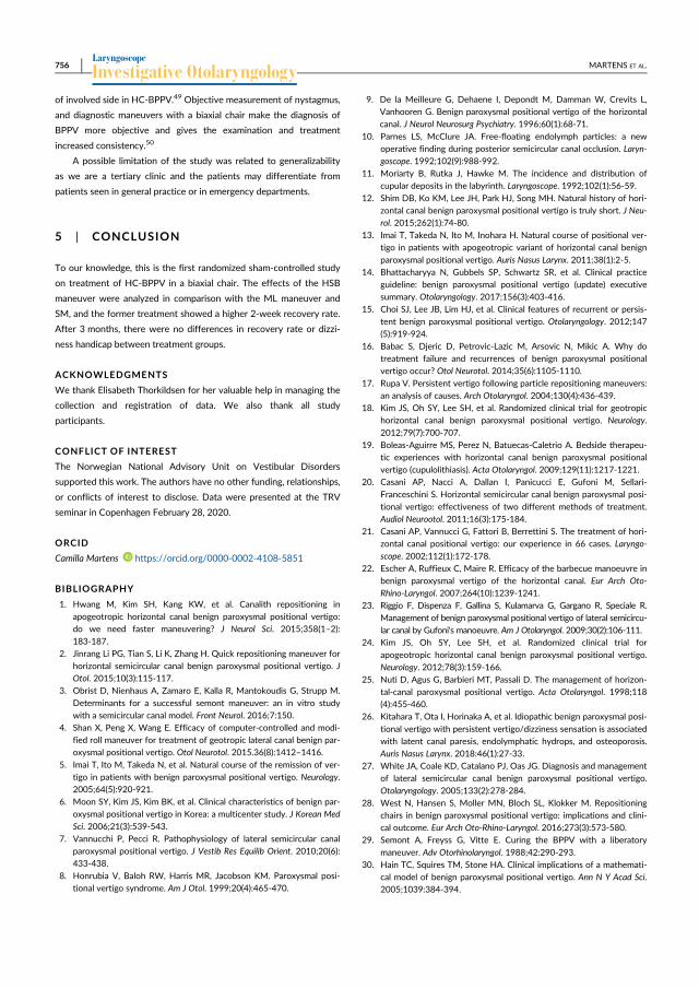
of involved side in HC-BPPV.49 Objective measurement of nystagmus,
and diagnostic maneuvers with a biaxial chair make the diagnosis of
BPPV more objective and gives the examination and treatment
increased consistency.50
A possible limitation of the study was related to generalizability
as we are a tertiary clinic and the patients may differentiate from
patients seen in general practice or in emergency departments.
5 | CONCLUSION
To our knowledge, this is the first randomized sham-controlled study
on treatment of HC-BPPV in a biaxial chair. The effects of the HSB
maneuver were analyzed in comparison with the ML maneuver and
SM, and the former treatment showed a higher 2-week recovery rate.
After 3 months, there were no differences in recovery rate or dizzi-
ness handicap between treatment groups.
ACKNOWLEDGMENTS
We thank Elisabeth Thorkildsen for her valuable help in managing the
collection and registration of data. We also thank all study
participants.
CONFLICT OF INTEREST
The Norwegian National Advisory Unit on Vestibular Disorders
supported this work. The authors have no other funding, relationships,
or conflicts of interest to disclose. Data were presented at the TRV
seminar in Copenhagen February 28, 2020.
ORCID
Camilla Martens https://orcid.org/0000-0002-4108-5851
BIBLIOGRAPHY
1. Hwang M, Kim SH, Kang KW, et al. Canalith repositioning in
apogeotropic horizontal canal benign paroxysmal positional vertigo:
do we need faster maneuvering? J Neurol Sci. 2015;358(1–2):183-187.
2. Jinrang Li PG, Tian S, Li K, Zhang H. Quick repositioning maneuver for
horizontal semicircular canal benign paroxysmal positional vertigo. J
Otol. 2015;10(3):115-117.
3. Obrist D, Nienhaus A, Zamaro E, Kalla R, Mantokoudis G, Strupp M.
Determinants for a successful semont maneuver: an in vitro study
with a semicircular canal model. Front Neurol. 2016;7:150.
4. Shan X, Peng X, Wang E. Efficacy of computer-controlled and modi-
fied roll maneuver for treatment of geotropic lateral canal benign par-
oxysmal positional vertigo. Otol Neurotol. 2015.36(8):1412–1416.5. Imai T, Ito M, Takeda N, et al. Natural course of the remission of ver-
tigo in patients with benign paroxysmal positional vertigo. Neurology.
2005;64(5):920-921.
6. Moon SY, Kim JS, Kim BK, et al. Clinical characteristics of benign par-
oxysmal positional vertigo in Korea: a multicenter study. J Korean Med
Sci. 2006;21(3):539-543.
7. Vannucchi P, Pecci R. Pathophysiology of lateral semicircular canal
paroxysmal positional vertigo. J Vestib Res Equilib Orient. 2010;20(6):
433-438.
8. Honrubia V, Baloh RW, Harris MR, Jacobson KM. Paroxysmal posi-
tional vertigo syndrome. Am J Otol. 1999;20(4):465-470.
9. De la Meilleure G, Dehaene I, Depondt M, Damman W, Crevits L,
Vanhooren G. Benign paroxysmal positional vertigo of the horizontal
canal. J Neurol Neurosurg Psychiatry. 1996;60(1):68-71.
10. Parnes LS, McClure JA. Free-floating endolymph particles: a new
operative finding during posterior semicircular canal occlusion. Laryn-
goscope. 1992;102(9):988-992.
11. Moriarty B, Rutka J, Hawke M. The incidence and distribution of
cupular deposits in the labyrinth. Laryngoscope. 1992;102(1):56-59.
12. Shim DB, Ko KM, Lee JH, Park HJ, Song MH. Natural history of hori-
zontal canal benign paroxysmal positional vertigo is truly short. J Neu-
rol. 2015;262(1):74-80.
13. Imai T, Takeda N, Ito M, Inohara H. Natural course of positional ver-
tigo in patients with apogeotropic variant of horizontal canal benign
paroxysmal positional vertigo. Auris Nasus Larynx. 2011;38(1):2-5.
14. Bhattacharyya N, Gubbels SP, Schwartz SR, et al. Clinical practice
guideline: benign paroxysmal positional vertigo (update) executive
summary. Otolaryngology. 2017;156(3):403-416.
15. Choi SJ, Lee JB, Lim HJ, et al. Clinical features of recurrent or persis-
tent benign paroxysmal positional vertigo. Otolaryngology. 2012;147
(5):919-924.
16. Babac S, Djeric D, Petrovic-Lazic M, Arsovic N, Mikic A. Why do
treatment failure and recurrences of benign paroxysmal positional
vertigo occur? Otol Neurotol. 2014;35(6):1105-1110.
17. Rupa V. Persistent vertigo following particle repositioning maneuvers:
an analysis of causes. Arch Otolaryngol. 2004;130(4):436-439.
18. Kim JS, Oh SY, Lee SH, et al. Randomized clinical trial for geotropic
horizontal canal benign paroxysmal positional vertigo. Neurology.
2012;79(7):700-707.
19. Boleas-Aguirre MS, Perez N, Batuecas-Caletrio A. Bedside therapeu-
tic experiences with horizontal canal benign paroxysmal positional
vertigo (cupulolithiasis). Acta Otolaryngol. 2009;129(11):1217-1221.
20. Casani AP, Nacci A, Dallan I, Panicucci E, Gufoni M, Sellari-
Franceschini S. Horizontal semicircular canal benign paroxysmal posi-
tional vertigo: effectiveness of two different methods of treatment.
Audiol Neurootol. 2011;16(3):175-184.
21. Casani AP, Vannucci G, Fattori B, Berrettini S. The treatment of hori-
zontal canal positional vertigo: our experience in 66 cases. Laryngo-
scope. 2002;112(1):172-178.
22. Escher A, Ruffieux C, Maire R. Efficacy of the barbecue manoeuvre in
benign paroxysmal vertigo of the horizontal canal. Eur Arch Oto-
Rhino-Laryngol. 2007;264(10):1239-1241.
23. Riggio F, Dispenza F, Gallina S, Kulamarva G, Gargano R, Speciale R.
Management of benign paroxysmal positional vertigo of lateral semicircu-
lar canal by Gufoni's manoeuvre. Am J Otolaryngol. 2009;30(2):106-111.
24. Kim JS, Oh SY, Lee SH, et al. Randomized clinical trial for
apogeotropic horizontal canal benign paroxysmal positional vertigo.
Neurology. 2012;78(3):159-166.
25. Nuti D, Agus G, Barbieri MT, Passali D. The management of horizon-
tal-canal paroxysmal positional vertigo. Acta Otolaryngol. 1998;118
(4):455-460.
26. Kitahara T, Ota I, Horinaka A, et al. Idiopathic benign paroxysmal posi-
tional vertigo with persistent vertigo/dizziness sensation is associated
with latent canal paresis, endolymphatic hydrops, and osteoporosis.
Auris Nasus Larynx. 2018:46(1):27-33.
27. White JA, Coale KD, Catalano PJ, Oas JG. Diagnosis and management
of lateral semicircular canal benign paroxysmal positional vertigo.
Otolaryngology. 2005;133(2):278-284.
28. West N, Hansen S, Moller MN, Bloch SL, Klokker M. Repositioning
chairs in benign paroxysmal positional vertigo: implications and clini-
cal outcome. Eur Arch Oto-Rhino-Laryngol. 2016;273(3):573-580.
29. Semont A, Freyss G, Vitte E. Curing the BPPV with a liberatory
maneuver. Adv Otorhinolaryngol. 1988;42:290-293.
30. Hain TC, Squires TM, Stone HA. Clinical implications of a mathemati-
cal model of benign paroxysmal positional vertigo. Ann N Y Acad Sci.
2005;1039:384-394.
756 MARTENS ET AL.

31. Begg C, Cho M, Eastwood S, et al. Improving the quality of reporting
of randomized controlled trials. The CONSORT statement. JAMA.
1996;276(8):637-639.
32. von Brevern M, Bertholon P, Brandt T, et al. Benign paroxysmal posi-
tional vertigo: diagnostic criteria. J Vestib Res Equilib Orient. 2015;25
(3–4):105-117.33. Johansson MS, Arlinger SD. Hearing threshold levels for an
otologically unscreened, non-occupationally noise-exposed popula-
tion in Sweden. Int J Audiol. 2002;41(3):180-194.
34. Choung YH, Shin YR, Kahng H, Park K, Choi SJ. ‘Bow and lean test’ todetermine the affected ear of horizontal canal benign paroxysmal
positional vertigo. Laryngoscope. 2006;116(10):1776-1781.
35. Tamber AL, Wilhelmsen KT, Strand LI. Measurement properties of
the dizziness handicap inventory by cross-sectional and longitudinal
designs. Health Qual Life Outcomes. 2009;7:101.
36. Wilhelmsen K, Strand LI, Nordahl SH, Eide GE, Ljunggren AE. Psycho-
metric properties of the vertigo symptom scale—short form. BMC Ear
Nose Throat Disord. 2008;8:2.
37. Martens C, Goplen FK, Nordfalk KF, Aasen T, Nordahl SH. Prevalence
and characteristics of positional nystagmus in Normal subjects. Oto-
laryngology. 2016;154:861-867.
38. Barin K. Head Angle During Static Position Test; 2010. https://www.
audiologyonline.com/ask-the-experts/head-angle-during-static-posi
tion-93. Accessed 25th of April 2018.
39. Gufoni M, Mastrosimone L, Di Nasso F. Repositioning maneuver in
benign paroxysmal vertigo of horizontal semicircular canal. Acta
Otorhinolaryngol Ital. 1998;18(6):363-367.
40. Tian L, Wang J, Sheng HB, et al. Comparative study on the roles of
the number of accelerations and rotation angle in the treatment
maneuvers for posterior semicircular canal benign paroxysmal posi-
tional vertigo. ORL J Otorhinolaryngol Relat Spec. 2015;78(1):36-45.
41. Steddin S, Ing D, Brandt T. Horizontal canal benign paroxysmal posi-
tioning vertigo (h-BPPV): transition of canalolithiasis to
cupulolithiasis. Ann Neurol. 1996;40(6):918-922.
42. Baloh RW, Jacobson K, Honrubia V. Horizontal semicircular canal
variant of benign positional vertigo. Neurology. 1993;43(12):2542-
2549.
43. Wang N, Zhou H, Huang HL. Comparison of automatic and manual
reposition treatment for horizontal semicircular canal benign
paroxysmal positional vertigo. J Biol Regul Homeost Agents. 2018;32
(3):719-723.
44. Wang NZ, Huang H, Geng D, Yang X, Yu C, Shi D. Efficacy of SRM-IV
vestibular function diagnosis and treatment system in treating benign
paroxysmal positional vertigo. Iran J Public Health. 2018;47(5):
641-647.
45. Levo H, Aalto H, Petteri HT. Nystagmus measured with video-
oculography: methodological aspects and normative data. ORL J
Otorhinolaryngol Relat Spec. 2004;66(3):101-104.
46. Bisdorff AR, Sancovic S, Debatisse D, Bentley C, Gresty MA,
Bronstein AM. Positional nystagmus in the dark in normal subjects.
Neuro-Ophtalmology. 2000;24(1):283-290.
47. Jeffery H, Hopkins M, Anderson R, Patel V, Rogers J. The interpreta-
tion of static positional nystagmus in a balance clinic. Int J Audiol.
2017;56(12):958-966.
48. Lee NH, Kwon HJ, Ban JH. Analysis of residual symptoms after treat-
ment in benign paroxysmal positional vertigo using questionnaire.
Otolaryngology. 2009;141(2):232-236.
49. Nuti D, Mandala M, Salerni L. Lateral canal paroxysmal positional ver-
tigo revisited. Ann N Y Acad Sci. 2009;1164:316-323.
50. Nakayama M, Epley JM. BPPV and variants: improved treatment
results with automated, nystagmus-based repositioning. Otolaryngol-
ogy. 2005;133(1):107-112.
SUPPORTING INFORMATION
Additional supporting information may be found online in the
Supporting Information section at the end of this article.
How to cite this article: Martens C , Goplen FK , Aasen T ,
Gjestad R , Nordfalk KF , Nordahl SHG . Treatment of
horizontal canal BPPV—a randomized sham-controlled trial
comparing two therapeutic maneuvers of different speeds.
Laryngoscope Investigative Otolaryngology. 2020;5:750–757.
https://doi.org/10.1002/lio2.420
MARTENS ET AL. 757


Errata for Benign paroxysmal positional vertigo and treatment in biaxial
rotational chair
Camilla Martens
Avhandling for graden philosophiae doctor (ph.d.)
ved Universitetet i Bergen
_____________________ _______________________
(dato sign. kandidat) (dato sign. fakultet)

2
Errata
Side vi Mangler Abbreviation PPPD- rettes til PPPD: Persistant Postural Perceptual Dizziness
Side vi Mangler Abbreviation TRV – rettes til TRV: Mechanical chair for treatment of BPPV named after the inventor Thomas Richard-Vitton.
Side 50 Stavefeil: “idiopathoc” – rettes til “idiopathic”
Side 50 Stavefeil: “taiwan” – rettes til “Taiwan”
Side 55 Stavefeil: “paroxysml” – rettes til “paroxysmal”
Side 61 Stavefeil: “ncidence” – rettes til “incidence”

Graphic design: Comm
unication Division, UiB / Print: Skipnes Komm
unikasjon AS
uib.no
ISBN: 9788230855133 (print)9788230852163 (PDF)