benign lesions of the subcutaneous soft tissue … · 2017-10-17 · lipoma is the most frequent...
TRANSCRIPT

Page 1 of 27
BENIGN LESIONS OF THE SUBCUTANEOUS SOFTTISSUE WITH CALCIFICATIONS. WHICH IS THE ROLE OFULTRASOUNDS IN DIAGNOSIS?
Poster No.: C-1404
Congress: ECR 2012
Type: Scientific Exhibit
Authors: E. GALLARDO AGROMAYOR; ES
DOI: 10.1594/ecr2012/C-1404
Any information contained in this pdf file is automatically generated from digital materialsubmitted to EPOS by third parties in the form of scientific presentations. Referencesto any names, marks, products, or services of third parties or hypertext links to third-party sites or information are provided solely as a convenience to you and do not inany way constitute or imply ECR's endorsement, sponsorship or recommendation of thethird party, information, product or service. ECR is not responsible for the content ofthese pages and does not make any representations regarding the content or accuracyof material in this file.As per copyright regulations, any unauthorised use of the material or parts thereof aswell as commercial reproduction or multiple distribution by any traditional or electronicallybased reproduction/publication method ist strictly prohibited.You agree to defend, indemnify, and hold ECR harmless from and against any and allclaims, damages, costs, and expenses, including attorneys' fees, arising from or relatedto your use of these pages.Please note: Links to movies, ppt slideshows and any other multimedia files are notavailable in the pdf version of presentations.www.myESR.org
Page 2 of 27
Purpose
The calcium is an obstacle for the US examination, due to the difference acousticimpedance with the rest of the tissues which provoke a very reflexive interface thatproduces an almost complete reflexion of the energy of the beam of ultrasoundand therefore an hyperrefringent line with acoustic shadow; although it is unableto assess an adequate evaluation of the central structure it allows the diagnosis ofa calcification easily. Moreover, most of these lesions appear as palpable lumpsand they frequently are evaluated with US as the first diagnostic tool. Therefore,we should be familiar with their appearance and the most frequent etiologies toguide additional diagnostic evaluation with other image modalities.
We will illustrate the most frequent entities which may appear as subcutaneousnodules with calcification with US, their diagnosis and the radiological correlation.
Methods and Materials
Dermal and subcutaneous soft tissue calcifications may be produced in a widerange of clinical entities. Calcinosis is due to a deposit of calcified hydroxyapatiteor classic amorphous phosphate in soft tissue. Calcifications can be divided infour basic types considering the etiopathogenic mechanism.
1- Dystrophic calcifications:
This is the most frequent type of calcification which is characterized byphosphocalcic deposits in previously injured subcutaneous tissue by diversemechanisms: traumatic, burns, venous insufficiency, radiotherapy, infections,tumors, insect bites and injections. Calcifications in these cases are usuallylocalized being known as circumscribed calcinosis. These calcifications are alsoproduced in some connective tissue diseases, especially in scleroderma anddermatomyositis, but also in SLE and others. This type of calcification is found indifferent soft tissue tumors, particularly frequent in the pilomatricoma, lipoma andadvanced neuroma between others.
2-Metastatic calcifications:

Page 3 of 27
These calcifications occur within the healthy soft tissue in patients with analtered phosphocalcic metabolism. The subcutaneous tissue involvement is notfrequent and when it occurs is characterized by hard subcutaneous nodulesand plaques, which occasionally are ulcerated with calcaric material extrusion.They are seen more frequently in chronic renal disease, primary and secondaryhyperparathyroidism, sarcoidosis, hypervitaminosis and alkali-milk syndrome.
3-Iatrogenic calcifications:
They usually appear in those locations where an invasive procedure has beenperformanced and are related with previous administration of intramusculardrugs, calcium gluconate or solutions with phosphates especially in previousextravasation. They can also appear in post-surgery scars, being particularlyfrequent in mid laparotomy.
4- Idiopathic calcifications:
These calcifications appear in absence of any cause or metabolic abnormality andtheir physiopathologic mechanism is unknown. The most representative entity ofthis group is tumoral calcinosis, nowadays it is referred as a hereditary conditionwith well defined massive periarticular calcifications, lobulated and non painfulwhich are usually localized in the extensor surfaces of the joints and in theanatomic distribution of the bursae.
Results
1- ISOLATED SUBCUTANEOUS CALCIFICATIONS:
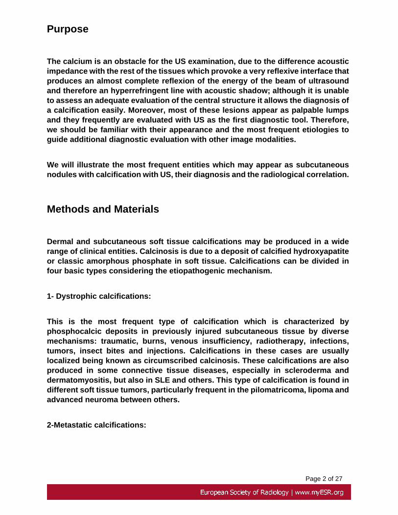
95% of the subcutaneous soft tissue calcifications are dystrophic calcifications.Post - injection gluteal calcifications, post-traumatic and fat necrosis are the mostfrequent types.
Post-injection calcifications have a typical radiologic appearance. They are wellrounded peripheral lineal calcifications with lower density centrally, located in thegluteal region superolaterally (Fig. 1). Ultrasound shows a convex hyperechogenicline with posterior acoustic shadow, which avoid evaluating both the central zoneand normal adjacent soft tissue.

Page 4 of 27
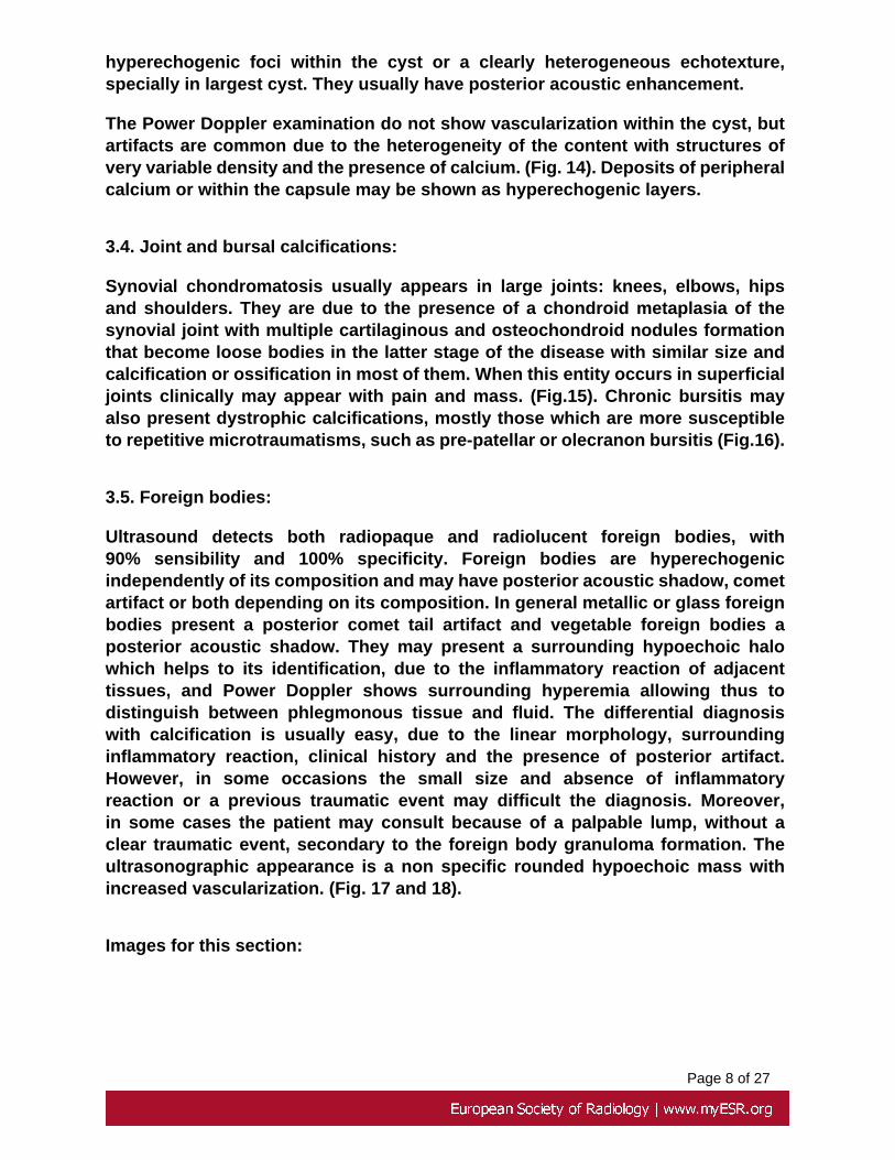
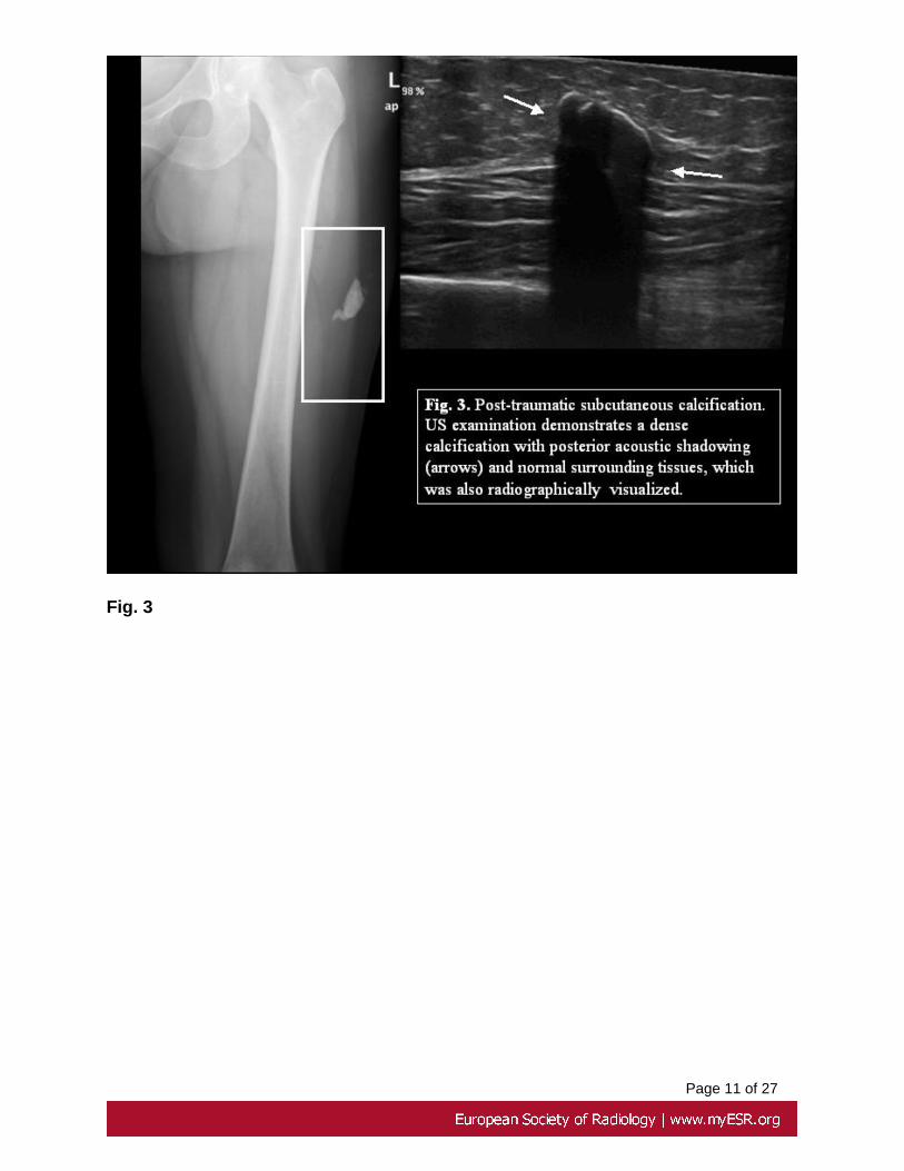
Post-traumatic calcifications may appear as small palpable nodules, with ahard consistency, with convex linear appearance in the ultrasound examination,homogeneous, with posterior acoustic shadow with a normal echotexture of theadjacent tissue (Fig.2).
Fat necrosis has different sonographic appearances. They can appear ashyperechogenic, ill-defined areas with an increase in consistency with ultrasoundtransducer pressure that may have lower echogenicity areas inside. They may alsoappear as lobulated tumors, similar to fat lobules but with a lower echogenicity,which are surrounded by a variable amount of fluid isolating them from the rest oftissue where they can freely move. Another appearance of fat necrosis is that ofrelatively hyperechogenic masses with a well defined hypoechoic capsule, with atendency to calcify (Figs. 3-4).
In connective soft tissue diseases, mostly in scleroderma and CREST syndrome,dermis or subcutaneous soft tissue circumscribed calcifications of the fingers, arefrequent. The ultrasound examination can easily identify calcifications and may beused in the surgical map when it is required (Fig. 5).
Sonographically calcifications are visualized as hyperechogenic images withsmooth border and lobulated contours and posterior acoustic shadow withthe largest diameter horizontally orientated related to the skin and a normallobular structure of the adjacent subcutaneous soft tissue. Ultrasound cannotdistinguish between the etiopathogenic mechanisms, however detects easily size,morphology and location in depth of the lesion and can diagnose a circumscribedcalcification in the skin (Fig. 5).
2.-CALCIFICATIONS WITHIN BENIGN SUBCUTANEOUS TUMORS:
Ultrasound can distinguish easily isolated calcifications and dystrophiccalcifications, chondroid or osseous within soft tissue tumors. The most frequenttumors that can calcify are the following ones:
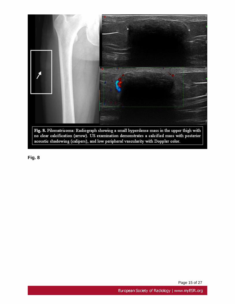
2.1.Pilomatricoma:
It is a benign tumor of dermal origin, from the primitive cells of the cellular matrix ofthe hair. It represents less than 1% of the subcutaneous tumors. However,it is themost frequent solid subcutaneous tumor in people younger than 20 years. Thereis a second peak of incidence in 50-65 year-old adults. Generally are small tumors,less than 3 cm, with a slow growth and confined to the subcutaneous tissue.The most frequent location is the face, neck and arms. One of the fundamental

Page 5 of 27
characteristic is the presence of a calcification or ossification which is usuallycentrally located, and can be appreciated in 85 % of the cases.
Sonographically they appear as hyperechogenic masses in relation with themuscle, with central calcification or ossification posterior acoustic shadow,rounded, with well-defined margins. The quantity of calcium may vary betweenisolated hyperechogenic foci to gross deposits of calcium or even completecalcification of the tumor. In most of the cases there is a hypoechoic halosurrounding the tumor. Doppler color or Power Doppler US examinations show apredominant peripheral high vascularization of the tumor (Fig. 6-8).
2.2.Hemangioma:
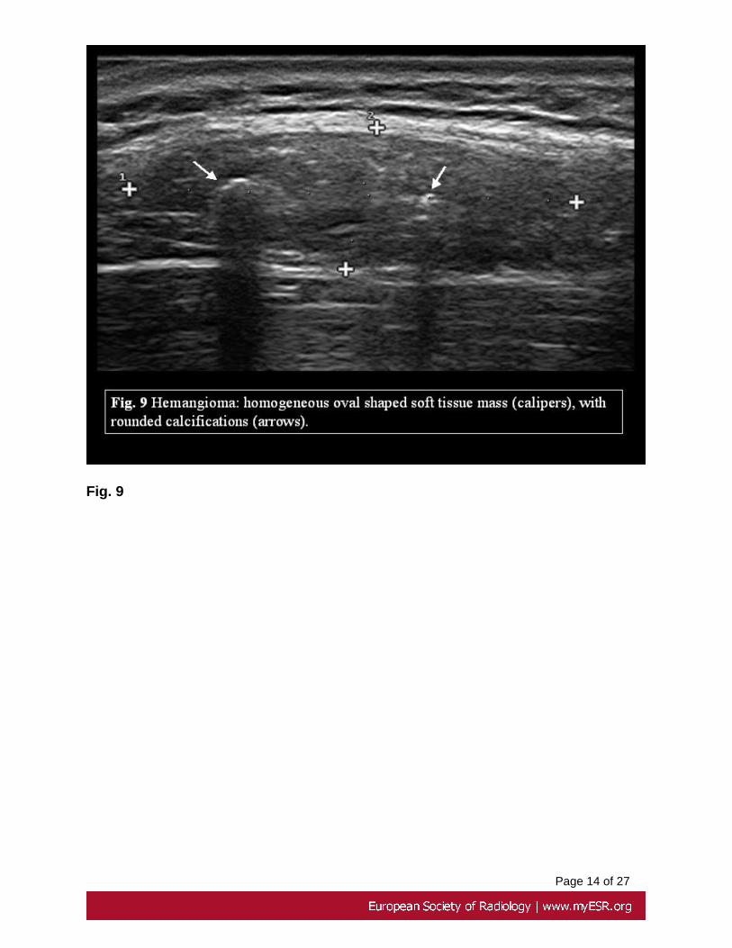
The term hemangioma includes a varied group of neoplasm of vascular origin,which are characterized by an increment of the number of normal and abnormalvessels. Both hemangiomas, which are real tumors that appear in the childhoodthat usually revert, and arteriovenous malformations, which show a dysplasticgrowth of blood vessels without associated cellular proliferation, are included.If considering the hemodynamic behavior, arteriovenous malformations can bedivided in those with high and low grade flow. The most frequent tumors ofthe subcutaneous soft tissue are capillary and cavernous hemangiomas. Inultrasound they appear as subcutaneous soft tissue mass rounded, well-defined,with varied echogenicity, homogeneously hypoechoic or highly heterogeneous.Hemangiomas may be circumscribed with well-defined margins or diffused withinterdigitation in the surrounding soft tissue, which difficults its delimitation.Characteristically, they show a wide vascular signal with high spectral peaks,which helps in the differential diagnosis with other subcutaneous masses. Theymay have serpiginous structures which are anechoic inside that depending on itsflow may fill with Power Doppler signal or respond to compression maneuvers.Low flow arteriovenous malformations are characterized by venous varicositieswith a normal arterial component. The spectral examination may demonstrate aslow monophasic or absent flow. It is frequent the presence of phleboliths, as smallhyperechogenic and convex linear images with posterior acoustic shadow. (Fig. 9).
2.3. Lipoma:
Lipoma is the most frequent soft tissue tumor. It represents 50 % of all soft tissue
tumors, with a prevalence of 2.1 per 100 habitants. They usually appear in the 5th-7th
decades of life with no male or female predilection. It is not clear if represents areal benign tumor or a focal hyperplasia of adipocytes. They may be superficiallylocated in the subcutaneous soft tissue or deeply, being subcutaneous lipomasextraordinary frequent. They may appear in any location but predominantly in theback, neck, proximal extremities and abdomen. They use to be solitary tumors, but

Page 6 of 27
may be multiple in 5-15 %, more frequently occurring in males with a family historyin 30% of the cases.
The ultrasound presentation is variable and generally diagnostic ,with two typesof common appearances:
a) Small sized lipomas are usually located in the more superficial subcutaneoussoft tissue. They are rounded, homogeneously hyperechogenic, well-defined withno visible capsule and generally avascular. This type of presentation is commonin the case of multiple lipomas which are mostly located in the trunk and extensorsurfaces of the arms. These lipomas may be spontaneously painful or undertransducer pression. In these cases, areas lower echogenicity within the tumor arefrequently visualized.
b) The other type of superficial lipomas appears as oval masses, with their largestdiameter parallel to the skin, well defined margins and in a hyperechogeniccapsule. The global echotexture may be hyperechogenic, isoechoic or hypoechoicrelated to the surrounding subcutaneous soft tissue, but the most frequentappearance is hyperechogenic. These tumors present linear hyperrefringentseptae inside, which are orientated to the largest axis of the tumor. They usuallyare easy compressed with ultrasound transducer pressure.
Typical lipomas may have necrosis, which produces an heterogeneous image:hypoechoic or hyperechoic areas with posterior acoustic shadow due todystrophic calcification. However, this is more frequent in deep, large size andlongstanding lipomas. Moreover, considering the different variants of lipoma,calcification is frequently present in chondroid lipomas and phleboliths inangiolipomas (Fig.10).
2.4 Extraskeletal chondroma:
This is a benign cartiloginous tumor, most common in hands and feet (80% infingers). They usually occur in 30-60 year-old adults. It appears as a subcutaneousnodule with a slow growth and rarely with spontaneous pain.
Plain films usually show irregular, ring or curvilinear calcifications.
On US a soft-tissue tumor with irregular hyperrefringent foci. (Fig.11)
2.5.Other tumors
Dystrophic calcifications may be seen in other soft tissue tumors, mostlylongstanding schwannomas and angioleiomyomas.

Page 7 of 27
3.- PSEUDOTUMORAL CALCIFICATIONS:
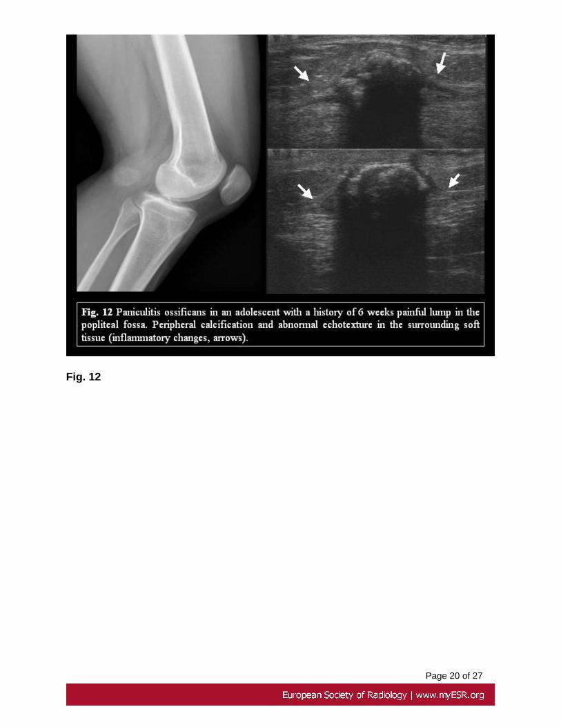
3.1.Panniculitis ossificans:
Myositis ossificans is a mass formed by heterotopic bone and cartilage, whichis typically located in the muscle. The circumscribed type of myositis ossificansis usually secondary to traumatic events, burns or abnormalities of the centralnervous system. Plain films show peripheral calcifications with a centripetalpattern of growth, which appear 4-6 weeks after injury. Panniculitis ossificansis a variant that appears in the subcutaneous soft tissue. They are also knownas osseous pseudomalignant soft tissue tumors. They occur frequently inadolescents and young adults, and 80% of the cases are located in lowerextremities. Ultrasound examination findings may vary depending on the momentof this process, detect peripheral calcification before is shown in plain films andassociated inflammatory changes of surrounding tissues (Fig.12).
3.2.Tophi:
Tophi are soft tissue conglomerates of urate crystals, which are preferentiallylocalized in the hands, feet and elbows. Ultrasonographically are depicted asheterogeneous masses with central hypo echoic areas which are surrounded byechogenic tissue. They usually appear as oval masses, which may be in occasionslobulated and avascular. If a deposit of calcium is associated, more echogenic fociwithin the mass with or without posterior acoustic shadow or completely calcifiedmasses are visualized being only the surface and the posterior acoustic shadowvisible. (Fig.13).
3.3: Inclusion cysts:
This is a cystic lesion which is covered by infundibular or epidermal cells thatkeratinize. Its origin is usually the traumatic implementation of epidermal cellsin the subjacent tissue. It has an heterogeneous content which is formed bykeratin and cholesterol granules, but also by epithelial rests and calcium. Generallyspeaking is named as sebaceous cyst, although this is an inappropriate term,being also named as epidermoid cyst.
Ultrasonographically it appears as a rounded or oval lesion, which issubcutaneously located, immediately beneath the dermis, sometimes with signsof umbilication with the dermis or associated with a hair follicle. The marginsare usually sharp and the capsule of the cyst may be identified. The contentis heterogeneous and its echotexture is mixed, usually hypoechogenic, with
Page 8 of 27
hyperechogenic foci within the cyst or a clearly heterogeneous echotexture,specially in largest cyst. They usually have posterior acoustic enhancement.
The Power Doppler examination do not show vascularization within the cyst, butartifacts are common due to the heterogeneity of the content with structures ofvery variable density and the presence of calcium. (Fig. 14). Deposits of peripheralcalcium or within the capsule may be shown as hyperechogenic layers.
3.4. Joint and bursal calcifications:
Synovial chondromatosis usually appears in large joints: knees, elbows, hipsand shoulders. They are due to the presence of a chondroid metaplasia of thesynovial joint with multiple cartilaginous and osteochondroid nodules formationthat become loose bodies in the latter stage of the disease with similar size andcalcification or ossification in most of them. When this entity occurs in superficialjoints clinically may appear with pain and mass. (Fig.15). Chronic bursitis mayalso present dystrophic calcifications, mostly those which are more susceptibleto repetitive microtraumatisms, such as pre-patellar or olecranon bursitis (Fig.16).
3.5. Foreign bodies:
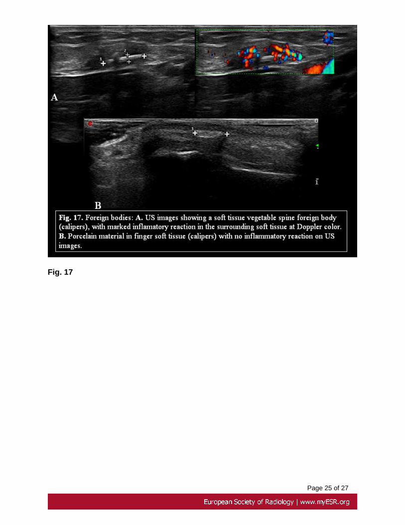
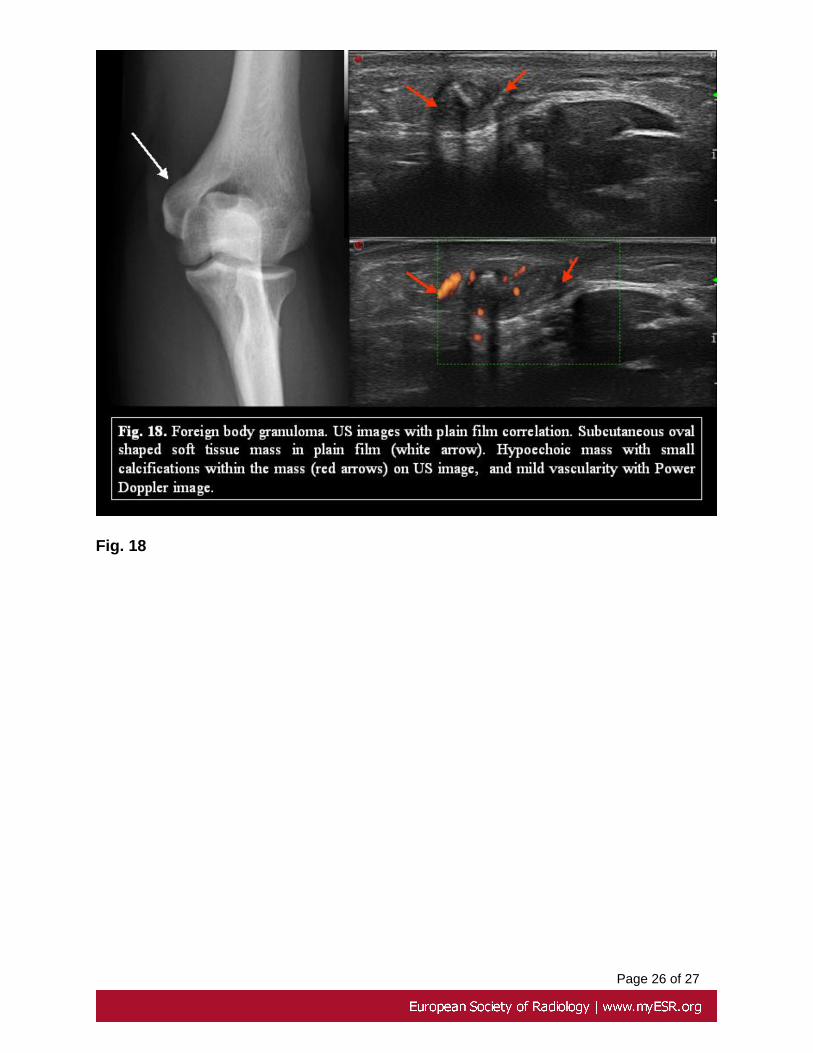
Ultrasound detects both radiopaque and radiolucent foreign bodies, with90% sensibility and 100% specificity. Foreign bodies are hyperechogenicindependently of its composition and may have posterior acoustic shadow, cometartifact or both depending on its composition. In general metallic or glass foreignbodies present a posterior comet tail artifact and vegetable foreign bodies aposterior acoustic shadow. They may present a surrounding hypoechoic halowhich helps to its identification, due to the inflammatory reaction of adjacenttissues, and Power Doppler shows surrounding hyperemia allowing thus todistinguish between phlegmonous tissue and fluid. The differential diagnosiswith calcification is usually easy, due to the linear morphology, surroundinginflammatory reaction, clinical history and the presence of posterior artifact.However, in some occasions the small size and absence of inflammatoryreaction or a previous traumatic event may difficult the diagnosis. Moreover,in some cases the patient may consult because of a palpable lump, without aclear traumatic event, secondary to the foreign body granuloma formation. Theultrasonographic appearance is a non specific rounded hypoechoic mass withincreased vascularization. (Fig. 17 and 18).
Images for this section:
Page 9 of 27
Fig. 1
Page 10 of 27
Fig. 2
Page 11 of 27
Fig. 3

Page 12 of 27
Fig. 4

Page 13 of 27
Fig. 5
Page 14 of 27
Fig. 9
Page 15 of 27
Fig. 8

Page 16 of 27
Fig. 7

Page 17 of 27
Fig. 6

Page 18 of 27
Fig. 10

Page 19 of 27
Fig. 11
Page 20 of 27
Fig. 12

Page 21 of 27
Fig. 13

Page 22 of 27
Fig. 14

Page 23 of 27
Fig. 15

Page 24 of 27
Fig. 16
Page 25 of 27
Fig. 17
Page 26 of 27
Fig. 18
Page 27 of 27
Conclusion
Ultrasound is usually the first diagnostic imaging method to evaluate a palpable softtissue mass. Calcifications within the mass can render the examination difficult. However,knowledge of ultrasound pattern can suggest a probable diagnosis, reduce the differentialdiagnosis, help to select additional tests and to avoid unnecessary or invasive procedures
References
1. Anolik R, Firoz B, Walters RF, Meehan SA, Tsou HC, Whitlow M, WainwrightB. Proloferating trichilemmal cyst with focal calcification, Dermatol Online J2008;14:25. 2. Bianchi S, Martinoli C. Ultrasound of themusculoskeletalsystem.Berlin, Germany: Springer-Verlag, 2007 3. Chang SJ, Sims J, Murtagh FR,McCaffrey JC, Messina JL. Proliferating Trichilemmal cyst of the scalp on CT.AJNR 2006; 27:712-1714. 4. Choo HJ, Lee SJ, Lee YH, Lee JH, Oh M, Kim MH,Lee EJ, Song JW, Kim SJ, Kim DW Pilomatricomas: the diagnostic value ofultrasound. Skeletal Radiol 2010; 39:243-250 5. Davae KC, Sofka CM, DiCarloE, Adler RS. Value of power Doppler Imaging and the hypoechoic halo in thesonographic detection of foreign bodies: correlation with histopathologicfindings. J Ultrasound Med 2003; 22: 1309 1313 6. De Orchis D, Ozonoff MB.Infiltrating angiolipoma with flebolith formation. Skeletal Radiol 1986;15:464-4677. Gomez-Dermit V, Gallardo E, Landeras R, Fernández Echevarría MA, GarcíaBarredo R. Subcutaneous angioleiomyomas: Gray- scale and Color Dopplersonographic appearances. J Clin Ultrasound 2006 ;34:50-54. 8. Haller JO,Kassner EG, Ostrowitz A, Kottmeler K, Perfschuck LP. Pilotarixoma (calcifyingepithelioma of Malherbe): radiographic features. Radiology1977:123:151-1539. Hoch B, Hermann G, Klein MJ, Abdelwahab IF. Ossifying chondroid lipoma.Skeletal Radiol 2008; 37:475-480 10. Horton LK, Jacobson JA, Powell A, FesselDP, Hayes CW. Sonography and radiography of soft tissue foreign bodies. AmJ Roentgenol 2001; 176:11559 11. Hwang JY, Lee SW, Lee SM. The commonultrasonographic features of pilomatricoma.J Ultrasound Med 2005;24:1397-140212. Jacobson JA, Powell A, Craig JG, Bouffard JA, van Holsbeck MT. Woodenforeign bodies in soft tissue: detection at US. Radiology 1998: 206:45-8 13.Kransdorf MJ, Meis JM. Extraskeletal osseus and cartilaginous tumors of theextremities. Radiographics 1993;13: 853-884. 14. Olsen K, Chew F. Tumoralcalcinosis: Pearls, polemics and Alternative Possibilities. Radiographics 2006;26: 871-875
Personal Information