benign breast lesions: ultrasound
TRANSCRIPT

Journal of Ultrasound (2011) 14, 55e65
ava i lab le at www.sc iencedi rec t .com
journal homepage: www.e lsev ier .com/ locate / jus
Benign breast lesions: Ultrasound
N. Masciadri a, C. Ferranti b,*
a IRCCS Multimedica, Sesto San Giovanni, Milan, Italyb Fondazione IRCCS Istituto Nazionale Tumori, Unit of Diagnostic Radiology 3, Senology, Milan, Italy
KEYWORDSBreast;Ultrasound;Benign breast lesions.
* Corresponding author. FondazionTumori, Unit of Diagnostic Radiology 3
E-mail address: claudio.ferra(C. Ferranti).
1971-3495/$ - see front matter ª 201doi:10.1016/j.jus.2011.03.002
Abstract Benign breast diseases constitute a heterogeneous group of lesions arising in themammary epithelium or in other mammary tissues, and they may also be linked to vascular,inflammatory or traumatic pathologies. Most lesions found in women consulting a physicianare benign. Ultrasound (US) diagnostic criteria indicating a benign lesion are described as wellas US findings in the most frequent benign breast lesions.
Sommario Le lesioni mammarie benigne costituiscono un gruppo eterogeneo di manifesta-zioni, sia proprie dell’epitelio mammario, sia con origine dagli altri tessuti che costituisconol’organo, sia con altra patogenesi (vascolare, flogistica e traumatica).Esse costituiscono il reperto piu frequente che si osserva nella maggior parte dei casi nei qualiuna donna pensa di avere un problema al seno e si rivolge al medico o al radiologo.Vengono riproposti da un punto di vista ecografico i criteri diagnostici che orientano per la be-nignita ed i quadri iconografici che si riscontrano nelle principali lesioni mammarie benigneecograficamente identificabili.ª 2011 Elsevier Srl. All rights reserved.
Introduction
Benign breast diseases constitute a heterogeneous group oflesions arising in the mammary epithelium or in othermammary tissues and they may also be linked to vascular,inflammatory or traumatic pathologies. (Table 1) [1]. Somelesions are palpable masses, which may be nodular, some-times with specific or unspecific characteristics, but often(particularly in lesions of greater prognostic significancesuch as atypical hyperplasia) there are no specific clinical
e IRCCS Istituto Nazionale, Senology, Milan, [email protected]
1 Elsevier Srl. All rights reserved.
signs, and detection is difficult also at diagnostic imagingexaminations.
This paper will not deal with mammary dysplasia orfibrocystic mastopathy which is the most common breastlesion in women of childbearing age. This denomination isinappropriate, as it does not refer to a specific histologicalcondition. It is a variable combination of some basicproliferative and regressive lesions induced by hormonalfactors affecting both the epithelium and the connectivetissue, and in the 1980s Hughes therefore introduced theconcept of ANDI (Aberrations of Normal Development andInvolution) [2].
It is important to recognize benign breast diseases fromthe clinical signs as well as mammographic and ultrasound(US) findings, since most lesions found in women consultinga physician are benign. In order to reduce the number ofunnecessary biopsies and to avoid inappropriate diagnostic

Table 1 Classification of benign breast lesions accordingto histological origin.
Terminal and lobular ductsa. Acinar distension
i. Cystb. Intralobular connective tissue proliferation
i. Sclerosing adenosisii. Fibroadenomaiii. Phyllodes tumoriv. Hamartoma
c. Epitelial changes in terminal duct lobularunits (TDLUs)
i. Apocrine metaplasiaii. Ductal and lobular hyperplasia, usual and atypicaliii. Papillomatosisiv. Intracystic papilloma
Ductal systemd. Ductal ectasiae. Intraductal papilloma
Lesions of different originf. Fatty tissue lesions
i. Lipomaii. Liponecrosis
g. Fibrous tissue lesionsi. Focal fibrosisii. Diabetic mastopathyiii. Pseudoangiomatous stromal hyperplasia (PASH)iv. Myofibroblastoma
h. Vascular origini. Hemangioma
i. Inflammatory origini. Mastitis/abscessii. Tuberculosis and sarcoidosisiii. Foreign body granuloma and siliconoma
j. Lymph node origini. Intramammary lymph nodes
56 N. Masciadri, C. Ferranti
delays, the breast radiologist should also be aware thatbenign breast diseases may present atypical findings andthat possible overlap of findings between certain types ofbenign and malignant diseases may occur.
Diagnostic US criteria indicating benign lesions aredescribed in this paper as well as US findings in the mostfrequent benign breast lesions.
US parameters in the diagnosis of a probablybenign lesion
US scanning of the breast has long been considered as justan additional examination for identifying the nature of anyabnormalities detected at physical examination or atmammography in order to classify them in the followingcategories:
� Normal tissue� Simple cyst� Complicated or complex cyst
� Indeterminate solid or cystic lesion� Solid lesion [3,4]
However, thanks to the technological progress, thediagnostic potential of US examination is far more completenow, and this procedure is very useful in the workup ofa lesion and essential in guiding interventional and biopticalprocedures [5].
In the field of US examinations, a systematic analysis ofthe findings in solid breast nodules in order to differentiatebenign from malignant lesions was proposed by Stavros in1995 [6,7], and in 2003 the Breast Imaging Reporting AndData System (BI-RADS) was developed by the AmericanCollege of Radiology [8].
Application of a standard terminology related to US, asindicated by the BI-RADS lexicon, should ensure unifor-mity of interpretation and consequently an appropriatemanagement of diagnostic US findings (follow-up or asc-ertainment) [9].
Simple cysts and some specific solid lesions havepathognomonic characteristics, so the diagnosis of benigndisease is easy and also BI-RADS classification in Category 2.However, in most solid lesions, the diagnosis of probablybenign lesion is based on the exclusion of suspicious signsbut also on the confirmation of certain parameters showingbenign lesions such as:
� elliptical shape and horizontal orientation;� well-defined curvilinear or only slightly lobatedmargins; the presence of a complete, thin echogeniccapsule;
� echotexture almost completely hyperechoic.
Classification in BI-RADS category 3 (<2% of malignancy)should lead to a follow-up of an appropriate duration,although a needle biopsy or surgical excision should beconsidered according to the patient and/or her physician’spreference. If there are no clearly suspicious signs, but thesigns of a benign lesion as mentioned above are not certain,the lesion should be classified as BI-RADS category 4a andbiopsy should be performed [3].
A standard examination carried out by experiencedoperators using equipment of the latest technology hasshown an elevated degree of sensitivity and specificity dueto the possibility of compound and harmonic imaging whichimproves the visibility of the margins and the echostructureof the lesion [10]. Technological progress has also over theyears provided software that allows second-level evalua-tion, which can further improve a non-invasive diagnosticapproach in order to avoid an excessive use of biopsy asa definitive diagnostic tool.
In the 1990s, color/power Doppler examination wasintroduced, and only more recently equipment has beendeveloped for three-dimensional (3D) studies andsonoelastography. US contrast agents do not seem to bewidely used, also due to the high cost involved; their useseems to be more appropriate particularly in the study ofbreast lesions undergoing neoadjuvant chemotherapy inorder to document the response to treatment [11e13]rather than in the differentiation between benign andmalignant lesions and in the detection of small impal-pable lesions.

Benign breast lesions 57
Doppler study was initially considered very promising inthe differential diagnosis between benign and malignantmasses on the basis of morphological criteria (number ofpenetrating vessels as well as central or peripheral distri-bution) and semiquantitative criteria (resistance and pul-satility indices, peak systolic velocity) which may identifythe characteristic neoangiogenesis of malignant lesions[14e16]. Power Doppler is better than color Doppler fordetecting small vessels with slow blood flow and is there-fore able to distinguish between solid and complicatedcystic lesions, but it is more sensitive to artifacts [17].However, the literature has revealed a substantial overlapof aspects in the vascularity of benign and malignantlesions. The hypothesis that more vascularization meansa higher probability that the lesion is malignant is abso-lutely not valid (for example, also benign papillary lesionsare highly vascularized). However, vascularization withina solid lesion detected at color/power Doppler shouldincrease the US operator’s attention and suspicion.
Software for 3D reconstruction of the superficial tissuesis also applicable to the study of breast lesions and can beuseful in the differential diagnosis between benign andmalignant nodules. Full thickness visualization of the regioncontaining the focal lesion and subsequent processing ofthe images allows assessment of the shape and margins ofthe lesion in three orthogonal planes evidencing expansiveor infiltrative growth [18,19]. After morphological evalua-tion, the 3D vascular image of the mass can be evaluated ina panoramic view that can facilitate quantification of thevessels and their relationship with the lesion.
Sonoelastography evaluates the different elastic prop-erties of the tissues and is best applied in the evaluation ofbreast lesions, as there is a substantial difference betweenfibroglandular tissue and nodules of different types; malig-nant lesions are generally less elastic than benign masses[20,21]. A slight rhythmic compression is applied using thetransducer positioned perpendicular to the breast, so thatthe relative deformation of the underlying tissues can bereconstructed and displayed on the monitor sometimes inblack and white images; otherwise in color images whichyield a better view of the degree of tissue elasticity. In thefirst case, benign lesions are white and malignant lesions aredark, while cysts show a bull’s eye appearance with a whitecenter and a surrounding dark halo. In color images, thelesions present characteristics which have been assigneda score from 1 to 5 (similar to the BI-RADS classification),where benign or probably benign lesions (more elastic) aregreen with possible blue points, whereas malignant lesions(less elastic) are almost entirely blue with a possible halowhich can be correlated with a desmoplastic reaction in thesurrounding tissues [22,23]. Typically, simple and compli-cated cystic lesions present a triphasic pattern with a blue,more rigid layer at the surface, an intermediate green layerand a deep layer which is red.
Sonoelastography is very useful in non-palpable lesions,where it assumes the role of clinical examination inassessing the consistency of the palpable mass, therebyimproving the perception of any US changes in shape andthickness of the lesion which become evident when thedegree of probe compression is changed. Sensitivity ofsonoelastography seems to improve in solid lesions of smallsize, and sensitivity is independent of the depth of the
lesion, thickness and echogenicity of the breast. Sensitivityvalues reported in the literature are 70%e96% and speci-ficity is 24%e90% [13].
Sonographic features of the most commonbenign breast lesions
Table 1 shows the classification of benign breast lesions onthe basis of histological origin [1].
Cysts
Cysts are caused by over-distension of the terminal ductlobular units (TDLU) due to progressive filling with liquid,fibrosclerosis of the loose connective intralobular tissueand coalescence of single dilated ductules in a polylobatedmass up to a single tensive cyst [3].
Cysts can be divided into three groups: simple, compli-cated and complex cysts [24,25].
Simple cysts present five basic characteristics: a well-circumscribed appearance, anechoic contents, a thinechogenic external capsule, enhanced through-trans-mission and subtle acoustic shadows at the edges (Fig. 1).Cysts with these features are very common in women of30e50 years of age, and unless they are symptomatic theydo not require evacuation or monitoring. If the mass ismammographically visible or palpable, it is important tomake sure that this finding corresponds to the cyst and thatthere are no adjacent, more important, solid lesions [3].
There may be possible echoes within the cyst due totechnical artifacts or improper adjustment of the gain ordue to excessive probe compression, which can generallybe eliminated in harmonic imaging. There are alsocomplicated cysts (so called because the content is notpurely anechoic), which show diffused echoes of lowamplitude, mainly due to the presence of amorphousmaterial with cellular debris, blood cells and macrophageswith foamy cytoplasm (Fig. 1B) or liquideliquid levels (e.g.galactocele) [24]. They should be evacuated if they aresymptomatic or if there are diagnostic doubts.
High-grade invasive ductal carcinomas and medullarycarcinomas, as well as extra mammary metastatic lesionscan show a marked hypoechoic image and have a roundedshape with enhanced through-transmission, thereby simu-lating a complicated cyst. However, a careful analysis ofthe shape and contour of the lesion as well as Dopplerevaluation will raise diagnostic suspicion [3].
Complex cysts (Fig. 1C), which are generally less worri-some when located in the breast than those arising in otherorgans, may indicate the presence of malignancy orinfection.
Morphological features which are suspicious for malig-nancy are thick isoechoic intracystic septations, muralnodules, fibrovascular stalk in the solid components anda microcystic appearance or microlobulated contour[3,24,25]. In 85%e90% of cases the lesion is a benignintracystic papilloma, and in the remaining 10%e15% ofcases it is a papillary lesion with atypia or intracysticpapillary carcinoma.
Definitive diagnosis requires histological analysis aftercore biopsy or preferably using vacuum-assisted device,

Fig. 1 Simple cyst (A): US shows a mass which appears well-circumscribed, with anechoic contents, thin echogenic exteriorcapsule, enhanced through-transmission, subtle acoustic shadowing at the edges. Complicated cyst (B): US shows that the contentis not purely anechoic (like a simple cyst), but there are diffused echoes of low amplitude. Complex cyst (C): US shows a well-circumscribed anechoic mass containing solid components. Cysts with sediment (D): US shows an anechoic mass with sharp marginsand posterior hyperechoic sediment.
58 N. Masciadri, C. Ferranti
leaving a marker to identify the sampling location. Morpho-logical features indicating inflammation or infection are:relatively uniform isoechoic circumferential cyst wallthickening, hyperemia of the cyst wall and presence of bloodin the cyst sediment (Fig. 1D) with the image showing fluid-debris level. If these three features coexist and the patient issymptomatic, the sediment is likely to be purulent. In thiscase the patient should undergo needle aspiration withsubsequent bacterial culture and antibiogram [3].
Abscess
Abscesses may originate from infection of the subareolarducts and/or preexisting galactocele (puerperal mastitis),or from ruptured ectatic ducts or cysts with initial chemicalinflammation and subsequent bacterial superinfection. US
Fig. 2 Abscess: US shows a hypo-anechoic mass with irre
features are similar (Fig. 2) and distinction is based onwhether or not the patient is breastfeeding [3,26,27].
Puerperal mastitis (Fig. 3) has an acute onset andalthough it is of lobar or sublobar origin, the classicinflammatory signs may involve the whole organ. Penetra-tion of germs - more commonly Staphylococcus e occursthrough a crack or fissure in the area of the nipple and itfinds an excellent culture medium in the milk contained inthe subareolar ducts or galactocele. If mastitis is nottreated properly, an abscess will develop with necrotictissue and denatured milk floating within the pus containedin the abscess cavity. When originating from a preexistinggalactocele, the abscess may develop earlier and tend tobe more demarcated with a roughly oval or multilobulatedshape. When the subareolar ducts are involved, the abscess
gular margins (A) and peripheral hypervascularity (B).

Fig. 3 Mastitis: US shows subcutaneous and parenchymal edema associated with small fluid collections (A) and diffuse hyper-vascularity (B).
Benign breast lesions 59
is often multiloculated due to the confluence of severalsmall abscesses.
When the original event is a focal chemical mastitiscaused by rupture of ducts or cysts and consequent releaseof lipid-rich secretions, it is an inflammatory conditioninvolving the large ducts, known as ductal ectasia, whichaffects perimenopausal women. This often causes multi-lobar bilateral involvement with episodes of focal peri-ductal mastitis and abscesses which are typically locatedaround the areola. Bacterial infection is a result of aspi-ration attempts or communication with ducts which arealready colonized by germs or hematogenous seeding.
US image of these abscesses is elongated and follows theaxis of the duct of origin, with markedly thickened walls,early involvement of the nipple and marked inflammationof the surrounding tissues.
Nonpuerperal abscesses tend to recur easily and becomechronic with cutaneous fistula formation which can bedifficult to eradicate. A marked fibrotic reaction causespermanent retraction of the nipple. In this case neoplasticdisorder should be included in a differential diagnosis.
However, several other benign breast conditions mayduring their evolution go through phases mimickingcomplex cysts, such as galactocele, seroma, hematoma,liponecrosis and hemangioma.
Galactocele
A painless lump developing during or a few weeks afterended breastfeeding is generally thought to be a gal-actocele. US monitoring may show spontaneous resolution,
Fig. 4 Galactocele in the intermediate stage: US showsmoderately echogenic contents with fat tending to form volu-minous unemulsified globules, which are distributed hetero-geneously in the liquid component.
or a targeted aspiration may be carried out in cases ofdiagnostic doubt [3,28].
A galactocele is a cystic dilatation of the terminal ductsand ductules containing milk, so the appearance of a gal-actocele may vary during the monitoring. At first it appearsas a anechoic cyst with possible septation, as the milk isfresh with homogeneously emulsified fat globules in a liquidcomponent. Later the content becomes moderately echo-genic, when the fat tends to form increasingly large andless emulsified globules, which are distributed unevenly orare suspended above the liquid component (Fig. 4), some-times forming the classic fat-fluid level which is alsomammographically visible in the mediolateral view. Whenthe milk is curdled, the galactocele may mimic a solidnodule; however, it is easily compressed, there is novascular signal at color Doppler, and the contents maymove according to the pressure. A chronic galactocele,whose liquid component has been completely reabsorbed,may appear as a simple or complex lipid cyst.
Seroma and hematoma
Seromas are collections of serous fluid arising unpredictablyafter interventional procedures [29], and they commonlyoccur after surgical resection or breast augmentation [3].After breast resection they occur at the level of the woundproportionally to the extension of the operation. Afterbreast augmentation they tend to occur between theprosthetic shell and the fibrous capsule, more or lesscompletely surrounding the prosthesis. Axillary seromasoccurring due to lymph node dissection are actually lym-phoceles. In both cases hemorrhagic extravasation mayoccur with consequent hematoma, particularly if thepatient is receiving anticoagulant treatment [30].
These collections may be round or oval shaped if theyare distended, whereas they may look angular and flat ifthe surgical cavity is slightly anfractuous.
Seromas may initially be anechoic or markedly hypo-echoic, but diffuse low-level echoes or thin fibrin septationssubsequently appear within them. The presence of bloodwhich tends to clot leads to sediment, pseudonodule, wallthickening and coarse septa formation showing no vascu-larity at power Doppler and a hypoechoic heterogeneouspattern at US becomes hyperechoic after coagulation.
Hematoma may resolve spontaneously or becomechronic with varying aspects mimicking also a solid mass,

60 N. Masciadri, C. Ferranti
and it may develop liponecrosis with possible wall calcifi-cation. In blunt trauma cases there may not be a realpseudocystic hematoma, but an edema in the adiposetissue may be observed in the presence of superficial skinbruising.
Liponecrosis
Liponecrosis is a nonsuppurative inflammatory processresulting from saponification of the adipose tissue afterbiopsy and/or surgical resection with seroma or hematomaformation and possibly exacerbated by subsequent radio-therapy [31,32]. Extravasation of blood causes edema andstromal thickening with ischemia and necrosis due toincreased local pressure and consequent adipocyte rupture.Accumulation of macrophages and plurinucleate giant cellscontaining necrotic lipid vacuoles results in an “oil cyst”,mammographically visible as a “soap bubble” image [33]. Ifthe initial US image shows a pseudonodular hyperechoicedematous area, fat liquefaction gives raise to a complexcyst (Fig. 5). It is sometimes multilocular and has theappearance already described for seroma and hematoma(presence of echogenic bands and mural pseudonodules,often mobile when the patient changes position, yielding novascular signals at color-power Doppler). During healing,cyst wall fibrosis develops, and parietal calcification occursshowing an egg-shell shape. Fibrous tissue may replace thefat content inside the cavity, and if the extent of necrosisand inflammation is important, the final stage may show anarea of well-circumscribed hyperechoic fibrosis of irregularshape and angular edges, sometimes with acousticabsorption and shrinking or deformation of the surroundingtissues. In this case scirrhous carcinoma should be includedin a differential diagnosis.
Hemangioma
This benign vascular tumor is often clinically invisible,although it is situated subcutaneously and can measuremore than 1 cm in diameter. It is an oval or polylobedpseudonodular mass, and US appearance depends on thecaliber of the blood vessels. Capillary hemangiomas arelargely homogeneously hyperechoic and cavernous heman-giomas present a mixed echotexture [34,35]. Both havea soft texture and can easily be compressed by probepressure. In case of thrombosis, hypoechoic areas can beobserved, often with phlebolithic calcifications, which aregranular or amorphous.
Fig. 5 Liponecrosis: US shows a nodule mimicking a complexcyst.
Fibroadenoma
Fibroadenomas are benign solid tumors developing froma terminal duct lobular unit due to uncoordinated prolif-eration of the epithelial and stromal component (presum-ably due to estrogen stimulation) which involves part of thesurrounding tissues. These tissues are partially compressedby the expansive growth, thereby creating a sort ofa pseudocapsule.
Fibroadenomas have an internal structure composed ofstromal and epithelial elements. The stromal element mayundergo a myxoid degeneration, such as sclerosis, hyalini-zation and calcification, whereas the epithelial elementmay present all possible proliferative and non-proliferativeaspects of the breast parenchyma, such as apocrine meta-plasia, ductal hyperplasia, sclerosing and florid adenosis[36e38]. Fibroadenomas characterized by apocrine meta-plasia, ductal hyperplasia, sclerosing adenosis or cysts aredefined as “complex” [39,40].
Fibroadenomas have two peaks of incidence: in the thirdand in the fifth decade of life, but they may also occur aftermenopause as a result of hormone replacement therapy.They can grow rapidly but usually up to max. 2e3 cm. Giantand juvenile fibroadenomas are exceptions which mayreach 6e10 cm. They have a highly cellular stroma andshould be distinguished from benign phyllodes tumor[41,42]. They can be multiple and bilateral in approxi-mately 20%e25% of patients. During pregnancy andbreastfeeding these lesions may become more irregular dueto episodes of infarctions and therefore more difficult todistinguish from carcinomas [43]. However, carcinomararely develops within a fibroadenoma; this occurs in 1 outof 1000 cases with an increased risk related to “complex”fibroadenomas, and in that case they are mainly in situwhile infiltrating carcinomas occur more rarely [44]. At USexamination, classic fibroadenomas, which are mobile andsmooth, present the following characteristics [36e38]:elliptical or slightly lobulated shape, horizontal orientation(transverse diameter greater than the anterior-posteriordiameter), isoechoic or mildly hypoechoic echotexture,well-defined curvilinear margin with a complete thin,echogenic capsule, unaltered US beam transmission beyondthe lesion and subtle acoustic shadows on both sides of thenodule (Fig. 6A).
If the nodule presents a microlobulated appearance, itdiffers from a classic or presumed fibroadenoma (BI-RADS 2or 3) and becomes suspicious for malignancy (BI-RADS 4a),and needle biopsy with micro-histological analysis istherefore required [3].
“Complex” fibroadenomas also require histologicalanalysis due to the presence of microcalcifications (asso-ciated with ductal hyperplasia), or heterogeneous echo-texture due to the presence of microcysts (apocrinemetaplasia) or small hyperechoic areas (sclerosing adeno-sis) (Fig. 6B) [3,39].
Fibroadenoma variants with evident epithelial hyper-plasia and a very little stromal component include tubularadenomas and lactating adenomas. The latter occursparticularly during breastfeeding and in the third trimesterof pregnancy; it is sometimes quite big, elastic in consis-tency and therefore compressible almost like a lipoma [3].
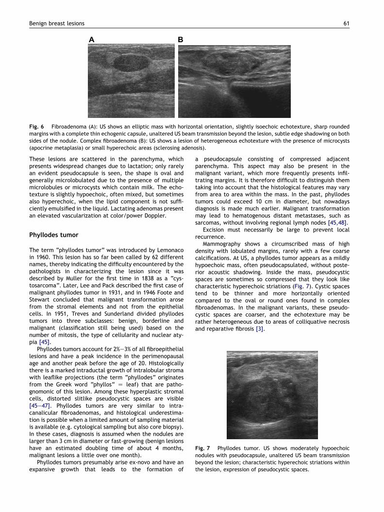
Fig. 6 Fibroadenoma (A): US shows an elliptic mass with horizontal orientation, slightly isoechoic echotexture, sharp roundedmargins with a complete thin echogenic capsule, unaltered US beam transmission beyond the lesion, subtle edge shadowing on bothsides of the nodule. Complex fibroadenoma (B): US shows a lesion of heterogeneous echotexture with the presence of microcysts(apocrine metaplasia) or small hyperechoic areas (sclerosing adenosis).
Benign breast lesions 61
These lesions are scattered in the parenchyma, whichpresents widespread changes due to lactation; only rarelyan evident pseudocapsule is seen, the shape is oval andgenerally microlobulated due to the presence of multiplemicrolobules or microcysts which contain milk. The echo-texture is slightly hypoechoic, often mixed, but sometimesalso hyperechoic, when the lipid component is not suffi-ciently emulsified in the liquid. Lactating adenomas presentan elevated vascularization at color/power Doppler.
Fig. 7 Phyllodes tumor. US shows moderately hypoechoicnodules with pseudocapsule, unaltered US beam transmissionbeyond the lesion; characteristic hyperechoic striations withinthe lesion, expression of pseudocystic spaces.
Phyllodes tumor
The term “phyllodes tumor” was introduced by Lemonacoin 1960. This lesion has so far been called by 62 differentnames, thereby indicating the difficulty encountered by thepathologists in characterizing the lesion since it wasdescribed by Muller for the first time in 1838 as a “cys-tosarcoma”. Later, Lee and Pack described the first case ofmalignant phyllodes tumor in 1931, and in 1946 Foote andStewart concluded that malignant transformation arosefrom the stromal elements and not from the epithelialcells. In 1951, Treves and Sunderland divided phyllodestumors into three subclasses: benign, borderline andmalignant (classification still being used) based on thenumber of mitosis, the type of cellularity and nuclear aty-pia [45].
Phyllodes tumors account for 2%e3% of all fibroepitheliallesions and have a peak incidence in the perimenopausalage and another peak before the age of 20. Histologicallythere is a marked intraductal growth of intralobular stromawith leaflike projections (the term “phyllodes” originatesfrom the Greek word “phyllos” Z leaf) that are patho-gnomonic of this lesion. Among these hyperplastic stromalcells, distorted slitlike pseudocystic spaces are visible[45e47]. Phyllodes tumors are very similar to intra-canalicular fibroadenomas, and histological underestima-tion is possible when a limited amount of sampling materialis available (e.g. cytological sampling but also core biopsy).In these cases, diagnosis is assumed when the nodules arelarger than 3 cm in diameter or fast-growing (benign lesionshave an estimated doubling time of about 4 months,malignant lesions a little over one month).
Phyllodes tumors presumably arise ex-novo and have anexpansive growth that leads to the formation of
a pseudocapsule consisting of compressed adjacentparenchyma. This aspect may also be present in themalignant variant, which more frequently presents infil-trating margins. It is therefore difficult to distinguish themtaking into account that the histological features may varyfrom area to area within the mass. In the past, phyllodestumors could exceed 10 cm in diameter, but nowadaysdiagnosis is made much earlier. Malignant transformationmay lead to hematogenous distant metastases, such assarcomas, without involving regional lymph nodes [45,48].
Excision must necessarily be large to prevent localrecurrence.
Mammography shows a circumscribed mass of highdensity with lobulated margins, rarely with a few coarsecalcifications. At US, a phyllodes tumor appears as a mildlyhypoechoic mass, often pseudocapsulated, without poste-rior acoustic shadowing. Inside the mass, pseudocysticspaces are sometimes so compressed that they look likecharacteristic hyperechoic striations (Fig. 7). Cystic spacestend to be thinner and more horizontally orientedcompared to the oval or round ones found in complexfibroadenomas. In the malignant variants, these pseudo-cystic spaces are coarser, and the echotexture may berather heterogeneous due to areas of colliquative necrosisand reparative fibrosis [3].

Fig. 8 Hamartoma: US shows heterogeneous appearance ofthe lesion with a mixture of isoechoic (adipose tissue andglandular lobules) and hyperechoic (fibrous tissue) elements.
62 N. Masciadri, C. Ferranti
Other stromal proliferations
Focal fibrosis corresponds to a focal area of homogeneousfibrous tissue with no glandular structures, and the USimage therefore shows an intensely and homogeneouslyhyperechoic mass, well-circumscribed but not encapsu-lated, which is drop-shaped or spindle-shaped with a hori-zontal axis [3].
Diabetic mastopathy, which typically occurs about 20years after diagnosis of type I diabetes, is a result of analtered collagen metabolism and it is a hard, palpable,painless nodule. Mammographic image is non-specific, butUS appearance is very suspicious, similar to spiculatedmalignant lesions (a taller than wide hypoechoic mass withirregular margins and acoustic absorbtion) [49]. Like lobularcarcinoma, diabetic mastopathy can be multifocal, multi-centric and bilateral. Color Doppler US and particularly MRImay show a lack of vascularity and enhancement, but theUS appearance still requires biopsy.
Pseudoangiomatous stromal hyperplasia (PASH), which isprobably caused by excessive progestinic stimulation, isfrequently amicroscopic incidental finding or itmay be a realmass. Histological analysis shows dense breast stromal tissuecontaining a complex pattern of linear spaces caused by theseparation of collagen fibrils, which resemble vascularspaces (hence the name “pseudoangiomatous”) and theymay suggest a lowgrademalignant angiosarcoma (distinctionis achieved by immunohistochemical markers for vasculartumors) [50,51].
At US a number of PASH nodules are similar to complexfibroadenomas with heterogeneous echotexture andsometimes a few microcysts, or similar to phyllodes tumors,so they can be classified as BI-RADS 3, but in most cases themasses have irregular or microlobulated margins and theyrequire biopsy.
Granular cell tumors (mioblastomas) are stromal tumorsprobably originating from Schwann cells, as they yielda positive reaction to S-100 protein. They may ariseanywhere in the body, particularly in the tongue, but also inthe breast, predominantly in the upper internal quadrant,i.e. the area which is innervated by the supraclavicularnerve [35]. US image shows a hypoechoic or slightlyhyperechoic nodule depending on whether the scan isparallel or perpendicular to the interior fibrils (anisotropy).The mass has an oval shape and a horizontal axis butseemingly the margins are infiltrative also at histologicalexamination, often resulting in overlying skin dimpling, andbiopsy is therefore always indicated.
Hamartoma
Breast hamartomas are roughly oval masses with a thinpseudocapsule. They can be of varying size and containvariable amounts of fat, glandular tissue and fibrousconnective tissue, all of normal histology. US image (Fig. 8)is usually heterogeneous with a variable mixture of iso-echoic elements (fat and glandular tissue) and hyperechoicelements (fibrous connective tissue); it sometimes showsa target or multilayered appearance which is pathogno-monic like the mammographic features [52,53]. Consis-tency and compressibility depend on the fat component,
which is extremely variable. Diagnosis may be difficultwhen the masses are small with a low component of fat andan incomplete pseudocapsule. Hamartomas are mostcommon in women over 40 years of age, and they aregenerally asymptomatic. They are not at risk of malignancy,so in cases of classic hamartomas further investigation ora specific follow-up is not required.
Papilloma
Papillomas are intraductal epithelial proliferations ofpapillary appearance; they have a fibrovascular stalk andare therefore well vascularized and highly cellular, beingextremely soft and fragile. A distinction is usually madebetween papillomas which arise as single lesions in thelarge retro-periareolar ducts, most frequently in the peri-menopausal period, and papillomas which arise in theperipheral ducts, most frequently seen in younger patientsand most often as multiple lesions. The latter are associ-ated with various proliferative aspects of the surroundingterminal ductal-lobular units, also with atypical charac-teristics; they are therefore considered at high risk ofmalignant transformation [3,54e56]. Papillomas occurringin the large ducts may vary in size from a few millimetersup to extending over a variable length of the duct lumeninvolving the ramifications. They tend to release secretionresulting in the expansion of the duct itself and frequentspontaneous secretion from the nipple. The secretion ismost commonly serous but due to partial infarction andnecrosis of the papilloma, secretion may contain blood. Asa result of hypersecretion and expansive growth of themass, duct obstruction may also occur resulting in cysticdilatation of the excretory duct and intracystic papilloma.Given the variable appearance and extent of intraductalpapillomas, US diagnosis requires the presence of circum-scribed ectasia of a milk duct whose lumen contains echoicmaterial. In the early stages it may look like an isoechoic orslightly hypoechoic nodule with a microlobulated or lobu-lated surface (Fig. 9) and Color Doppler examination willshow a marked vascular signal at the fibrovascular stalk.Later there will be signs linked to the expansion of thepapilloma along the duct with involvement of the ramifi-cations, and subsequently transformation to intracysticpapilloma. Intraductal papilloma should be studied withscans performed along radial and antiradial planes and withalternated compression and decompression of the ductusing the US probe to differentiate it from any mobile
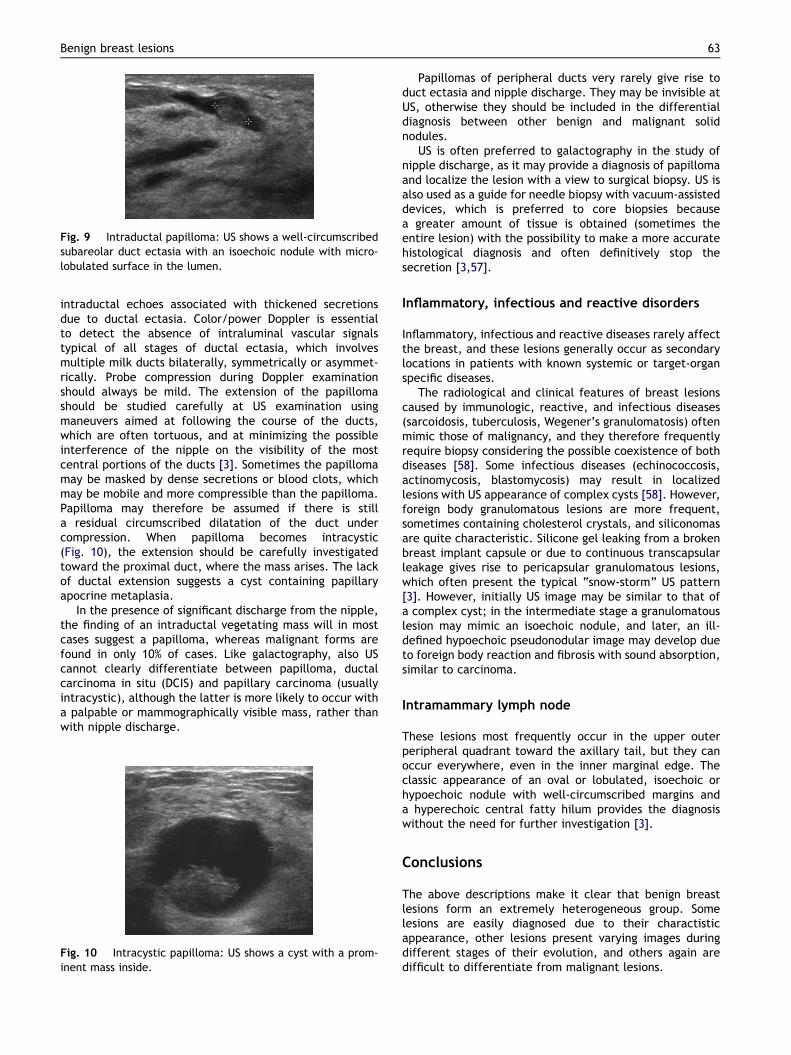
Fig. 9 Intraductal papilloma: US shows a well-circumscribedsubareolar duct ectasia with an isoechoic nodule with micro-lobulated surface in the lumen.
Benign breast lesions 63
intraductal echoes associated with thickened secretionsdue to ductal ectasia. Color/power Doppler is essentialto detect the absence of intraluminal vascular signalstypical of all stages of ductal ectasia, which involvesmultiple milk ducts bilaterally, symmetrically or asymmet-rically. Probe compression during Doppler examinationshould always be mild. The extension of the papillomashould be studied carefully at US examination usingmaneuvers aimed at following the course of the ducts,which are often tortuous, and at minimizing the possibleinterference of the nipple on the visibility of the mostcentral portions of the ducts [3]. Sometimes the papillomamay be masked by dense secretions or blood clots, whichmay be mobile and more compressible than the papilloma.Papilloma may therefore be assumed if there is stilla residual circumscribed dilatation of the duct undercompression. When papilloma becomes intracystic(Fig. 10), the extension should be carefully investigatedtoward the proximal duct, where the mass arises. The lackof ductal extension suggests a cyst containing papillaryapocrine metaplasia.
In the presence of significant discharge from the nipple,the finding of an intraductal vegetating mass will in mostcases suggest a papilloma, whereas malignant forms arefound in only 10% of cases. Like galactography, also UScannot clearly differentiate between papilloma, ductalcarcinoma in situ (DCIS) and papillary carcinoma (usuallyintracystic), although the latter is more likely to occur witha palpable or mammographically visible mass, rather thanwith nipple discharge.
Fig. 10 Intracystic papilloma: US shows a cyst with a prom-inent mass inside.
Papillomas of peripheral ducts very rarely give rise toduct ectasia and nipple discharge. They may be invisible atUS, otherwise they should be included in the differentialdiagnosis between other benign and malignant solidnodules.
US is often preferred to galactography in the study ofnipple discharge, as it may provide a diagnosis of papillomaand localize the lesion with a view to surgical biopsy. US isalso used as a guide for needle biopsy with vacuum-assisteddevices, which is preferred to core biopsies becausea greater amount of tissue is obtained (sometimes theentire lesion) with the possibility to make a more accuratehistological diagnosis and often definitively stop thesecretion [3,57].
Inflammatory, infectious and reactive disorders
Inflammatory, infectious and reactive diseases rarely affectthe breast, and these lesions generally occur as secondarylocations in patients with known systemic or target-organspecific diseases.
The radiological and clinical features of breast lesionscaused by immunologic, reactive, and infectious diseases(sarcoidosis, tuberculosis, Wegener’s granulomatosis) oftenmimic those of malignancy, and they therefore frequentlyrequire biopsy considering the possible coexistence of bothdiseases [58]. Some infectious diseases (echinococcosis,actinomycosis, blastomycosis) may result in localizedlesions with US appearance of complex cysts [58]. However,foreign body granulomatous lesions are more frequent,sometimes containing cholesterol crystals, and siliconomasare quite characteristic. Silicone gel leaking from a brokenbreast implant capsule or due to continuous transcapsularleakage gives rise to pericapsular granulomatous lesions,which often present the typical “snow-storm” US pattern[3]. However, initially US image may be similar to that ofa complex cyst; in the intermediate stage a granulomatouslesion may mimic an isoechoic nodule, and later, an ill-defined hypoechoic pseudonodular image may develop dueto foreign body reaction and fibrosis with sound absorption,similar to carcinoma.
Intramammary lymph node
These lesions most frequently occur in the upper outerperipheral quadrant toward the axillary tail, but they canoccur everywhere, even in the inner marginal edge. Theclassic appearance of an oval or lobulated, isoechoic orhypoechoic nodule with well-circumscribed margins anda hyperechoic central fatty hilum provides the diagnosiswithout the need for further investigation [3].
Conclusions
The above descriptions make it clear that benign breastlesions form an extremely heterogeneous group. Somelesions are easily diagnosed due to their charactisticappearance, other lesions present varying images duringdifferent stages of their evolution, and others again aredifficult to differentiate from malignant lesions.

64 N. Masciadri, C. Ferranti
A last comment should be dedicated to high risk lesionsand particularly atypical hyperplasia, which involve a moreimportant prognostic significance albeit with unspecific UScharacteristics. Macroscopic findings show those prolifera-tive conditions in whose context atypical hyperplasia has itsorigin, i.e. papillary hyperplasia, intraductal papilloma andradial scar. In atypical hyperplasia, diagnosis is exclusivelyhistological and this pathology is more easily detectedduring mammographically guided investigation of clusteredmicrocalcifications. However, the findings are difficult tointerpret and also histological analysis may becontroversial.
Technologically advanced US equipment providesa better evaluation of lesions and therefore a potentiallyreduced number of diagnostic biopsies. However, whenfurther investigation is appropriate, US guided biopsy isperformed and micro-histological diagnosis is obtained.This procedure is still based mainly on core biopsy butrecently also sampling using vacuum-assisted biopsydevices with a larger caliber has been introduced to ensurea better sample for histological evaluation, and in some
AppendixSupplementary data
Supplementary data related to this article can befound online at doi:10.1016/j.jus.2011.03.002
cases of a circumscribed benign lesion this procedure hasperformed definitive resection of the mass.
Conflict of interest statement
The authors have no conflict of interest.
References
[1] Lanyi M. Mammography. Diagnosis and pathological analysis.Berlin, Heidelberg, New York: Springer-Verlag; 2003.
[2] Hughes LE, Mansel RE, Webster DJ. Aberrations of normaldevelopment and involution (ANDI); a new perspective onpathogenesis and nomenclature of benign breast disorders.Lancet 1987;2(8571):1316e9.
[3] Stavros AT. Breast ultrasound. Philadelphia: LippincottWilliams & Wilkins; 2004.
[4] Dennis MA, Parker SH, Klaus AJ, Stavros AT, Kaske TJ, Clark SB.Breast biopsy avoidance: the value of normal mammogramsand normal sonograms in the setting of a palpable lump.Radiology 2001;219:186e91.
[5] Jackson VP. Management of solid nodules: what is the role ofsonography? Radiology 1995;196:14e5.
[6] Stavros AT, Thickman D, Rapp CL, Dennis MA, Parker SH,Sisney GA. Solid breast nodules: use of sonography to distin-guish between benign and malignant nodules. Radiology 1995;196:123e34.
[7] Rahbar G, Sie AC, Hansen GC, Prince JS, Melany ML,Reynolds HE, et al. Benign versus malignant solid breastmasses: US differentiation. Radiology 1999;213:889e94.
[8] American College of Radiology (ACR). Illustrated breastimaging reporting and data system (BI-RADS). 4th ed. Reston,VA: American College of Radiology; 2003.
[9] Levy L, Suissa M, Chiche JF, Teman G, Martin B. BIRADSultrasonography. Eur J Radiol 2007;61:202e11.
[10] Szopinski KT, Pajk AM, Wysocki M, Amy D, Szopinska M,Jakubowski W. Tissue harmonic imaging utility in breastsonography. J Ultrasound Med 2003;22:479e87.
[11] Sehgal CM, Weinstein SP, Arger PH, Conant EF. A review ofbreast ultrasound. J Mammary Gland Biol Neoplasia 2006;11:113e23.
[12] Athanasiou A, Tardivon A, Ollivier L, Thibault F, El Khoury C,Neuenschwander S. How to optimize breast ultrasound. Eur JRadiol 2009;69:6e13.
[13] Piccoli CW, Forsberg F. Advanced ultrasound techniques forbreast imaging. Semin Roentgenol 2011;46:60e7.
[14] Cosgrove DO, Kedar RP, Bamber JC, al-Murrani B, Davey JB,Fisher C, et al. Breast diseases: color Doppler US in differen-tial diagnosis. Radiology 1993;189:99e104.
[15] Calliada F, Raieli G, Sala G, Conti MP, Bottinelli O, La Fianza A,et al. Doppler color-echo in the echographic evaluation ofsolid neoplasms of the breast: 5 years of experience. RadiolMed 1994;87:28e35.
[16] Draghi F, Coopmans de Yoldi G. Atlante eco color-Dopplerdella mammella. Milano: Poletto; 1995.
[17] Hayashi N, Miyamoto Y, Nakata N, Irie T, Ikegami M, Asao K,et al. Breast masses: color Doppler, power Doppler, andspectral analysis findings. J Clin Ultrasound 1998;26:231e8.
[18] Cho KR, Seo BK, Lee JY, Pisano ED, Je BK, Lee JY, et al. Acomparative study of 2D and 3D ultrasonography for evalua-tion of solid breast masses. Eur J Radiol 2005;54:365e70.
[19] Abbattista T, Serri L, Busilacchi P. Studio ecografico 3D dellelesioni mammarie. Three dimensional sonographic study ofbreast nodules. J Ultrasound 2007;10:93e8.
[20] Samani A, Zubovits J, Plewes D. Elastic moduli of normal andpathological human breast tissues: an inversion-technique-basedinvestigation of 169 samples. Phys Med Biol 2007;52:1565e76.
[21] Ginat DT, Destounis SV, Barr RG, Castaneda B, Strang JG,Rubens DJ. US Elastography of breast and prostate lesions.RadioGraphics 2009;29:2007e16.
[22] Itoh A, Ueno E, Tohno E, Kamma H, Takahashi H, Shiina T,et al. Breast disease: clinical application of US elastographyfor diagnosis. Radiology 2006;239:341e50.
[23] Scaperrotta G, Ferranti C, Costa C, Mariani L, Marchesini M,Suman L, et al. Role of sonoelastography in non-palpablebreast lesions. Eur Radiol 2008;18:2381e9.
[24] Berg WA, Campassi CI, Ioffe OB. Cystic lesions of the breast:sonographic-pathologic correlation. Radiology 2003;227:183e91.
[25] Houssami N, Irwig L, Ung O. Review of complex breast cysts:implications for cancer detection and clinical practice. ANZ JSurg 2005;75:1080e5.
[26] Hayes R, Michell M, Nunnerley HB. Acute inflammation of thebreast: the role of breast ultrasound in diagnosis andmanagement. Clin Radiol 1991;44:253e6.
[27] Boisserie-Lacroix M, Lafitte JJ, Sirben C, Latrabe V, Grelet P,Zeinoun R, et al. Inflammatory and infectious lesions of thebreast: contribution of ultrasonography. J Chir (Paris) 1993;130:408e15.
[28] Salvador R, Salvador M, Jimenez JA, Martinez M, Casas L.Galactocele of the breast: radiologic and ultrasonographicfindings. Br J Radiol 1990;63:140e2.
[29] Harlow CL, Schackmuth EM, Bregman PS, Zeligman BE,Coffin CT. Sonographic detection of hematomas and fluid afterimaging-guided core breast biopsy. J Ultrasound Med 1994;13:877e82.
[30] Pignatelli V, Basolo F, Bagnolesi A, Cartei F, Grassi L, Savino A,et al. Hematoma and fat necrosis of the breast: mammo-graphic and echographic features. Radiol Med 1995;89:36e41.

Benign breast lesions 65
[31] Soo MS, Kornguth PJ, Hertzberg BS. Fat necrosis in the breast:sonographic features. Radiology 1998;206:261e9.
[32] Tan PH, Lai LM, Carrington EV, Opaluwa AS, Ravikumar KH,Chetty N, et al. Fat necrosis of the breast - A review. TheBreast 2006;15:313e8.
[33] Harvey JA, Moran RE, Maurer EJ, De Angelis GA. Sonographicfeatures of mammary oil cysts. J Ultrasound Med 1997;16:719e24.
[34] Webb LA, Young JR. Case report: haemangioma of the breast-appearances on mammography and ultrasound. Clin Radiol1996;51:523e4.
[35] Porter GJ, Evans AJ, Lee AH, Hamilton LJ, James JJ. Unusualbenign breast lesions. Clin Radiol 2006;61:562e9.
[36] Cole-Beuglet C, Soriano RZ, Kurtz AB, Goldberg BB. Fibroa-denoma of the breast: sonomammography correlated withpathology in 122 patients. AJR 1983;140:369e75.
[37] Jackson VP, Rothschild PA, Kreipke DL, Mail JT, Holden RW.The spectrum of sonographic findings of fibroadenoma of thebreast. Invest Radiol 1986;21:34e40.
[38] Fornage BD, Lorigan JG, Andry E. Fibroadenoma of the breast:sonographic appearance. Radiology 1989;172:671e5.
[39] Sklair-Levy M, Sella T, Alweiss T, Craciun I, Libson E, Mally B.Incidence and management of complex fibroadenomas. AJR2008;190:214e8.
[40] Kuijper A, Mommers EC, van der Wall E, van Dienst PJ.Histopathology of fibroadenoma of the breast. Am J ClinPathol 2001;115:736e42.
[41] Steinbock RT, Stomper PC, Meyer JE, Kopans DB. The ultra-sound appearance of giant fibroadenoma. J Clin Ultrasound1983;11:451e4.
[42] Kronemer KA, Rhee K, Siegel MJ, Sievert L, Hildebolt CF. Grayscale sonography of breast masses in adolescent girls.J Ultrasound Med 2001;20:491e6.
[43] Skaane P, Engedal K. Analysis of sonographic features in thedifferentiation of fibroadenoma and invasive ductal carci-noma. AJR 1998;170:109e14.
[44] DuPont WD, Page DL, Parl FF, Vnencak-Jones CL,Plummer JrWD,RadosMS, et al. Long-termrisk of breast cancerin women with fibroadenoma. N Engl J Med 1994;331:10e5.
[45] Giri D. Recurrent challenges in the evaluation of fibroepithe-lial lesions. Arch Pathol Lab Med 2009;133:713e21.
[46] Cole-Beuglet C, Soriano R, Kurtz AB, Meyer JE, Kopans DB,Goldberg BB. Ultrasound, X-ray mammography and histopa-thology of cystosarcoma phylloides. Radiology 1983;146:481e6.
[47] Buchberger W, Strasser K, Heim K, Muller E, Schrocksnadel H.Phylloides tumor: findings on mammography, sonography andaspiration cytology in 10 cases. AJR 1991;157:715e9.
[48] Liberman L, Bonaccio E, Hamele-Bena D, Abramson AF,Cohen MA, Dershaw DD. Benign and malignant phyllodestumors mammographic and sonographic findings. Radiology1996;198:121e4.
[49] Thorncroft K, Forsyth L, Desmond S, Audisio RA. The diagnosisand management of diabetic mastopathy. Breast J 2007;13:607e13.
[50] Mercado C-L, Naidrich SA, Hamele-Bena D, Fineberg SA,Buchbinder SS. Pseudoangiomatous stromal hyperplasia of thebreast: sonographic features with histopathologic correlation.Breast J 2004;10:427e32.
[51] Jones KN, Glazebrook KN, Reynolds C. Pseudoangiomatousstromal hyperplasia: imaging findings with pathologic andclinical correlation. AJR 2010;195:1036e42.
[52] Adler DD, Jeffries DO, Helvie MA. Sonographic features ofbreast hamartomas. J Ultrasound Med 1990;9:85e90.
[53] Georgian-Smith D, Kricun B, McKee G, Yeh E, Rafferty EA,D’Alessandro HA, et al. The mammary hamartoma: apprecia-tion of additional imaging characteristics. J Ultrasound Med2004;23:1267e73.
[54] Han BK, Choe YH, Ko YH, Yang JH, Nam SJ. Benign papillarylesions of the breast: sonographic-pathologic correlation.J Ultrasound Med 1999;18:217e23.
[55] Ganesan S, Karthik G, Joshi M, Damodaran V. Ultrasoundspectrum in intraductal papillary neoplasms of breast. Br JRadiol 2006;79:843e9.
[56] Brookes MJ, Bourke AG. Radiological appearances of papillarybreast lesions. Clin Radiol 2008;63:1265e73.
[57] Puglisi F, Zuiani C, Bazzocchi M, Valent F, Aprile G, Pertoldi B,et al. Role of mammography, ultrasound and large core biopsyin the diagnostic evaluation of papillary breast lesions.Oncology 2003;65:311e5.
[58] Sabate JM, Closet M, Gomez A, De Las Heras P, Torrubia S,Salinas T. Radiologic evaluation of uncommon inflammatoryand reactive breast disorders. RadioGraphics 2005;25:411e24.