bench aids - who
TRANSCRIPT

second edition
Bench aidsfor the diagnosis of intestinal parasites
These bench aids were planned and produced by
Professor Marco Genchi, Department of Veterinary Sciences, University of Parma, Parma, Italy
Mr Idzi Potters, BSc, Department of Clinical Sciences, Institute of Tropical Medicine, Antwerp, Belgium
Ms Rina G. Kaminsky, MSc, Department of Paediatrics, School of Medical Sciences, National Autonomous University, Honduras and Parasitology Service, Department of Clinical Laboratory, University Hospital, Honduras
Dr Antonio Montresor, Preventive Chemotherapy and Transmission Control, Department of Control of Neglected Tropical Diseases, World Health Organization, Geneva, Switzerland
Dr Simone Magnino, Istituto Zooprofilattico Sperimentale della Lombardia e dell’Emilia Romagna, Bruno Ubertini, Pavia, Italy
Acknowledgements
The World Health Organization (WHO) thanks the Istituto Zooprofilattico Sperimentale della Lombardia e dell’Emilia Romagna “Bruno Ubertini” for financially and technically supporting the preparation of this document. Many thanks also go to the Institute of Tropical Medicine of Antwerp for making available its extensive image library.
Thanks are also due to
Dr Shaali Ame, WHO Collaborating Centre for Neglected Tropical Diseases, Parasitology Unit, Public Health Laboratory Ivo de Carneri, Pemba, United Republic of Tanzania
Professor Giuseppe Cringoli, Department of Veterinary Medicine and Animal Productions, University of Naples Federico II, Naples, Italy
Dr Yvette Endriss, Swiss Tropical and Public Health Institute, Basel, Switzerland
Professor Laura Rinaldi, Department of Veterinary Medicine and Animal Productions, University of Naples Federico II, Naples, Italy
Dr Francesca Tamarozzi, WHO Collaborating Centre for the Epidemiology, Detection and Control of Cystic and Alveolar Echinococcosis, Istituto Superiore di Sanità (National Institute of Health), Rome, Italy
Dr Fabio Tosini, European Union Reference Laboratory for Parasites, Istituto Superiore di Sanità, Rome, Italy
Dr Jürg Utzinger, Swiss Tropical and Public Health Institute, Basel, Switzerland
First edition, 1994, revised and reprinted, 2000, 2003, 2012 with corrections
ISBN 978-92-4-151534-4
© World Health Organization 2019
Some rights reserved. This work is available under the Creative Commons Attribution-NonCommercial-ShareAlike 3.0 IGO licence (CC BY-NC-SA 3.0 IGO; https://creativecommons.org/licenses/by-nc-sa/3.0/igo).
Under the terms of this licence, you may copy, redistribute and adapt the work for non-commercial purposes, provided the work is appropriately cited, as indicated below. In any use of this work, there should be no suggestion that WHO endorses any specific organization, products or services. The use of the WHO logo is not permitted. If you adapt the work, then you must license your work under the same or equivalent Creative Commons licence. If you create a translation of this work, you should add the following disclaimer along with the suggested citation: “This translation was not created by the World Health Organization (WHO). WHO is not responsible for the content or accuracy of this translation. The original English edition shall be the binding and authentic edition”.
Any mediation relating to disputes arising under the licence shall be conducted in accordance with the mediation rules of the World Intellectual Property Organization.
Suggested citation. Bench aids for the diagnosis of intestinal parasites, second edition. Geneve: World Health Organization; 2019. Licence: CC BY-NC-SA 3.0 IGO.
Cataloguing-in-Publication (CIP) data. CIP data are available at http://apps.who.int/iris.
Sales, rights and licensing. To purchase WHO publications, see http://apps.who.int/bookorders. To submit requests for commercial use and queries on rights and licensing, see http://www.who.int/about/licensing.
Third-party materials. If you wish to reuse material from this work that is attributed to a third party, such as tables, figures or images, it is your responsibility to determine whether permission is needed for that reuse and to obtain permission from the copyright holder. The risk of claims resulting from infringement of any third-party-owned component in the work rests solely with the user.
General disclaimers. The designations employed and the presentation of the material in this publication do not imply the expression of any opinion whatsoever on the part of WHO concerning the legal status of any country, territory, city or area or of its authorities, or concerning the delimitation of its frontiers or boundaries. Dotted and dashed lines on maps represent approximate border lines for which there may not yet be full agreement.
The mention of specific companies or of certain manufacturers’ products does not imply that they are endorsed or recommended by WHO in preference to others of a similar nature that are not mentioned. Errors and omissions excepted, the names of proprietary products are distinguished by initial capital letters.
All reasonable precautions have been taken by WHO to verify the information contained in this publication. However, the published material is being distributed without warranty of any kind, either expressed or implied. The responsibility for the interpretation and use of the material lies with the reader. In no event shall WHO be liable for damages arising from its use.
Printed in France
Design and illustration by Andrea Iacobuzio
Front and back cover: graphic design by Marco Genchi, based on the identification keys in the first edition.
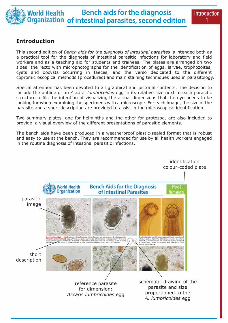
schematic drawing of the parasite and size
proportioned to the A. lumbricoides egg
parasitic image
short description
identification colour-coded plate
reference parasite for dimension:
Ascaris lumbricoides egg
Introduction1
Introduction
This second edition of Bench aids for the diagnosis of intestinal parasites is intended both as a practical tool for the diagnosis of intestinal parasitic infections for laboratory and field workers and as a teaching aid for students and trainees. The plates are arranged on two sides: the recto with microphotographs for the identification of eggs, larvae, trophozoites, cysts and oocysts occurring in faeces, and the verso dedicated to the different copromicroscopical methods (procedures) and main staining techniques used in parasitology.
Special attention has been devoted to all graphical and pictorial contents. The decision to include the outline of an Ascaris lumbricoides egg in its relative size next to each parasitic structure fulfils the intention of visualizing the actual dimensions that the eye needs to be looking for when examining the specimens with a microscope. For each image, the size of the parasite and a short description are provided to assist in the microscopical identification.
Two summary plates, one for helminths and the other for protozoa, are also included to provide a visual overview of the different presentations of parasitic elements.
The bench aids have been produced in a weatherproof plastic-sealed format that is robust and easy to use at the bench. They are recommended for use by all health workers engaged in the routine diagnosis of intestinal parasitic infections.
Bench aids for the diagnosis of intestinal parasites, second edition
Good laboratory practices and biosafety
Some basic practices and biosafety principles that must be followed in the laboratory are presented below. For more detailed information, refer to: https://apps.who.int/iris/handle/10665/42981A laboratory of parasitology is generally classified as a basic biosafety 2 laboratory (BSL-2) (see http://www.hse.gov.uk/pubns/misc208.pdf). This requires the application of good laboratory practices, the use of personal protective equipment and the display of an international biological hazards sign. The laboratory must also be equipped with safety hoods and specific disinfection and treatment procedures for biological materials, to be used also in case of accidental spillage. It should have regulated access and there should be enough space for laboratory benches and equipment, which must be arranged to allow for adequate cleaning. Facilities for storing personal clothing and items must be provided for all personnel, and storage areas for specimens, reagents and equipment should be available. Moreover, it is crucial that “clean” and “dirty” areas are clearly distinguished, adequately lit and ventilated; that barriers against arthropods are in place if the windows can be opened; that an easily accessible water source is available; and that the benches, walls and floors are smooth, water-repellent, and easy to clean and disinfect. Finally, the laboratory must be separated from any changing rooms and recreational areas provided to the staff.The disinfectants normally used are sodium hypochlorite (bleach), 70% ethanol or isopropanol, and quaternary ammonium compounds. Bleach is easily available and inexpensive. When diluted at 5–10%, bleach is suitable for disinfecting benches and work areas. Alcohols are effective in decontaminating stainless steel surfaces and removing bleach residues from metals to minimize corrosion. Quaternary ammonium compounds must be used after removal of organic matter, which reduce their effectiveness. For waste disposal, under ideal conditions all infected or potentially infected material should be decontaminated, autoclaved or incinerated in the laboratory. Contaminated waste containers, including those provided for sharps waste disposal, must be easily identifiable and fit for purpose.
Basic laboratory rules can be summarized as follows:
Introduction2
1. Keep the work areas uncluttered (e.g. never place backpacks, bags, books, etc. on the laboratory bench).
2. Always wash your hands with soap and water when you enter and leave the laboratory.3. Always wear your laboratory coat when in the laboratory and remove it when leaving;
laboratory coats and personal clothing should not be stored in the same locker.4. Always wear gloves when handling potentially dangerous biological or chemical substances.5. Wear safety glasses for protection against splashes, sprays and UV radiation.6. Use proper shoes (no sandals).7. Handle toxic substances (e.g. formalin) under a safety hood.8. Unequivocally label all preparations and samples to be analysed.9. Dispose of all waste appropriately and safely.
10. Clean and disinfect the work area at the beginning and end of each laboratory session.11. Do not take out of the laboratory the clipboard/notepad/pen/pencil used because they are
potentially contaminated.12. Do not store food and/or drinks in the laboratory.13. Do not eat and/or drink in the laboratory, nor bring hands or other objects (e.g. pencils,
make-up, contact lenses) to your mouth or eyes.
Bench aids for the diagnosis of intestinal parasites, second edition

Introduction3
- Microscope, objective 4x, 10x, 40x, 100x - Supplementary objective 20x, 60x- Ocular scale and stage micrometer for microscope calibration- Centrifuge (if possible, with rotor for microtiter plates)
Basic laboratory supplies in medical parasitology
- Laboratory scale- Hot plate with magnetic stirrer- Fridge- Stereomicroscope (if possible)
- Adhesive tape (transparent) and paper: 2 cm wide- Beakers (plastic and glass): 250 mL, 500 mL, 1000 mL- Conical glasses- Bottles (plastic or glass): 25 mL, 30 mL, 50 mL, 100 mL, 250 mL, 500 mL, 1000 mL, with stoppers or dropper-tops and screw-caps- Centrifuge tubes, conical, flat-top, graduated: 15 mL, 50 mL- Centrifuge tubes, conical, plastic disposable: 12 mL- Graduated cylinder: 10 mL, 25 mL, 50 mL, 100 mL, 250 mL, 1000 mL- Detergents and disinfectants- Dishes for staining, Coplin jars- Dropper bottle for saline, iodine, etc.- Forceps and scissors- Funnel (plastic and glass)- Gauze - Hot plate - Hydrometer (specific gravity 1.10–1.40)- Immersion oil, low viscosity- Permanent markers, pens, pencils
- Mortar and pestle (laboratory porcelain) - Gloves (latex or nitrile), disposable - Membrane filter (12 µm or 15 µm) and filter holder- Microscope slide and coverslips- Paper towels- Pasteur pipettes and rubber bulbs, electronic pipette- Petri dishes (plastic and glass)- Pipettes (plastic droppers) disposable: full pipette capacity 7 mL- “Squeeze” plastic: 100 ml, 250 mL, 500 mL- Rod (plastic) - Slide tray (plastic)- Stirring rods- Rack for centrifuge tubes - Record forms and notepaper - Self-adhesive labels- Strainer, metal (tea strainer), 7.5 cm diameter- Timer- Wooden applicator sticks, cotton swabs and tongue depressors
- Chromotrope 2R- Glacial acetic acid- Distilled water- Ethanol: 70%, 95%, 100%- Ethyl acetate- Formaldehyde (37–40%)- Glycerol- Hydrochloric acid (HCl)
- Iodine crystals (I2)- Light green SF- Malachite green- Potassium iodide (KI)- Saline solution- Sodium acetate- Methylene blue
- Carbol fuchsin: liquefy 5 g of phenol crystals with a small amount of distilled water, using a warm water bath at 95 °C. Dissolve 1 g of basic fuchsin in the liquefied phenol. Add 10 mL of 95% ethanol and mix. Add 100 mL of distilled water. Filter and store in a dark flask, well labelled. The solution is ready for use.- Formalin 5%: 50 mL formaldehyde + 950 mL distilled water or saline (recommended for all-purpose use and for preservation of protozoan cysts).- Formalin 10%: 100 mL formaldehyde + 900 mL distilled water or saline (recommended for helminth eggs and larvae).
- Lugol’s solution: 2 g potassium iodide (KI) + 1.5 g powered iodine crystals (add after KI dissolves) + 100 mL distilled water. Store in a brown, glass-stoppered bottle at room temperature and in the dark; the expiration date is 1 year. The solution is ready to use. For routine use, put 20 mL in a brown dropper bottle for 10–14 days.- SAF (sodium acetate-acetic acid-formalin fixative): sodium acetate 1.5 g + acetic acid, glacial 2.0 mL + formalin 4 mL + distilled water 92.0 mL).
Saturated sodium chloride (NaCl), specific gravity – 1.20: warm water 1000 mL + NaCl 500 g, dissolve overnight by magnetic stirrer. Check the specific gravity with a hydrometer.
Zinc sulfate (ZnSO4 7H2O), specific gravity 1.35: water 685 mL + zinc sulfate 685 g, dissolve overnight by magnetic stirrer. Check the specific gravity with a hydrometer.
Saturated sodium chloride (NaCl), sg 1.20Hookworms(Ascaris lumbricoides)(Trichuris spp.)Trichostrongylus spp.Strongyloides stercoralisEnterobius vermicularis
Ascaris lumbricoidesTrichuris spp.Fasciola hepaticaSchistosoma mansoniDicrocoelium dendriticumBalantidium coli
Dientamoeba fragilisBlastocystis hominisEntamoeba spp.Endolimax nanaGiardia duodenalisEnteromonas hominis
Zinc sulfate (ZnSO4 7H2O), sg - 1.35
Flotation solutions
Solutions
Reagents
Materials
Equipment
Hymenolepis spp.Taenia spp.
Bench aids for the diagnosis of intestinal parasites, second edition

Introduction4
The search for eggs and larvae of helminths (and of ciliates) is classically done using the 10x objective. The entire preparation is examined. To accomplish this, one should work systematically. Always start at a corner of the cover slip and work in a straight line from the chosen corner towards the opposite side. Once there, move one row aside and work back until the entire preparation has been examined. Always proceed by looking at the next microscopic field with a small overlap: when a field has been examined, an object in this field is chosen, and is brought towards the opposite side of the field. This second field is then examined. When parasitic structures are found, details are examined at 40x objective.
Microscopic examination
For searching most of the protozoans, the 40x objective is used. In the same way as described above, a few overlapping rows (3 or 4) should be examined. For morphological identification, oil immersion can be used with the Lugol’s solution smear. For differentiation of species, trophozoites and/or cysts must be measured.
Ocular scale
0 10 20 30 40 50
0 10 20 30 40 50
0 01 02 03 04
0 01 02 03 04
Stage
Disc-type micrometer
Adjust the stage micrometer to align 0 lines
Stage micrometer
Calibration of ocular micrometer
In order to measure structures in the microscopic field, it is necessary to have a measuring scale in the eye- piece of the microscope. Before it can be used, the scale must be calibrated.
Instructions1. Remove the eyepiece (10x or other) from the micro-scope and place the ocular scale on the diaphragm within the eyepiece. Screw back the lens and re-insert the eyepiece into the microscope.2. Place the stage micrometer on the microscope stage and focus on the scale.3. Adjust the stage micrometer by moving the stage so that the 0 line of the ocular micrometer is exactly super- imposed on the 0 line of the stage micrometer.4. Without moving the stage micrometer, find another point at the extreme right where two other lines are exactly superimposed. This second set of superimposed lines should be as far to the right as possible from the 0 lines. 5. Count the number of division lines on the ocular micrometer between the 0 line and the point where the second set of lines is superimposed. In the example provided in the figure, this number, indicated by the black arrows, equal 27 ocular units.6. Then count the number of 0.1 mm division lines between the 0 line and the second superimposed line on the stage micrometer; in the figure, this number, indicated by the red arrow, equals 0.2 mm.7. To calculate the length represented by one ocular unit: 1 ocular unit = (0.2 mm/27) x 1000 = 7.4 µm.8. Thus, 1 ocular unit = 7.4 µm for this specific objec- tive. Each objective on the microscope must be calibrated separately.9. When all objectives have been calibrated, prepare a simple chart that displays the calibration factor for each objective.
Bench aids for the diagnosis of intestinal parasites, second edition
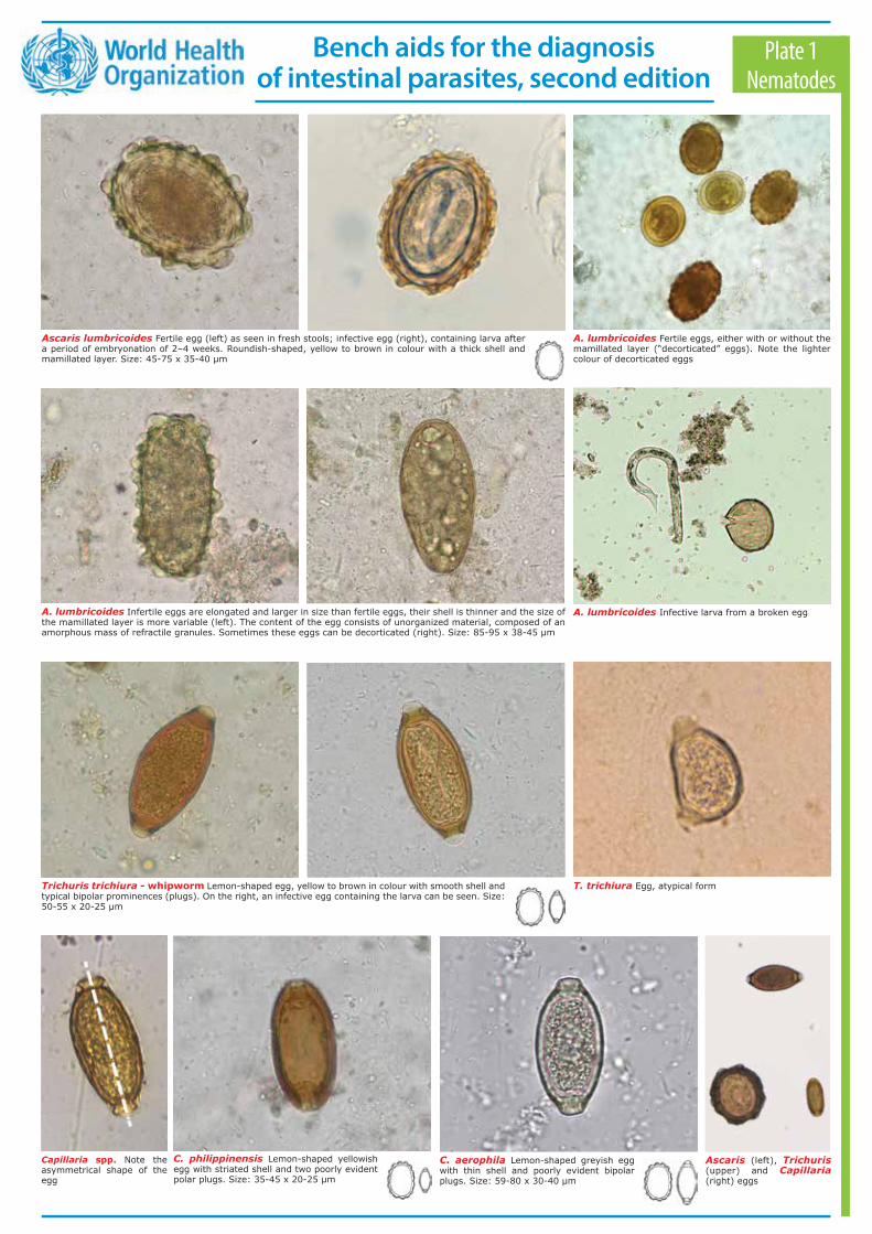
Ascaris lumbricoides Fertile egg (left) as seen in fresh stools; infective egg (right), containing larva after a period of embryonation of 2–4 weeks. Roundish-shaped, yellow to brown in colour with a thick shell and mamillated layer. Size: 45-75 x 35-40 µm
A. lumbricoides Fertile eggs, either with or without the mamillated layer (“decorticated” eggs). Note the lighter colour of decorticated eggs
A. lumbricoides Infective larva from a broken eggA. lumbricoides Infertile eggs are elongated and larger in size than fertile eggs, their shell is thinner and the size of the mamillated layer is more variable (left). The content of the egg consists of unorganized material, composed of an amorphous mass of refractile granules. Sometimes these eggs can be decorticated (right). Size: 85-95 x 38-45 μm
Trichuris trichiura - whipworm Lemon-shaped egg, yellow to brown in colour with smooth shell and typical bipolar prominences (plugs). On the right, an infective egg containing the larva can be seen. Size: 50-55 x 20-25 µm
T. trichiura Egg, atypical form
C. philippinensis Lemon-shaped yellowish egg with striated shell and two poorly evident polar plugs. Size: 35-45 x 20-25 µm
Capillaria spp. Note the asymmetrical shape of the egg
Ascaris (left), Trichuris (upper) and Capillaria (right) eggs
C. aerophila Lemon-shaped greyish egg with thin shell and poorly evident bipolar plugs. Size: 59-80 x 30-40 µm
Plate 1Nematodes
Bench aids for the diagnosis of intestinal parasites, second edition
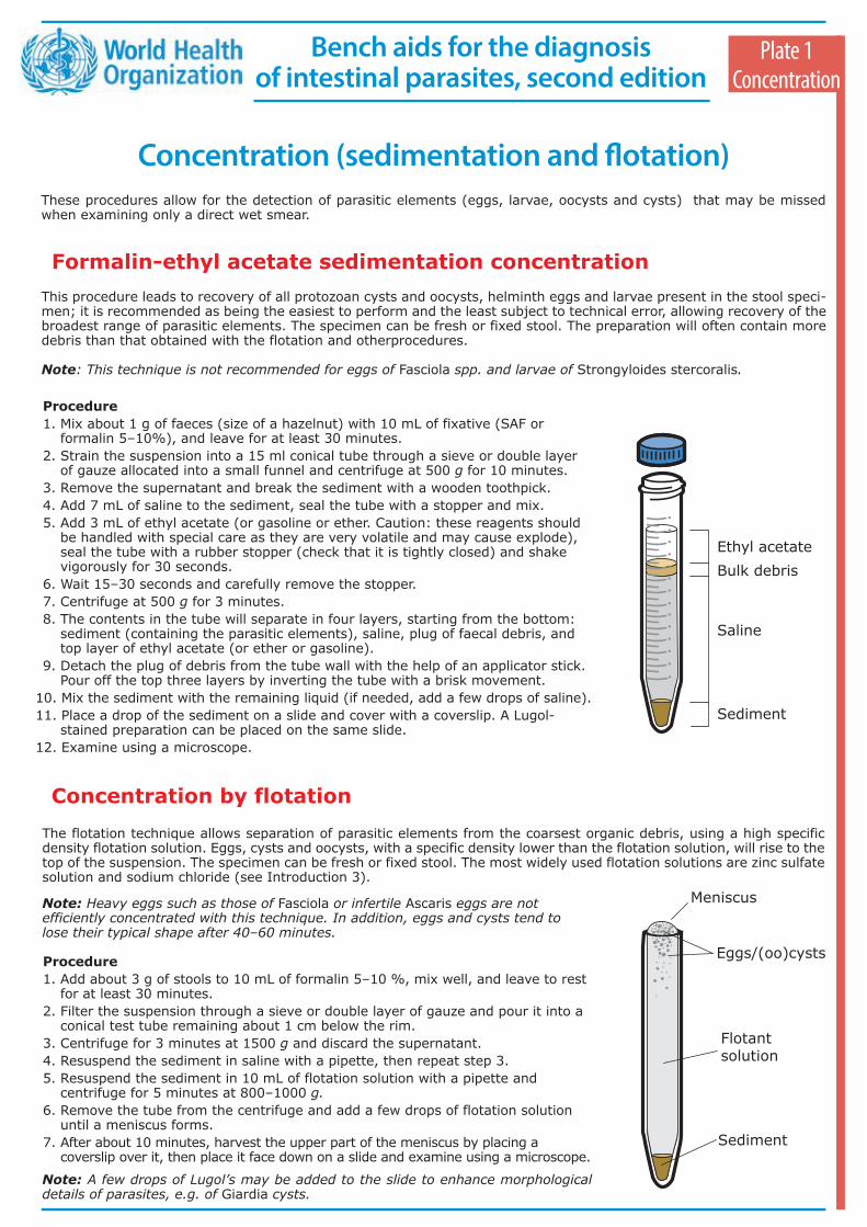
Concentration (sedimentation and flotation) These procedures allow for the detection of parasitic elements (eggs, larvae, oocysts and cysts) that may be missed when examining only a direct wet smear.
Formalin-ethyl acetate sedimentation concentrationThis procedure leads to recovery of all protozoan cysts and oocysts, helminth eggs and larvae present in the stool speci-men; it is recommended as being the easiest to perform and the least subject to technical error, allowing recovery of the broadest range of parasitic elements. The specimen can be fresh or fixed stool. The preparation will often contain more debris than that obtained with the flotation and otherprocedures.
Note: This technique is not recommended for eggs of Fasciola spp. and larvae of Strongyloides stercoralis.
Concentration by flotationThe flotation technique allows separation of parasitic elements from the coarsest organic debris, using a high specific density flotation solution. Eggs, cysts and oocysts, with a specific density lower than the flotation solution, will rise to the top of the suspension. The specimen can be fresh or fixed stool. The most widely used flotation solutions are zinc sulfate solution and sodium chloride (see Introduction 3).
Procedure 1. Mix about 1 g of faeces (size of a hazelnut) with 10 mL of fixative (SAF or
formalin 5–10%), and leave for at least 30 minutes.2. Strain the suspension into a 15 ml conical tube through a sieve or double layer
of gauze allocated into a small funnel and centrifuge at 500 g for 10 minutes.3. Remove the supernatant and break the sediment with a wooden toothpick.4. Add 7 mL of saline to the sediment, seal the tube with a stopper and mix.5. Add 3 mL of ethyl acetate (or gasoline or ether. Caution: these reagents should
be handled with special care as they are very volatile and may cause explode), seal the tube with a rubber stopper (check that it is tightly closed) and shake vigorously for 30 seconds.
6. Wait 15–30 seconds and carefully remove the stopper.7. Centrifuge at 500 g for 3 minutes.8. The contents in the tube will separate in four layers, starting from the bottom:
sediment (containing the parasitic elements), saline, plug of faecal debris, and top layer of ethyl acetate (or ether or gasoline).
9. Detach the plug of debris from the tube wall with the help of an applicator stick. Pour off the top three layers by inverting the tube with a brisk movement.
10. Mix the sediment with the remaining liquid (if needed, add a few drops of saline).11. Place a drop of the sediment on a slide and cover with a coverslip. A Lugol-
stained preparation can be placed on the same slide.12. Examine using a microscope.
Procedure 1. Add about 3 g of stools to 10 mL of formalin 5–10 %, mix well, and leave to rest
for at least 30 minutes.2. Filter the suspension through a sieve or double layer of gauze and pour it into a
conical test tube remaining about 1 cm below the rim.3. Centrifuge for 3 minutes at 1500 g and discard the supernatant.4. Resuspend the sediment in saline with a pipette, then repeat step 3.5. Resuspend the sediment in 10 mL of flotation solution with a pipette and
centrifuge for 5 minutes at 800–1000 g.6. Remove the tube from the centrifuge and add a few drops of flotation solution
until a meniscus forms.7. After about 10 minutes, harvest the upper part of the meniscus by placing a
coverslip over it, then place it face down on a slide and examine using a microscope.
Note: A few drops of Lugol’s may be added to the slide to enhance morphological details of parasites, e.g. of Giardia cysts.
Plate 1Concentration
Ethyl acetateBulk debris
Saline
Sediment
Meniscus
Eggs/(oo)cysts
Flotantsolution
Sediment
Note: Heavy eggs such as those of Fasciola or infertile Ascaris eggs are not efficiently concentrated with this technique. In addition, eggs and cysts tend to lose their typical shape after 40–60 minutes.
Bench aids for the diagnosis of intestinal parasites, second edition

Ancylostomidae – hookworm (Ancylostoma braziliense, A. caninum, A. duodenale, Necator americanus) Eggs of these parasites cannot be differentiated microscopically. They are ovoid, yellow-greyish and have a thin shell. In fresh faeces, they contain a 4–8 cell morula. If left at room temperature for a few hours, a higher number of cells (right) will develop. Size: 60-75 x 35-40 µm
A. duodenale and N. americanus Buccal capsules of adult parasites. Note the difference between the sharp teeth of A. duodenale and the semilunar cutting plates of N. americanus. Fixed in formalin and cleared in 70% alco- hol and glycerine
Trichuris (left) and Ancylostomidae (right) Eggs in the same microscopic field illustrate the different sizes
Trichostrongylus spp. (T. orientalis, T. colubriformis, T. axei) Eggs ovoid in shape, pointed at one end, yellow-greyish in colour, thin-walled, very similar to hookworm eggs but slightly larger. The morula inside may consist of a variable number of blastomeres. Size: 75-95 x 40-50 µm
Strongyloides stercoralis Ovoid in shape, greyish in colour, thin-walled eggs, already harbouring a larva when passed in faeces, where they occasionally can be seen. Sizes: 50-58 x 30-34 µm
S. stercoralis Rhabditiform larva (L1) stained with Lugol. Note the genital primordium (arrow, left). Close-up of the cephalic end (right), note the rhabditoid oesopha-gus and the short buccal cavity. Size: 180-380 x 14-20 µm
S. stercoralis Rhabditiform larvae (L1) after Baermann sedimentation (unstained, left) and filariform larvae (L3) in expectorate (Ziehl-Neelsen stain, right)
E. vermicularis Adult female; note the cephalic expansions and the pointed tail. Females adhering to the anus are replete with eggs. Female size is 8-13 x 0.3-0.5 mm; male size is 2.5 x 0.1-0.2 mm
Enterobius vermicularis - pinworm Oval-shaped eggs, asymmetrical, usually containing a larva. On the right, eggs collected by the tape-test. Size: 50-60 x 20-30 µm
S. stercoralis Detail of the caudal end of a rhabditiform larva (L1) with tapered tail (left, Lugol) and filariform larva (L3) with notched tail (right, unstained)
Plate 2Nematodes
Bench aids for the diagnosis of intestinal parasites, second edition
McMaster techniqueThe McMaster technique is used for the identification and quantification of the number of parasitic elements per gram of faeces: eggs per gram (epg), oocysts per gram (opg), cysts per gram (cpg), larvae per gram (lpg). The specimen can be fresh or fixed stool. This test uses a special microscope slide with a grid, which makes counting easier.
Procedure without speciment centrifugation 1. Homogenize the stool specimen. 2. Weigh 2 g of faeces and dilute it in 28 mL of flotation solution (see Introduction 3).3. Strain the suspension through a sieve for 3 times.4. Mix the suspension by pouring it from one glass to another for 10 times.5. Fill the first chamber of the McMaster slide using a pipette. Leave no fluid in the pipette, as the eggs will rise
quickly in the flotation fluid.6. Repeat step 4 and fill the second chamber (Fig. 1).7. Wait 2 minutes.8. Examine one chamber under a microscope and multiply the number of parasitic elements under one etched area
by 100 (Fig. 2). Alternatively, examine both chambers and multiply by 50 to obtain the number of parasitic elements per gram of faeces: epg, opg, cpg, lpg.
Note: At the end of the procedure, place the chamber in soapy water with disinfectant, then, clean, rinse and dry.
Procedure with specimen centrifugation 1. Homogenize the stool specimen. 2. Weigh 2 g of faeces into a beaker on a scale and dilute it in 28 mL of tap water.3. Strain the suspension through a tea strainer or double layer of gauze.4. Mix the suspension by pouring it from one beaker to another for 10 times and fill a 15 mL test tube to a few cm
below the rim.5. Centrifuge at 1500 g for 3 minutes.6. Pour off supernatant, and fill tube to previous level with flotation solution (see Introduction 3).7. Mix very well the suspension thoroughly with a pipette and fill the first chamber (“A”) of the McMaster slide. Leave
no fluid in the pipette, as the eggs will rise quickly in the flotation fluid.8. Repeat step 7 and fill the second chamber (“B”).9. Wait 2 minutes.
10. Examine one chamber under a microscope and multiply the number of parasitic elements under one etched area by 100. Alternatively, examine both chambers and multiply by 50 to obtain the number of parasitic elements per gram of faeces: epg, opg, cpg, lpg.
If a centrifuge is not available, the procedure can be carried out without centrifugation, but slide examination might be more difficult due to the presence of more debris.
Plate 2Concentration
Concentration (flotation)
x 100
x 50
A B
Fig. 2Fig. 1
Bench aids for the diagnosis of intestinal parasites, second edition
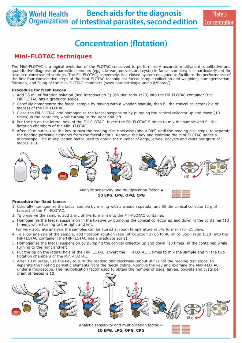
Schistosoma mansoni Oval-shaped elongated egg with a peculiar lateral spine which can or cannot be seen depending on the egg position. One polar end is tapered and slightly curved. They are yellowish-grey in colour, thin-walled and contain a miracidium. Right, calcified egg. Size: 114-180 x 45-70 µm
S. mansoni (centre) and ancylostomidae (upper and lower). Eggs in the same microscopic field illustrate the different sizes. Lugol stain
S. mekongi Eggs almost spherical in shape, similar to those of S. japonicum but smaller. They have a small inconspicuous spine (arrow, left), which is often not visible (right). Eggs are grey-yellowish in colour, their wall is thicker than that of other schistosomal eggs. They contain a miracidium. Size: 50-80 x 40-65 µm
S. japonicum Eggs rounded in shape, similar to those of S. mansoni but smaller. They have a small inconspicuous lateral spine (arrow). Eggs are grey-yellowish in colour, thin-walled and contain a miracidium. Often, the egg’s orientation may obscure the spine (right). Size: 70-100 x 55-64 µm
S. japonicum Egg elongated in shape. The lateral spine is often hidden
S. intercalatum Eggs are rhomboid in shape, sometimes with an equatorial bulge. They have a prominent terminal spine, are thin-walled and contain a miracidium. Size:104-203 µm
S. mansoni Adults, the thin female resides in the gynecophoral canal of the thicker male. Adults reside in venous plexi
S. haematobium These eggs are usually found in the urine but occasionally occur also in faeces. Size:110-170 µm
S. mansoni Adults, thin female and thicker male
Plate 3Trematodes
Bench aids for the diagnosis of intestinal parasites, second edition
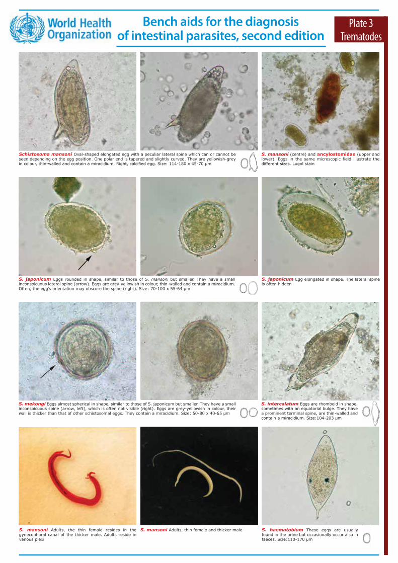
The Mini-FLOTAC is a logical evolution of the FLOTAC conceived to perform very accurate multivalent, qualitative and quantitative diagnosis of parasitic elements (eggs, larvae, oocysts and cysts) in faecal samples; it is particularly apt for resource-constrained settings. The Fill-FLOTAC, conversely, is a closed system designed to facilitate the performance of the first four consecutive steps of the Mini-FLOTAC techniques: faecal sample collection and weighing, homogenization, filtration, and filling of the Mini-FLOTAC chambers (www.parassitologia.unina.it/flotac/).
Procedure for fresh faeces 1. Add 38 mL of flotation solution (see Introduction 3) (dilution ratio 1:20) into the Fill-FLOTAC container (the
Fill-FLOTAC has a graduate scale).2. Carefully homogenize the faecal sample by mixing with a wooden spatula, then fill the conical collector (2 g of
faeces) of the Fill-FLOTAC.3. Close the Fill FLOTAC and homogenize the faecal suspension by pumping the conical collector up and down (10
times) in the container, while turning to the right and left.4. Put the tip on the lateral hole of the Fill-FLOTAC. Invert the Fill-FLOTAC 5 times to mix the sample and fill the
flotation chambers of the Mini-FLOTAC.5. After 10 minutes, use the key to turn the reading disc clockwise (about 90°) until the reading disc stops, to separate
the floating parasitic elements from the faecal debris. Remove the key and examine the Mini-FLOTAC under a microscope. The multiplication factor used to obtain the number of eggs, larvae, oocysts and cysts per gram of faeces is 10.
Procedure for fixed faeces 1. Carefully homogenize the faecal sample by mixing with a wooden spatula, and fill the conical collector (2 g of
faeces) of the Fill-FLOTAC.2. To preserve the sample, add 2 mL of 5% formalin into the Fill-FLOTAC container.3. Homogenize the faecal suspension in the fixative by pumping the conical collector up and down in the container (10
times), while turning to the right and left. For very accurate analysis the samples can be stored at room temperature in 5% formalin for 21 days.4. To allow analysis of the sample, add flotation solution (see Introduction 3) up to 40 ml (dilution ratio 1:20) into the
Fill-FLOTAC container (the Fill-FLOTAC has a graduate scale).5. Homogenize the faecal suspension by pumping the conical collector up and down (10 times) in the container, while
turning to the right and left.6. Put the tip on the lateral hole of the Fill-FLOTAC. Invert the Fill-FLOTAC 5 times to mix the sample and fill the two
flotation chambers of the Mini-FLOTAC. 7. After 10 minutes, use the key to turn the reading disc clockwise (about 90°) until the reading disc stops, to
separate the floating parasitic elements from the faecal debris. Remove the key and examine the Mini-FLOTAC under a microscope. The multiplication factor used to obtain the number of eggs, larvae, oocysts and cysts per gram of faeces is 10.
Plate 3Concentration
Concentration (flotation) Mini-FLOTAC techniques
(A)
(B)
Analytic sensitivity and multiplication factor =10 EPG, LPG, OPG, CPG
(A)
(B)
Analytic sensitivity and multiplication factor =10 EPG, LPG, OPG, CPG
Bench aids for the diagnosis of intestinal parasites, second edition
Fasciola hepatica Eggs ellipsoidal in shape, yellowish in colour and thin-walled. The operculum may often be indistinct. Eggs are unembryonated when passed in faeces and cannot be easily differentiated from those of Fasciolopsis buski, F. gigantica, Echinostoma spp. and Gastrodiscoides hominis Size: 130-145 x 70-90 µm
Clonorchis sinensis Eggs are operculated and have a miracidium inside. At the abopercular end, sometimes a small knob can be seen. Opisthor-chis and Metagonimus eggs are very similar to those of C. sinensis. Size: 27-35 x 11-20 µm
Opisthorchis spp. Eggs are operculated and have a miracidium inside. At the abopercu-lar end, sometimes a small knob can be seen. Clonorchis and Metagonimus eggs are very similar to those of Opisthorchis. Size: 26-30 x 11-15 µm
Metagonimus yokogawai Eggs are opercula-ted and have a miracidium inside. At the abopercu-lar end, sometimes a small knob can be seen. Clonorchis and Opisthorchis eggs are very similar to those of M. yokogawai. Size: 26-30 x 15-20 µm
Paragonimus westermani Eggs are so- metimes asymmetrical, with a prominent operculum. Eggs are unembryonated and can be found in expectorations. They are occasio-nally found in faeces. Size: 80-120 x 45-70 µm
Paragonimus uterobilateralis Eggs are oval-shaped, usually smaller and with a less prominent operculum than those of P. westermani. Size: 50-95 x 35-55 µm
Dicrocoelium dendriticum Oval-shaped asymmetrical eggs, dark brown in colour, thick-walled, operculated and containing a miracidium. Size: 35-45 x 20-30 µm
From left to right: C. sinensis, P. westermani, D. dendriticum, F. hepatica and F. buski adults. Bar = 1 cm
F. hepatica After flotation with zinc sulphate solution, the egg is coarted and it is difficult to recognize
F. hepatica Cercariae (left),present in the aquatic environment and the infective stage metacercaria (right), encysted on water vegetation
Plate 4Trematodes
Bench aids for the diagnosis of intestinal parasites, second edition
This technique allows the concentration and detection of larvae of Strongyloides spp. and trophozoites of Balantidium spp. in stools. The Baermann apparatus consists of a glass funnel (14 cm in diameter) equipped with a soft rubber 10-cm tubing, pinched at one end with a clamp.
Note: Only fresh unfixed and unrefrigerated faecal specimens can be processed. Always wear gloves during all procedures.
Procedure 1. Homogenize the stool specimen; if faeces are very hard, add a few mL of saline.2. Collect about 10 g (size of an apricot) of faeces and place them inside two layers of gauze. Place a filter paper disc
with a medium flow rate over the gauze in case of diarrhoeic faeces. Ensure that the gauze remains suspended inside the funnel using a stick, such as a wooden tongue depressor or by placing it the steel tea strainer.
3. Fill the funnel almost to the brim with lukewarm water or saline.4. A light source may be placed under the Baermann apparatus to enhance larvae migration. 5. After about 2–3 hours, loosen the clamp at the end of the tubing and transfer about 10 mL of the sediment into a
conical tube.6. Centrifuge the tube at 500 g for 10 minutes.*7. Discard the supernatant.8. Transfer a few drops of the sediment with a pipette onto a slide.9. Before examining the slide with the microscope, a drop of Lugol’s iodine solution may be added for staining and
immobilizing the larvae.10. Examine using a microscope with 10x objective.
* If a centrifuge is not available, a small amount of sediment can be transferred directly onto a slide, if desired with a drop of Lugol’s iodine solution.
Plate 4Concentration
Concentration (sedimentation) Baermann technique
Glass funnel
Soft rubber tube
Gauze
Funnel support
Clip Tongue depressor
Two alternatives for Baermann apparatus. In the left image, a plastic sedimentation cup is used; in this case the larvae are collected from the bottom using a long pipette. In the right image, a 50 mL syringe and an intravenous tube is used.
Bench aids for the diagnosis of intestinal parasites, second edition

Diphyllobothrium spp. Adults are ivory coloured and measure up to 10 m long and 1.5 cm wide. Mature gravid proglottids are wider than long and show a typical rosette-shaped uterus, visible in the middle of each proglottid. Eggs leave the mature proglottid through the genital pore and can be seen at copromicroscopic examination. Unstained
T. solium Stained proglottids. Adults are ivory coloured, up to 2-7 m long. Mature proglottids are longer than wide and characterized by 7-13 unilateral uterine branches. Mature proglottids are passed with the faeces. Carmine stain
T. saginata Stained proglottid. Adults are ivory coloured, up to 5 m long. Mature proglottids are longer than wide and characterized by 14-32 unilateral uterine branches. Mature proglottids actively leave their host. Carmine stain
Diphyllobothrium latum Operculated egg (arrow head), never embryonated when passed in the faeces. At the abopercular end, a knob (arrow) can sometimes be seen. Size: 67-71 x 40-51 µm
Dipylidium caninum Three unstained proglottids. Adults are ivory-coloured, up to 50 cm long, with typical proglottids that resemble rice grains
Hymenolepis nana Egg spherical to oval in shape, grey in colour with a thin external membrane. Left: unstained preparation; middle: Lugol preparation. On the internal membrane, two polar thickenings can sometimes be seen. Three pairs of hooks can be seen inside the oncosphere. Size: 30-50 µm. Adult worm showing armed scolex (right). Size: 1.5-4.4 cm
Hymenolepis diminuta Egg spherical to oval in shape, grey-yellowish in colour with a darker and sometimes striated external membrane. Three pairs of hooks can be seen inside the oncosphere. Left: egg in Lugol preparation; right: cephalic end of the adult. Size of the eggs: 70-85 x 60-80 µm. Size of the adults: 20-60 cm
D. caninum Details of a mature proglottid (left) where the egg capsules (centre) can be seen, containing the oncospheres with hooks (right). Size of the egg containing the hexacanth embryo: 25-50 µm
Plate 5Cestodes
Taenia spp. Egg has a very thick radiated shell (embryophore), sometimes surrounded by vitellum (arrow). Three pairs of hooks can be seen inside the larva (onchosphere. Taenia species egg cannot be differentiated. Size: 30-35 µm
Bench aids for the diagnosis of intestinal parasites, second edition

Harada–Mori filter paper strip culture
Procedure1. Add saline to solid or semi-solid stools to make them pasty. 2. Approximately double the stool volume by adding granulated hardwood charcoal
and mix thoroughly.*3. Cut a 12 cm strip as wide as a 15 mL tube out of a blotting paper disc4. Pour 3–4 mL of saline into a 15 mL tube.5. Spread the stool sample with a wooden spatula over 4–5 cm in the middle of the
paper strip and leave both ends clean (about 4 cm each side). 6. Insert the strip into the tube avoiding contact of faeces with saline.7. Cut the blotting paper in excess outside the tube and close the tube with a
stopper or cotton plug.8. Store the tube in a rack at 24–28 °C for 10 days and ensure each day that the
level of saline is maintained. 9. Open the tube and discard the paper strip.
10. Transfer the solution with a pipette into a new tube and add 12 mL of formalin 5%.
11. After 1 hour, centrifuge at 500 g for 2 minutes and discard the supernatant.12. Resuspend the sediment with a pipette, transfer a drop of the solution onto a
slide, cover it with a coverslip and examine with a microscope. A drop of Lugol’s iodine solution may be added for colour contrast and enhancement of morphological details.
Plate 5Culture
Koga–agar plate technique
Procedure1. Add saline to solid or semi-solid stools to make them pasty. 2. Approximately double the stool volume by adding granulated hardwood
charcoal and mix thoroughly.* 3. Place about 6–7 g of faeces in the middle of a Petri plate with agar medium
(1.5% agar, 0.5% meat extract, 1.0% peptone and 0.5% NaCl). Add no more than 10 ml of medium per plate, so as to make a thin medium.
4. Seal the plate with adhesive tape and store it for 48 hours in the dark at 26-33 °C. For the detection of hookworms, extend the incubation time to 7 days.
5. Check the plate with a stereomicroscope for larvae and/or their tracks in the medium. If positive, cover the agar with formalin 5% and allow to stand for 30–60 minutes.
6. Transfer the liquid with a pipette into a tube, centrifuge at 500 g for 2 minutes and discard the supernatant.
7. Resuspend the sediment with a pipette, transfer a drop of the solution onto a slide, cover it with a coverslip and examine with a microscope. A drop of Lugol’s iodine solution may be added for enhancement of morphological details.
Culture of larval-stage nematodes
These techniques are used to identify infections by hookworms, S. stercoralis, Trichostrongylus spp.Note: only fresh unfixed and unrefrigerated faecal specimens can be processed..
Filterpaper
Faeces
Salinesolution
Agar plate culture is considered to be the most efficient method for the detection of S. stercoralis larvae and this technique should be the test of choice, especially in immunocompromised patients.
* Charcoal may also not be added, however, adding it increases the sensitivity of the technique.
* Charcoal may also not be added, however, adding it increases the sensitivity of the technique.
Bench aids for the diagnosis of intestinal parasites, second edition
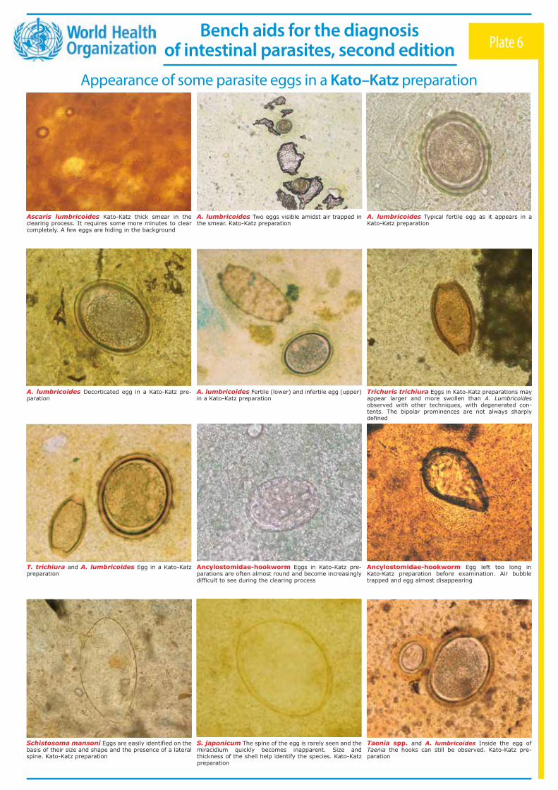
Schistosoma mansoni Eggs are easily identified on the basis of their size and shape and the presence of a lateral spine. Kato-Katz preparation
Taenia spp. and A. lumbricoides Inside the egg of Taenia the hooks can still be observed. Kato-Katz pre- paration
Ancylostomidae-hookworm Egg left too long in Kato-Katz preparation before examination. Air bubble trapped and egg almost disappearing
Ascaris lumbricoides Kato-Katz thick smear in the clearing process. It requires some more minutes to clear completely. A few eggs are hiding in the background
Plate 6
A. lumbricoides Two eggs visible amidst air trapped in the smear. Kato-Katz preparation
A. lumbricoides Typical fertile egg as it appears in a Kato-Katz preparation
A. lumbricoides Decorticated egg in a Kato-Katz pre- paration
A. lumbricoides Fertile (lower) and infertile egg (upper) in a Kato-Katz preparation
Trichuris trichiura Eggs in Kato-Katz preparations may appear larger and more swollen than A. Lumbricoides observed with other techniques, with degenerated con- tents. The bipolar prominences are not always sharply defined
T. trichiura and A. lumbricoides Egg in a Kato-Katz preparation
Ancylostomidae-hookworm Eggs in Kato-Katz pre- parations are often almost round and become increasingly difficult to see during the clearing process
S. japonicum The spine of the egg is rarely seen and the miracidium quickly becomes inapparent. Size and thickness of the shell help identify the species. Kato-Katz preparation
Appearance of some parasite eggs in a Kato–Katz preparation
Bench aids for the diagnosis of intestinal parasites, second edition

Kato–Katz technique for soil-transmitted helminths
Materials and reagents1. Wooden applicator sticks.2. Screen (stainless steel, nylon or plastic: 60-105 µm mesh) (Fig. 1).3. Template (stainless steel, plastic, or cardboard) (Fig. 1). A hole of 9 mm on a
1 mm thick template will deliver about 50 mg of faeces; a hole of 6 mm on a 1.5 mm thick template, 41.7 mg; and a hole of 6.5 mm on a 0.5 mm thick template, 20 mg. The same size of templates should always be used to ensure repeatability and comparability of prevalence and intensity data.
4. Spatula (plastic) (Fig. 1).5. Microscope slides (75 x 25 mm).6. Hydrophilic cellophane (40–50 µm thick, strips 25 x 30 or 25 x 35 mm in size). 7. Flat-bottom jar with lid, forceps and toilet paper or absorbent tissue. 8. Newspaper.9. Glycerol-malachite green (1 mL of 3% aqueous malachite green is added to 100
mL of glycerol and 100 ml of distilled water and mixed well). This solution is poured onto the cellophane strips in a jar and left for at least 24 hours prior to use.
Plate 6Kato-Katz
Procedure1. Place a small amount of the faecal sample on a newspaper and press a piece of
nylon screen on top. Using a spatula, scrape the sieved faecal material from the screen (Fig. 2).
2. Label a glass slide with the sample number and place a template with hole on the centre of a microscope slide. Fill the hole in the template with the sired faecal material, avoiding air bubbles and levelling the faeces off to remove any excess material (Fig. 3).
3. Carefully lift off the template and place it in a bucket of water mixed with concentrated detergent and disinfectant so that it can be reused.
4. Place one piece of cellophane, which has been soaked overnight in glycerol solution, over the faecal sample (Fig. 4).
5. Invert the microscope slide (Fig. 5) and firmly press the sample against the cellophane strip on another microscope slide or on a smooth hard surface to spread the faeces in a circle (Fig. 6).
6. Carefully pick up the slide again by gently sliding it sideways to avoid separating the cellophane strip or lifting it off. Place the slide on the bench with the cellophane upwards. Water evaporates while glycerol clears the faeces. When clarified it should be possible to read newspaper print through the stool smear (Fig. 7).
7. For all except hookworm eggs, keep the slide for one or more hours at room temperature to clear the faecal material prior to examination under the microscope. To speed up clearing and examination, the slide can be placed in a 40 °C incubator or kept in direct sunlight for several minutes.
8. A. lumbricoides and T. trichiura eggs will remain visible and recognizable for many months. Hookworm eggs clear rapidly and will no longer be visible after 30–60 minutes. Schistosome eggs may be recognizable for up to several months but it is preferable to examine the slide preparations within 24 hours.
9. The smear should be examined systematically. Then, multiply by the appropriate number to give the number of eggs per gram of faeces (by 20 if using a 50 mg template; by 50 for a 20 mg template; and by 24 for a 41.7 mg template).
Cellophane faecal thick smear
The Kato–Katz technique is the diagnostic method recommended for monitoring large-scale treatment programmes implemented for the control of soil-transmitted helminth infections because of its simple format and ease of use in the field. Alternative techniques are McMaster and FLOTAC.
Commercial diagnostic kits are available for immediate use in the field.
000Abcd Efghi
000
Abcd
Efgh
i
Fig. 1
Fig. 2
Fig. 3
Fig. 4
Fig. 5
Fig. 6
Fig. 7
000
Abcd
Efgh
i
Bench aids for the diagnosis of intestinal parasites, second edition
E. histolytica Haematophagous trophozoite containing red blood cells. Trophozoites usually measure 15-20 µm (range 10-60 µm), tending to be more elongated in diarrhoeic stool. Phagocytosed red blood cells can also be present in E. dispar
Entamoeba histolytica/E. dispar Immatu-re cyst, 1 nucleus, unstained. Size: 12-15 µm
E. histolytica/E. dispar Immature cysts, 1 nucleus, with glycogen vacuole. Iron-haematoxylin stain according to Kinyoun
E. histolytica/E. dispar Immature cyst, 2 nuclei. Note that mature cysts have 4 nuclei. Lugol
E. histolytica Haematophagous trophozoite showing a typical small nucleus and compact karyosome centrally located, the peripheral chromatin is evenly distributed on the nuclear membrane and many ingested red blood cells. Iron-haematoxylin stain according to Kinyoun
E. histolytica/E. dispar Trophozoite and Giardia duodenalis cyst (size: 8-14 x 6-10 µm), in iron-haema-toxylin stain according to Kinyoun. Cysts and non-haema-tophagous trophozoites of E. histolytica and E. dispar cannot be distinguished microscopically
E. hartmanni Trophozoites. Iron-haematoxylin stain according to Kinyoun. Size: 5-15 µm
E. coli Mature cysts. Lugol. Size: 10-35 μmE. coli Mature cysts with more than 4 nuclei visible. Left: unstained; right: Lugol
E. coli Trophozoites. Iron-haematoxylin stain according to Kinyoun. Note the thicker central karyosome compared to E. histolytica/E. dispar trophozoites. Size: 15-50 μm
E. hartmanni Cysts are similar to those of E. histolytica but smaller (range 5-10 µm); this is a fundamental element for the differential dia- gnosis. Lugol
Plate 7Protozoa amoebae
Bench aids for the diagnosis of intestinal parasites, second edition
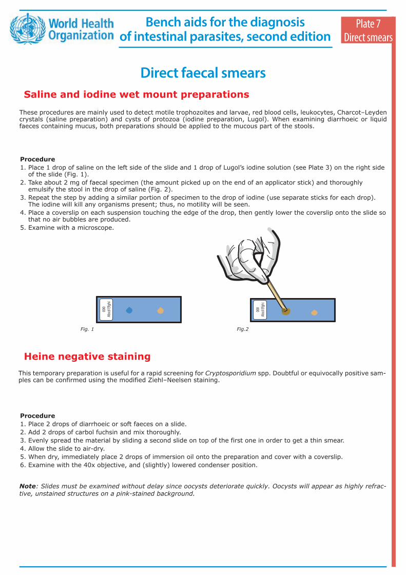
These procedures are mainly used to detect motile trophozoites and larvae, red blood cells, leukocytes, Charcot–Leyden crystals (saline preparation) and cysts of protozoa (iodine preparation, Lugol). When examining diarrhoeic or liquid faeces containing mucus, both preparations should be applied to the mucous part of the stools.
Procedure 1. Place 1 drop of saline on the left side of the slide and 1 drop of Lugol’s iodine solution (see Plate 3) on the right side
of the slide (Fig. 1).2. Take about 2 mg of faecal specimen (the amount picked up on the end of an applicator stick) and thoroughly
emulsify the stool in the drop of saline (Fig. 2).3. Repeat the step by adding a similar portion of specimen to the drop of iodine (use separate sticks for each drop).
The iodine will kill any organisms present; thus, no motility will be seen.4. Place a coverslip on each suspension touching the edge of the drop, then gently lower the coverslip onto the slide so
that no air bubbles are produced. 5. Examine with a microscope.
Plate 7Direct smears
Direct faecal smearsSaline and iodine wet mount preparations
Heine negative staining This temporary preparation is useful for a rapid screening for Cryptosporidium spp. Doubtful or equivocally positive sam-ples can be confirmed using the modified Ziehl–Neelsen staining.
Procedure 1. Place 2 drops of diarrhoeic or soft faeces on a slide.2. Add 2 drops of carbol fuchsin and mix thoroughly.3. Evenly spread the material by sliding a second slide on top of the first one in order to get a thin smear.4. Allow the slide to air-dry.5. When dry, immediately place 2 drops of immersion oil onto the preparation and cover with a coverslip. 6. Examine with the 40x objective, and (slightly) lowered condenser position.
Note: Slides must be examined without delay since oocysts deteriorate quickly. Oocysts will appear as highly refrac- tive, unstained structures on a pink-stained background.
000
Abcd
Efgh
i00
0Ab
cd Ef
ghi
000
Abcd
Efgh
i00
0Ab
cd Ef
ghi
Fig. 1 Fig.2
Bench aids for the diagnosis of intestinal parasites, second edition

Endolimax nana Cysts. Left: with 4 nuclei each almost entirely occupied by a large karyosome, which appear refractive in wet mounts, unstained; right: Lugol. Size: 5-10 µm
Iodamoeba bütschlii Left: cyst unstained, glycogen vacuole barely visible; right: cysts, dark staining glycogen vacuoles clearly visible, Lugol. Size: 5-20 µm
E. nana Cyst. Three of the four nuclei are visible. Iron-haematoxylin stain according to Kinyoun
E. nana Trophozoite showing a large nucleus with round karyosome. The nuclear membrane characteristically lacks peripheral chromatin. Iron-haematoxylin stain according to Kinyoun. Size: 6-12 µm
E. polecki Uninucleate cyst. Trichrome stain. E. polecki is, together with E. moshkovskii and E. bangladeshi, morphologically identical to E. histolytica/E. dispar
Entamoeba gingivalis Trophozoite. Eccentric nucleus containing a punctate karyosome and fine, evenly distributed peripheral chromatin. Many endocytotic vacuoles are present in the cytoplasm. Iron-Hematoxylin stain according to Kinyoun. Size: 10-20 µm
Enteromonas hominis Cysts are similar those of E. nana but with 2 nuclei. Trichrome stain. Size: 6-8 x 3-4 μm
I. bütschlii Cysts. Note the single nucleus and glycogen vacuole: iron-haematoxylin stain according to Kinyoun
I. bütschlii Trophozoite, unstained. Size: 8-20 µm
E. polecki Trophozoite. Trichrome stain. E. polecki is, together with E. moshkovskii and E. bangladeshi, morphologically identical to E. histolytica/E. dispar. Size 10-25 µm
Entamoeba polecki Uninucleate cyst. Trichrome stain. E. polecki is, together with E. moshkovskii and E. bangladeshi, morphologically identical to E. histolytica/E. dispar. Size: 9-25 µm
Plate 8Protozoa amoebae
E. hominis Trophozoite and cyst (left). Usually the 4 trophozoite flagellae are hardly visible. Mature cyst typically has 4 nuclei, 2 at each end (right). Iron-haematoxylin stain according to Kinyoun. Trophozoite size: 3-6 μm
Bench aids for the diagnosis of intestinal parasites, second edition

Staining procedures for protozoa in faeces The use of Lugol’s iodine for staining wet mount preparations from fresh or formalin-preserved faecal specimens is described on Plate 7. The following are some procedures for permanent staining of smears prepared from fresh or pre- served faecal material.
Permanent stains for faecal smearsAll solutions must be stored in clean, labelled and dated bottles.
Trichrome stainVery good stain for fresh and unpreserved stools. SAF and formalin preserved material does not yield satisfactory results.
PreparationAdd 10 mL of glacial acetic acid to 6 g of chromotrope 2R, 3 g of light green SF and 7 g of phosphotungstic acid in a clean flask. Swirl to mix and leave to stand for 30 minutes. Add 1000 mL of distilled water and mix thoroughly; the stain should be a deep purple. Store in a glass-stoppered bottle; the stain is stable and is used undiluted. Staining procedurePlace fixed slides into 70% alcohol for 2 minutes. Add Lugol’s diluted iodine solution to 70% ethanol to produce a colour of strong tea: place slides in the solution for 5 minutes. Place slides in two changes of 70% alcohol. Stain slides in undiluted trichrome stain for 10 minutes. Remove slides, drain thoroughly, and place them in 90% acidified alcohol (prepared by adding 4.5 mL of glacial acetic acid to 1 L of 90% ethanol) for 2–3 seconds. Dip slides in 95% alcohol to rinse and then dehydrate through 100% ethanol and xylene or through carbol–xylene mixture. Using resinous moun-ting medium, place a coverslip on the smear.
Iron–haematoxylin stainVery good stain for fresh, unpreserved stool, SAF (sodium acetate acetic acid formalin) and formalin-preserved faecal smears.
Preparation Stock solution A: dissolve 1 g of haematoxylin crystals in 100 mL of 95% alcohol; allow solution to stand in daylight for 1 week and then filter. Stock solution B: mix 1 g of ferrous ammonium sulfate, 1 g of ferric ammonium sulfate and 1 mL of hydrochloric acid in 97 mL of distilled water. Prepare a working solution by combining 25 mL each of stock solutions A and B; prepare at least 3–4 hours prior to staining. Prepare picric acid solution for destaining by adding 25 mL of saturated aqueous picric acid to 25 mL of distilled water.
Staining procedurePlace slides into 70% alcohol for 5 minutes; into 50% alcohol for 2 minutes; into tapwater for 5 minutes; into working haematoxylin stain solution for 10 minutes; into distilled water for 1 minute; into picric acid solution for 1 minute; into running tapwater for 10 minutes; into 70% alcohol containing 1 drop of ammonia for 5 minutes; and into 95% alcohol for 5 minutes. Dehydrate through 100% ethanol and xylene or through carbol–xylene mixture. Using resinous mounting medium, place a coverslip on the smear.
Modified Ziehl–Neelsen technique (acid-fast stain)For detection of Cryptosporidium, Cyclospora, and other coccidian infections.
Reagents 50% ethanol: add 50 ml of absolute ethanol to 50 mLof distilled water.Carbol fuchsin solution A: dissolve 4 g of basic fuchsin in 20 mL of 95% ethanol. Solution B: dissolve 8 g of phenol crystals in 100 mL of distilled water. Mix solutions A and B.1% sulfuric acid: add 1 mL of concentrated sulfuric acid to 99 ml of distilled water.Alkaline methylene blue: dissolve 0.3 g of methylene blue in 30 mL of 95% ethanol. Add 100 mL of dilute (0.01%) potassium hydroxide.Store all the solutions at room temperature. They are stable for 1 year. Important: indicate the expiration date on the label.
ProcedureMake a thin stool smear on a clear and dry slide. Allow to dry. Fix with absolute methanol for 30 seconds. Allow to dry. Stain the slide with carbol fuchsin for 5 minutes. Rinse for 3–5 seconds with 50% ethanol. Rinse in tap water. Decolor- ize with 1% sulfuric acid until the flow of pink colour disappears (1–2 minutes, depending on thickness of preparation). Rinse in tap water and drain. Stain with methylene blue for 1 minute. Rinse, allow to dry and examine using oil immersion.
Note: This technique is not recommended for eggs of Fasciola spp. and larvae of Strongyloides stercoralis.
Plate 8Staining
Bench aids for the diagnosis of intestinal parasites, second edition

Chilomastix mesnili Cyst, typically pear- shaped. Left: unstained. Right: cysts, Lugol. Nuclei and cytostome vaguely outlined. Size: 7-9 µm
Giardia duodenalis Left: cysts, unstained. Right: cysts and trophozoite (arrow), Lugol. Size: 8-12 x 7-10 µm
C. mesnili Trophozoite with 3 free flagella, nucleus and vaguely outlined cytostome at anterior end. Lugol. Size: 12-20 x 5-6 µm
C. mesnili Left, trophozoites and cyst: unstained. Right: trophozoites, iron-haematoxylin stain according to Kinyoun
G. duodenalis Cysts. It is possible to observe 3 of the 4 nuclei present, the axonems that divide the cyst (left) and some disk fragments (arrow, right). Iron-haematoxylin stain according to Kinyoun
G. duodenalis Trophozoites. Left: Iron-hae-matoxylin stain according to Kinyoun. Right: Giemsa, which stained the 4 pairs of flagella distinctly and 2 large nuclei. Size: 10-20 µm
Dientamoeba fragilis Trophozoites, Lugol. They appear like polymorphic refractive bodies and may therefore be misdiagnosed as artefacts (especially leukocytes). Moreover, trophozoites must be morphologically differentia-ted from other small nonpathogenic amoebae. Size: 5-15 µm
D. fragilis Binucleate trophozoites with different mor- phology. In D. fragilis the cystic form is difficult to identify. Iron-haematoxylin stain according to Kinyoun
D. fragilis Mononucleate trophozoite. The fragmented karyosomes are readily seen. Iron-haematoxylin stain according to Kinyoun
B. coli Trophozoites, left: unstained; right: Lugol. In both organisms cilia are visible on the surface, cytostome at the anterior end (arrow head), and cytopyge posteriorly (arrow). Size: 40-200 x 40-70 µm
B. coli trophozoite and Trichuris trichiura egg. Un- stained
Balantidium coli Left: cyst barely show- ing horseshoe-shaped macronucleus, un- stained. Right: focused at surface to show cilia elements (arrow). Size: 50-70 µm
Plate 9Protozoa amoebae
flagellatesciliates
Bench aids for the diagnosis of intestinal parasites, second edition
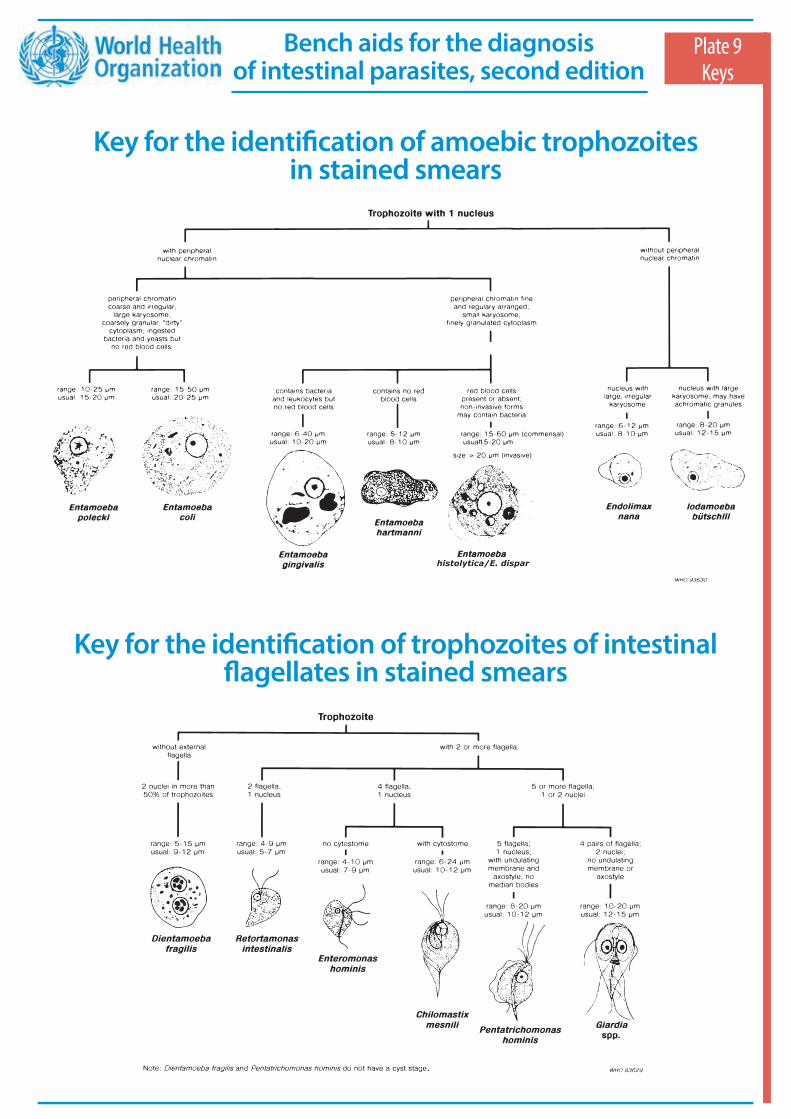
Key for the identification of amoebic trophozoites in stained smears
Plate 9Keys
Key for the identification of trophozoites of intestinal flagellates in stained smears
histolytica/E. dispar
Bench aids for the diagnosis of intestinal parasites, second edition
.
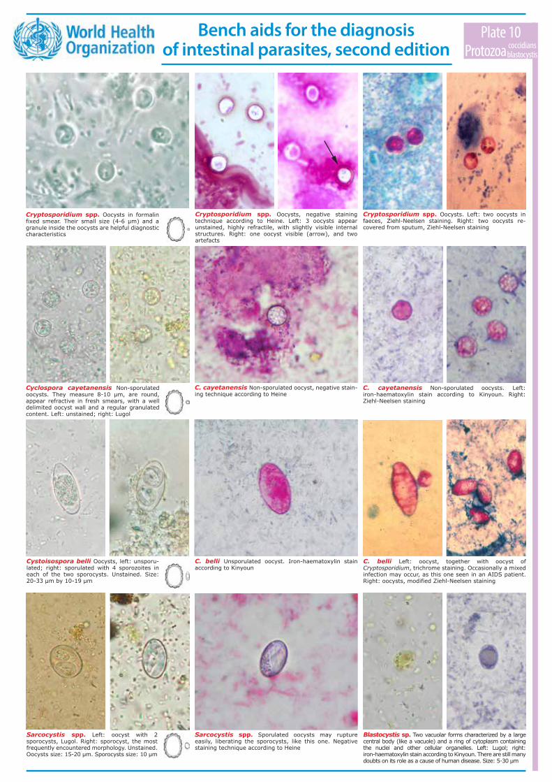
Cyclospora cayetanensis Non-sporulated oocysts. They measure 8-10 µm, are round, appear refractive in fresh smears, with a well delimited oocyst wall and a regular granulated content. Left: unstained; right: Lugol
Cryptosporidium spp. Oocysts in formalin fixed smear. Their small size (4-6 µm) and a granule inside the oocysts are helpful diagnostic characteristics
C. cayetanensis Non-sporulated oocyst, negative stain- ing technique according to Heine
Cryptosporidium spp. Oocysts, negative staining technique according to Heine. Left: 3 oocysts appear unstained, highly refractile, with slightly visible internal structures. Right: one oocyst visible (arrow), and two artefacts
Cystoisospora belli Oocysts, left: unsporu-lated; right: sporulated with 4 sporozoites in each of the two sporocysts. Unstained. Size: 20-33 µm by 10-19 µm
Sarcocystis spp. Sporulated oocysts may rupture easily, liberating the sporocysts, like this one. Negative staining technique according to Heine
Blastocystis sp. Two vacuolar forms characterized by a large central body (like a vacuole) and a ring of cytoplasm containing the nuclei and other cellular organelles. Left: Lugol; right: iron-haematoxylin stain according to Kinyoun. There are still many doubts on its role as a cause of human disease. Size: 5-30 µm
Sarcocystis spp. Left: oocyst with 2 sporocysts, Lugol. Right: sporocyst, the most frequently encountered morphology. Unstained. Oocysts size: 15-20 μm. Sporocysts size: 10 μm
C. cayetanensis Non-sporulated oocysts. Left: iron-haematoxylin stain according to Kinyoun. Right: Ziehl-Neelsen staining
C. belli Unsporulated oocyst. Iron-haematoxylin stain according to Kinyoun
C. belli Left: oocyst, together with oocyst of Cryptosporidium, trichrome staining. Occasionally a mixed infection may occur, as this one seen in an AIDS patient. Right: oocysts, modified Ziehl-Neelsen staining
Plate 10Protozoa amoebae
coccidiansblastocystis
Cryptosporidium spp. Oocysts. Left: two oocysts in faeces, Ziehl-Neelsen staining. Right: two oocysts re- covered from sputum, Ziehl-Neelsen staining
Bench aids for the diagnosis of intestinal parasites, second edition
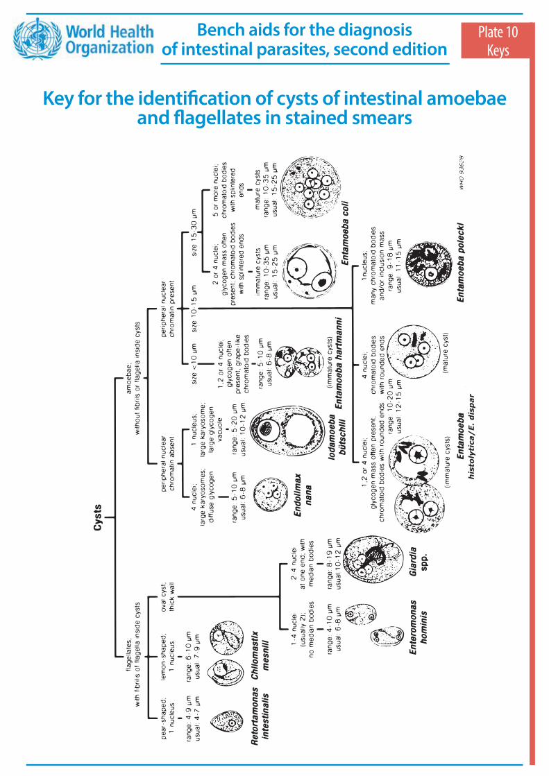
Key for the identification of cysts of intestinal amoebae and flagellates in stained smears
Plate 10Keys
his
toly
tica
/E
. d
isp
ar
Bench aids for the diagnosis of intestinal parasites, second edition

Plant hairs may be confused with parasite larvae. Note their usually straight appearance and non-structured central shaft. One end is often frayed
Plant material can often be found in faeces and may sometimes strongly resemble parasitic elements. It can mimick e.g. Hymenolepis nana eggs (left, note the absence of the typical hooks inside) or Taenia eggs (right, note the absence of hooks inside)
Polymorphonuclear leukocytes are seen clustered in trichrome-stained smear. Although these may be mistaken for amoebae, the large size of nuclei in relation to the cytoplasm of the cell and their structure indicate that these are inflammatory cells
Ascospores are spores from Ascomycetes, a type of fungi of which some are edible (e.g. morel mushrooms). Due to their size and oval shape they can be confused with Giardia duodenalis. Note the absence of typical inner structures
Heterodera spp. or Meloidogyne spp. are parasitic plant nematodes. Their eggs can be confused with hookworm eggs but are generally larger and bean-shaped. Sometimes a larva has formed inside
Acarina eggs may resemble hookworm eggs. Their size, however, always exeeds 100µm. Usually they are completely filled. Sometimes typical structures (e.g. legs) can be distinguished inside
Epithelial cells Lugol
Plate 11Artefacts
Charcot-Leyden crystals have a typical “compass needle” appearance (left). They are a degradation product of eosinophilic leukocytes and their presence should be reported. They should not be confused with crystals that can be produced in faeces by some fruit types (right)
Oligochetes (or earthworms) are sometimes confused with Ascaris adults (or juveniles). Oligochetes will have small bristles, which can be visualized using a binocular loupe (top) or by examining a fragment of its skin with a microscope (bottom)
Bench aids for the diagnosis of intestinal parasites, second edition
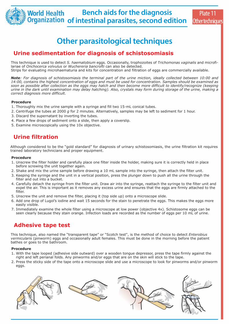
This technique is used to detect S. haematobium eggs. Occasionally, trophozoites of Trichomonas vaginalis and microfi-lariae of Onchocerca volvulus or Wuchereria bancrofti can also be detected. Strips for evaluating microhaematuria and kits for concentration and filtration of eggs are commercially available.
Note: For diagnosis of schistosomiasis the terminal part of the urine miction, ideally collected between 10:00 and 14:00, contains the highest concentration of eggs and must be used for concentration. Samples should be examined as soon as possible after collection as the eggs may hatch and then become more difficult to identify/recognize (keeping urine in the dark until examination may delay hatching). Also, crystals may form during storage of the urine, making a correct diagnosis more difficult.
Procedure 1. Thoroughly mix the urine sample with a syringe and fill two 15-mL conical tubes.2. Centrifuge the tubes at 2000 g for 2 minutes. Alternatively, samples may be left to sediment for 1 hour.3. Discard the supernatant by inverting the tubes.4. Place a few drops of sediment onto a slide, then apply a coverslip. 5. Examine microscopically using the 10x objective.
Plate 11Other techniques
Other parasitological techniquesUrine sedimentation for diagnosis of schistosomiasis
Urine filtration Although considered to be the “gold standard” for diagnosis of urinary schistosomiasis, the urine filtration kit requires trained laboratory technicians and proper equipment.
Procedure 1. Unscrew the filter holder and carefully place one filter inside the holder, making sure it is correctly held in place
before screwing the unit together again.2. Shake and mix the urine sample before drawing a 10 mL sample into the syringe, then attach the filter unit.3. Keeping the syringe and the unit in a vertical position, press the plunger down to push all the urine through the
filter and out into a bucket.4. Carefully detach the syringe from the filter unit. Draw air into the syringe, reattach the syringe to the filter unit and
expel the air. This is important as it removes any excess urine and ensures that the eggs are firmly attached to the filter.
5. Unscrew the unit and remove the filter, placing it (top side up) onto a microscope slide.6. Add one drop of Lugol’s iodine and wait 15 seconds for the stain to penetrate the eggs. This makes the eggs more
easily visible.7. Immediately examine the whole filter using a microscope at low power (objective 4x). Schistosome eggs can be
seen clearly because they stain orange. Infection loads are recorded as the number of eggs per 10 mL of urine.
Adhesive tape test This technique, also named the “transparent tape” or “Scotch test", is the method of choice to detect Enterobius vermicularis (pinworm) eggs and occasionally adult females. This must be done in the morning before the patient bathes or goes to the bathroom.
Procedure 1. With the tape looped (adhesive side outward) over a wooden tongue depressor, press the tape firmly against the
right and left perianal folds. Any pinworms and/or eggs that are on the skin will stick to the tape.2. Press the sticky side of the tape onto a microscope slide and use a microscope to look for pinworms and/or pinworm
eggs.
Bench aids for the diagnosis of intestinal parasites, second edition
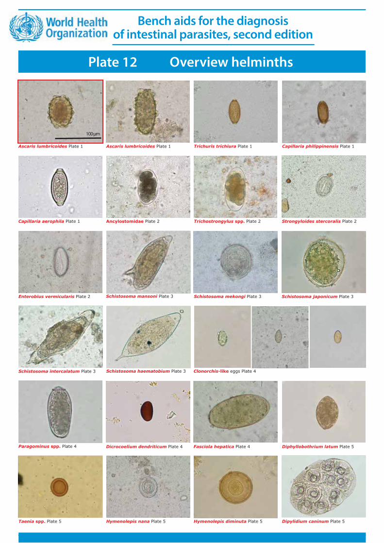
Schistosoma intercalatum Plate 3
Schistosoma mekongi Plate 3 Schistosoma japonicum Plate 3Schistosoma mansoni Plate 3
Strongyloides stercoralis Plate 2Trichostrongylus spp. Plate 2Ancylostomidae Plate 2
Capillaria philippinensis Plate 1Trichuris trichiura Plate 1Ascaris lumbricoides Plate 1
Clonorchis-like eggs Plate 4Schistosoma haematobium Plate 3
Paragominus spp. Plate 4 Dicrocoelium dendriticum Plate 4 Fasciola hepatica Plate 4
Enterobius vermicularis Plate 2
Diphyllobothrium latum Plate 5
Taenia spp. Plate 5 Hymenolepis diminuta Plate 5Hymenolepis nana Plate 5
Ascaris lumbricoides Plate 1
Capillaria aerophila Plate 1
Plate 12 Overview helminths
Dipylidium caninum Plate 5
100 µm
Bench aids for the diagnosis of intestinal parasites, second edition

Plate 12 Overview protozoa
Entamoeba histolytica/E. dispar Plate 7 Left: cyst; right: trophozoite
Entamoeba hartmanni Plate 7 Left: cyst; right: trophozoite
Entamoeba coli Plate 7 Left: cyst; right: trophozoite
Iodamoeba bütschlii Plate 8 Left: cyst; right: trophozoite
Endolimax nana Plate 8 Left: cyst; right: trophozoite
Entamoeba polecki Plate 8 Left: cyst; right: trophozoite
Entamoeba gingivalis Plate 8 Trophozoite
Enteromonas hominis Plate 8 Giardia duodenalis Plate 9 Left: cyst; right: trophozoite
Chilomastix mesnili Plate 9 Left: cyst; right: trophozoite
Dientamoeba fragilis Plate 9 Trophozoite
Balantidium coli Plate 9. Left: cyst; right: trophozoite
Ascaris lumbricoides Plate 1
Cryptosporidium spp. Plate 10
Cyclospora cayetanensis Plate 10. Oocysts unsporulated
Cystoisospora belli Plate 10. Oocyst unsporulated
Sarcocystis spp. Plate 10. Left: oocyst; right: sporocyst
Blastocystis sp. Plate 10
Cryptosporidium spp. Plate 10
100 µm
Bench aids for the diagnosis of intestinal parasites, second edition


These Bench aids for the diagnosis of intestinal parasites are intended both as a guide for
laboratory and field workers in endemic countries and as a teaching aid for students and trainees. They provide guidance on the
choice of preparation to the different copromicroscopical methods and main staining technique for the diagnosis of
intestinal parasites (nematodes, trematodes, cestodes and protozoa).
Photomicrographs demonstrate the appearance and diagnostic features of the
various parasites in the different preparations.
The bench aids have been produced in a weatherproof plastic-sealed format that is
robust and easy to use at the bench. They are recommended for use by all health workers
engaged in the routine diagnosis of intestinal parasitic infections.
Bench aidsfor the diagnosis
of intestinal parasitessecond edition
ISBN 978-92-4-151534-4