behavioral/cognitive sphingosine-1-phosphate ... ·...
TRANSCRIPT
Behavioral/Cognitive
Sphingosine-1-Phosphate-Induced Nociceptor Excitationand Ongoing Pain Behavior in Mice and Humans Is LargelyMediated by S1P3 Receptor
María Camprubí-Robles,1,2 Norbert Mair,1 Manfred Andratsch,1 Camilla Benetti,1 Dimitra Beroukas,3 Roman Rukwied,4Michiel Langeslag,1 Richard L. Proia,5 Martin Schmelz,4 Antonio V. Ferrer Montiel,2 Rainer V. Haberberger,3and Michaela Kress1
1Department of Physiology and Medical Physics, Division of Physiology, Medical University of Innsbruck, A-6020 Innsbruck, Austria, 2Institute ofMolecular and Cellular Biology, Miguel Hernandez University, 03202 Elche, Alicante, Spain, 3Department of Anatomy and Histology and Centre forNeuroscience, Flinders University, Adelaide, Bedford Park SA 5042, South Australia, Australia, 4Department of Anaesthesiology and Intensive CareMedicine, Medical Faculty Mannheim, Heidelberg University, 68167 Mannheim, Germany, and 5National Institute of Diabetes and Digestive and KidneyDiseases, Bethesda, Maryland 20892
The biolipid sphingosine-1-phosphate (S1P) is an essential modulator of innate immunity, cell migration, and wound healing. It isreleased locally upon acute tissue injury from endothelial cells and activated thrombocytes and, therefore, may give rise to acute post-traumatic pain sensation via a yet elusive molecular mechanism. We have used an interdisciplinary approach to address this question,and we find that intradermal injection of S1P induced significant licking and flinching behavior in wild-type mice and a dose-dependentflare reaction in human skin as a sign of acute activation of nociceptive nerve terminals. Notably, S1P evoked a small excitatory ioniccurrent that resulted in nociceptor depolarization and action potential firing. This ionic current was preserved in “cation-free” solutionand blocked by the nonspecific Cl � channel inhibitor niflumic acid and by preincubation with the G-protein inhibitor GDP-�-S. Notably,S1P3 receptor was detected in virtually all neurons in human and mouse DRG. In line with this finding, S1P-induced neuronal responsesand spontaneous pain behavior in vivo were substantially reduced in S1P3
�/� mice, whereas in control S1P1 floxed (S1P1fl/fl) mice and
mice with a nociceptor-specific deletion of S1P1�/� receptor (SNS-S1P1
�/�), neither the S1P-induced responses in vitro nor the S1P-evoked pain-like behavior was altered. Therefore, these findings indicate that S1P evokes significant nociception via G-protein-dependent activation of an excitatory Cl � conductance that is largely mediated by S1P3 receptors present in nociceptors, and point tothese receptors as valuable therapeutic targets for post-traumatic pain.
IntroductionOngoing pain regularly results as a consequence of surgical inter-ventions or trauma. Although this represents a major clinicalproblem and severely affects the patients’ recovery, pain reliefstrategies so far are restricted to the classical opioids or nonopioid
analgesics. To develop better strategies based on mechanistic in-sight, the sequelae of mediators and interactions of injured tissuewith the nociceptive system leading to postoperative pain need tobe elucidated. Upon tissue injury and damage to blood vessels,vascular leakage, accumulation, and activation of thrombocytesand plasma extravasation occur. Injection of plasma and experi-mentally activated thrombocytes excite nociceptors and inducepain (Armstrong et al., 1957; Ringkamp et al., 1994). Humanplatelets contain high concentrations of sphingomyelin-derivedsphingosine-1-phosphate (S1P), which can be released upon ac-tivation and shape change (Dahm et al., 2006; Ulrych et al., 2011).In blood plasma, S1P levels may reach up to micromolar concen-trations, most of which is bound to plasma proteins (Murata etal., 2000; Schmidt et al., 2006; Ohkawa et al., 2008). S1P acts as alipid growth factor and induces robust endothelial cell activation,resulting in cellular locomotion, vascular maturation, and angio-genesis (Daum et al., 2009). Furthermore, S1P has a general rolein postoperative wound healing via either intracellular mode ofaction or specific targeting of metabotropic membrane receptors(Vogler et al., 2003; Watterson et al., 2007). In general, S1P is thenatural ligand of five different G-protein-coupled receptors (GP-
Received Sept. 20, 2012; revised Nov. 27, 2012; accepted Dec. 4, 2012.Author contributions: M.C.-R., N.M., C.B., R.R., M.S., R.V.H., and M.K. designed research; M.C.-R., N.M., M.A., C.B.,
D.B., R.R., M.L., and R.V.H. performed research; R.L.P. contributed unpublished reagents/analytic tools; M.C.-R.,N.M., M.A., C.B., D.B., R.R., M.L., M.S., R.V.H., and M.K. analyzed data; M.C.-R., M.L., R.L.P., A.V.F.M., R.V.H., and M.K.wrote the paper.
The authors thank K. Braun, T. Martha, and M. Doblander for expert technical assistance. This work was supportedby la Generalitat Valenciana and the Ministerio de Economia y Competitividad (A.V.F.M.), the Australian NationalHealth and Medical Research Council Project Grant 535055 to R.V.H., the Intramural Research Programs of theNational Institutes of Health, National Institute of Diabetes and Digestive and Kidney Diseases to R.L.P., and theAustrian Research Funding Agency FWF Project Grants P20562, P25345, and SPIN to M.K.
The authors declare no competing financial interests.Correspondence should be addressed to either of the following: Dr. María Camprubí-Robles (at current address),
Miguel Hernandez University, Institute of Molecular and Cellular Biology, Avda. de la Universidad s/n, 03202 Elche,Alicante, Spain, E-mail: [email protected]; or Dr. Michaela Kress, Medical University Innsbruck, Department ofPhysiology and Medical Physics, Division of Physiology, Fritz-Pregl-Str. 3, A-6020 Innsbruck, Austria. E-mail:[email protected].
DOI:10.1523/JNEUROSCI.4479-12.2013Copyright © 2013 the authors 0270-6474/13/332582-11$15.00/0
2582 • The Journal of Neuroscience, February 6, 2013 • 33(6):2582–2592

CRs S1P1-S1P5) that are expressed in various tissues, includingneurons and nociceptors (Spiegel and Milstien, 2003; Herr andChun, 2007; Chi and Nicol, 2010; Mair et al., 2011). ExogenousS1P enhances the excitability of DRG neurons, potentiates heat-sensitive transducer channels and modifies nociceptor heat sen-sitivity via neuronally expressed S1P1 receptor (Mair et al., 2011).In addition, this receptor has been associated with opioid-induced hyperalgesia (Muscoli et al., 2010; Mair et al., 2011).Because local increases in S1P levels arise from acute tissue injury,damage to blood vessels and activation of platelets after trauma orsurgical procedures, we hypothesized that S1P may have a signif-icant role in the mechanism causing post-traumatic pain. In thispaper, we address this issue with an interdisciplinary approachand investigate S1P-induced nociceptor excitation in a cellularmodel. In addition, the relevance of this finding is addressed invivo in mice and humans.
Materials and MethodsEthics statement. All animal experiments have been performed withpermission of the Austrian Bundesministerium fur Wissenschaft undForschung (BMWF) ministry (BMWF-66.011/0051-II/10b2008;BMWF-66.011/0113-II/3b/2010; GZ 66.011/85-C/GT/2007) and ac-cording to ethical guidelines of the IASP (International Association forthe Study of Pain) (Zimmermann, 1986).
Flinching and licking measurement. All behavioral measurements weredone in age-matched awake, unrestrained, male C57BL/6J mice (� 8weeks old) by individuals who were blinded to the genotype of the micebeing analyzed. Spontaneous pain-like behavior was monitored after in-tracutaneous injection of S1P (15 �l, 500 �M) or vehicle (15 �l, 10%methanol in PBS) into the left hindpaw of wt, S1P3
�/�, S1P1fl/fl, or
SNS-S1P1�/� mice, which selectively lack the S1P1 receptor subtype in
Nav1.8 channel-expressing nociceptive neurons (Mair et al., 2011). Micewere placed in the plastic chamber and allowed to habituate for at least1 h before the experiment. Flinching and licking behavioral responseswere scored over a 30 min period after injection. When mice rapidlyshook their hindpaw, it was scored as a positive flinching response andthen the number of flinches was assessed manually. Licking behavior wasassessed by counting manually the number of licks and the overall dura-tion of all scored licks.
Mouse model of incisional plantar pain. All mice were anesthetized withketamine (2 mg/kg) and xylasol (0.2 mg/kg) (both from Ogris Pharma).The plantar aspect of the hindpaw was disinfected with polyvidone-iodine solution (Wundesin, Gebro Pharma) and a 1-cm-longitudinalincision was made with a sterile surgical blade (no. 10), through skin andfascia of the plantar aspect of the paw, starting 0.5 cm from the proximaledge of the heel and extending toward the toes. The plantaris muscle wasalso incised longitudinally. The skin was sutured with two thread of 5-0polypropylene on a TF needle (Prolene, Ethicon, Johnson & JohnsonMedical Products) and the wound covered with Wundesin. The sutureswere removed under anesthesia on postoperative day 2, and typicallywounds healed within 5– 6 d.
Heat sensitivity. Heat sensitivity was assessed using the Hargreaves test(Hargreaves et al., 1988). A radiant heat source delivering an increasingheat stimulus was focused under the plantar surface of the hindpaw. Thetime from initiation of the radiant heat until paw withdrawal (paw with-drawal latency [PWL] in seconds) was measured automatically with analgesiometer (Ugo Basile). Each hindpaw was tested at least three times,and the mean withdrawal latency was calculated. The time interval be-tween two trials on the same hindpaw was at least 1 min.
Mechanical sensitivity. At least 1 h before testing, mice were placed in aPlexiglas chamber (10.5 � 10.5 � 14 cm) with a metal grid floor. Me-chanical sensitivity was tested manually before and after surgery by usingcalibrated von Frey monofilaments with defined bending forces (1.4, 4, 8,16, 22.6, 32, and 45.3 mN). Two baseline measurements were taken at 2and 1 d before surgery. Each filament was applied five times with at least60 s between applications. Data are expressed as the percentage of pawwithdrawals in response to each filament (mechanical response fre-
quency). Positive responses were counted when the paw was sharplywithdrawn and flinching of the paw was observed immediately afterremoval of the filament. All other movements of the paw were consideredas unclear responses, and the stimulus application was repeated.
Transgenic mice. Global S1P3 receptor null mutant mice (S1P3�/�)
and mice homozygous for the floxed exon 2 allele of the S1P1 receptorgene (S1P1
fl/fl), which encodes for the entire receptor protein, have beendescribed previously (Allende et al., 2003; Kono et al., 2004). S1P1
fl/fl
mice were cross-bred with SNS-Cre-mice (Agarwal et al., 2004) to obtainhomozygous SNS-Cre:S1P1
fl/fl (hereinafter referred to as SNS-S1P1�/�)
and S1P1fl/fl mice (control littermates; the Cre-recombinase is homozy-
gous or heterozygous) as previously published (Mair et al., 2011).Flare-reaction measurement. Five healthy subjects (1 woman and 4
men; mean age 32 years; range, 23 years) participated in this randomized,double-blinded study. Each subject was informed and gave written con-sent to take part in the study; the experimental protocol was approved bythe Ethics Committee of the Medical Faculty of Mannheim of the Uni-versity Heidelberg. Vehicle solution (saline, 15% cyclodextrin) and S1P(100 �l of 0.125, 0.25, 0.5, and 1 mM) were injected in the central volarforearm at distances of 4 cm between the injection sites. Superficial bloodflow around the injection site was assessed before the injection and im-mediately thereafter repeatedly in 4 min intervals using a laser-Dopplerimager (Moor Instruments). An area of 9 � 34.5 cm around the injectionsites was scanned. The area of the axon-reflex erythema was determinedas the number of cm 2 increasing �2-fold compared with the baselinescan by dedicated software (Moorldi Version 5.0, Moor Instruments).Pain scores were assessed for the injections of S1P and vehicle (saline,15% cyclodextrin); however, the pain upon injection did not differ sig-nificantly between vehicle and S1P and there was no lasting pain sensa-tion after retraction of the cannula (data not shown).
Primary sensory neuron culture. Lumbar (L1-L6) DRG with the cellbodies of primary afferents that project into the hindpaw were harvestedfrom adult C57BL/6J or transgenic mice and dissociated as previouslypublished (Mair et al., 2011). Briefly, ganglia were treated enzymaticallywith collagenase (Liberase, Roche) and trypsin-EDTA (Invitrogen), anddissociated mechanically with a fire-polished Pasteur pipette. The result-ing cell suspension was washed, plated on glass coverslips coated withpoly-L-lysine/laminin (Sigma) and cultivated in synthetic, serum-freemedium (supplemented TNB, Biochrom) supplemented with nervegrowth factor (NGF; 100 ng/ml) at 37°C in 5% CO2 in a humidifiedincubator for 18 –32 h.
Electrophysiology. Using the whole-cell voltage-clamp configuration ofthe patch-clamp technique, ionic currents were recorded from isolatedneurons at a holding potential of �80 mV as previously published (Ob-reja et al., 2002, 2005). The external control solution (ECS-low Ca 2�)contained the following (in mM): 150 NaCl, 5 KCl, 0.1 CaCl2, 1 MgCl2 (allSigma), 10 glucose and 10 HEPES (Merck), at pH 7.3 adjusted withNaOH (Merck). Borosilicate glass micropipettes (Science Products)pulled with a horizontal puller (Sutter Instruments) were filled withinternal solution (ICS, in mM): 148 KCl, 2 MgCl2, 2 Mg-ATP, 0.2 Na-GTP, 0.1 CaCl2, 1 EGTA (all Sigma) and 10 HEPES (Merck), at pH 7.3adjusted with KOH (Merck). After filling, electrode resistance was 2.5–3.5 M�. Currents were filtered at 2.9 kHz, sampled at 100 Hz, and re-corded using an EPC-9 (HEKA) and the Pulse Version 8.74 software(HEKA). The “cation free” extracellular solution contained the following(in mM): 163 NMDG, 5 EGTA, 10 HEPES, 10 D-glucose, 1 MgCl2 at pH7.3 adjusted with HCl (Merck). The membrane potential was recorded
Table 1. Antigen characteristics
Antigen Host Dilution Source
S1P2 (directed against C-terminus) Rabbit 1:100 S. MandalaS1P3 (directed against N-terminus) Rabbit 1:100 S. MandalaNF200 Mouse, clone N52 1:1000 SigmaI-B4 Bandeira simplifolicia 1:1000 SigmaSecondary antisera
Cy3 anti-rabbit Ig Donkey 1:100 JacksonCy5 anti-mouse Ig Donkey 1:50 Jackson
Camprubí-Robles et al. • S1P Induces Spontaneous Pain J. Neurosci., February 6, 2013 • 33(6):2582–2592 • 2583

using current-clamp configuration (Ihold � 0pA) using an ECS containing the following (inmM): 145 NaCl, 5 KCl, 2 CaCl2, 1 MgCl2 (allSigma), 10 D-glucose and 10 HEPES (Merck),at pH 7.3 adjusted with NaOH (Merck). Thepipette solution was composed (in mM) of 45KCl, 98 K-gluconate, 0.5 CaCl2, 5 EGTA, 10HEPES, 2 MgATP, 0.2 NaGTP, pH 7.3 adjustedwith KOH (Merck). The membrane potenialwas sampled at 4 kHz. Experiments were per-formed at room temperature (�22°C), andonly a single neuron was tested per coverslip.An automated seven-barrel system with com-mon outlet at 100 �m distance of the recordedcell was used for fast S1P administration (Dit-tert et al., 1998).
Microfluorimetric calcium measurements.Cells were plated on glass-bottom dishescoated with poly-L-lysine hydrobromide (10�g/ml) and laminin (10 �g/ml) (both fromSigma). One day cultured cells were incubatedwith 3 �M of the calcium (Ca 2�) sensitive dyeFura-2-acetoxymethyl ester (Invitrogen) inECS consisting of the following (in mM): 145NaCl, 5 KCl, 2 CaCl2, 1 MgCl2, 10 D-glucose(all from Sigma) and 10 HEPES (Merck), at pH7.3 adjusted with NaOH (Merck) and were in-cubated for 25 min at 37°C in 5% CO2, in ahumidified incubator. Then cells were washedtwice with PBS (PAA) and finally kept in ECSfor experiments. For the Ca 2�-free solution,CaCl2 was replaced by 5 mM EGTA. Ca 2� re-cordings were performed using an OlympusIX71 microscope (Olympus) with a 20�/0.85N.A. oil-immersion objective (Olympus).Fura-2 was excited consecutively at 340 and380 nm (excitation time: 25 ms) with a poly-chrome IV monochromator (TILL Photonics),and fluorescence intensities were filtered by a510 nm LP filter and recorded with a CCDcamera (TILL Photonics). The changes of in-tracellular Ca 2� concentration [Ca 2�]i are de-picted as ratio of fluorescence intensitiescollected at 340 nm and 380 nm (�F340/380)at a time interval of 1 s (Linhart et al., 2003).For data acquisition, MetaFluor4.6r8 (Univer-sal Imaging) was used and off-line analysis wasperformed with OriginPro7.SR2 (Origin Lab).The threshold for S1P-positive cells was set to fourfold the SD of theCa 2� signal evoked by 0.1% methanol (�F340/380 � 0.04). All chemi-cals were purchased from Sigma.
Immunohistochemistry. Murine DRGs were dissected, fixed in Zambo-ni’s fixative for 24 h, dehydrated through a graded series of etha-nol and DMSO, and subsequently embedded in polyethylene glycol(1450 MW: Sigma-Aldrich) (Murphy et al., 1998). DRGs were sectionedat 12 �m and stored in PBS containing 0.01% sodium azide at 4°C beforemultiple-labeling immunohistochemistry. Free-floating sections wereblocked using 10% normal donkey serum and incubated for 48 h with theS1P3 antiserum (a generous gift from S. Mandala). After washing in PBS,the sections were incubated for 2 h with secondary antiserum (Cy3-labeled donkey anti-rabbit IgG, Jackson ImmunoResearch Laborato-ries), washed in PBS, and coverslipped in buffered glycerol. Likewise,indirect immunofluorescence was performed in cultured neurons fixedin Zamboni’s fixative for 2 h and stored in PBS.
Human DRGs (N � 2) were obtained from the South Australian andVictorian Brain Bank from motor neuron disease patients 12–16 h post-mortem. DRGs were immersion fixed in Zamboni’s fixative for 24 –72 h.The use of human material was approved by the Flinders Clinical Re-search Ethics Committee. Human DRGs were embedded in polyethylene
glycol as described previously (Murphy et al., 1998), cut at a thickness of10 �m, and the free floating sections were incubated for 48 h with S1P3
antiserum. After washing in PBS, the sections were incubated overnightwith secondary antiserum, washed in PBS, and coverslipped in bufferedglycerol. Images were taken using a confocal laser scanning microscope(Leica SP5).
Immunocytochemistry. DRG neurons were cultured on coverslips,fixed for 2 h in Zamboni’s fixative, washed in PBS, and incubated withprimary antisera overnight, followed by washing and 1 h incubation withsecondary antisera. Finally, the coverslips were washed in PBS and cov-erslipped in buffered glycerol (Mair et al., 2011). Preabsorption of theS1P3 antiserum with the corresponding antigen led to the absence ofstaining (Table 1).
mRNA in situ hybridization. The protocol used was modified from anearlier method used by Obernosterer et al. (2007). Cryostat sections ofmice lumbar DRGs were fixed with Zamboni’s fixative for 24 h, washedin PBS, digested with proteinase K (2 mg/ml), fixed with 4% PFA, acety-lated (triethanolamine/HCl/acetic anhydride), and incubated with pre-hybridization buffer. Target mRNAs were hybridized with 0.34 pmol ofspecific antisense double digoxigenin-labeled mRNA detection probes(S1P3, 5DigN/ACTGATGAGGAAGGCGATGTAT/3Dig_N) or scram-
Figure 1. Contribution of S1P to pain-like behavior in mice and axon reflex in humans. Intracutaneous injection of S1P into theipsilateral hindpaw of wt mice significantly enhanced the number of flinches (A), licks (B), and increased the time of lickingresponse during 10 min upon administration (C) (N � 7–10 mice). D, Specimen (before and 4 min after S1P injections) of the flarereaction recorded by laser Doppler imaging indicating an increased blood flow by lighter colors (flux in arbitrary units [AU], and ina pseudo-color scale) after intradermal injection of increasing concentrations of S1P (100 �l of 0.125, 0.25, 0.5, 1 mM) and vehicle(100 �l, saline, 15% cyclodextrin). Larger flare areas were developed around the injection sites of the two highest concentrations(red arrows). E, Quantitative analysis of the flare reaction area produced by S1P in human skin by repeated laser-Doppler images ofthe flare profile. The graph represents the time course of the axon reflex flare development (flare area in cm 2) around the injectionsites of vehicle and S1P (injection after minute 1) at 4 min intervals. *p 0.05. **p 0.01. ***p 0.001.
2584 • J. Neurosci., February 6, 2013 • 33(6):2582–2592 Camprubí-Robles et al. • S1P Induces Spontaneous Pain

bled probes (Exiqon) or hybridization buffer as negative controls(Obernosterer et al., 2007) overnight at 55°C. Washing with sodium–saline citrate was followed by blocking and overnight incubation withalkaline phosphatase-coupled anti-digoxigenin antiserum. Incubationwith secondary antisera for 1 h was followed by washing in PBS. Slideswere coverslipped in buffered glycerol and images taken using an epiflu-orescence microscope (BX50, Olympus). Subsequently coverslips wereremoved, and the sections incubated with BCIP/NBT (Roche) to visual-ize S1P3 receptor mRNA.
Data analysis and statistics. Statistical analysis was performed per ani-mal ( N) and per number of cells (n) for the patch-clamp and calciumimaging data. For detailed statistical analysis, the Sigma Stat 3 andGraphPad Prism 5 software were used, and the data are presented asmean SEM. Statistical tests were used depending on sample size, dis-tribution, and number of variables: two-way repeated-measures (RM)ANOVA with post Tukey test, the nonparametric Mann–Whitney U test,or the parametric unpaired two-sided t test for comparison betweengroups; for intraindividual comparisons, the Wilcoxon matched-pairssigned-rank test or the parametric Student’s paired t test was used. Fish-er’s test was used where stated. Differences were considered statisticallysignificant at p 0.05.
ResultsS1P evoked pain-like behavior in mice and flare-reactionin humansTo determine whether S1P was involved in the spontaneous paininduction in vivo, we examined flinching and licking responses inwt mice after S1P intradermal injection. We measured the num-ber of flinches for 10 min after S1P (500 �M, 15 �l) or vehicle(10% methanol in PBS, 15 �l) injection into the left hindpaw(ipsilateral) of wt mice. Interestingly, we found that upon S1Pinjection mice developed a significant increase in the number offlinches (4.9 2.02 compared with vehicle, p � 0.0007, Mann–
Whitney U test, N � 10 mice per condi-tion, Fig. 1A). This was accompanied byan increase in S1P-induced number oflicks (5.31 1.53 S1P-injected vs vehicle-injected mice, p � 0.0011, Mann–Whit-ney U test, N � 7 mice per condition, Fig.1B) and licking time (26.14 8.27 s com-pared with vehicle, p � 0.001, Mann–Whitney U test, N � 7 mice per condition,Fig. 1C). No effect on flinching and lickingbehavior was observed in vehicle-injectedcontrol mice (Fig. 1A–C) or at lower S1Pdoses (10 and 100 �M; data not shown).Furthermore, in control experiments,methanol did not produce any depolariza-tion, Ca2� rise, or pain-like behavior up toconcentrations of 10%. These data indicatethat S1P can evoke acute spontaneous pain-like behavior in vivo.
To investigate whether these findingsbear relevance to pain-related behavior inhumans, increasing doses of S1P (0.125,0.25, 0.5, and 1 mM) were injected intrad-ermally. As objective assessment of skinnociceptor excitation the increase of skinblood flow (in pixels) and the correspond-ing size of the axon-reflex flare area (incm 2) around the different injected siteswere monitored with laser Doppler imag-ing. Cutaneous S1P challenges produced adose-dependent flare response by increas-ing the blood flow in the skin immediatelyafter injection (increased intensity in arbi-
trary units is indicated by red arrows in Fig. 1D, right). Painscores were assessed for the injections of S1P and vehicle (saline,15% cyclodextrin); however, the pain upon injection did notdiffer significantly between vehicle and S1P, and there was nolonger lasting pain sensation after retraction of the cannula (datanot shown). S1P provoked a massive increase of the axon reflex-mediated blood flow showing the most pronounced effect at 0.5and 1 mM concentration at 4 min after injection (flare area �5.82 0.95 cm 2 and 8.58 1.16 cm 2, respectively) that wassignificantly higher compared with the vehicle-injected site (flarearea � 2.23 0.28 cm 2) (p � 0.0000113 compared with 0.5 and1 mM S1P-injented subjects within the time, two-way RMANOVA with post Tukey test, N � 5 subjects; p � 0.0079 for 0.5mM S1P vs vehicle at 4 min after injection, p � 0.0079 for 1 mM
S1P vs vehicle at 4 min after injection, Mann–Whitney U test, Fig.1E). The S1P-evoked flare response was detectable until 12 minafter injection, indicating a long-lasting effect of S1P on skinvasodilatation (Fig. 1E). These results support the hypothesis thatS1P has a significant role in spontaneous pain behavior indicatedby pain like-behavior in mice and skin flare reaction as a sign ofnociceptor activation in humans.
S1P-induced membrane depolarization and generation ofaction potentials in neuronsIn the whole-cell current-clamp configuration of the patch-clamp technique, exposure of single dissociated mouse DRG neu-rons to S1P (1 �M, 10 s) induced a significant membranedepolarization from a resting membrane potential of �54.39 1.02 mV (Ctrl) to �35.68 1.99 mV (S1P) in n � 34 neuronstested (p � 0.0001, t(33) � 10.297, paired t test, N � 3 mice each
Figure 2. Effect of S1P on the membrane potential in murine DRG neurons. A, B, Short application of S1P (1 �M, 10 s)depolarized DRG neurons by changing the membrane potential (Em) from �54.4 1.0 mV in control (Ctrl) condition to �36 2 mV in the presence of S1P. The Em recovery value was measured 10 min after S1P application (�50 1 mV). C, D, Most neurons(64.7%) generated APs in response to S1P (1 �M, 10 s) with a mean firing rate of 1.7 0.4 AP s �1. ***p 0.001.
Camprubí-Robles et al. • S1P Induces Spontaneous Pain J. Neurosci., February 6, 2013 • 33(6):2582–2592 • 2585

condition, Fig. 2A,B). Depolarization wassufficient to induce firing of action po-tentials (APs) upon S1P stimulation in22 of 34 neurons [64.7%] (fAP � 1.67 0.04 s �1, p � 0.0000004768, T� � 253,Wilcoxon matched-pairs signed-ranktest, Fig. 2C,D). Both depolarizationand AP discharge recovered to pre-stimulation values within 10 min ofwashout (recovery Em � �49.94 1.23mV vs S1P application, p � 0.0000511,t(32) � 8.357 and recovery fAP � 0.076 0.068 s �1 vs S1P application, p �0.00002384, T� � 8, respectively).These results suggest a direct excitatoryeffect of S1P on primary afferentneurons.
S1P-induced slowly activating andslowly inactivating excitatoryionic conductanceUsing the whole-cell voltage-clamp con-figuration of the patch-clamp technique,we observed that S1P (1 �M, 10 s) gener-ated currents with an amplitude of0.538 0.036 nA (n � 34 cells, Fig. 3A) inmost sensory neurons. S1P-evoked cur-rents reached a maximum value after100.4 4.14 s (n � 57 cells) and wereslowly deactivating (� � 177.90 15.16s). S1P-activated currents increased in adose-dependent manner with an EC50 of0.160 �M (Fig. 3B).
Because S1P reportedly increasesintracellular calcium concentrations([Ca 2�]i) (Hopson et al., 2011), we hy-pothesized that excitatory currents wereat least partially carried by Ca 2� (andNa�) ions. We performed Fura-2-basedCa 2� imaging on cultured neurons andfound that S1P-induced [Ca 2�]i rises in182 of 308 [59.1%] neurons (Fig. 3C top,D). Neurons usually responded to S1Pwithin 15 s, and mean ratio amplitudes ofevoked Ca 2� responses were �F340/380 � 0.14 0.01 (N � 6 mice, n � 308cells, Fig. 3E). To determine the Ca 2�
source of S1P-induced [Ca 2�]i elevation,Ca 2� measurements were performed inCa 2� free solution (5 mM EGTA), andwe found S1P-induced Ca 2� transientsfully abrogated (�F340/380 � 0.0043 0.0005; p � 0.00002 vsS1P, t(126) � 8.791, unpaired t test, N � 3 mice each, n � 127 cells,Fig. 3C bottom, D,E). These results were further corroborated byrecordings from isolated DRG neurons after depletion of intra-cellular Ca 2� stores with thapsigargin (TG), an inhibitor of theendoplasmic reticulum Ca 2�-ATPase SERCA (Rogers et al.,1995). Cultured neurons exposed to TG (1 �M, 5 min) did notshow any Ca 2� alteration mediated by high potassium-induceddepolarization (data not shown). As expected, the percentageof S1P reacting neurons was unchanged (79 of 136 responders[58.1%]; p � 0.917, Fisher’s test, Fig. 3D) and neuronal re-sponses to S1P after TG treatment (�F340/380 � 0.12 0.01)
were similar to untreated controls ( p � 0.264, Fisher’s test,N � 4 mice, n � 136 cells, Fig. 3E). Together, these datasuggest that S1P-evoked Ca 2� transients are the result of aCa 2� influx rather than a release from intracellular stores.
S1P-activated chloride conductance in primarysensory neuronsTo determine the relative contribution of cations to the overallcurrent induced by S1P, whole-cell voltage-clamp recordingswere performed using “cation-free” external solution (0 Na�, 0K�, and 0 Ca 2�). Surprisingly, we observed that the S1P-evokedcurrent was fully preserved in “cation-free” ECS (�IS1P: 6.18
Figure 3. Ionic nature of the excitatory currents induced by S1P. A, Typical recording of a responsive neuron after a short pulseof S1P (1 �M, 10 s). B, The graph shows the dose–response relationship of the S1P-induced conductance. The EC50 was 0.160 �M
S1P. C, Top, Representative Ca 2� imaging measurement of the S1P-evoked Ca 2� transient in DRG neurons in ECS (containing 2mM Ca 2�). Bottom, The Ca 2� rise induced by S1P was completely abolished in Ca 2� free condition (5 mM EGTA, �3.5 minexposure) but lightly recovered after washout (2 min). D, E, A small change in [Ca 2�]i induced by short application of S1P (1 �M,60 s) was observed in most DRG neurons. In presence of TG (1 �M, 5 min), the percentage of S1P-responding neurons and the Ca 2�
increase magnitude was not significantly different from control conditions. For each Ca 2� measurement, 25–30 neurons (N � 3mice) were randomly selected in the field. F, Top, Whole-cell recording from S1P-activated current in “cation free” condition (0Ca 2�, 0 K �, 0 Na �, 1 min). Bottom, Quantification of the S1P-induced current density for recordings in F that remained unalteredafter neuron exposure to “cation free” solution (3 min after S1P application). n.s., Not significant ( p � 0.05). ***p 0.001.
2586 • J. Neurosci., February 6, 2013 • 33(6):2582–2592 Camprubí-Robles et al. • S1P Induces Spontaneous Pain

1.08 pA/pF control vs 5.85 1.38 pA/pF after 1 min “cation free”;p � 0.138, t(15) � 1.568, paired t test N � 3 mice, n � 18 cells, Fig.3F). This result indicated that excitatory currents were not car-ried by cations and that Ca 2� influx was likely the result ofvoltage-gated Ca 2� channel activation secondary to the Ca 2�-independent membrane depolarization. Because chloride (Cl�)channels have been associated with the activation of ionic cur-rents by S1P in neuroblastoma cells (Postma et al., 1996), elec-trophysiological and microfluorometric Ca 2� measurementswere performed in the presence of the nonspecific Cl� channelblocker niflumic acid (NFA). We found significant inhibition ofIS1P by NFA by reducing the deactivation time constant (�) from177.90 15.16 s in control to 44.46 5.38 s with NFA (100 �M)
(p � 0.000100839, T � 36, Mann–Whit-ney U test, N � 3 mice each, n � 18 cells,Fig. 4A,B). Moreover, incubation withNFA (100 �M, 9 min) almost fully pre-vented S1P-evoked Ca 2� transients(�F340/380 � 0.023 0.003, p �0.0000021, t(434) � 7.581, unpaired t test,N � 4 mice each, n � 128 cells, Fig. 4C,E)and only 18 of 128 (14%) neurons stillresponded to S1P (p � 0.0000025, Fish-er’s test, Fig. 4D). Both results support theidea that Cl� is the main ion carryingS1P-induced currents in sensory neurons.
S1P-induced conductance andexcitation is reduced in S1P3
receptor-deficient neuronsTwo possible mechanisms of activationmay account for S1P-induced currentactivation: (1) S1P may act as a directligand at a channel protein, or (2) chan-nel gating may occur indirectly and re-quires G-protein coupled S1P receptors.To discriminate between both possibil-ities, neurons were internally perfusedwith GDP-�-S (a general G-protein in-hibitor; 3 mM, �9 min). This treatmentfully abolished IS1P in DRG neurons,suggesting a G-protein-dependent mech-anism (p � 0.0000015, T � 11, Mann–Whitney U test, N � 3 mice, n � 13neurons, Fig. 4F).
At the moment, five G-protein-coupled S1P receptors are known: S1P1
receptors were found predominantly ex-pressed in small peptidergic and nonpep-tidergic (isolectin B4-positive [I-B4�])sensory neurons, which are important inregulating behavioral sensitivity duringinflammation (Chi and Nicol, 2010; Mairet al., 2011). In addition, S1P2 and S1P3
mRNAs are also detected in DRG neurons(Mair et al., 2011). To address expressionpatterns for S1P receptors in certain neu-ronal populations, indirect immunofluo-rescence was performed with specificantisera for S1P receptors, which were agenerous gift from S. Mandala, and cola-beling with a marker for large nonnocice-ptive neurons (Neurofilament 200
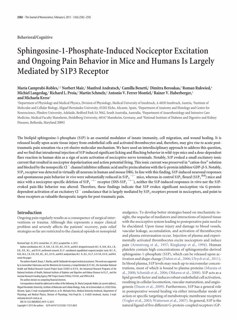
[NF200]). S1P2 receptor immunoreactivity was exclusively de-tected in large (NF200�) peptidergic neurons (Fig. 5A, top).More importantly, S1P3 was the most abundant S1P receptorexpressed in virtually all small and large mouse DRG neurons(Fig. 5A, bottom).
To first address the functional significance of the S1P1 recep-tor in neuronal responses to S1P, we performed microfluorimet-ric Ca 2� measurements in DRG neurons from transgenic micewith conditional deletion of S1P1 in nociceptive neurons (SNS-S1P1
�/�). S1P-evoked Ca 2� transients in neurons from SNS-S1P1
�/� (1 �M, 60 s) were similar to those from S1P1fl/fl mice
(p � 0.9688, t(278) � 0.03921, unpaired t test, N � 3 mice pergenotype, Fig. 5B,C). This suggests that S1P1 receptor activation
Figure 4. Contribution of Cl � to the ionic conductance of S1P-activated currents. A, B, Application of the nonspecific Cl �
channel blocker NFA (100 �M, 5 min) drastically diminished the time constant (�) of S1P-evoked currents. C, Application of NFA(100 �M, 9 min) prevented the S1P-induced Ca 2� influx. D, Only 14% of neurons [18 of 128] responded to S1P in the presence ofNFA. E, S1P-induced Ca 2� response was fully inhibited by NFA. For each Ca 2� measurement, 25–30 neurons from N � 3 micewere selected at random in the field. F, Incubation of DRG neurons with the general G-protein inhibitor GDP-�-S (3 mM, �9 min)fully blocked S1P-induced current density. ***p 0.001.
Camprubí-Robles et al. • S1P Induces Spontaneous Pain J. Neurosci., February 6, 2013 • 33(6):2582–2592 • 2587
may not be quantitatively relevant in S1P-induced pain-like behavior. This is fur-ther supported by the small population ofS1P1-expressing neurons (Mair et al.,2011), which was in obvious contrast tothe finding that IS1P could be evoked invirtually all neurons. Nonetheless, S1P1
may contribute to neuronal responses toS1P in the S1P1-expressing subpopulationof sensory neurons.
In contrast to several reports sup-porting a role for S1P1 in nociceptorsensitization and hyperexcitability, thesignificance of S1P3 receptors for nocice-ption has so far not been addressed. Tofirst characterize S1P3 expression in sen-sory neurons, we performed mRNA in situhybridization using riboprobes recogniz-ing S1P3 receptor on mouse DRG sec-tions. Distinct, specific signals over thesoma of virtually all large- and small-diameter wt DRG neurons were obtained(black arrows in Fig. 6A, left top). Controlscramble probes used in wt and S1P3
�/�
DRG sections (Fig. 6A, right top and bot-tom) and the prehybridization buffer(data not shown) did not yield any appre-ciable signals. No S1P3 labeling was de-tectable in neurons from S1P3
�/� (Fig.6A, left bottom). Importantly, S1P3 recep-tor immunoreactivity was expressed invirtually all small and large sensory neu-rons in human and mouse DRG sections(Fig. 6B). When we assessed S1P-inducedchanges of membrane potential (Em) inS1P3
�/� neurons, we observed that S1P-evoked depolarization was drastically de-creased compared with wt neurons(�Em
S1P in S1P3�/� � 4.84 1.1 mV vs
wt � 18.71 2.1 mV; p � 0.00000379306,T � 351, Mann–Whitney U test, N � 5mice, n � 28 neurons, Fig. 6C). In ratio-metric Ca 2� measurements, the number of neurons responding toS1P was decreased to �20% in S1P3
�/� mice (p � 0.0002 vs wt,Fisher’s test, N � 6 mice, n � 228 neurons, Fig. 6D) and S1P-evokedCa2� transients were significantly diminished (S1P3
�/� �F340/380 � 0.069 0.006 vs wt � 0.14 0.01; p � 0.00000013, t(622) �5.327, unpaired t test, Fig. 6E). These data introduce S1P3 receptor asa significant component of S1P-induced nociceptor excitation.
Spontaneous S1P-induced pain-like behavior is attenuated inS1P3
�/� miceFinally, we addressed the role of S1P receptors in the generationof spontaneous pain in vivo. After intradermal injection of S1P,licking and flinching behavior responses were similar betweenSNS-S1P1
�/� and S1P1fl/fl mice (p � 0.937, Mann–Whitney U
test, N � 7 mice per genotype, Fig. 7A–C). In contrast, the num-ber of flinches was substantially reduced in S1P3
�/� mice com-pared with wt littermates (0.40 0.31 vs 4.9 2.02; p � 0.0075,Mann–Whitney U test, N � 10 mice per genotype, Fig. 7D). Inthe same line, licking behavior after S1P injection was almost fullyprevented in S1P3
�/� mice (number of licks S1P3�/� � 1.35
1.10 vs wt � 5.31 1.53; p � 0.0132, Mann–Whitney U test, N �
8 mice, Fig. 7E; and licking time for S1P3�/� mice � 3.13 2.22 s
vs wt � 26.14 8.27 s; p � 0.0066, N � 10 mice per genotype,Fig. 7F). The absence of S1P-evoked pain-like behavior inS1P3
�/� mice suggests that S1P-induced pain generation islargely mediated by S1P3 receptors in vivo.
Role of S1P3 receptors in postoperative painBecause S1P can be released as a consequence of tissue injury, theincision model of postoperative pain with assessment of respon-siveness to mechanical and thermal stimuli was used to addressthe role of S1P in a more clinically relevant preclinical model(Pogatzki and Raja, 2003). Plantar incision decreased the PWL inwt mice from 8.88 0.37 s (24 h before incision as baseline) to4.32 0.42 s on postoperative day 1 (Fig. 8A). S1P3
�/� miceshowed markedly reduced thermal hypersensitivity 24 h after theincision surgery (PWL � 8.14 0.36 s) compared with wt litter-mates (PWL � 4.32 0.42 s, p � 0.000012, two-way RMANOVA with post Tukey test, N � 8 mice per genotype, Fig. 8A).Likewise, mechanical hypersensitivity was significantly dimin-ished in S1P3
�/� compared with wt mice (p � 0.032 for 4 mN,p � 0.000035 for 8 and 16 mN, two-way RM ANOVA with post
Figure 5. Expression of S1P2 and S1P3 receptors in different populations of mice DRG neurons. A, Top, Representative confocalimages of cultured sensory neurons show immunoreactivity for S1P2 receptor subtype present in large myelinated, NF200 � (blue;Cy5 anti-mouse Ig) but not in peptidergic or IB-4 binding sensory neurons. A, Bottom, S1P3 immunoreactivity in cultured DRGneurons colocalized with peptidergic, IB-4 binding (IB-4 �), and large diameter (NF200 �) sensory neurons. Scale bar, 20 �m. B,C, Characterization of S1P-mediated Ca 2� transients in SNS-S1P1
�/� sensory neurons. Typical ratiometric Ca 2� recordings fromS1P1
fl/fl and SNS-S1P1�/� sensory neurons revealed no difference in the magnitude of the S1P-induced Ca 2� responses (1 �M
S1P, 10 s). n.s., Not significant ( p � 0.05).
2588 • J. Neurosci., February 6, 2013 • 33(6):2582–2592 Camprubí-Robles et al. • S1P Induces Spontaneous Pain

Tukey test, N � 8 mice per genotype, Fig. 8B). At later timepoints, both mouse strains recovered similarly to baseline. Theseresults suggest that S1P via S1P3 receptor has a critical role inpostoperative pain.
Together with our reported results in acellular model of nociception and thefindings obtained in humans, this studyprovides data supporting the hypothesisthat S1P-induced activation of S1P3 re-ceptor and a GPCR-dependent Cl� con-ductance in mice may notably contributeto acute post-traumatic pain.
DiscussionThe sphingolipid S1P is a multifacetedimmune modulator that can act extra-cellularly in an autocrine or paracrinemanner or intracellularly as a secondmessenger molecule (Mitra et al., 2006;Alvarez et al., 2007; Takabe et al., 2008).Extracellular and intracellular levels ofS1P are tightly regulated by sphingosinekinases (SphKs) and S1P degrading en-zymes (Spiegel and Milstien, 2003; vanEchten-Deckert and Herget, 2006). Acti-vation of SphK1 is the key event in elevat-ing S1P levels (Maceyka et al., 2002; Tahaet al., 2006; Alemany et al., 2007). S1P isdeactivated by S1P-phosphatases or de-graded by S1P-lyase (Pyne and Pyne,2000; Le Stunff et al., 2004; Bandhuvulaand Saba, 2007). Mice deficient in bothSphK1 and SphK2 are not viable, indicat-ing an essential cellular requirement forS1P (Mizugishi et al., 2005).
Chemoattractants, such as TNF� orNGF, can stimulate S1P production inimmune cells, epithelia, and even neurons(Zhang et al., 2006b; Alemany et al.,2007), which can release S1P presumablyvia the specific multidrug resistance-associated protein ABCC1 (Mitra et al.,2006). Although S1P in blood plasma mayreach up to micromolar concentrations, itis largely bound to plasma proteins, andfree active S1P concentrations are proba-bly very low (Murata et al., 2000; Schmidtet al., 2006; Ohkawa et al., 2008). How-ever, high concentrations of free S1P can,however, arise locally at inflammationsites and presumably at sites of thrombo-cyte activation (Mitra et al., 2006;Hammad et al., 2008; Ulrych et al., 2011).Together with a previous work of ourgroup on nociceptor excitation by acti-vated platelets that can release S1P uponthromboxane stimulation as it occursduring acute injury of blood vessels(Ringkamp et al., 1994; Ulrych et al.,2011), these findings encouraged us to in-vestigate the significance of S1P in nocice-ption. S1P locally induces nociceptorsensitization and thermal hypersensitiv-ity, which is largely mediated by S1P1 re-
ceptor expressed in nociceptors (Mair et al., 2011). In the CNS,the spinal cord analgesic effects have been reported (Coste et al.,2008). However, a number of reports support a proalgesic action
Figure 6. Role of S1P3 receptor in S1P-mediated neuronal excitability. A, mRNA in situ hybridization demonstrates the expression of S1P3
receptormRNAinpopulationsofsmall-andlarge-diameterneuronsinmouseDRGsections.A,Bottomleft,NosignaloflabelinginDRGsectionsfromS1P3
�/� mice. Right, S1P3 scramble probes (negative control) did not show any noticeable signal in both genotypes. Scale bar, 100 �m. B,ImmunohistochemistryrevealedthepresenceofS1P3 receptorproteininsmallandlargeneuronsinmurineandhumanDRG.Scalebar,100�m.C,The depolarizing effect of S1P was significantly decreased in S1P3
�/� sensory neurons. D, Analysis of the percentage of neurons that showed aCa 2� rise evoked by S1P in wt and S1P3
�/� sensory neurons. The number of S1P-responsive neurons in S1P3-deficient mice was significantlyreduced. E,ThemagnitudeoftheS1P-mediatedCa 2� transientswasseverelydiminishedinS1P3
�/�miceDRGneurons.***p0.001.
Camprubí-Robles et al. • S1P Induces Spontaneous Pain J. Neurosci., February 6, 2013 • 33(6):2582–2592 • 2589

of S1P when acting at peripheral nerve ter-minals via S1P1 (Zhang et al., 2006a,b; Chiand Nicol, 2010; Doyle et al., 2011; Mair etal., 2011), and even an involvement inopioid-induced hyperalgesia at spinalcord level (Muscoli et al., 2010). In addi-tion to the S1P1-mediated proalgesic ac-tion, we observed activation of a slowlyactivating and deactivating inward cur-rent in the majority of neurons that wereexposed to S1P, and this issue was ad-dressed in the present study. S1P induceda membrane depolarization, which in ap-proximately two thirds of sensory neu-rons was sufficient to evoke actionpotentials in vitro, and spontaneous pain-like behavior in vivo in mice. In contrast tothe S1P1-mediated thermal sensitization(Mair et al., 2011), the excitatory effect didnot require TRPV1 (data not shown) butstill was mediated by a GPCR. In general,five metabotropic S1P receptors areknown up to date (Spiegel and Milstien,2003). Several reports support expressionof S1P1, S1P2, and S1P3 GPCRs in DRGneurons, whereas expression of S1P4 andS1P5 is controversially discussed (Chi andNicol, 2010; Mair et al., 2011). Primaryafferent neurons in murine DRG are di-vided into at least three distinctsubclasses: sensory neurons expressingNF200 giving rise to myelinated neuronsfor touch sensation and those being noci-ceptors. The nociceptor population canbe subdivided in neurons expressing neuro-peptides, especially calcitonin gene-relatedpeptide-positive, and nonpeptidergic neu-rons, which express binding sites for I-B4(isolectin B4, I-B4� neurons). In a re-cently published work by our group, weonly found evidence of expression ofS1P1, S1P2, and S1P3 receptors in miceDRG neurons (Mair et al., 2011). S1P1 re-ceptors were mainly expressed by I-B4�
nociceptors but were also present in a sub-population of calcitonin gene-relatedpeptide-positive neurons (Mair et al.,2011). In contrast, S1P2 receptor immu-noreactivity is dominant in neurons ex-pressing the large nonnociceptive neuronmarker NF200. Last, with a combination of in situ hybridizationand indirect immune fluorescence, our current results demon-strated that S1P3 receptors were most widely distributed andpresent in virtually all neurons of all neuron types. The S1PmRNA and protein distribution pattern closely matched thepresence of IS1P (S1P current) in virtually all recorded neurons.Furthermore, IS1P required activation of a GPCR and this sup-ported the possibility that S1P-induced excitatory effects may bemediated by S1P3. As expected, the number of S1P responsiveneurons was strongly reduced, although in mice with a global nullmutation for S1P3, �20% of neurons responded to S1P with,however, a small Ca 2� transient. This can be explained by a con-tribution of S1P1 receptor, which is expressed in �20% of sen-
sory neurons (Mair et al., 2011). In vivo, the S1P-inducedspontaneous pain behavior was to a large extent abolished in micelacking S1P3 suggesting that S1P3 largely mediates nociceptorexcitation by S1P. Moreover, S1P3-deficient mice were largelyprotected from thermal and mechanical hyperalgesia inducedafter plantar incision, revealing a prominent role of S1P3 receptorin postoperative pain.
Regarding the nature of S1P-induced currents, several ionchannels may be involved: in particular, lipid sensing is an im-portant aspect of transient receptor potential canonical (TRPC)channel biology enabling integration with other cellular signalingsystems (Beech, 2012). There are seven mammalian genes encod-ing for TRPC channels, of which six are expressed in DRG
Figure 7. Spontaneous pain behavior in SNS-S1P1�/� and S1P3
�/� mice. The number of flinches (A) and licking response(B,C) after S1P injection (500 �M, 15 �l) into the ipsilateral hindpaw of SNS-S1P1
�/� mice was not significantly different fromcontrol S1P1
fl/fl mice. N � 8 –10 mice per genotype. D, A significant reduced flinching response was observed in S1P3�/� mice
compared with wt mice. E, F, Licking behavior was almost absent in S1P3�/� mice. N � 10 mice per genotype. n.s., Not
significant ( p � 0.05). *p 0.05. **p 0.01.
Figure 8. Heat and mechanical sensitivity of S1P3�/� and wt littermates after plantar incision. A, In wt mice, plantar incision
induced a significant decrease of withdrawal latency (PWL) in response to a noxious heat stimulus at the incision site. The decreasewas significantly reduced in the S1P3
�/� mice 24 h after plantar incision. B, Likewise, mechanical response frequency wassignificantly lower in S1P3
�/� versus wt mice on postoperative day 1. *p 0.05. ***p 0.001.
2590 • J. Neurosci., February 6, 2013 • 33(6):2582–2592 Camprubí-Robles et al. • S1P Induces Spontaneous Pain

neurons (Zhu et al., 1996; Montell et al., 2002; Kress et al., 2008;Talavera et al., 2008). TRPC1 and TRPC5 sense the presence ofS1P and contribute to S1P effects on Ca 2� homeostasis in a num-ber of cells (Formigli et al., 2009; Beech, 2012). TRPC1 togetherwith C3 and C6 are the most abundant in DRG cells (Kress et al.,2008), and S1P-induced activation of TRPC1 but also TRPC5could contribute to Ca 2� transients after neuron exposure to S1Pin the current study. In contrast to, for example, endothelial cellswhere S1P promotes endothelial junctional integrity by activat-ing the release of endoplasmic reticulum-Ca 2� (Mehta et al.,2005), Ca 2� transients in the current study fully depended on thepresence of extracellular Ca 2�, suggesting the Ca 2� influx as amain source of intracellular Ca 2� increase possibly via TRPC1 orTRPC5. However, S1P-induced currents were unaltered involtage-clamp recordings using nominally “cation free” solution,and this, together with reversal potential shifts observed withvariation of extracellular Cl� concentrations (data not shown),strongly supports the idea that IS1P in DRG neurons is probably aCl� conductance. Like lysophosphatidic acid, S1P activates Cl�
currents in keratinocytes and fibroblasts (Postma et al., 1996;Wang et al., 2002). Mature sensory neurons express cation-Cl�
cotransporters and maintain high intracellular Cl� concentra-tions (Mao et al., 2012). Inflammatory mediators and kinasesregulate these cotransporters under inflammatory conditions,and activation of Cl� conductances results in neuron excitationrather than inhibition as generally observed in mature neurons inthe brain (Funk et al., 2008; Delpire and Austin, 2010). BecauseS1P induces increases in [Ca 2�]i, members of the TMEM16 ionchannel subfamily may contribute to S1P-activated Cl� conduc-tance (Yang et al., 2008). Although activation of TMEM16/ANO1amplifies depolarization and thereby may increase Ca 2� influx(Cho et al., 2012), our observation that S1P-induced Ca 2� tran-sients were fully inhibited with the Cl� channel blocker NFAdoes not support this possibility: In our experiments, Ca 2� tran-sients in response to S1P were probably evoked by S1P-inducedactivation of a Cl� conductance and membrane depolarizationand consecutive activation of voltage-gated Ca 2� currents as asource of Ca 2� influx.
Direct activation of Cl� channels by GPCRs has not beenreported so far; however, first evidence for S1P activation of G�13
links the RhoA GTPase pathway with activation of intracellularCl� channels (Postma et al., 2001; Ponsioen et al., 2009). Thesereports are consistent with the substantial inhibition of S1P-induced currents and Ca 2� influxes in the presence of the generalCl� channel blocker NFA in the present study and support theidea that a depolarizing Cl� conductance is activated by S1P insensory neurons. The clinical relevance of a Cl� current for acti-vation of nociceptive primary afferents and the ensuing axonreflex flare response has been shown in human skin by blockingthe NaK2Cl cotransporter, which reduced histamine-evokeditch-and-flare responses (Willis et al., 2004).
In conclusion, we show, for the first time to our knowledge,that the S1P-induced activation of an excitatory Cl � conduc-tance downstream of S1P3 receptor is a significant inducer ofperipheral nociception in mice and humans. Together withrecent results, the concept that modulation of S1P receptorscan be used to direct neuroprotective and regenerative actionsin the CNS is supported, as well as its anti-inflammatory ef-fects (Soliven et al., 2011). Thus, our study supports the idea toextend the pharmacological targeting of S1P receptors intopain therapy.
ReferencesAgarwal N, Offermanns S, Kuner R (2004) Conditional gene deletion in
primary nociceptive neurons of trigeminal ganglia and dorsal root gan-glia. Genesis 38:122–129. CrossRef Medline
Alemany R, van Koppen CJ, Danneberg K, Ter Braak M, Meyer Zu Hering-dorf D (2007) Regulation and functional roles of sphingosine kinases.Naunyn Schmiedebergs Arch Pharmacol 374:413– 428. CrossRef Medline
Allende ML, Yamashita T, Proia RL (2003) G-protein-coupled receptorS1P1 acts within endothelial cells to regulate vascular maturation. Blood102:3665–3667. CrossRef Medline
Alvarez SE, Milstien S, Spiegel S (2007) Autocrine and paracrine roles ofsphingosine-1-phosphate. Trends Endocrinol Metab 18:300 –307.CrossRef Medline
Armstrong D, Jepson JB, Keele CA, Stewart JW (1957) Pain-producing sub-stance in human inflammatory exudates and plasma. J Physiol 135:350–370.Medline
Bandhuvula P, Saba JD (2007) Sphingosine-1-phosphate lyase in immunityand cancer: silencing the siren. Trends Mol Med 13:210 –217. CrossRefMedline
Beech DJ (2012) Integration of transient receptor potential canonical chan-nels with lipids. Acta Physiol (Oxf) 204:227–237. CrossRef Medline
Chi XX, Nicol GD (2010) The sphingosine 1-phosphate receptor, S1PR(1),plays a prominent but not exclusive role in enhancing the excitability ofsensory neurons. J Neurophysiol 104:2741–2748. CrossRef Medline
Cho H, Yang YD, Lee J, Lee B, Kim T, Jang Y, Back SK, Na HS, Harfe BD,Wang F, Raouf R, Wood JN, Oh U (2012) The calcium-activated chlo-ride channel anoctamin 1 acts as a heat sensor in nociceptive neurons. NatNeurosci 15:1015–1021. CrossRef Medline
Coste O, Brenneis C, Linke B, Pierre S, Maeurer C, Becker W, Schmidt H, GaoW, Geisslinger G, Scholich K (2008) Sphingosine 1-phosphate modu-lates spinal nociceptive processing. J Biol Chem 283:32442–32451.CrossRef Medline
Dahm F, Nocito A, Bielawska A, Lang KS, Georgiev P, Asmis LM, Bielawski J,Madon J, Hannun YA, Clavien PA (2006) Distribution and dynamicchanges of sphingolipids in blood in response to platelet activation.J Thromb Haemost 4:2704 –2709. CrossRef Medline
Daum G, Grabski A, Reidy MA (2009) Sphingosine 1-phosphate: a regula-tor of arterial lesions. Arterioscler Thromb Vasc Biol 29:1439 –1443.CrossRef Medline
Delpire E, Austin TM (2010) Kinase regulation of Na �-K �-2 Cl � cotrans-port in primary afferent neurons. J Physiol 588:3365–3373. CrossRefMedline
Dittert I, Vlachova V, Knotkova H, Vitaskova Z, Vyklicky L, Kress M, ReehPW (1998) A technique for fast application of heated solutions of differ-ent composition to cultured neurones. J Neurosci Methods 82:195–201.CrossRef Medline
Doyle T, Finley A, Chen Z, Salvemini D (2011) Role for peroxynitrite insphingosine-1-phosphate-induced hyperalgesia in rats. Pain 152:643–648. CrossRef Medline
Formigli L, Sassoli C, Squecco R, Bini F, Martinesi M, Chellini F, Luciani G,Sbrana F, Zecchi-Orlandini S, Francini F, Meacci E (2009) Regulation oftransient receptor potential canonical channel 1 (TRPC1) by sphingosine1-phosphate in C2C12 myoblasts and its relevance for a role of mechano-transduction in skeletal muscle differentiation. J Cell Sci 122:1322–1333.CrossRef Medline
Funk K, Woitecki A, Franjic-Wurtz C, Gensch T, Mohrlen F, Frings S (2008)Modulation of chloride homeostasis by inflammatory mediators in dorsalroot ganglion neurons. Mol Pain 4:32. CrossRef Medline
Hammad SM, Crellin HG, Wu BX, Melton J, Anelli V, Obeid LM (2008)Dual and distinct roles for sphingosine kinase 1 and sphingosine 1 phos-phate in the response to inflammatory stimuli in RAW macrophages.Prostaglandins Other Lipid Mediat 85:107–114. CrossRef Medline
Hargreaves K, Dubner R, Brown F, Flores C, Joris J (1988) A new and sen-sitive method for measuring thermal nociception in cutaneous hyperal-gesia. Pain 32:77– 88. CrossRef Medline
Herr DR, Chun J (2007) Effects of LPA and S1P on the nervous system andimplications for their involvement in disease. Curr Drug Targets 8:155–167. CrossRef Medline
Hopson KP, Truelove J, Chun J, Wang Y, Waeber C (2011) S1P activatesstore-operated calcium entry via receptor- and non-receptor-mediatedpathways in vascular smooth muscle cells. Am J Physiol Cell Physiol 300:C919 –C926. CrossRef Medline
Camprubí-Robles et al. • S1P Induces Spontaneous Pain J. Neurosci., February 6, 2013 • 33(6):2582–2592 • 2591

Kono M, Mi Y, Liu Y, Sasaki T, Allende ML, Wu YP, Yamashita T, Proia RL(2004) The sphingosine-1-phosphate receptors S1P1, S1P2, and S1P3function coordinately during embryonic angiogenesis. J Biol Chem 279:29367–29373. CrossRef Medline
Kress M, Karasek J, Ferrer-Montiel AV, Scherbakov N, Haberberger RV(2008) TRPC channels and diacylglycerol-dependent calcium signalingin rat sensory neurons. Histochem Cell Biol 130:655– 667. CrossRefMedline
Le Stunff H, Milstien S, Spiegel S (2004) Generation and metabolism ofbioactive sphingosine-1-phosphate. J Cell Biochem 92:882– 899.CrossRef Medline
Linhart O, Obreja O, Kress M (2003) The inflammatory mediators sero-tonin, prostaglandin E2 and bradykinin evoke calcium influx in rat sen-sory neurons. Neuroscience 118:69 –74. CrossRef Medline
Maceyka M, Payne SG, Milstien S, Spiegel S (2002) Sphingosine kinase,sphingosine-1-phosphate, and apoptosis. Biochim Biophys Acta 1585:193–201. CrossRef Medline
Mair N, Benetti C, Andratsch M, Leitner MG, Constantin CE, Camprubí-Robles M, Quarta S, Biasio W, Kuner R, Gibbins IL, Kress M, HaberbergerRV (2011) Genetic evidence for involvement of neuronally expressedS1P(1) receptor in nociceptor sensitization and inflammatory pain. PLoSOne 6:e17268. CrossRef Medline
Mao S, Garzon-Muvdi T, Di Fulvio M, Chen Y, Delpire E, Alvarez FJ, Alvarez-Leefmans FJ (2012) Molecular and functional expression of cation-chloride-cotransporters in dorsal root ganglion neurons during postnatalmaturation. J Neurophysiol 108:834 – 852. CrossRef Medline
Mehta D, Konstantoulaki M, Ahmmed GU, Malik AB (2005) Sphingosine1-phosphate-induced mobilization of intracellular Ca 2� mediates rac ac-tivation and adherens junction assembly in endothelial cells. J Biol Chem280:17320 –17328. CrossRef Medline
Mitra P, Oskeritzian CA, Payne SG, Beaven MA, Milstien S, Spiegel S (2006)Role of ABCC1 in export of sphingosine-1-phosphate from mast cells.Proc Natl Acad Sci U S A103:16394 –16399. CrossRef Medline
Mizugishi K, Yamashita T, Olivera A, Miller GF, Spiegel S, Proia RL (2005)Essential role for sphingosine kinases in neural and vascular development.Mol Cell Biol 25:11113–11121. CrossRef Medline
Montell C, Birnbaumer L, Flockerzi V, Bindels RJ, Bruford EA, Caterina MJ,Clapham DE, Harteneck C, Heller S, Julius D, Kojima I, Mori Y, Penner R,Prawitt D, Scharenberg AM, Schultz G, Shimizu N, Zhu MX (2002) Aunified nomenclature for the superfamily of TRP cation channels. MolCell 9:229 –231. CrossRef Medline
Murata N, Sato K, Kon J, Tomura H, Yanagita M, Kuwabara A, Ui M, Oka-jima F (2000) Interaction of sphingosine 1-phosphate with plasma com-ponents, including lipoproteins, regulates the lipid receptor-mediatedactions. Biochem J 352:809 – 815. CrossRef Medline
Murphy SM, Pilowsky PM, Llewellyn-Smith IJ (1998) Pre-embeddingstaining for GAD67 versus postembedding staining for GABA as markersfor central GABAergic terminals. J Histochem Cytochem 46:1261–1268.CrossRef Medline
Muscoli C, Doyle T, Dagostino C, Bryant L, Chen Z, Watkins LR, Ryerse J,Bieberich E, Neumman W, Salvemini D (2010) Counter-regulation ofopioid analgesia by glial-derived bioactive sphingolipids. J Neurosci 30:15400 –15408. CrossRef Medline
Obernosterer G, Martinez J, Alenius M (2007) Locked nucleic acid-based insitu detection of microRNAs in mouse tissue sections. Nat Protoc 2:1508 –1514. CrossRef Medline
Obreja O, Rathee PK, Lips KS, Distler C, Kress M (2002) IL-1 � potentiatesheat-activated currents in rat sensory neurons: involvement of IL-1RI,tyrosine kinase, and protein kinase C. FASEB J 16:1497–1503. CrossRefMedline
Obreja O, Biasio W, Andratsch M, Lips KS, Rathee PK, Ludwig A, Rose-JohnS, Kress M (2005) Fast modulation of heat-activated ionic current byproinflammatory interleukin 6 in rat sensory neurons. Brain 128:1634 –1641. CrossRef Medline
Ohkawa R, Nakamura K, Okubo S, Hosogaya S, Ozaki Y, Tozuka M, OsimaN, Yokota H, Ikeda H, Yatomi Y (2008) Plasma sphingosine-1-phosphate measurement in healthy subjects: close correlation with redblood cell parameters. Ann Clin Biochem 45:356 –363. CrossRef Medline
Pogatzki EM, Raja SN (2003) A mouse model of incisional pain. Anesthesi-ology 99:1023–1027. CrossRef Medline
Ponsioen B, van Zeijl L, Langeslag M, Berryman M, Littler D, Jalink K, Mool-enaar WH (2009) Spatiotemporal regulation of chloride intracellular
channel protein CLIC4 by RhoA. Mol Biol Cell 20:4664 – 4672. CrossRefMedline
Postma FR, Jalink K, Hengeveld T, Bot AG, Alblas J, de Jonge HR, MoolenaarWH (1996) Serum-induced membrane depolarization in quiescent fi-broblasts: activation of a chloride conductance through the G-protein-coupled LPA receptor. EMBO J 15:63–72. Medline
Postma FR, Jalink K, Hengeveld T, Offermanns S, Moolenaar WH (2001)Galpha(13) mediates activation of a depolarizing chloride current thataccompanies RhoA activation in both neuronal and nonneuronal cells.Curr Biol 11:121–124. CrossRef Medline
Pyne S, Pyne NJ (2000) Sphingosine 1-phosphate signalling in mammaliancells. Biochem J 349:385– 402. CrossRef Medline
Ringkamp M, Schmelz M, Kress M, Allwang M, Ogilvie A, Reeh PW (1994)Activated human platelets in plasma excite nociceptors in rat skin, invitro. Neurosci Lett 170:103–106. CrossRef Medline
Rogers TB, Inesi G, Wade R, Lederer WJ (1995) Use of thapsigargin to studyCa 2� homeostasis in cardiac cells. Biosci Rep 15:341–349. CrossRefMedline
Schmidt H, Schmidt R, Geisslinger G (2006) LC-MS/MS-analysis ofsphingosine-1-phosphate and related compounds in plasma samples.Prostaglandins Other Lipid Mediat 81:162–170. CrossRef Medline
Soliven B, Miron V, Chun J (2011) The neurobiology of sphingosine1-phosphate signaling and sphingosine 1-phosphate receptor modula-tors. Neurology 76:S9 –S14. CrossRef Medline
Spiegel S, Milstien S (2003) Sphingosine-1-phosphate: an enigmatic signal-ling lipid. Nat Rev Mol Cell Biol 4:397– 407. CrossRef Medline
Taha TA, Hannun YA, Obeid LM (2006) Sphingosine kinase: biochemicaland cellular regulation and role in disease. J Biochem Mol Biol 39:113–131. CrossRef Medline
Takabe K, Paugh SW, Milstien S, Spiegel S (2008) “Inside-out” signaling ofsphingosine-1-phosphate: therapeutic targets. Pharmacol Rev 60:181–195.CrossRef Medline
Talavera K, Nilius B, Voets T (2008) Neuronal TRP channels: thermome-ters, pathfinders and life-savers. Trends Neurosci 31:287–295. CrossRefMedline
Ulrych T, Bohm A, Polzin A, Daum G, Nusing RM, Geisslinger G, Hohlfeld T,Schror K, Rauch BH (2011) Release of sphingosine-1-phosphate fromhuman platelets is dependent on thromboxane formation. J ThrombHaemost 9:790 –798. CrossRef Medline
van Echten-Deckert G, Herget T (2006) Sphingolipid metabolism in neuralcells. Biochim Biophys Acta 1758:1978 –1994. CrossRef Medline
Vogler R, Sauer B, Kim DS, Schafer-Korting M, Kleuser B (2003)Sphingosine-1-phosphate and its potentially paradoxical effects oncritical parameters of cutaneous wound healing. J Invest Dermatol120:693–700. CrossRef Medline
Wang J, Carbone LD, Watsky MA (2002) Receptor-mediated activation of aCl(�) current by LPA and S1P in cultured corneal keratocytes. InvestOphthalmol Vis Sci 43:3202–3208. Medline
Watterson KR, Lanning DA, Diegelmann RF, Spiegel S (2007) Regulation offibroblast functions by lysophospholipid mediators: potential roles inwound healing. Wound Repair Regen 15:607– 616. CrossRef Medline
Willis EF, Clough GF, Church MK (2004) Investigation into the mecha-nisms by which nedocromil sodium, frusemide and bumetanide inhibitthe histamine-induced itch-and-flare response in human skin in vivo.Clin Exp Allergy 34:450 – 455. CrossRef Medline
Yang YD, Cho H, Koo JY, Tak MH, Cho Y, Shim WS, Park SP, Lee J, Lee B,Kim BM, Raouf R, Shin YK, Oh U (2008) TMEM16A confersreceptor-activated calcium-dependent chloride conductance. Nature455:1210 –1215. CrossRef Medline
Zhang YH, Fehrenbacher JC, Vasko MR, Nicol GD (2006a) Sphingosine-1-phosphate via activation of a G-protein-coupled receptor(s) enhances theexcitability of rat sensory neurons. J Neurophysiol 96:1042–1052.CrossRef Medline
Zhang YH, Vasko MR, Nicol GD (2006b) Intracellular sphingosine1-phosphate mediates the increased excitability produced by nervegrowth factor in rat sensory neurons. J Physiol 575:101–113. CrossRefMedline
Zhu X, Jiang M, Peyton M, Boulay G, Hurst R, Stefani E, Birnbaumer L(1996) trp, a novel mammalian gene family essential for agonist-activated capacitative Ca 2� entry. Cell 85:661– 671. CrossRef Medline
Zimmermann M (1986) Ethical considerations in relation to pain in animalexperimentation. Acta Physiol Scand 128:221–233. Medline
2592 • J. Neurosci., February 6, 2013 • 33(6):2582–2592 Camprubí-Robles et al. • S1P Induces Spontaneous Pain