bbrc - home | john innes centre ketone and aldehyde group-containing meta-fission products. we have...
TRANSCRIPT

www.elsevier.com/locate/ybbrc
Biochemical and Biophysical Research Communications 351 (2006) 675–681
BBRC
A novel meta-cleavage product hydrolase from Flavobacterium sp.ATCC27551
Syed Khajamohiddin a, Pakala Suresh Babu a, Deviprasanna Chakka a, Mike Merrick b,Anirban Bhaduri c, Ramanathan Sowdhamini c, Dayananda Siddavattam a,*
a Department of Animal Sciences, School of Life Sciences, University of Hyderabad, Hyderabad 500046, Indiab Department of Molecular Microbiology, John Innes Centre, NR4 7UH Norwich, UK
c National Centre for Biological Sciences, Tata Institute of Fundamental Research, UAS-GKVK Campus, Bangalore 560065, India
Received 15 October 2006Available online 24 October 2006
Abstract
The organophosphate degrading (opd) gene cluster of plasmid pPDL2 of Flavobacterium sp. ATCC27551 contains a novel open-read-ing frame, orf243. This was predicted to encode an a/b hydrolase distantly related to the meta-fission product (MFP) hydrolases such asXylF, PhnD, and CumD. By homology modeling Orf243 has most of the structural features of MFP hydrolases including the charac-teristic active site catalytic triad. The purified protein (designated MfhA) is a homotetramer and shows similar affinity for 2-hydroxy-6-oxohepta-2,4-dienoate (HOHD), 2-hydroxymuconic semialdehyde (HMSA), and 2-hydroxy-5-methylmuconic semialdehyde (HMMSA),the meta-fission products of 3-methyl catechol, catechol, and 4-methyl catechol. The unique catalytic properties of MfhA and the pres-ence near its structural gene of cis-elements required for transposition suggest that mfhA has evolved towards encoding a common hydro-lase that can act on meta-fission products containing either aldehyde or ketone groups.� 2006 Elsevier Inc. All rights reserved.
Keywords: Flavobacterium sp. ATCC27551; Hydrolase; meta-Fission product; a/b Hydrolase; meta-Pathway
meta-Fission product (MFP) hydrolases catalyze a rareclass of reaction, namely hydrolytic cleavage of a carbon–carbon bond [1]. Despite showing only weak primarysequence homology, these proteins have unique structuralfeatures typically found in enzymes that belong to thea/b hydrolase family [2]. They typically possess a catalytictriad comprising a serine, an aspartate, and a histidineresidue, and they do not have any organic or inorganiccompounds as cofactors [2].
MFP hydrolases involved in the degradation of mono-aromatic compounds act primarily on the ketone group-containing compound 2-hydroxy-6-oxohepta-2,4-dienoate(HOHD), a meta-fission product of 3-methylcatechol. Theyshow very low activity towards the aldehyde group-
0006-291X/$ - see front matter � 2006 Elsevier Inc. All rights reserved.
doi:10.1016/j.bbrc.2006.10.080
* Corresponding author. Fax: +91 40 23010120.E-mail address: [email protected] (D. Siddavattam).
containing compounds hydroxymuconic semialdehyde(HMSA) and 2-hydroxy-5-methylmuconic semialdehyde(HMMSA), meta-fission products (MFPs) of catecholand 4-methylcatechol [3] HMSA and HMMSA are prefer-entially catabolized by hydroxymuconic semialdehydedehydrogenase (HMSD) [3].
Hence two divergent pathways have evolved to catabolizemeta-fission products [3]; a hydrolytic pathway mediatedby a hydroxymuconic semialdehyde hydrolase (HMSH),and the 4-oxalocrotonate/dehydrogenase pathway, medi-ated by a hydroxymuconic semialdehyde dehydrogenase(HMSD). These two divergent pathways can be found inthe same organism and are induced non-specifically by anyof the monophenolic substrates [4]. However, the non-specific induction of these two pathway enzymes imposes ametabolic burden on the cell and raises the question as towhy a high-affinity hydrolase for MFPs of differentcatechols has not evolved [3,4].

676 S. Khajamohiddin et al. / Biochemical and Biophysical Research Communications 351 (2006) 675–681
We now report that the open-reading frame, orf243,encoded in the organophosphate degrading (opd) gene clus-ter of plasmid pPDL2 of Flavobacterium sp. ATCC27551encodes an MFP hydrolase that shows high affinity forboth ketone and aldehyde group-containing meta-fissionproducts. We have consequently designated this open-read-ing frame mfhA (meta-fission hydrolase A).
Materials and methods
Bacterial strains and culture conditions. Cultures of Escherichia coli andPseudomonas aeruginosa PAO1161 were grown in LB medium at 37 �Cand 30 �C, respectively. When necessary, antibiotics such as ampicillin(100 lg/ml), chloramphenicol (30 lg/ml), and kanamycin (25 lg/ml) wereadded to the growth medium.
Construction of plasmids. To express MfhA with an N-terminal His-tagin E. coli, the structural gene, mfhA, was amplified from pSM2 [5] using a5 0 primer 5 0CGCGGATCCATGGCGCATGCCCGCGTTC3 0 with aBamHI site (in bold type) and a 3 0 primer 5 0GCCCTGCAGCTGTGACTAACCCGGCGCGG3 0. The PCR product was cloned intopRSETA (Invitrogen) using BamHI to give pSM12. To express mfhA in P.
aeruginosa [6], it was cloned in a broad-host-range vector, pMMB206 [7].The gene was amplified from pSM12 using the primer 5 0AACAAGATC
TAGGGAGACCACAACGGTTTCCC3 0 with a BglII restriction site (inbold type) and a T7 terminator primer. The PCR product was digestedwith BglII and cloned in pMMB206 digested with BamHI. A recombinantplasmid with the correct insert orientation was designated pSM13. Theplasmid pSM13 was mobilized into P. aeruginosa by following standardprotocols [8] and the presence of plasmid in exconjugants was confirmedby the method of Kado and Liu [9].
Expression and purification of MfhA. Overexpression of mfhA in E.coli
BL21(DE3) [10] or in P. aeruginosa was carried out in LB medium. Onemillimolar IPTG was added to a culture at an OD600 of 0.5 and 2 h laterinduction of MfhA was monitored using 12.5% SDS–PAGE. The cellpellet was resuspended in 50 mM sodium phosphate buffer, pH 7.2, con-taining 0.5 M NaCl and lysed by a freeze–thaw method [11,14]. The sol-uble fraction containing the expressed protein was used for furtherpurification. As MfhA contains N-terminal his-tag the protein was affinitypurified using Hi-Trap chelating affinity column (Amersham Biosciences)following manufacturers, protocols. Active fractions were pooled andanalyzed on 12.5% SDS–PAGE to assess purity and subunit molecularweight. The molecular mass of native MfhA was estimated by gel-filtrationchromatography using Sephacryl S-200 (75 · 1.5 cm). Blue dextran(2000 kDa), Alcohol dehydrogenase (150 kDa), b-galactosidase (116 kDa),and albumin (67 kDa) were used as molecular size markers.
Enzyme assays. MfhA activity was determined at 28 �C in 100 mMsodium phosphate buffer (pH 7.5), using a 1 ml reaction volumecontaining appropriate concentrations of MFPs of catechol (HMSA),3-methylcatechol (HOHD), and 4-methylcatechol (HMMSA). The reac-tion was initiated by addition of pure MfhA and the decrease in absor-bance at wavelength 389 nm (HOHD), 376 nm (HMSA), and 382 nm(HMMSA) was recorded. The molar extinction coefficients used for meta-cleavage products HOHD, HMSA, and HMMSA were 11.9 mM�1 cm�1,40.0 mM�1 cm�1, and 24.5 mM�1 cm�1, respectively [30]. The MFPs wereprepared fresh daily using E.coli DH5a (pWW0-6000) and proceduresdescribed elsewhere [13].
Kinetic properties of MfhA. The steady-state kinetic properties ofMfhA were determined using MFPs HOHD, HMSA, and HMMSA assubstrates at concentrations over the range of 1–50 lM in 100 mM sodiumphosphate buffer (pH 7.5) at 28 �C. The enzyme concentration wasadjusted so that the reaction was linear for at least 2 min and six inde-pendent experiments were carried out at each concentration for eachsubstrate. Kinetic parameters were calculated using Graphpad prismsoftware (www.graphpad.com). One unit of enzyme activity was defined asthe amount of enzyme required to convert 1 lmole of substrate per min.To estimate the optimum pH for MfhA, the enzyme activity was measured
over a pH range of 6.0–10.0 in 50 mM Na2HPO4 (pH 6–8) buffer or50 mM Tris (pH 8–10). The molar extinction coefficient of MFPs at dif-ferent pH was taken as described elsewhere [37]. The effect of temperatureon enzyme activity was measured by varying the temperature from 5 to85 �C in 50 mM sodium phosphate buffer (pH 7.5). The effect of metal ionson MfhA activity was determined using a 10 lM concentration of chlo-ride, sulfate or nitrate salts of Fe2+, Ca2+, Cu2+, Mg2+, Mn2+, Co2+, Li2+,and Zn2+. The effects of chelating agents such as EDTA and group-specificinhibitors such as PMSF, DEPC, N-ethylmaleimide and, p-chloro-mercuribenzoate on MfhA activity were all assessed using a concentrationof 10 lM.
Modeling the structure of MfhA. Threefold prediction methods, Gen-Threader [15], hybrid fold prediction [16], and 3DPSSM [17], were con-sidered for structure prediction. Homology modeling was performed usingMODELLER [18] starting from the best structural template. Modelsgenerated were validated using VERIFY3D [19] and the best model wasselected. Structure-annotation was performed on the sequence alignmentbetween query and template using the program JOY [20]. Interactionswith possible ligands were simulated using the docking program,GRAMM [21,22], which predicts structure of the complex formed byusing their atomic coordinates without any prior information as to theirbinding sites. Structural comparisons were performed after best fit usingSUPER [23]. The electrostatic surface representations were projectedusing GRASP [24].
Results and discussion
Structural modeling of MfhA
Our previous studies suggested that orf243 (mfhA),encoded in the organophosphate degrading (opd) gene clus-ter of plasmid pPDL2 of Flavobacterium sp. ATCC27551,encodes an a/b hydrolase somewhat distantly related toMFP hydrolases such as XylF, PhnD, and CumD [5]. Adendrogram for known MFP-hydrolases shows MfhA isvery distantly related to the rest of the MFP-hydrolases(Fig. 1), creating a new subfamily, designated as subfamilyIV based on the classification described previously [25].
Given this distant relationship to MFP-hydrolases weundertook structural modeling of MfhA to examinewhether it has all the expected features of this protein fam-ily. All threefold prediction methods suggested an a/bhydrolase as the most probable fold with high confidence(90% probability). A meta-cleavage product hydrolase,CumD, from Pseudomonas fluorescens (PDB code: 1iuo)was the best template, with the highest score for compati-bility of structure (lowest e-value) according to 3DPSSM.CumD was therefore used as the template for modelingthe putative hydrolase. Alignment of MfhA with the tem-plate sequence (see Fig. 2) confirmed our earlier predictions[5] that residues Ser58, Asp183, and His215 were likely can-didates for the catalytic triad residues of the enzyme.
The structure of CumD was therefore used as the tem-plate for modeling and the resultant MfhA model(Fig. 3A), which was validated using the HARMONYwebserver (Fig. 3B) [35], showed very good agreement withthe 3D structure of CumD [26]. It has the distinctive coreand lid domains characteristic of the MFP hydrolases[27] and the predicted catalytic triad residues, Ser58,Asp183, and His215 [5], are indeed located within the

Fig. 1. Phylogenetic Tree of the meta-fission product hydrolases. Tree was constructed by the neighbor-joining method by aligning the sequences of 52MFP-hydrolases. MFP-hydrolases were grouped into four subfamilies. MfhA is shown as part of subfamily IV and is shown in a box. Scale representsdistance expressed as percentage of divergence. Bootstrap values are not shown. GenBank accession numbers for MFP-hydrolases are shown after theenzyme name.
S. Khajamohiddin et al. / Biochemical and Biophysical Research Communications 351 (2006) 675–681 677
potential substrate-binding pocket between those domains.As coordinates of the precise substrates of MfhA (HOHD,HMSA, and HMMSA) were not available we probed theinteraction of the protein with coordinates of the relatedmolecules catechol, 3-methylcatechol, and 4-methylcate-chol, using GRAMM. In each instance, facile dockingwas observed in the vicinity of the putative active siteand the meta-fission products of these molecules could beexpected to behave similarly.
Despite high structural similarity between CumD andMfhA, the cavities at the substrate-binding site (interfacebetween core and lid regions) are rather different when pro-jected by surface representation (Fig. 4). The cavities in theMfhA model are more pronounced owing to several resi-due differences. For instance, the residue immediately afterthe active site near the glycine-rich loop is Phe in CumDbut is replaced by a smaller residue (Ala) in MfhA. Per-haps, additional interactions are required between Pheand methyl groups of the substrate in CumD. Further-more, the electrostatics distributions in the two proteinsare remarkably different (Fig. 4) indicating that their inter-actions with other protein molecules could be distinct.Whereas CumD is highly negatively charged (total char-ge = �12) (Fig. 4A), MfhA is slightly positively charged(total charge = +1) (Fig. 4B) as measured using GRASP[24].
When compared to some other MFP hydrolases, appar-ently MfhA is truncated at the N-terminus (Fig. 2). As aconsequence the first three b-strands seen in CumD areabsent from MfhA. However, the absence of these threeb-strands in this superfamily is not uncommon. We findthat nearly one-fourth of the structural entries at 40%sequence identity cut-off in this superfamily, as recordedin PASS2 database [28], are shorter and can be alignedwithout the N-terminal peripheral beta-strands. Theseinclude nine PDB entries, 1ehya (epoxide hydrolase),1ei9a (palmitoyl thioesterase), 1fj2a (acyl thioesterase),1i6wa (lipase A), 1qj4A (hydroxynitrile lyase), 1auoA(carboxylesterase), 1brt (bromoperoxidase A2), 1c4x(BPHD), and 1cvl (triacylglycerol hydrolase).
Heterologous expression of MfhA
Overexpression of MfhA with an N-terminal His-tagwas carried out using pSM12 in E. coli BL21(DE3). A30 kDa band was observed only in IPTG-induced culturesbut most of the expressed protein was found in the partic-ulate fraction indicating that MfhA was accumulated ininclusion bodies (data not shown). Attempts were madeto reconstitute active enzyme by solubilizing the inclusionbodies in the denaturing agents 8 M urea or 8 M sodiumlauroylsarcosine followed by dialysis in progressively

Fig. 3. (A) Overlay of the MfhA modeled structure with the known structure of the CumD meta-fission product hydrolase from P. fluorescens (PDB code:1iuo). CumD is shown in blue and MfhA in green. Major regions of difference identified from the initial alignment (Fig. 2) are indicated as are the activesite residues of MfhA (S58, D183, H215). (B) Structure validation of MfhA model using HARMONY webserver [35]. HARMONY validation scores aremapped on the structure using MOLSCRIPT [36]. Red and orange colored regions indicate possible local errors in the structure. (For interpretation of thereferences to color in this figure legend, the reader is referred to the web version of this paper.)
Fig. 2. Alignment between MfhA and the P. fluorescens CumD hydrolase (PDB code: 1iuo) employed as the template for modeling. The alignment hasbeen structure-annotated using JOY [20] where upper case, lower case, blue text, and red text denote solvent-buried, solvent accessible, helical, and b-strands, respectively. Hydrogen bonds to main chain amide, main chain carbonyl are marked by bold and underline, respectively. Residues with positive-phi are marked in italics. The catalytic residues are indicated with yellow boxes and the lid region is marked in pale blue. Regions (1–4) of significantinsertion or deletion are indicated. (For interpretation of the references to color in this figure legend, the reader is referred to the web version of this paper.)
678 S. Khajamohiddin et al. / Biochemical and Biophysical Research Communications 351 (2006) 675–681

Fig. 4. Electrostatic surface representation of (A) CumD and (B) the MfhA model. View from the ‘‘top’’ surface of the proteins with the ‘‘lid’’ on the top.Figure generated using GRASP [24].
S. Khajamohiddin et al. / Biochemical and Biophysical Research Communications 351 (2006) 675–681 679
decreased concentrations of these agents [11]. Whilst someMfhA was then found to be soluble, suggesting successfulrefolding, this protein showed no catalytic activity whenHOHD, HMSA, and HMMSA were used as substrates.Interestingly most of the MfhA encoded by the plasmidpSM13 in P. aeruginosa PAO1161 was not only found tobe in soluble form but also showed activity when theabove-mentioned meta-fission products were used as assaysubstrates.
Biochemical properties of MfhA
The molecular weight of MfhA was determined in dena-turing and native conditions using SDS–PAGE and gel fil-tration chromatography, respectively. In denaturingconditions MfhA had a molecular weight of 27 kDa onSDS–PAGE [5] which was increased to 30 kDa by theN-terminal histidine tag used in this study. The molecularmass of MfhA was found to be �120 kDa by gel-filtrationchromatography (data not shown), indicating that it is ahomotetramer, a structure previously reported for otherMFP-Hydrolases [29–31]. MfhA purified from P. aerugin-osa successfully hydrolyzed meta-fission products such asHOHD, HMSA, and HMMSA and it obeyed classicMichaelis-Menten kinetics for all of the substrates tested(data not shown). The apparent steady-state kinetic param-eters were calculated from Lineweaver-Burk plots and areshown in Table 1. MfhA showed similar activities forHOHD and HMSA, and low activity towards HMMSA.
The hydrolase activity had a pH optimum of 8.5 andwhen assessed over a temperature range of 5–85 �C usingHOHD as substrate, it showed a maximum activity at
75 �C, after which activity declined rapidly. At this temper-ature optimum MfhA lost just 21% of its activity afterincubation for 2 h. MfhA activity was unaffected by diva-lent metal ions like Fe2+, Ca2+, Cu2+, Mg2+, Mn2+,Co2+, Li2+, and Zn2+ or by the chelating agent EDTA.However, activity was strongly inhibited by group-specificagents such as DEPC (82%) and PMSF (75%) suggestingthe presence of serine and histidine as part of the catalyticresidues. This is also in agreement with our earlier predic-tions that the residues Ser58, Asp183, and His215 couldconstitute the catalytic triad [5]. Consistent with theabsence of cysteine residues in the deduced primarysequence, the thiol group modifying agents such as N-eth-ylmaleimide (7%) and p-chloromercuribenzoate (5%)showed no significant effect on enzyme activity. Theenzyme showed no significant loss in activity when storedfor 15 days at 4 �C or �20 �C in the presence of 20% glyc-erol, but after 30 days storage lost 20% activity at �20 �C,and 55% activity at 4 �C.
MFP-hydrolases are normally substrate specific, show-ing high activity towards ketone group-containing HOHDand low activity towards aldehyde group-containingHMSA and HMMSA [3,32]. By contrast MfhA shows sim-ilar affinities to both aldehyde and ketone group-containingMFPs; the Kms for HOHD, HMSA, and HMMSA being4.8, 5.7, and 7.4 lM, respectively (Table 1). As expectedMfhA showed high activity with HOHD as a substratebut surprisingly also with HMSA as substrate. However,despite showing a high affinity towards HMMSA, MfhAhad a relatively low activity with this substrate (Table 1).Nevertheless, the activity of MfhA towards HMMSA wasconsiderably higher than those reported for other MFP
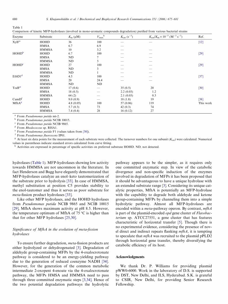
Table 1Comparison of kinetic MFP-hydrolases (involved in mono-aromatic compounds degradation) purified from various bacterial strains
Enzyme Substrate Km (lM) Vmaxh Kcat (s�1) Kcat/Km · 10�5 (M�1 s�1) Ref.
XylFa HOHD 36 100 — — [12]HMSA 6.7 6.9 —HMMSA 10 3.2 — —
HOHDb HOHD 6.7 100 — — [29]HMSA ND 7 — —HMMSA ND 3 — —
HOHDc HOHD 27 100 — — [29]HMSA ND 3 — —HMMSA ND 1 — —
EtbD1d HOHD 4.5 100 — — [37]HMSA 20 24.4 — —HMMSA ND ND
TodFe HOHD 17 (0.6) — 35 (0.5) 20 [30]HMSA 18 (0.5) — 2.2 (0.03) 1.2HMMSA 64 (2) — 2.1 (0.03) 0.3
CumDf HOHD 9.0 (0.9) — 18 (1.8) 19 [38]MfhAg HOHD 4.8 (0.05) 100 57 (0.06) 119 This work
HMSA 5.7 (0.3) 73 42 (0.3) 74HMMSA 7.4 (0.4) 28 16 (0.12) 27
a From Pseudomonas putida mt-2.b From Pseudomonas putida NCIB 10015.c From Pseudomonas putida NCIB 9865.d From Rhodococcus sp. RHA1.e From Pseudomonas putida F1 (values taken from [30]).f From Pseudomonas fluorescens IP01.g At least six data points for the measurement of each substrate were collected. The turnover numbers for one subunit (Kcat) were calculated. Numerical
values in parentheses indicate standard errors calculated from curve fitting.h Activities are expressed in percentage of specific activities on preferred substrate HOHD. ND, not detected.
680 S. Khajamohiddin et al. / Biochemical and Biophysical Research Communications 351 (2006) 675–681
hydrolases (Table 1). MFP-hydrolases showing low activitytowards HMMSA are not uncommon in the literature. Infact Henderson and Bugg have elegantly demonstrated thatMFP-hydrolases catalyze an enol–keto tautomerization ofthe substrate prior to hydrolysis [33]. In case of HMMSA,methyl substitution at position C5 provides stability tothe enol-tautomer and thus it serves as poor substrate formeta-fission product hydrolases [33].
Like other MFP hydrolases, and the HOHD hydrolasesfrom Pseudomonas putida NCIB 9865 and NCIB 10015[29], MfhA shows maximum activity at pH 8.5. However,the temperature optimum of MfhA of 75 �C is higher thanthat for other MFP hydrolases [25,30].
Significance of MfhA in the evolution of meta-fission
hydrolases
To ensure further degradation, meta-fission products areeither hydrolyzed or dehydrogenated [3]. Degradation ofaldehyde group-containing MFPs by the 4-oxalocrotonatepathway is considered to be an energy-yielding pathwaydue to the generation of reduced coenzyme NADH [34].However, for the generation of the common metabolicintermediate 2-oxopent 4-enoate via the 4-oxalocrotonatepathway, the MFPs HMSA and HMMSA need to passthrough three committed enzymatic steps [3,34]. Hence ofthe two potential degradation pathways the hydrolytic
pathway appears to be the simpler, as it requires onlyone committed enzymatic step. In view of the catabolicdivergence and non-specific induction of the enzymesinvolved in degradation of MFPs it has been proposed thatit should be advantageous to have a unique hydrolase withan extended substrate range [3]. Considering its unique cat-alytic properties, MfhA is potentially an MFP-hydrolasewith the capability to degrade both aldehyde and ketonegroup-containing MFPs by channeling them into a simplehydrolytic pathway. Almost all MFP-hydrolases areencoded within a meta-pathway operon. By contrast, mfhA
is part of the plasmid-encoded opd gene cluster of Flavobac-
terium sp. ATCC27551, a gene cluster that has featurescharacteristic of horizontal transfer [5]. Though there isno experimental evidence, considering the presence of nov-el direct and indirect repeats flanking mfhA, it is temptingto speculate that mfhA was recruited to the plasmid pPLD2through horizontal gene transfer, thereby diversifying thecatabolic efficiency of its host.
Acknowledgments
We thank Dr. P. Williams for providing plasmidpWW0-6000. Work in the laboratory of D.S. is supportedby DST, New Delhi, and ILS, Hyderabad. S.K. is gratefulto CSIR, New Delhi, for providing Senior ResearchFellowship.

S. Khajamohiddin et al. / Biochemical and Biophysical Research Communications 351 (2006) 675–681 681
References
[1] J.M. Horn, S. Harayama, K.N. Timmis, DNA sequence determina-tion of the TOL plasmid (pWWO) xylGFJ genes of Pseudomonas
putida: implications for the evolution of aromatic catabolism, Mol.Microbiol. 5 (1991) 2459–2474.
[2] D.L. Ollis, E. Cheah, M. Cygler, B. Dijkstra, F. Frolow, S.M.Franken, M. Harel, S.J. Remington, I. Silman, J. Schrag, J.L.Sussman, K.H.G. Verscheuren, A. Goldman, The alpha/beta hydro-lase fold, Protein. Eng. 5 (1992) 197–211.
[3] J.M. Sala-Trepat, K. Murray, P.A. Williams, The metabolic diver-gence in the meta cleavage of catechols by Pseudomonas putida NCIB10015. Physiological significance and evolutionary implications, Eur.J. Biochem. 28 (1972) 347–356.
[4] J.R. van der Meer, W.M. de Vos, S. Harayama, A.J. Zehnder,Molecular mechanisms of genetic adaptation to xenobiotic com-pounds, Microbiol. Rev. 56 (1992) 677–694.
[5] D. Siddavattam, S. Khajamohiddin, B. Manavathi, S.B. Pakala, M.Merrick, Transposon-like organization of the plasmid-borne organo-phosphate degradation (opd) gene cluster found in Flavobacterium sp,Appl. Environ. Microbiol. 69 (2003) 2533–2539.
[6] K.D. Schmidt, B. Tummler, U. Romling, Comparative genomemapping of Pseudomonas aeruginosa PAO with P. aeruginosa C,which belongs to a major clone in cystic fibrosis patients and aquatichabitats, J. Bacteriol. 178 (1996) 85–93.
[7] V.M. Morales, A. Backman, M. Bagdasarian, A series of wide-host-range low-copy-number vectors that allow direct screening forrecombinants, Gene 97 (1991) 39–47.
[8] D.H. Figurski, D.R. Helinski, Replication of an origin containingderivative of plasmid RK2 dependent on a plasmid function providedin trans, Proc. Natl. Acad. Sci. USA 73 (1976) 1648–1652.
[9] C.I. Kado, S.T. Liu, Rapid procedure for detection and isolation oflarge and small plasmids, J. Bacteriol. 145 (1981) 1365–1373.
[10] F.W. Studier, B.A. Moffatt, Use of bacteriophage T7 RNA polymer-ase to direct selective high-level expression of cloned genes, J. Mol.Biol. 189 (1986) 113–130.
[11] K. Tsumoto, D. Ejima, I. Kumagai, T. Arakawa, Practical consid-erations in refolding proteins from inclusion bodies, Protein Expr.Purif. 28 (2003) 1–8.
[12] C.J. Duggleby, P.A. Williams, Purification and some properties of 2-hydroxy-6-oxo-2,4-heptadienoate hydrolase from two strains ofPseudomonas putida, J. Gen. Microbiol. 132 (1986) 717–726.
[13] E. Diaz, K.N. Timmis, Identification of functional residues in a 2-hydroxymuconic semialdehyde hydrolase. A new member of thealpha/beta hydrolase-fold family of enzymes which cleaves carbon–carbon bonds, J. Biol. Chem. 270 (1995) 6403–6411.
[14] C.H. Schein, M.H.M. Noteborn, Formation of soluble recombinantproteins in Escherichia coli is favoured by lower growth temperature,Biotechnology 6 (1988) 291–294.
[15] D.T. Jones, GenTHREADER: an efficient and reliable protein foldrecognition method for genomic sequences, J. Mol. Biol. 287 (1999)797–815.
[16] D. Fischer, Hybrid fold recognition: combining sequence derivedproperties with evolutionary information, Pac. Symp. Biocomput.(2000) 119–130.
[17] L.A. Kelley, R.M. MacCallum, M.J. Sternberg, Enhanced genomeannotation using structural profiles in the program 3D-PSSM, J. Mol.Biol. 299 (2000) 499–520.
[18] A. Sali, T.L. Blundell, Comparative protein modelling by satisfactionof spatial restraints, J. Mol. Biol. 234 (1993) 779–815.
[19] D. Eisenberg, R. Luthy, J.U. Bowie, VERIFY3D: assessment ofprotein models with three-dimensional profiles, Methods Enzymol.277 (1997) 396–404.
[20] K. Mizuguchi, C.M. Deane, T.L. Blundell, M.S. Johnson, J.P.Overington, JOY: protein sequence–structure representation andanalysis, Bioinformatics 14 (1998) 617–623.
[21] E. Katchalski-Katzir, I. Shariv, M. Eisenstein, A.A. Friesem, C.Aflalo, I.A. Vakser, Molecular surface recognition: determination ofgeometric fit between proteins and their ligands by correlationtechniques, Proc. Natl. Acad. Sci. USA 89 (1992) 2195–2199.
[22] I.A. Vakser, Protein docking for low-resolution structures, ProteinEng. 8 (1995) 371–377.
[23] N. Srinivasan, Conformational analysis on globular proteins:dataanalysis. Molecular Biophysics Unit, Indian Institute of Science,Bangalore, India. Ref Type: Thesis/Dissertation, 1991.
[24] A. Nicholls, K.A. Sharp, B. Honig, Protein folding and association:insights from the interfacial and thermodynamic properties ofhydrocarbons, Proteins 11 (1991) 281–296.
[25] M.J. Hernaez, E. Andujar, J.L. Rios, S.R. Kaschabek, W. Reineke, E.Santero, Identification of a serine hydrolase which cleaves the alicyclicring of tetralin, J. Bacteriol. 182 (2000) 5448–5453.
[26] S. Fushinobu, T. Saku, M. Hidaka, S.Y. Jun, H. Nojiri, H. Yamane,H. Shoun, T. Omori, T. Wakagi, Crystal structures of a meta-cleavage product hydrolase from Pseudomonas fluorescens IP01(CumD) complexed with cleavage products, Protein Sci. 11 (2002)2184–2195.
[27] N. Nandhagopal, A. Yamada, T. Hatta, E. Masai, M. Fukuda, Y.Mitsui, T. Senda, Crystal structure of 2-hydroxyl-6-oxo-6-phenyl-hexa-2,4-dienoic acid (HPDA) hydrolase (BphD enzyme) from theRhodococcus sp. strain RHA1 of the PCB degradation pathway, J.Mol. Biol. 309 (2001) 1139–1151.
[28] A. Bhaduri, L. Krishnaswamy, G.R. Ullal, M.R. Panicker, R.Sowdhamini, Fold prediction and comparative modeling of Bdm1:a probable alpha/beta hydrolase associated with hot water epilepsy, J.Mol. Model. (Online) 9 (2003) 3–8.
[29] R.C. Bayly, B.D. Di, Purification and properties of 2-hydroxy-6-oxo-2,4-heptadienoate hydrolase from two strains of Pseudomonas putida,J. Bacteriol. 134 (1978) 30–37.
[30] S.Y. Seah, G. Terracina, J.T. Bolin, P. Riebel, V. Snieckus, L.D. Eltis,Purification and preliminary characterization of a serine hydrolaseinvolved in the microbial degradation of polychlorinated biphenyls, J.Biol. Chem. 273 (1998) 22943–22949.
[31] K. Furukawa, J. Hirose, A. Suyama, T. Zaiki, S. Hayashida, Genecomponents responsible for discrete substrate specificity in themetabolism of biphenyl (bph operon) and toluene (tod operon), J.Bacteriol. 175 (1993) 5224–5232.
[32] S. Harayama, N. Mermod, M. Rekik, P.R. Lehrbach, K.N. Timmis,Roles of the divergent branches of the meta-cleavage pathway in thedegradation of benzoate and substituted benzoates, J. Bacteriol. 169(1987) 558–564.
[33] I.M. Henderson, T.D. Bugg, Pre-steady-state kinetic analysis of 2-hydroxy-6-keto-nona-2,4-diene-1,9-dioic acid 5,6-hydrolase: kineticevidence for enol/keto tautomerization, Biochemistry 36 (1997)12252–12258.
[34] K. Murray, C.J. Duggleby, J.M. Sala-Trepat, P.A. Williams, Themetabolism of benzoate and methylbenzoates via the meta-cleavagepathway by Pseudomonas arvilla mt-2, Eur. J. Biochem. 28 (1972)301–310.
[35] G. Pugalenthi, K. Shameer, N. Srinivasan, R. Sowdhamini, HAR-MONY: a web-server for the assessment of protein structures,Nucleic Acids Res. 34 (2006) W231–W234.
[36] P.J. Kraulis, Molscript – a program to produce both detailed andschematic plots of protein structures, J. Appl. Crystal. 24 (1991) 946–950.
[37] T. Hatta, T. Shimada, T. Yoshihara, A. Yamada, E. Masi, M.Fukuda, H. Kiyohara, meta-Fission product hydrolases from a strongPCB degrader Rhodococcus sp. RHA1, J. Ferment. Bioeng. 85 (1998)174–179.
[38] T. Saku, S. Fushinobu, S.Y. Jun, N. Ikeda, H. Nojiri, H. Yamane, T.Omori, T. Wakagi, Purification, characterization, and steady-statekinetics of a meta-cleavage compound hydrolase from Pseudomonas
fluorescens IPO1, J. Biosci. Bioeng. 93 (2002) 568–574.