bayliff cbc 101 2016 - uk healthcare cecentral - bayliff_cbc 101_2016.pdf · (hypoproliferative)...
TRANSCRIPT

1
CBC 101“Cracking the Code”
Sherry L. Bayliff, MD, MPHAssistant Professor of Pediatrics
Kentucky Children’s Hospital
Family Medicine Review/Contemporary Pediatrics ConferenceNovember 10th, 2016
Faculty Disclosure
• My spouse or I have not had any relevant financial relationships during the past 12 months.
Objectives1) Clarify a systematic approach to the
interpretation of pediatric CBCs that identifies abnormalities of concern.
2) Distinguish abnormalities on the CBC that indicate further evaluation is warranted and the appropriate timeline for initiation of that workup.
3) Identify those CBC abnormalities that warrant referral for further evaluation to establish an appropriate plan of care.
Educational Need/Practice Gap
• Interpreting the complete blood count report can often be the source of great confusion
• Misinterpretation of the reported results may lead to great anxiety for both the provider and the patient/family
Expected Outcome
• Our goal today is to “crack the code” of the CBC report and increase the ability to identify when to be concerned and how/where/when to act on that concern
• By empowering you to read beyond the “H”s and “L”s, you can return to your calm and cool demeanor (and so will your patients/families!)
Active Learning
• Please try and incorporate some activity participation into your presentation either through case study discussions or audience response questions.

2
What am I working with? What is the CBC?
• a.k.a. Complete Blood Count• 3 parts:
– red blood cells– white blood cells– platelets
• 10 tests: RBC, Hgb, HCT, MCV, MCH, MCHC, RDW, WBC, Platelets, MPV
• the differential (just do it!)
Definitions x 10• RBC – number of RBCs per microL of blood (or number of RBCs x 1012/L• HGB – Hemoglobin is the concentration of hemoglobin in whole blood, in
grams/deciliter (g/dL). • HCT- The Hematocrit is the packed spun volume of blood made up of RBC,
expressed as a percentage of total blood volume. It can be measured or calculated as Hct = (RBC x MCV)/10.
• MCV – Mean corpuscular volume is the average volume (size) of the patient's RBCs. It can be measured or calculated as above .
• MCH – Mean corpuscular hemoglobin is the average hemoglobin content in a RBC. It is calculated as MCH (pg/red cell) = Hgb (g/dL) x 10 ÷ RBC (millions/microL).
• MCHC – Mean corpuscular hemoglobin concentration is the average hemoglobin concentration per RBC, in grams/dL. It is calculated as MCHC (g/dL) = Hgb (g/dL) X 100 ÷ Hct (percent).
• RDW – Red cell distribution width is a measure of the variation in RBC size, which is reflected in the degree of anisocytosis on the peripheral blood smear.
• WBC – The white blood cell count is the number of WBC per microL of blood (or number of WBC x 109/L).
• PLT – The platelet count is the number of platelets per microL of blood (or number of platelets x 109/L).
• MPV – Mean platelet volume is the average volume (size) of the patient's platelets as measured in femtoliters (fL).
Where we were . . .
• Manual techniques– Hemocytometer– Cyanomethemoglobin method– Wintrobe tube
• Imprecise and laborious
Automated Coulter Counters
• electric impedance aperture• multi-chamber coulters• modern day “channelizers”
– plot of frequency vs channel size=RBC distribution curve
• calculations
3
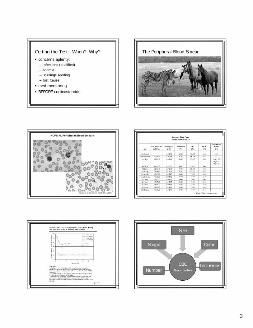
Getting the Test: When? Why?
• concerns aplenty:– Infections (qualified)– Anemia– Bruising/Bleeding– Just Cause
• med monitoring• BEFORE corticosteroids
The Peripheral Blood Smear
• Gold (non-invasive) Standard• automated processes are imperfect• extreme importance:
– Hemolytic anemia– Thrombocytopenia– WBC disorders
• complicated process
NORMAL Peripheral Blood Smears
Courtesy of Carola von Kapff, SH (ASCP). Mayo Clinic Laboratories
CBCAbnormalities
Shape
Size
Color
NumberInclusions

4
RBC “Inclusion” Etiology Conditions Special Staining?
Reticulocytes Polyribosomes Increased RBC production
*supravital stains
Nucleated RBC Nuclear Remnants Severe hemolysis, Profound stress,MDS/MPS
*NOT NORMAL on peripheral smear
Howell Jolly Bodies Nuclear remnants Post-splenectomyAsplenism
Heinz Body Denatured Hgbaggregates
G6PD, oxidant exposure, unstable Hgb
*supravital stains
Basophilic Stippling Cytoplasmic Ribosomes
Thalassemias, ETOH, lead/heavy metals, Pyrimidine 5’ Nucleotidase deficiency
Pappenheimer Bodies Iron Sideroblastic anemia
Hgb crystals Artifacts Hgb C diseaseHgb SC disease
*dehydrated sample
Red cell GHOSTS Lysed Red Cells Fulminant bacteremia *NEVER NORMAL
RBC organisms Parasites, Bacteria Malaria, TrypansomesC. perfringens
RBC Abnormalities: Shape
• Normal: biconcave disc• Poikilocytosis• MCH/MCHC• Other:
– sickle cells, target cell– fragmented red cells, bite cells, helmet cells– teardrop– Spiculated (echinocytes, acanthocytes)
Pictures by Carola von Kapff, SH (ASCP)
RBC Abnormalities: Size
• Normal: 7-8 microns/MCV 90 fL • Anisocytosis• MCV /• RDW
RDW is normal
RDW > 20%
RBC Abnormalities: Color• Normal: central pallor 1/3 dm of width• Hypochromia/Hyperchromia• MCHC• Absence of pallor: spherocytes• Bluish tint: reticulocytes
Classification of AnemiaBlood Loss Impaired
Production(Hypoproliferative)
Increased Destruction(Hemolytic)
Acute/Chronic Iron deficiency Extrinsic:
Perinatal(placental/umbilical)
Megaloblastic(Vitamin B12 or
Folate)
-auto-immune or iso-immune-infections-chemical or physical agents
Postnatal(gut, lung, nose,
GU, trauma)
Myelophthistic(infiltrative or
myelodysplastic)
-hypersplenism
Intrinsic:
Chronic Disease -membrane defects-enzyme deficiencies
Aplastic(congenital or
acquired)

5
Nathan & Oski, 8th ed., 2015
MCHC
Courtesy of Carola von Kapff, SH (ASP)
History & Physical
Targeted History• Onset/Severity of symptoms• Symptoms related to hemolysis• Prior history of events• Possible blood loss• Underlying medical conditions• Drug/toxin exposure• Diet• Birth/developmental history• Family history
Physical Exam• General Appearance• Skin• Eyes• Mouth• Cardiac• Abdomen• Musculoskeletal

6
Reticulocyte response• Absolute reticulocyte count (ARC)
– ARC= % reticulocytes x RBC/L– ARC: active erythropoiesis response
• hemolysis, acute blood loss
– ARC: suboptimal response • marrow aplasia, marrow infiltration, toxic insult
Courtesy of Stanley L. Schrier, MD
Physiologic classification
Reticulocytosis• Hemorrhage• Membranopathies• Enzymopathies• Hemoglobinopathies• AIHA• Microangiopathic HA
Reticulocytosis• Hypoplastic anemias
– Congenital– Acquired
• TEC• Systemic disease• Marrow infiltration• Medications• Infections
Carola von Kapff, SH (ASCP)
2.317.820.467232617.3
PoikilocytosisAnisocytosisHypochromiaMicrocytosis
Supportive Labs iron, TIBC transferrin satferritin NORMAL reticthrombocytosisstool guaiac +/-
*Gold Standard Test: RESPONSE to Iron
Iron Deficiency Anemia
*most common nutritional deficiency worldwide
• stores• NL HCT, NL
serum Fe
Prelatent Iron
Deficiency
• serum iron, TIBC
• NL HCT
LatentIron
Deficiency
• MCV• MCHC
Frank Iron Deficiency Anemia
Dietary factors that increase risk . . .
• Insufficient Intake– Normal Infants: 1 mg/kg/day*– LBW Infants: double* (*max=15mg/kg/d)– 1 kg in body wt requires 35-45 mg body iron
• Poor Dietary Practices– early cow’s milk, early solid food intake, tea, Vit C
intake, meat intake• Breast feeding > 6 mos w/o supplements• Low socioeconomic status
> 1 quart/day of Cow’s Milk
Management of IDA
Dietary Counseling
• Review good choices:– liver, other meats, seafood– dried fruits, nuts, beans– green leafy vegetables– whole grains
• iron-fortified products
Implemented Changes
• Breast feed 6 mos minimum
• Iron-fortified formula• < 1 pint/day whole milk• WEAN!• Iron-fortified cereals• Supplemental iron
– Facilitators: citrus juice, fruits, Vit C pills, tomatoes, potatoes, meat, fish, poultry
– Inhibitors: coffee, tea, egg yolks, milk, fiber, soy protein, antacids, H2-blockers, PPIs

7
Supplemental Iron• Oral
– Ferrous iron– 3-6 mg Elemental
iron/kg/day divided BID
– between meals– continue for 2-3
months AFTER correction of the Hgb
– side effects: GI*
• Parenteral– failure of oral tx– IV– Iron sucrose– reduced SE
White Blood Cells
CBCAbnormalities
Shape
Size
Color
NumberInclusions
WBC NumbersLeukocytosis• INFECTION
– viral– bacterial
• reactive• malignant
Leukocytopenias• NEUTROPENIA
• Lymphocytopenia

8
Toxic granulations and Döhle bodies
Courtesy of Carola von Kapff, SH (ASCP)
Neutrophilia• Eosinophilia
– allergic state, parasitic infections
• Basophilia– very unusual
• Monocytosis– EBV, reactive processes, post-chemotherapy
Lymphocyte—abnormal shape/size
Bone Marrow Examination
Stanley Schrier, MD
cancerjournal.net
cancer.gov

9
WBC Abnormality Condition Pictures
Auer rods (cytoplasmic granules)
Acute Myeloid Leukemia
Dohle bodies (cytoplasmic granules)
Infection or Inflammation
GIANT cytoplasmic granules Chediak-Higashi
Other:Small Cleaved Lymphocytes Follicular Lymphoma
Bipolar Villous Projections
Ragged “Hairy” Cytoplasm
Splenic marginal zone lymphomaHairy Cell Leukemia
“Cloverleaf” Nuclei Adult T Cell Leukemia
Parasites
Platelets
CBCAbnormalities
Shape
Size
Color
NumberInclusions
Platelet Numbers
Thrombocytopenia• Pseudo-• Confirmed
– Destruction ( platelets)• ITP, DIC, TTP, HUS
– Decreased Production • Viral• Congenital (TAR)• Replacement
Thrombocytosis (>450K)• Reactive
– cytokine driven– 3 primary qualities
• not MPD/MDD• has a likely cause• expected normalization
• Autonomous– clonal overproduction
• PV, PMF, CML, myelodysplasia
• Essential Thrombocytosis
N=91 (plts>600K)RT70%AT22%Combo8%
31% infection27% infection + post op16% post surgical9% malignancy9% post splenectomy8% acute blood loss or iron deficiency
Tefferi et al. Am J Med 1994;96: 247

10
Extreme Thrombocytopenia (>1,000,000/microL)
• N=280• RT 82%• AT 14%• Unknown 4%
31% infection19% post splenectomy/hyposplenism14% malignancy14% trauma9% inflammation6% blood loss3% rebound thrombocytosis4% uncertain
Buss DH, et al. Am J Med 1994; 96:247
Symptoms of Thrombocytosis
• Vasomotor– HA, visual, lightheadedness, atypical chest
pain, acral dysesthesias, erythromelalgia
• Thrombotic• Bleeding
ET 24% (each)RT 1% thrombosis
3% bleeding
Buss DH, et al. Am J Med 1994; 96:247
Evaluation of Thrombocytosis• Confirm
– repeat testing– peripheral blood smear
• H & P– establish the duration of the thrombocytosis
• Labs– CBCd, smear, ferritin– CRP, ESR, fibrinogen, ferritin– Gene expression (platelet RNA)
Essential Thrombocytosis
• *rate of cytogenetic abnormalities < 5%• JAK2 V617F in ~50%
Management of Thrombocytosis
Bleeding• stop anti-platelet meds• FFP/Platelets
– Acquired Factor V
• Platelet apheresis– Acquired Factor II vWd
• treat the underlying dz– Iron therapy, direct
endoscopy/imaging, myelosuppressive agent or plt-lowering agent
Thrombosis
• Platelet apheresis(>800K)• Platelet lowering agent• Anticoagulant therapy
Reviewing the basics. . .
• a CBC is meant to HELP . . . NOT hinder• always get a DIFFERENTIAL• a SMEAR gains you exponential info• Phone-a-Friend• BUT—if it feels wrong
REFER