basics of cell biology literature reading -
TRANSCRIPT
BME 42-620 Engineering Molecular Cell Biology
Lecture 06:Lecture 06:
Basics of Cell Biology Literature Reading
Methods of Cell Biology (I): Imaging
Basics of the Diffusion Theory
1BME42-620 Lecture 06, September 15, 2011

A Case Study: Malaria & Artemisininy• Malaria is a mosquito-borne
infectious disease caused by yparasite Plasmodia.
225 illi h 1• 225 million cases each year; 1 million deaths in 2010 (World malaria report).
• 4000 recipes• 380 extracts from 200 herbs
http://www.laskerfoundation.org/
• 380 extracts from 200 herbs• Compound 191 artemisinin
Youyou Tu
2
http://www.who.int/malaria/world_malaria_report_2010/en/index.html
y
Outline
• Basics of cell biology literature reading
• Imaging (I): light microscopy
• Imaging (II): electron microscopyImaging (II): electron microscopy
• Basics of the diffusion theory
3

Outline
• Basics of cell biology literature reading
• Imaging (I): light microscopy
• Imaging (II): electron microscopyImaging (II): electron microscopy
• Basics of the diffusion theory
4
Why Focus on Literature Reading?
• Biology is a scientific discipline undergoing rapid development.
• For in-depth understanding of cell biology, it is essential to read primary research literature.
• The cell biology information that can be covered in our textbook is rather limitedtextbook is rather limited.
• Basic skills for effective reading of cell biology research literature can be learned.
5
An Overview of Cell Biology Literaturegy• Journals
– Examples of general purpose journalsExamples of general purpose journals- Science- Nature - PNAS
– Examples of specialized journals- Cell and associated journals- Journal of Cell Biology
N t i t d j l- Nature associated journals- Plos journals
• Commercial versus noncommercial journalsCommercial versus noncommercial journals- subscription versus open access
• Review journals and review articles
6
Review journals and review articles
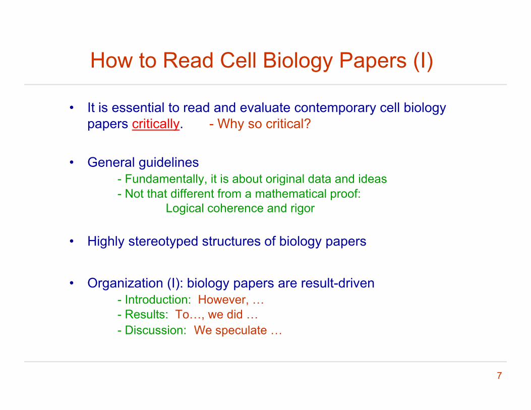
How to Read Cell Biology Papers (I)
• It is essential to read and evaluate contemporary cell biology papers critically. - Why so critical?papers critically. Why so critical?
• General guidelines- Fundamentally it is about original data and ideas- Fundamentally, it is about original data and ideas- Not that different from a mathematical proof:
Logical coherence and rigor
• Highly stereotyped structures of biology papers
• Organization (I): biology papers are result driven• Organization (I): biology papers are result-driven - Introduction: However, …- Results: To…, we did …- Discussion: We speculate …
7
p
How to Read Cell Biology Papers (II)
• Organization (II): - Every figure must tellEvery figure must tell- Logical flow: connection between result sections
• Our aims- To be able to effectively read papers in cell biology- To be able to effectively communicate cell biology resultsTo be able to effectively communicate cell biology results
8

General Process of Publication• Journal selection
- What are the messages: short vs long formatU ll l bl j l h f- Usually several comparable journals to choose from
Similar paper formatsSimilar review standards
Keep a rational perspective: vanity journals- Keep a rational perspective: vanity journals- Keep doing good science, the record will show
S b i i d i• Submission and review process- Pre-submission inquiry: usually for vanity journals - Editorial review- External reviewExternal review- Outcome I: preliminary acceptance
Point-to-point response to reviews- Outcome II: rejection
P i t i t f t b t ll k
9
- Peer-review system is not perfect but generally works
Outline
• Basics of cell biology literature reading
• Imaging (I): light microscopy
• Imaging (II): electron microscopyImaging (II): electron microscopy
• Basics of the diffusion theory
10

Visualizing Cellular Structure by Imaging• Microscopy makes it possible to
visualize cell structure and d idynamics.
• Light microscopy permits live imaging of cellular processes.
• Below 100nm, electron microscopypy
• Below 1 nm: crystallography, NMR, spectroscopyspectroscopy
• Imaging modalities are generally non-contact and non-invasive
11
non-contact and non-invasive.

Invention of Light Microscopes• Light microscope was invented more than three hundred years ago.
(Micrographia, Robert Hooke, 1665)
12
http://micro.magnet.fsu.edu/index.html Molecular expressions: microscopy world
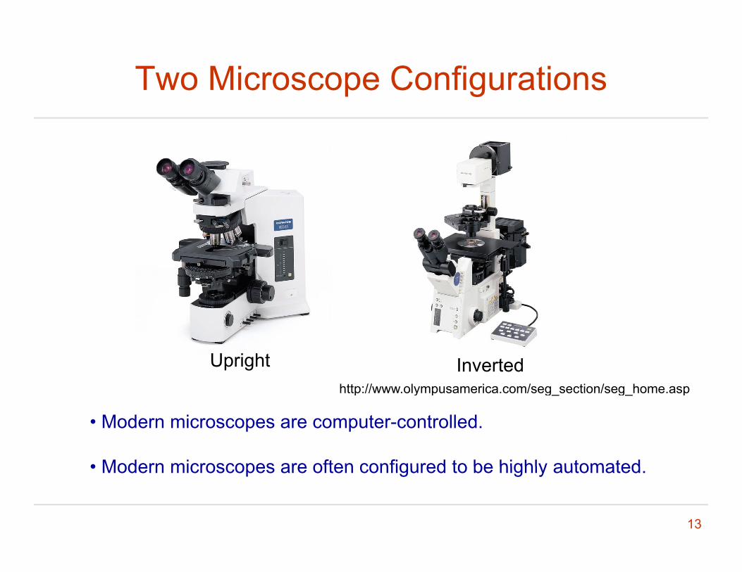
Two Microscope Configurations
Upright Invertedhttp://www.olympusamerica.com/seg section/seg home.aspp y p g_ g_ p
• Modern microscopes are computer-controlled.
• Modern microscopes are often configured to be highly automated
13
• Modern microscopes are often configured to be highly automated.
Sources of Information (I)• Major microscope manufacturers
( )
• Basic microscope structures and performance from different suppliers are very similar.
14
Sources of Information (II)
http://micro.magnet.fsu.edu/index.html Molecular expressions: microscopy world
Michael W. DavidsonFlorida State University
http://www.olympusmicro.com/
http://www microscopyu com/http://www.microscopyu.com/
http://zeiss campus magnet fsu edu/index html
15
http://zeiss-campus.magnet.fsu.edu/index.html
Light Microscopy (I)
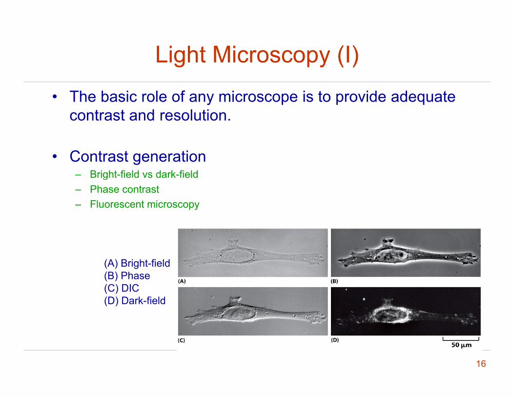
• The basic role of any microscope is to provide adequate contrast and resolution.
• Contrast generationBright field vs dark field– Bright-field vs dark-field
– Phase contrast– Fluorescent microscopy
(A) Bright-field(B) Phase(B) Phase(C) DIC(D) Dark-field
16

Light Microscopy (II)
• Under dark-field contrast, by using a special condenser,using a special condenser, only the light scattered by the specimen can enter the objective lensobjective lens.
17
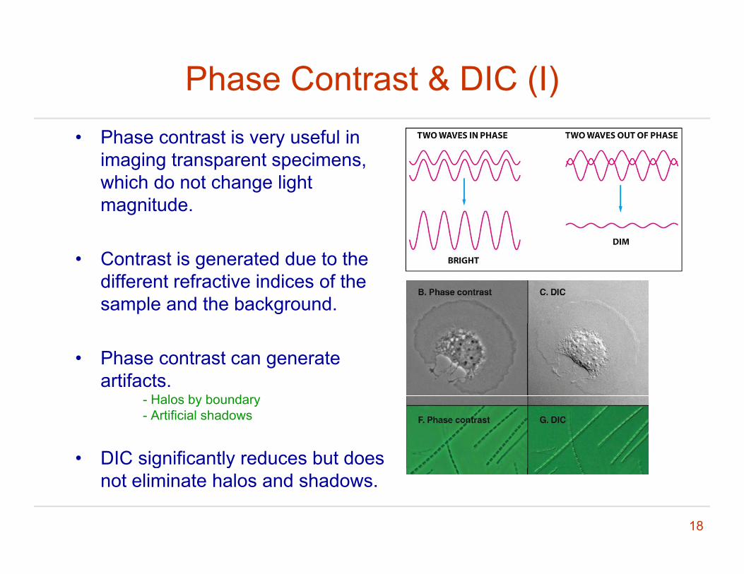
Phase Contrast & DIC (I)• Phase contrast is very useful in
imaging transparent specimens, hi h d t h li htwhich do not change light
magnitude.
• Contrast is generated due to the different refractive indices of the sample and the background.
• Phase contrast can generate artifacts.
H l b b d- Halos by boundary- Artificial shadows
• DIC significantly reduces but does
18
g ynot eliminate halos and shadows.
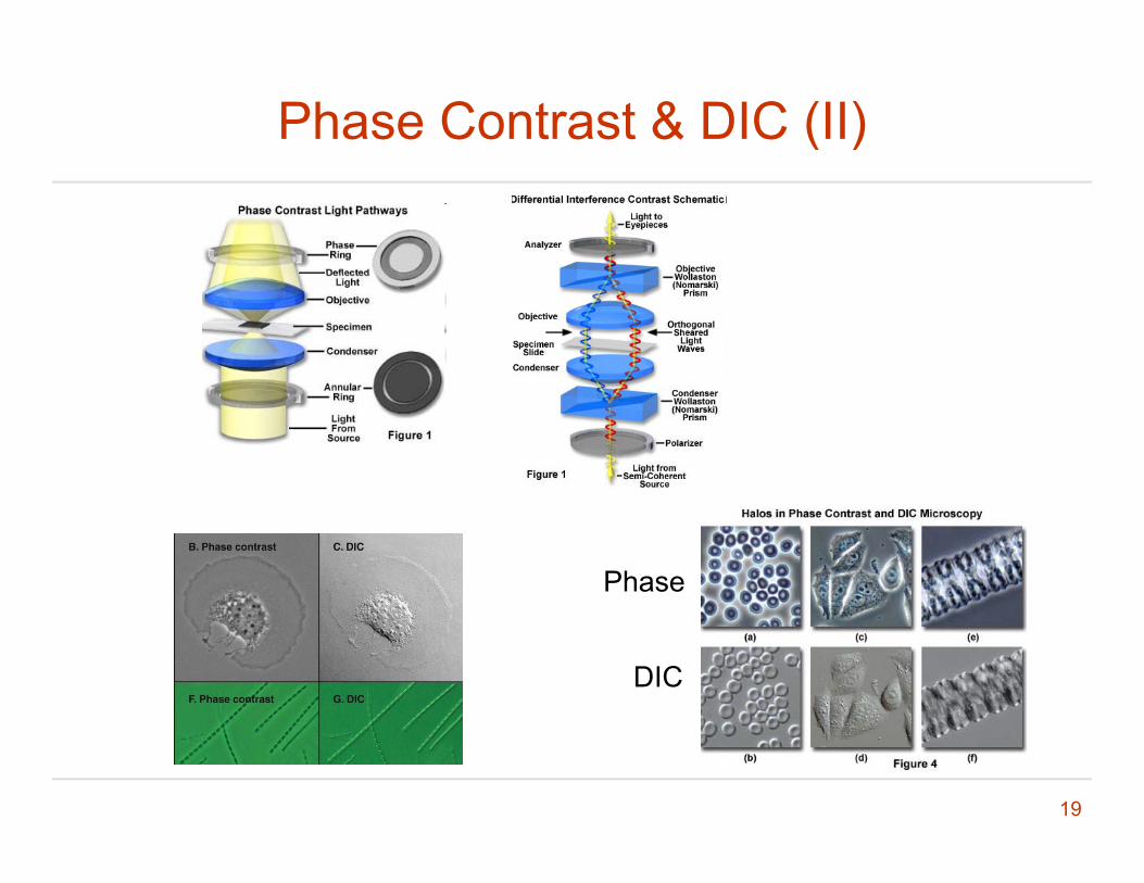
Phase Contrast & DIC (II)
Phase
DIC
19

What is missing from these images?
20
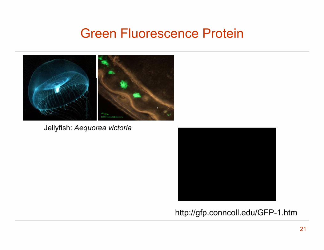
Green Fluorescence Protein
Jellyfish: Aequorea victoria
21
http://gfp.conncoll.edu/GFP-1.htm

Tagging a Protein Using GFPgg g g
http://gfp.conncoll.edu/GFP-1.htm
22
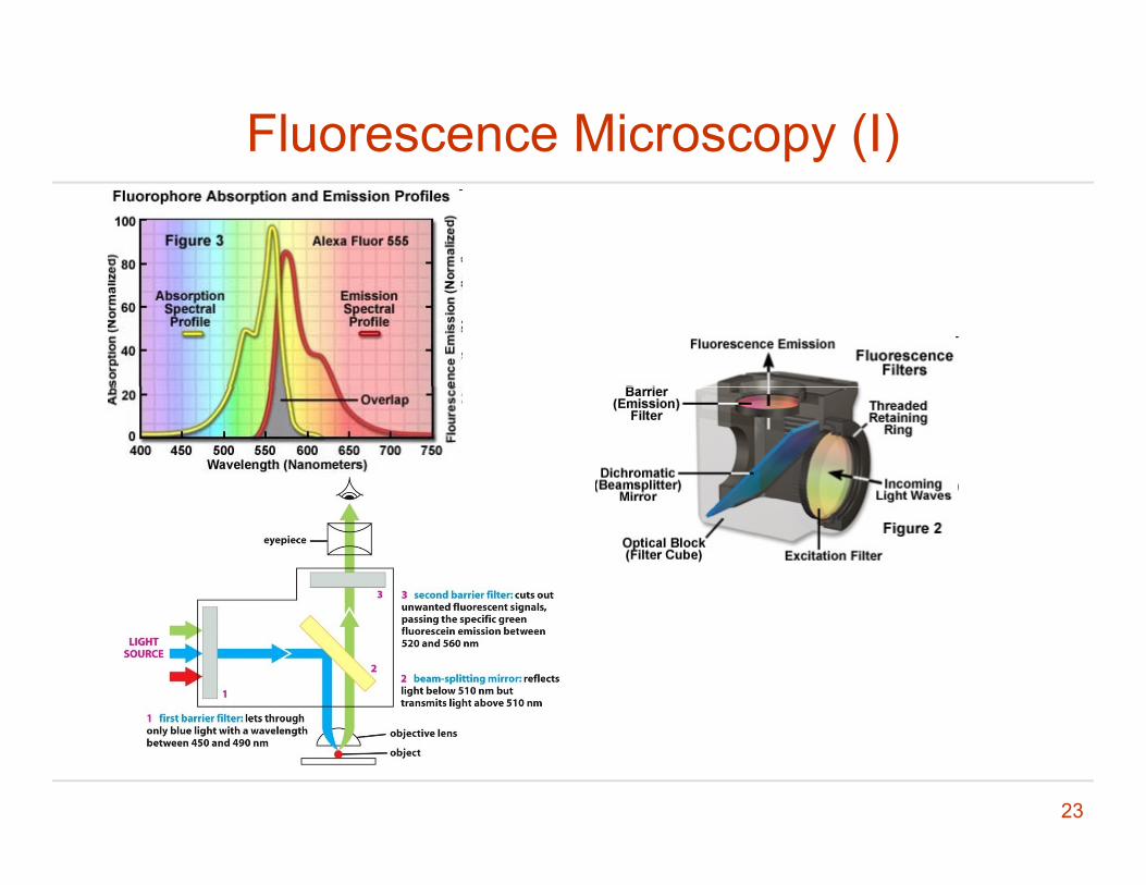
Fluorescence Microscopy (I)
23
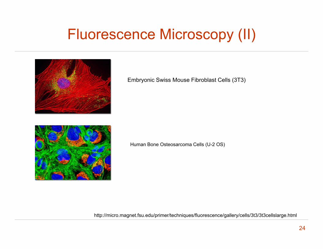
Fluorescence Microscopy (II)
Embryonic Swiss Mouse Fibroblast Cells (3T3)
Human Bone Osteosarcoma Cells (U-2 OS)
24
http://micro.magnet.fsu.edu/primer/techniques/fluorescence/gallery/cells/3t3/3t3cellslarge.html

Fluorescence Imaging of Neurons in Cerebral Cortex Sections
25
Livet J, Weissman TA, Kang H, et al. Nature 450: 56–62, 2007
Outline
• Basics of cell biology literature reading
• Imaging (I): light microscopy
• Imaging (II): electron microscopyImaging (II): electron microscopy
• Basics of the diffusion theory
26
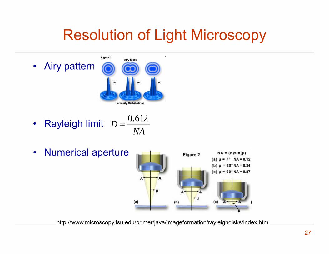
Resolution of Light Microscopy
• Airy pattern
• Rayleigh limit 0 61.DNA
• Numerical aperture
27
http://www.microscopy.fsu.edu/primer/java/imageformation/rayleighdisks/index.html
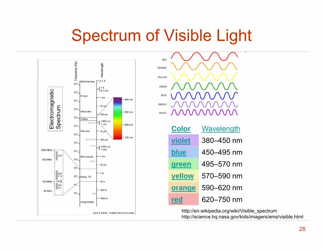
Spectrum of Visible Light
Color Wavelengthviolet 380–450 nmviolet 380–450 nmblue 450–495 nmgreen 495–570 nmyellow 570–590 nmorange 590–620 nmred 620–750 nm
28
http://en.wikipedia.org/wiki/Visible_spectrumhttp://science.hq.nasa.gov/kids/imagers/ems/visible.html
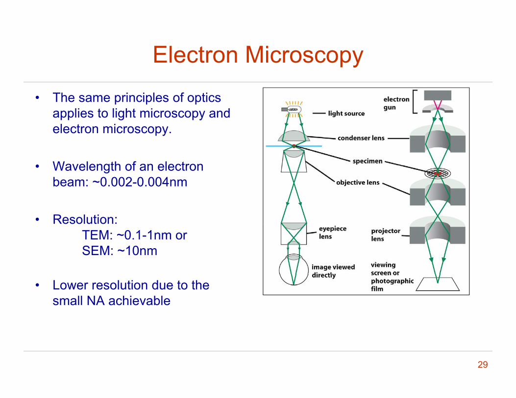
Electron Microscopy
• The same principles of optics applies to light microscopy and electron microscopy.
• Wavelength of an electron beam: ~0.002-0.004nm
• Resolution: TEM: ~0.1-1nm or SEM: ~10nm
• Lower resolution due to the small NA achievable
29
Different Electron Microscopy Modalities
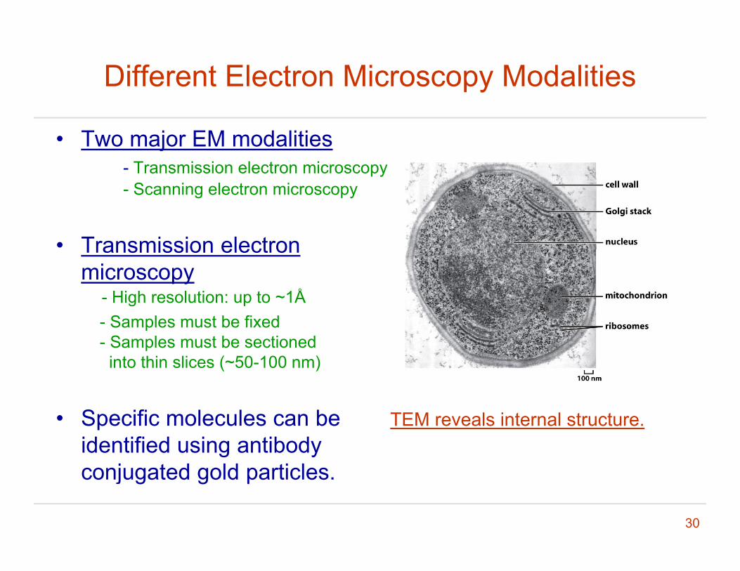
• Two major EM modalities- Transmission electron microscopy- Scanning electron microscopy
• Transmission electronTransmission electron microscopy
- High resolution: up to ~1ÅSamples must be fixed- Samples must be fixed
- Samples must be sectionedinto thin slices (~50-100 nm)
• Specific molecules can be identified using antibody
j t d ld ti l
TEM reveals internal structure.
30
conjugated gold particles.
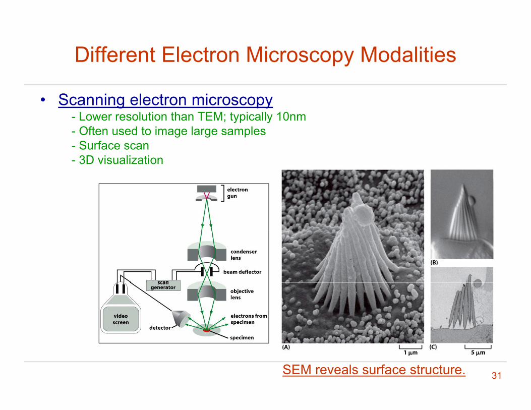
Different Electron Microscopy Modalities
• Scanning electron microscopy- Lower resolution than TEM; typically 10nm
Oft d t i l l- Often used to image large samples- Surface scan- 3D visualization
31SEM reveals surface structure.

Cryo-EM; 3D Reconstruction
• Preservation of samples by rapidly freezing the sample to a very low temperatureto a very low temperature.
• Electron-tomography: g p y- Images from different angles are integrated for 3D reconstruction in a tomography fashion.
- The crystal structure, if available, can often facilitate this process.
32
Hepatitis B virus
Comments: Methods in Cell Biology
• Cell biology is experimental science and is strongly dependent on technological advancement.dependent on technological advancement.
• Importance of protocols. - It is common to see different or even conflicting resultsIt is common to see different or even conflicting results in different studies on the same subject.
• Importance of control experiments• Importance of control experiments.
• Cultural differences from engineering
33
Outline
• Basics of cell biology literature reading
• Imaging (I): light microscopy
• Imaging (II): electron microscopyImaging (II): electron microscopy
• Basics of the diffusion theory
34
References
• Howard Berg, Random Walks in Biology, Princeton University Press 1993Princeton University Press, 1993.
• Jonathon Howard, Mechanics of Motor ProteinsJonathon Howard, Mechanics of Motor Proteins and the Cytoskeleton, Sinauer Associated, 2001.
35

Introduction
Cell lar molec les are s bject to thermal force d e• Cellular molecules are subject to thermal force due to collisions with water and other molecules.
• The resulting motion and energy are called thermal motion and thermal energy.
36
Movement of a Free Molecule (I)
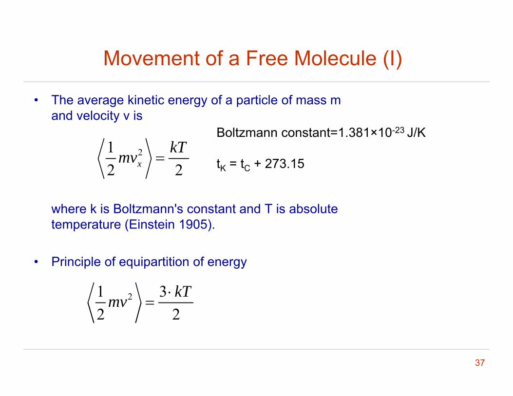
• The average kinetic energy of a particle of mass m and velocity v is
212 2x
kTmv
Boltzmann constant=1.381×10⁻²³ J/K
tK = tC + 273.15
where k is Boltzmann's constant and T is absolute temperature (Einstein 1905)
2 2
temperature (Einstein 1905).
• Principle of equipartition of energy
21 32 2
kTmv
37
Movement of a Free Molecule (II)
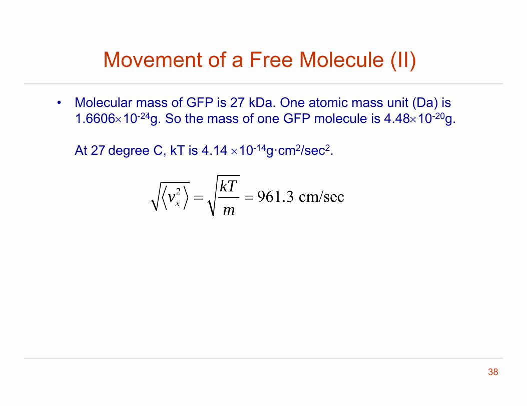
• Molecular mass of GFP is 27 kDa. One atomic mass unit (Da) is 1.660610-24g. So the mass of one GFP molecule is 4.4810-20g.
At 27 degree C, kT is 4.14 10-14g·cm2/sec2.
2 961 3 cm/secxkTv .m
38
1D Random Walk in Solution (I)
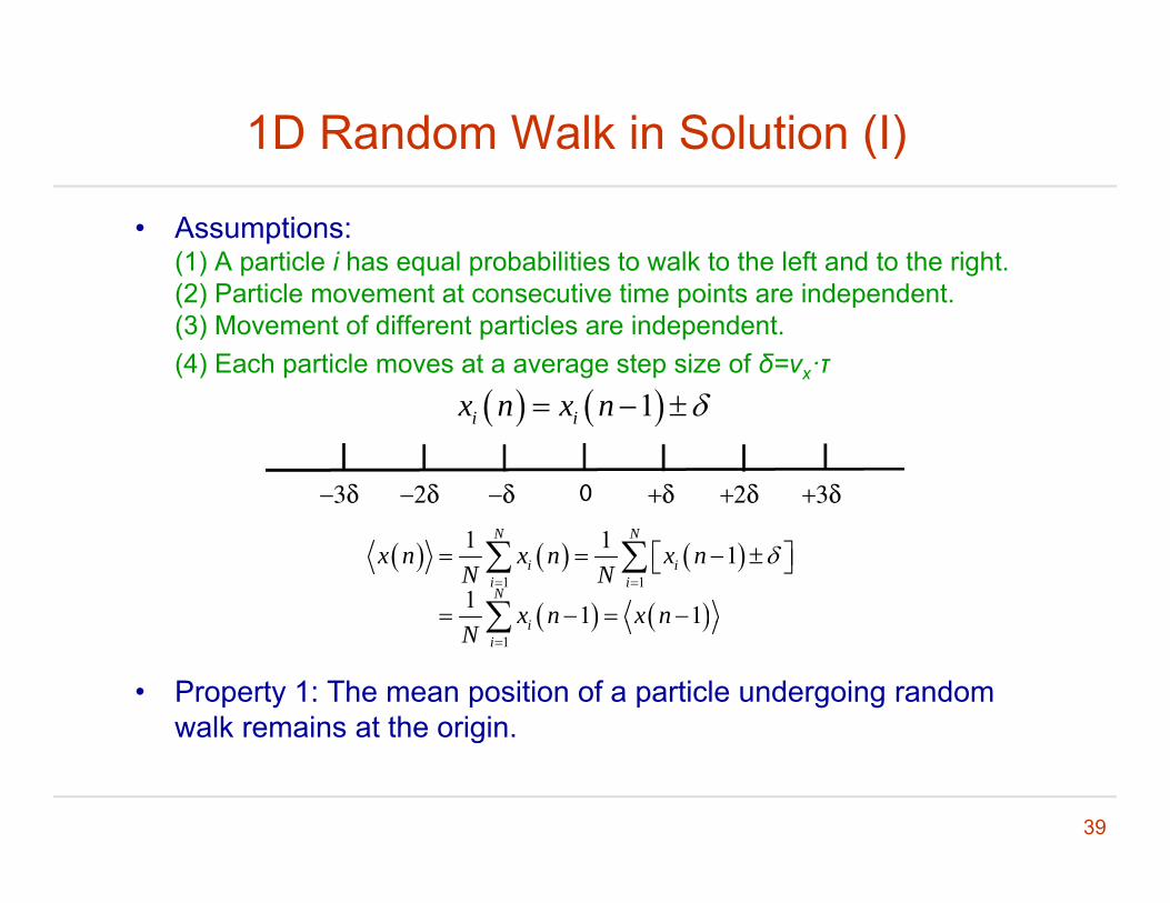
• Assumptions:(1) A particle i has equal probabilities to walk to the left and to the right.(2) Particle movement at consecutive time points are independent.(3) Movement of different particles are independent. (4) Each particle moves at a average step size of δ=vx·τ
1 1i ix n x n
1 1
1 1 1
1 1 1
N N
i ii iN
i
x n x n x nN N
x n x n
• Property 1: The mean position of a particle undergoing random walk remains at the origin.
1
1 1ii
x n x nN
39
walk remains at the origin.

1D Random Walk in Solution (II)
• Property 2: The mean square displacement of a particle undergoing random walk increases linearly w r t timeundergoing random walk increases linearly w.r.t. time.
2 2 2 2
1 12 2
1 1 1 2 1
1
N N
i i ii i
x n x n x n x nN N
2 21x n
2 2 2 2tx n n Dt
2 2 2 4r n x n y n Dt
P t 3 Th di l t f ti l f ll l
2 2 2 2 6r n x n y n z n Dt
• Property 3: The displacement of a particle follows a normal distribution.
40
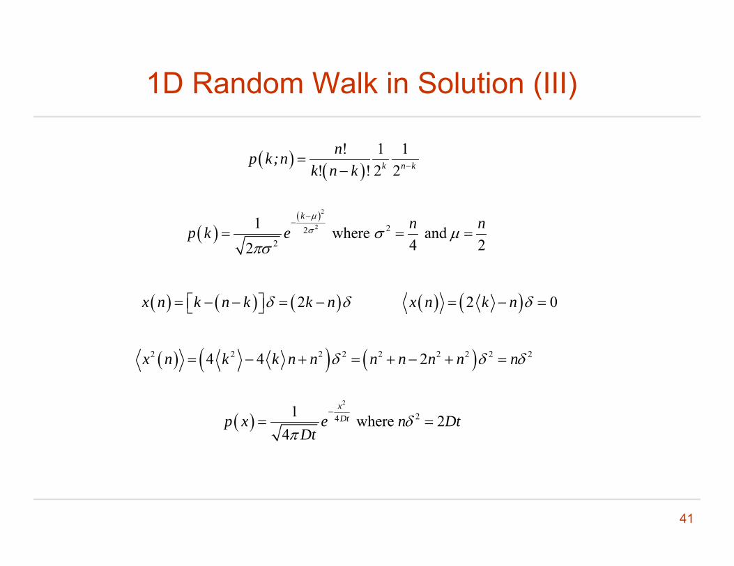
1D Random Walk in Solution (III)
! 1 1
! ! 2 2k n k
np k;nk n k
2
2 222
1 where and 4 22
k n np k e
2x n k n k k n 2 0x n k n
4 22
2 2 2 2 2 2 2 2 24 4 2x n k k n n n n n n n
2
241 where 24
xDtp x e n Dt
Dt
41
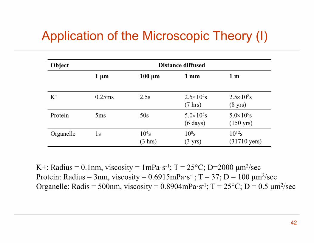
Application of the Microscopic Theory (I)
Object Distance diffused
1 μm 100 μm 1 mm 1 m1 μm 100 μm 1 mm 1 m
K+ 0.25ms 2.5s 2.5104s(7 hrs)
2.5108s (8 yrs)(7 hrs) (8 yrs)
Protein 5ms 50s 5.0105s (6 days)
5.0109s (150 yrs)
Organelle 1s 104s 108s 1012sg(3 hrs) (3 yrs) (31710 yers)
K+: Radius = 0 1nm viscosity = 1mPa·s-1; T = 25°C; D=2000 μm2/secK+: Radius = 0.1nm, viscosity = 1mPa s ; T = 25 C; D=2000 μm /secProtein: Radius = 3nm, viscosity = 0.6915mPa·s-1; T = 37; D = 100 μm2/secOrganelle: Radis = 500nm, viscosity = 0.8904mPa·s-1; T = 25°C; D = 0.5 μm2/sec
42
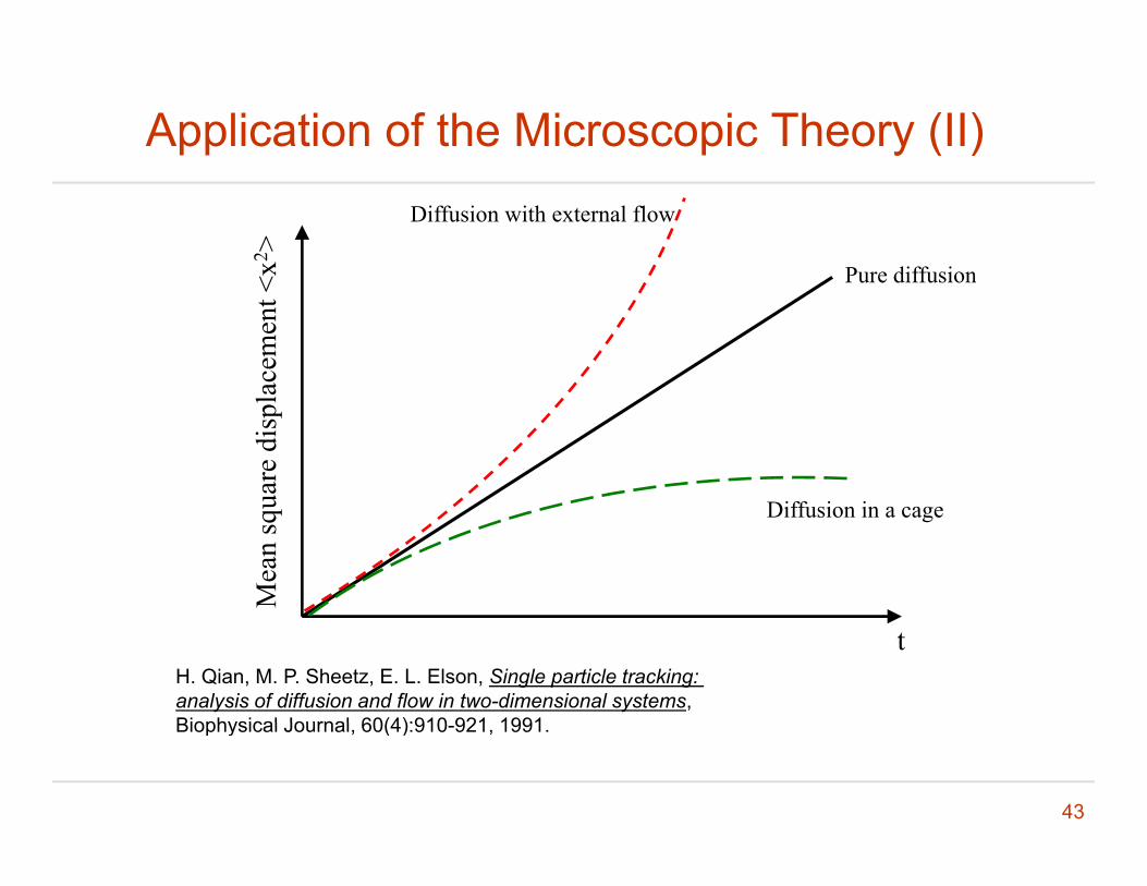
Application of the Microscopic Theory (II)
Pure diffusionx2 >
Diffusion with external flow
Pure diffusionac
emen
t <ua
re d
ispl
aM
ean
squ Diffusion in a cage
H. Qian, M. P. Sheetz, E. L. Elson, Single particle tracking: analysis of diffusion and flow in two-dimensional systems, Biophysical Journal 60(4):910 921 1991
t
43
Biophysical Journal, 60(4):910-921, 1991.

Questions ?
44
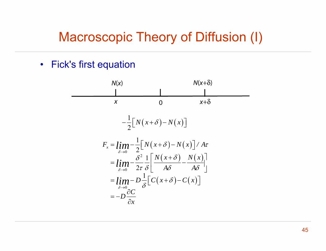
Macroscopic Theory of Diffusion (I)
• Fick's first equation
12
N x N x
1
0
2
0
12
12
xF N x N x / A
N x N xA A
lim
lim
0
1D C x C x
CDx
lim
45

Macroscopic Theory of Diffusion (II)
• Fick's second equation
1x xC t C t F x F x A
A
1 1 1
1x xC t C t F x F x A
AF x F x
x xF x F x
2xFC CD
2x D
t x x
46
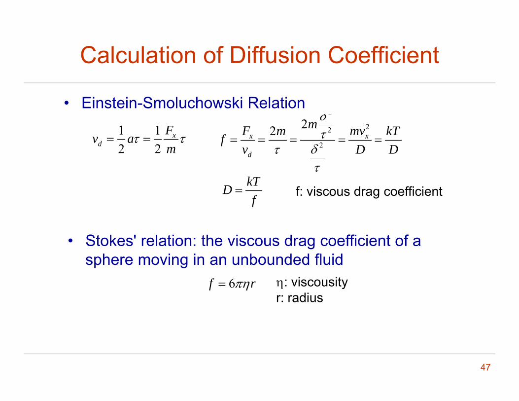
Calculation of Diffusion Coefficient
2
22m • Einstein-Smoluchowski Relation
1 12 2
xd
Fv am
22
2
22x x
d
mF mvm kTfv D D
kTDf
f: viscous drag coefficient
• Stokes' relation: the viscous drag coefficient of a sphere moving in an unbounded fluid
6f r : viscousityr: radius
47
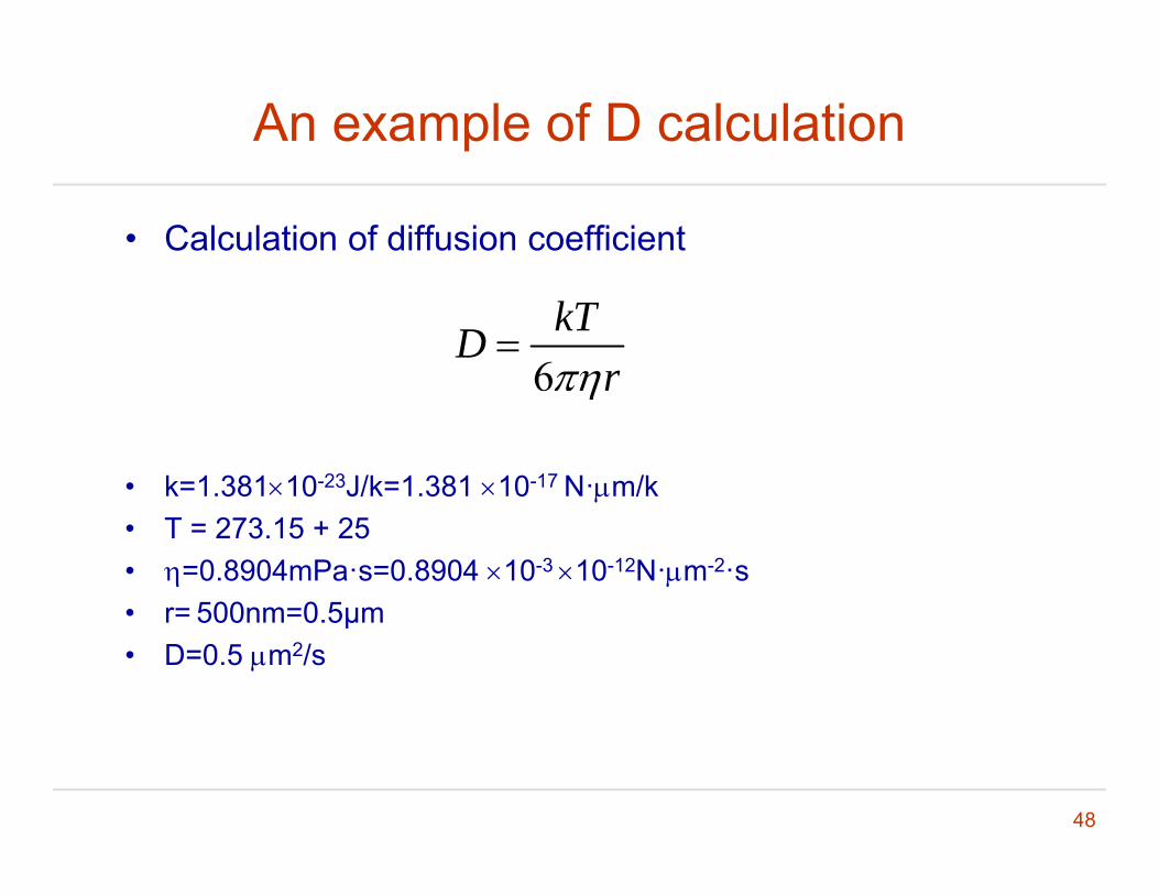
An example of D calculation
• Calculation of diffusion coefficient
6kTD
r
• k=1.38110-23J/k=1.381 10-17 N·m/k
• T = 273.15 + 25• =0.8904mPa·s=0.8904 10-3 10-12N·m-2·s• r= 500nm=0 5μmr 500nm 0.5μm• D=0.5 m2/s
48
An example of direct measurement of DAn example of direct measurement of D
M. B. Elowitz et al, Protein mobility in the cytoplasm of E. coli,J B t i l 181 197 203 1999J. Bacteriology, 181:197-203, 1999
49