basic interpretation of abdominal radiographs (1) · the diagnosis was abdominal aortic aneurysm...
TRANSCRIPT
1
Basic Interpretation of Abdominal Radiographs (1)
2 2222222222222222222222222222
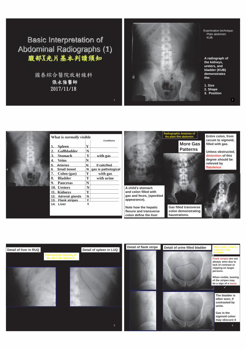
A radiograph of the kidneys, ureters, and bladder (KUB) demonstrates the: 1. Size 2. Shape 3. Position
Examination technique: Plain abdomen KUB
3
What is normally visible 1. Spleen Y 2. Gallbladder N 3. Stomach Y with gas 4. Veins N 5. Arteries N if calcified 6. Small bowel N gas is pathological 7. Colon (gas) Y with gas 8. Bladder Y with urine 9. Pancreas N 10. Ureters N 11. Kidneys Y 12. Adrenal glands N 13. Flank stripes Y 14. Liver Y
Conditions
4
Gas filled transverse colon demonstrating haustrations.
Entire colon, from cecum to sigmoid, filled with gas. Unless obstructed, distention of this degree should be relieved by flatulence
More Gas Patterns
Radiographic Anatomy of the plain film abdomen
A child’s stomach and colon filled with gas and feces, (speckled appearance). Note how the hepatic flexure and transverse colon define the liver
5
Detail of liver in RUQ Detail of spleen in LUQ Radiographic Anatomy of the plain film abdomen
6
The bladder is often seen, if contrasted by urine. Gas in the sigmoid colon may obscure it
Detail of flank stripe Detail of urine filled bladder Radiographic Anatomy of the plain film abdomen
Flank stripes are not always seen due to lack of contrast or clipping on larger persons. When visible, bowing of the stripes may be a sign of a mass.
7
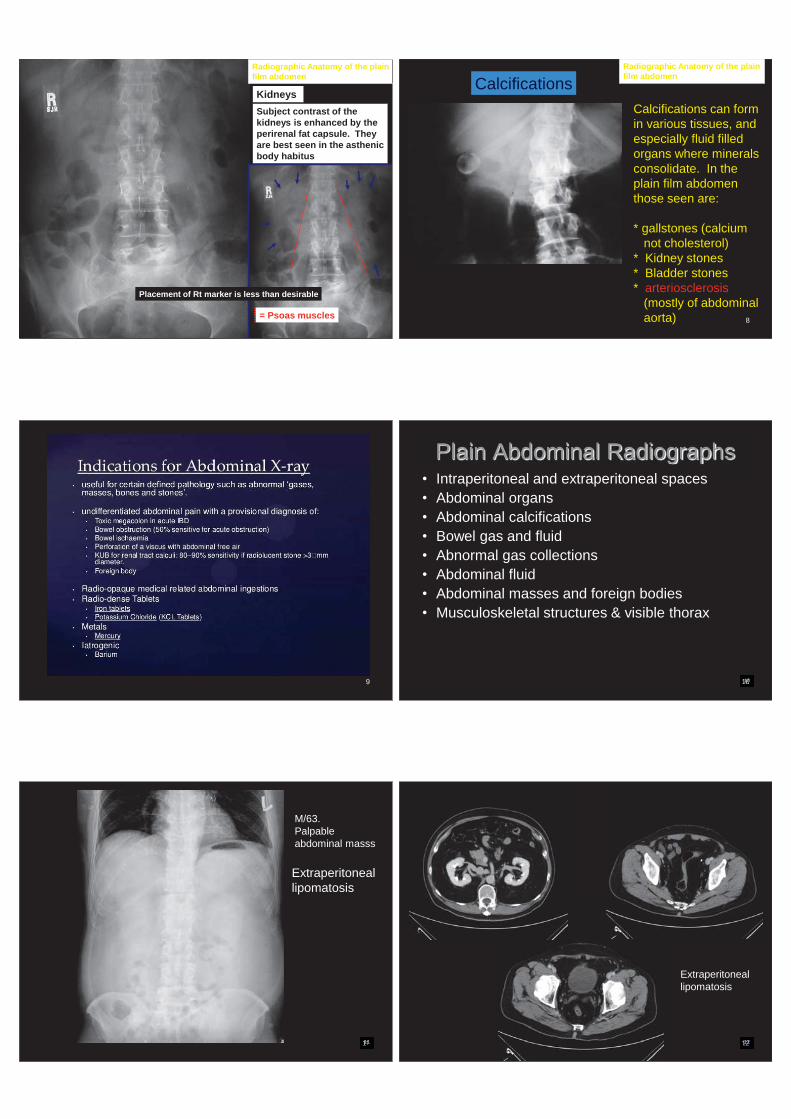
Subject contrast of the kidneys is enhanced by the perirenal fat capsule. They are best seen in the asthenic body habitus
Kidneys
Radiographic Anatomy of the plain film abdomen
Placement of Rt marker is less than desirable
= Psoas muscles 8
Calcifications can form in various tissues, and especially fluid filled organs where minerals consolidate. In the plain film abdomen those seen are: * gallstones (calcium not cholesterol) * Kidney stones * Bladder stones * arteriosclerosis (mostly of abdominal aorta)
Calcifications Radiographic Anatomy of the plain film abdomen
Large gallstone in RUQ If not in the RUQ, where else could it be?
9 10 10000000000000000000000000000010
Plain Abdominal Radiographs • Intraperitoneal and extraperitoneal spaces • Abdominal organs • Abdominal calcifications • Bowel gas and fluid • Abnormal gas collections • Abdominal fluid • Abdominal masses and foreign bodies • Musculoskeletal structures & visible thorax
11 11111111111111111111
Extraperitoneal lipomatosis
M/63. Palpable abdominal masss
12 12222222222222222222222222222222222222212
Extraperitoneal lipomatosis
13 1333333333333333333333333333333333313
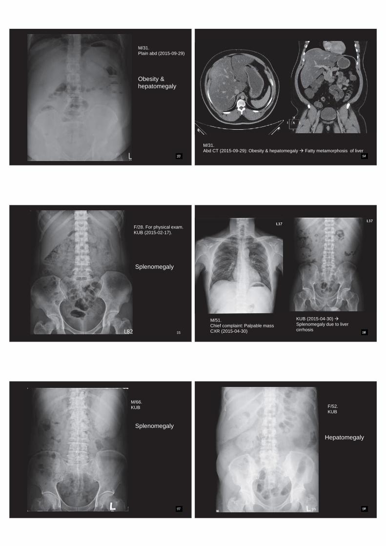
M/31. Plain abd (2015-09-29)
Obesity & hepatomegaly
14 144444444444444444444444444444444444444444414
M/31. Abd CT (2015-09-29): Obesity & hepatomegaly Fatty metamorphosis of liver
15
F/28. For physical exam. KUB (2015-02-17).
Splenomegaly
16 1666666666666666666666666666666666666616
M/51. Chief complaint: Palpable mass CXR (2015-04-30)
KUB (2015-04-30) Splenomegaly due to liver cirrhosis
17 17777777777777777777777777777717
M/66. KUB
Splenomegaly
18 188888888888888888888888888888888888818
F/52. KUB
Hepatomegaly
19 19999999999999999999999999999999999999919
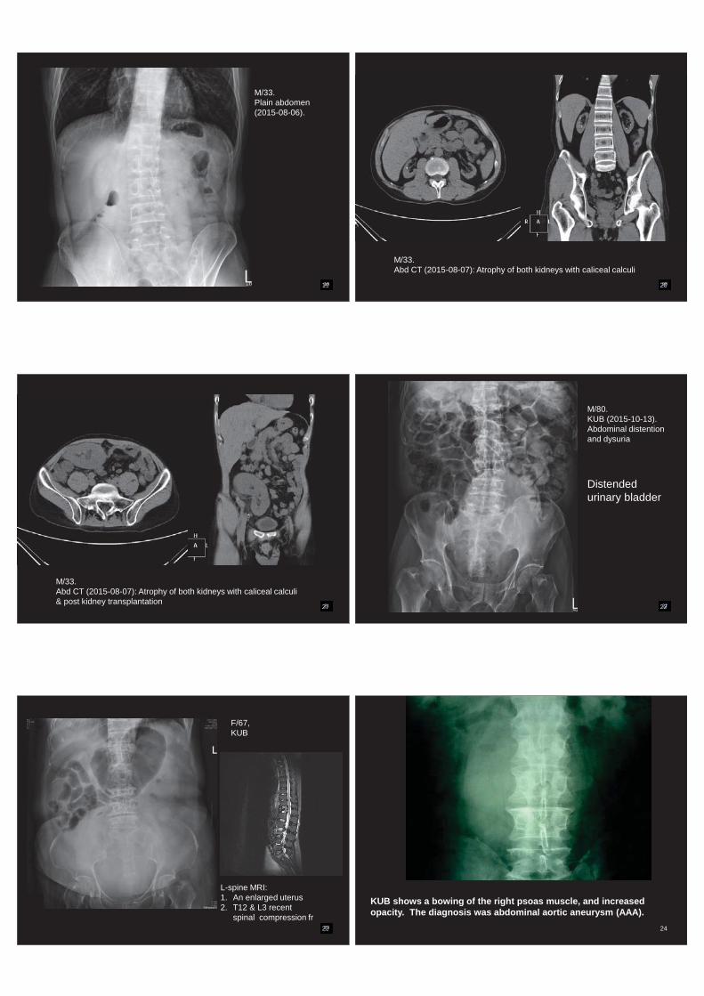
M/33. Plain abdomen (2015-08-06).
20 20220202202202220202200222222222000000000000000000000000000000020
M/33. Abd CT (2015-08-07): Atrophy of both kidneys with caliceal calculi
21 22222222222222222222211111111111111111111121
M/33. Abd CT (2015-08-07): Atrophy of both kidneys with caliceal calculi & post kidney transplantation
22 2222222222222222222222222222222222222222222222222222222222222222222222222
M/80. KUB (2015-10-13). Abdominal distention and dysuria
Distended urinary bladder
23 232322322322323223232332222233333333333333333333333333333323
F/67, KUB
L-spine MRI: 1. An enlarged uterus 2. T12 & L3 recent
spinal compression fr
24
KUB shows a bowing of the right psoas muscle, and increased opacity. The diagnosis was abdominal aortic aneurysm (AAA).
25 25222252252522252522525252522222255555555555555555555555555555555555525
Intra-abdominal calcifications
• Calculi • Vascular calcifications – arteries, veins • Post infectious or inflammatory
calcificatons • Dystrophic calcifications • Calcifications of neoplasm
26 262262622622622262622662222222226666666666666666666666666666666666626
Acute calculous cholecystitis
M/73 Abdominal pain
27 2722227222727222722772727272222777777777777777777777777777727
Gallbladder stone, M/58
28 2822282282282822828228282222222888888888888888888888888888888828
1. Acute calculous Cholecystitis 2. Bladder stones
M/73 Abdominal pain
29 292922922922929229292992222299999999999999999999999999999999929
F/85. KUB (2012-11-28).
Porcelain gallbladder
30 3030303330333033030303303330000000000000000000000000000000000000030
F/81.
Splenic arterial aneurysm

31 333333333333333333333311111111111111111111131
M/72. Abdominal pain KUB (2013-02-11)
M/72. KUB 2013-03-28) Abdominal aortic aneurym, post stenting, Lt retroperitoneal abscess
32 32323332323333233322333232333233332222222222222222222222222222222222232
Chronic pancreatitis
Typical coarse calcifications at the pancreas on plain abdominal presentation
M/43 Abdominal pain
33 3333333333333333333333333333333333333333333333333333333333333333333333
F/47 CXR (2015-11-05) for physical exam
1. Cardiomegaly with left ventricular hypertrophy
2. A subsegmental atelectasis at left lower lung field.
3. Chronic pancreatitis with coarse calcifications
34 3434333434343334443433434343343433434334444444444444444444444444444444444434
M/57. Acute abdominal pain, KUB (2015-10-01)
Abnormal coarse calcifications at upper abdomen
35 35353535335353355335353535533533555555555555555555555555555555555555535
M/57. Acute abdominal pain 10 year ago ER Abdominal CT (2005-05-15): Chronic pancreatitis with relapse episode
36 363636333633363363636336333666666666666666666666666666666666666636
M/40. KUB (2015-09-28)
Bil renal calculi & Lt ureteral calculi

37
Bilateral renal stones and ureteral stones. Bilateral enlarged kidneys (hydronephroses)
M/44
38 383833383833338333883338383338333388888888888888888888888888888888838
F/83. Standing abdomen X-ray
Staghorn stones in both kidneys.
39 393933939393339933399933399393933939999999999999999999999999999999999999939
M/44
Acute abdominal pain
40 404044040404440444444440440000000000000000000000000000000000040
Lt UV junctional stone
M/44 Abdominal pain
41 4444444444444444441111111111111111141
M/61. KUB
Calcified mesenteric lymph nodes
42 42424242442242442244244244424224422222222222222222222222222222222242
M/61. KUB
Calcified left paraaortic lymph nodes
43 433443444344344444434433333333333333333333333333333333343
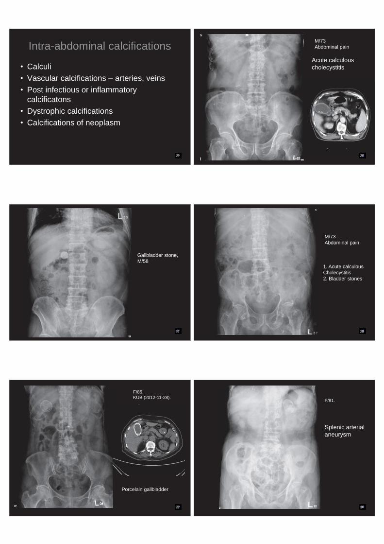
Diverticulosis of descending & sigmoid colon
Barium retention or calcifications
F/88. KUB (2015-09-24)
44
Thank you for your attentions!