basic information and product report on composite … · basic information and product report on...
TRANSCRIPT

Basic Information and Product Report on Composite Resin for Dental Fillings
Edited byYAMAKIN CO., LTD.
Yamada B, Ph.D. in Engineering Kato T, Ph.D. in EngineeringSato Y, Ph.D.INTERNATIONAL20171013
Head Of f ice: 3-7 Sanadayama-cho Tennoji-ku Osaka 543-0015, JapanBranch Of f ice: Tokyo, Osaka, Sendai, Nagoya, Fukuoka, JAPAN
Factory and R&D: Kochi, JAPANP: +81-887-55-0281 F: +81-887-55-0053
Head Of f ice: 3-7 Sanadayama-cho Tennoji-ku Osaka 543-0015, JapanBranch Of f ice: Tokyo, Osaka, Sendai, Nagoya, Fukuoka, JAPAN Factory and R&D: Kochi, JAPANP: +81-887-55-0281 F: +81-887-55-0053
Yamamoto Precious Metal Co., Ltd.
From Production Skills to Scientific Knowledge, and onward to Fusion with Medical Science
YAMAKIN CO., LTD.
YAMAKIN CO., LTD.Head Office: 3-7 Sanadayama-cho Tennoji-ku Osaka 543-0015, JapanBiological Science Safety Laboratory: Laboratory in the Department of Oral and Maxillofacial Surgery, Kochi Medical School, Kochi University Kohasu, Oko-cho, Nankoku-shi, Kochi 783-8505, JAPANBranch Office: Tokyo, Osaka, Sendai, Nagoya, Fukuoka, JAPAN P: +81-887-55-0281 F: +81-887-55-0053E: [email protected]://www.yamakin-global.com

Index
1. Foreword
2. Basic information on composite for dental fillings 2.1 What is a composite ?
2.2 Mechanism of a photo-radical polymerization reaction by a visble-light photoinitiator
2.3 Initiation reaction and photo-irradiation device
2.4 Scope of application of composites
3. Our new product “iGOS” 3.1 Product concept
3.2 SEM observation of iGOS
3.3 Fluoride sustained release
3.4 The special characteristics of fluorine recharge
3.5 The risk of fluoride ions
3.6 Strength
3.7 Vickers hardness
3.8 Occlusal tooth abrasion
3.9 X-ray imageability
3.10 Colour
3.11 Workability
3.12 RestrictingStreptococcus mutans adhesion test
3.13 Stain resistant test
3.14 Clinical cases
3.15 Biological safety
4. Afterword
2
3
3
4
6
7
8
8
9
9
10
10
12
13
14
15
16
17
17
18
20
22
25
・ ・ ・ ・ ・ ・ ・ ・ ・ ・ ・ ・ ・ ・ ・ ・ ・ ・ ・ ・ ・ ・ ・ ・ ・ ・ ・ ・ ・ ・ ・ ・ ・ ・
・ ・ ・ ・ ・ ・ ・・ ・ ・ ・ ・ ・ ・ ・ ・ ・ ・ ・ ・ ・ ・ ・ ・ ・ ・ ・ ・ ・ ・ ・ ・ ・ ・ ・ ・
・ ・・ ・ ・ ・ ・ ・ ・ ・ ・ ・ ・ ・ ・ ・ ・ ・ ・ ・
・ ・ ・ ・ ・ ・ ・ ・ ・ ・ ・ ・ ・ ・ ・ ・ ・ ・ ・ ・ ・ ・ ・ ・
・ ・ ・ ・ ・ ・ ・ ・ ・ ・ ・ ・ ・ ・ ・ ・ ・ ・ ・ ・ ・ ・ ・・ ・ ・ ・ ・ ・ ・ ・ ・ ・ ・ ・ ・ ・ ・ ・ ・ ・ ・ ・ ・ ・ ・ ・ ・ ・ ・ ・ ・ ・ ・ ・
・ ・ ・ ・ ・ ・ ・ ・ ・ ・ ・ ・ ・ ・ ・ ・ ・ ・ ・ ・ ・ ・ ・ ・ ・ ・ ・・ ・ ・ ・ ・ ・ ・ ・ ・ ・ ・ ・ ・ ・ ・ ・ ・ ・ ・ ・ ・ ・ ・ ・ ・ ・ ・ ・
・ ・ ・ ・ ・ ・ ・ ・ ・ ・ ・ ・ ・ ・ ・ ・ ・ ・ ・・ ・ ・ ・ ・ ・ ・ ・ ・ ・ ・ ・ ・ ・ ・ ・ ・ ・ ・ ・ ・ ・ ・ ・ ・ ・ ・ ・
・ ・ ・ ・ ・ ・ ・ ・ ・ ・ ・ ・ ・ ・ ・ ・ ・ ・ ・ ・ ・ ・ ・ ・ ・ ・ ・ ・ ・ ・ ・ ・ ・ ・ ・・ ・ ・ ・ ・ ・ ・ ・ ・ ・ ・ ・ ・ ・ ・ ・ ・ ・ ・ ・ ・ ・ ・ ・ ・ ・ ・ ・ ・ ・ ・ ・
・ ・ ・ ・ ・ ・ ・ ・ ・ ・ ・ ・ ・ ・ ・ ・ ・ ・ ・ ・ ・ ・ ・ ・ ・ ・ ・ ・ ・・ ・ ・ ・ ・ ・ ・ ・ ・ ・ ・ ・ ・ ・ ・ ・ ・ ・ ・ ・ ・ ・ ・ ・ ・ ・ ・ ・ ・ ・ ・
・ ・ ・ ・ ・ ・ ・ ・ ・ ・ ・ ・ ・ ・ ・ ・ ・ ・ ・ ・ ・ ・ ・ ・ ・ ・ ・ ・ ・ ・ ・ ・ ・ ・ ・・ ・ ・ ・ ・ ・ ・ ・ ・ ・ ・ ・ ・ ・ ・ ・ ・ ・ ・ ・ ・ ・ ・ ・ ・ ・ ・ ・ ・ ・ ・ ・ ・
・ ・ ・ ・ ・ ・ ・ ・ ・ ・ ・ ・ ・ ・ ・ ・ ・ ・ ・・ ・ ・ ・ ・ ・ ・ ・ ・ ・ ・ ・ ・ ・ ・ ・ ・ ・ ・ ・ ・ ・ ・ ・ ・ ・ ・ ・ ・ ・ ・
・ ・ ・ ・ ・ ・ ・ ・ ・ ・ ・ ・ ・ ・ ・ ・ ・ ・ ・ ・ ・ ・ ・ ・ ・ ・ ・ ・ ・ ・ ・ ・ ・・ ・ ・ ・ ・ ・ ・ ・ ・ ・ ・ ・ ・ ・ ・ ・ ・ ・ ・ ・ ・ ・ ・ ・ ・ ・ ・ ・ ・ ・ ・ ・
・ ・ ・ ・ ・ ・ ・ ・ ・ ・ ・ ・ ・ ・ ・ ・ ・ ・ ・ ・ ・ ・ ・ ・ ・ ・ ・ ・ ・ ・ ・ ・ ・ ・

1. Foreword
�e conventional basic dental restoration technique is to remove around tooth substance infected by dental caries. �e objective is to thoroughly remove tooth substance infected by tooth decay, to pulpectomy as necessary, and to prevent the prognistic risk of secondary dental caries. However, such a restoration technique involves the removal of sound tooth substance. One of restoration techniques for teeth damaged by dental caries etc. is called the composite resin restoration technique. �is technique has been first introduced in 1960s, uses a filling meterials called composite for direct restoration. Genarally, decayed tooth was removed caries and tooth substance infected by tooth decay. �e cavity was filled with paste form of composite and irradiated by visble light to cure it. Composite resin restoration, unlike the previous conventional basic dental restoration techniques represented by restoration with metal materials, is a major advantage from the point of view of saving sound tooth substance, and allows composite resin to match natural tooth colour varying from person to person. �e clinical concept “Minimal Intervention” (MI) – infected tooth substance is removed as little as possible, and sound tooth is retained as much as possible – has been widely used since it was proposed by Federation dental international (FDI) in 2000. From the adventage mentioned above, it can be said that the composite resin restoration would comply with the MI concept, and meet needs of both patients and dental professionals for a esthetic restoration. From the above background, the composite resin restoration is widely practiced today as one of the main choices for the treatment of tooth decay. Composites is required for various mechanical, physical and chemical properties : strength, durability, workability, fluoride sustained release, colour, and stability, it is important that it is comprehensively excellent each of the properties. However, there are many technically problems to be solved to fulfill all of the properties equally. For example, it is known that the process of fluoride sustained release of dental material causes lower of its strength and influences on its colour. Our product, “iGOS”, was succeed in achieving an accomodation with each the required mechanical, physical and chemical properties of composite. �is has been made possible by our ceramics cluster filler technique, where was involued to an unique process in relation to surface treatment. Accually our porduct, “TWiNY”, which is excellence strength composite, is contained the fillers produced by our technique. �is report provide a basic information on a photo radical polymerization system, chemical characteristics of componets consisted in composite for dental fillings, and also discusses physical characteristics of our product iGOS, along with clinical cases. We would be delighted if this report is of help to practitioners of restorative dental treatment and to their patients.
― 2 ― ― 3 ―
Basic Information and Product Report on Composite Resin for Dental Fillings
2. Basic information on composite resin for dental fillings
2.1 What is composite resin? Resin-based composite used in its restoration, which is classifiable to an organic-inorganic hybrid material, is compoed with organic materials, which are several mixed monomers as precursor of polymer matrix by radical polymerization, inorganic materials, which are fillers mainly contained with glass component, and interface both of materials. Monomer is organic compound possesses the property of converting to polymer (resin) when they undergo curing by polymerization. Especially, mixtures of several monomers containing cross-linking (meta) acrylate monomers are common in their use as a dental material. A chemical characteristic of polymerization; fluid monomer is converted to a solid-state polymer, is concerned with a important function of composite in related to its forming resin to cure. In other words, monomers are responsible for this distinctive feature; their mixture that can be freely changed its shape before polymarization, retains its shape as polymer matrix after polymerization. Urethane dimethacrylate (UDMA) and bisphenol A-glycidyl methacrylate (Bis-GMA) is shown in Fig. 1 Both of monomers are widely used for dental restoratives. A wide range of other monomers are also in use, and different monomers are mixed to make a coordination for a target viscocity, desirable degree of contraction in polymerization and other propoties.
Inorganic filler, which is made of particulated alumina, zirconia or silica, is filled into mixture of monomers to produce unhardened composite. Addition of the filler into monomers provides flexural strength and a high degree of mechanical strength to the matrix properties. Fillers with a wide range of characteristics and particle sizes are used for dental materials: inorganic fillers with range from the sub-micron size to single micron size, nano-sized colloidal silica, and organic hybrid fillers, manufactured to shatter curing material that is made to be filled nano-filles into the monomers, and then to be polymerized it previously. By changing the chemical composition and form of the inorganic filler composite made of it extend ranges of its functoins where can be expected to enhance qualities of its polishability, workability and fluorine sustained release etc.,. Also, the workability of the paste in which these fillers are mixed can be adjusted (Figure 2). As shown, both of monomer and inorganic filler play individually important roles. However, strength and durability of composite will not be sufficient if they are not tightly bonded on the interface. In estimating critical damage or fraction of hybrid materials, it should be assumed on the interface between matrix and filler. Usually, affinity of most of monomers and inorganic fillers is not effective because most of monomers are hydrophobic, surface of inorganic filler is hydrophiric. Treatment of organosilane coupling is adopted to solute this problem of
thier affinity; the surfaces of hydrophiric inorganic fillers is converted to hydrophobic surface, then enhancing their affinity with monomers. A high filling rate of inorganic filler of composite is made possible as a result, it allows for firm bonding with the polymer matrix after polymerization curing, making a major contribution to the strength and high-level durability of composite. So, as we have seen, in order for composite resin to achieve the performance required of it as a dental material, monomers, inorganic fillers and the interface between them must all play their part properly.
Figure 1 Monomers used for dental materials
OO
O
OO N
H
NH
O O
O
OO
HO
O
Bis-GMA
UDMA
O O
OH
O

― 4 ― ― 5 ―
thier affinity; the surfaces of hydrophiric inorganic fillers is converted to hydrophobic surface, then enhancing their affinity with monomers. A high filling rate of inorganic filler of composite is made possible as a result, it allows for firm bonding with the polymer matrix after polymerization curing, making a major contribution to the strength and high-level durability of composite. So, as we have seen, in order for composite resin to achieve the performance required of it as a dental material, monomers, inorganic fillers and the interface between them must all play their part properly.
2.2 Mechanism of a photo-radical polymerization reaction by a visble-light photoinitiator A particular reaction called the polymerization reaction is required in order to achieve polymers from monomers. Polymerization reactions are classified into successive reactions and chain reactions. In chain reactions, when polymerization reaction intermediates are produced from monomers, the immediately react with other monomers, and further rapid reactions with still other monomers then continue to subsequently occur (Figure 3). As a result of chain reaction, monomers are instantly converted to the mixture of high molecular weight polymer and remaining monomers. On the other hand, the monomers are rapidly consumed by the step reaction, the molecular weight of polymer is slower propagaed than the molecular weight of polymer by radical reaction. Monomers posseses several polymerzing group in their molecular (cross-linking monomers), are formed polymer network by a chain reaction. Processing the reaction, the density of the polymer network is higher, the respective polymer become insoluve to solvent which is smaller moleclar weight. Also, in the case of free radical polymerization of the multiple monomers inside the molecules (cross-linking monomers), by polymerization the polymers are formed into a networked (cross-linked) structure, and as polymerization proceeds, the density of the polymer network structure increases, and the molecular weight are not dissolved by small-scale solvents. In other words, by means of chain reaction, monomers can be vulcanized in an extremely short period of time. Radical polymerization is one form of these kinds of chain reactions; other forms of polymerization reaction also exhibit a broad range of monomers and reaction conditions. Radical polymerization is the most suitable method to instantaneously vulcanize dental materials, including monomers in the highly humid conditions of the intraoral area, and this polymerization reaction is commonly used for dental materials. Composite for dental fillings is used with the monomers in combination with photoinitiator , because it is cure byvisible-light irradiation. Radical polymerization of monomers occurs on exposure to light, and polymers are generated and vulcanized. Normally, radical polymerization does not proceed on monomers alone; if polymerization is conducted by the application of heat, heat intensifier agents are used, and if polymerization is conducted by exposure to light, photosensitizers are used as a primer. Photosensitizers such as camphor quinone (CQ) and tertiary amines are
used as accelerants for most composite resins for dental use, with polymerization curing by exposure to light. Radical polymerization involves four reactions: initiation, propagation, termination and chain transfer reaction as shown in Figure 4. In the case of polymerization by exposure to light, photosensitizers generate initial radicals by exposure to light, and propagation radicals are generated by the addition of monomers to the initial radicals. �ese two types of reaction constitute initiation. In propagation, the successive addition of propagation radicals to the monomers develops a higher molecular weight. In termination, propagation radicals are re-bonded or disproportioned by two molecule propagation radicals to inert, and 1-molecule or 2-molecule macromolecule chains are generated.
Figure 2 Example figure of filler being placed
1-10μmmicron-sized inorganic filler of irregular shape, 10-100 nm globular nano-filler
200-300 nm sub-micron-sized globular inorganic filler, 20-30μm organic composite filler containing 200-300 nm sub-micron filler
0.5-1μm micron-sized globular inorganic filler, 10-50μm organic composite filler containing 10-30 nm nano-filler
Figure 4 Radical polymerization elementary reactions
(Initiation)
(Termination)
(Propagation)
(Chain transfer)
2 +
propagation radicals in shortest period of time
Initial radicals monomers Monomers
n
propagation radicals
(re-bonded)
(disproportonation)
2
-H
Or
+
chain transfer agentpropagation radicals Inertization re-start radicals
Figure 3 Chain reactions (radical polymerization):
In short period of time
Mixture of polymer (insoluble) and monomers
Before reaction

― 6 ― ― 7 ―
2.3 Initial reaction and photo-irradiation device Radical polymerization of composite resin is intitalized by means of exposure to light from a photo-irradiation device; the light employed in intialization must be absorbed by photosensitizers. CQ is widely used as a photopolymerizationa primer for dental materials; because it displays an absorption maxi-mum of around 470 nm, it undergoes excitation by exposure to light in this wavelength range. Photoexcited CQ takes hydrogen from hydrogen donors such as tertiary amines, generating initial radicals. Two types of radicals are generated by the exposure of CQ/amine primer to visible light; because the steric hindrance of CQH・is large, addition to monomers cannot occur. Meanwhile, addition to monomers occurs for R”CH(・) N(R)(R’), forming initial radicals (Figure 5). In other words, resin material using CQ as a photosensitizer is polymerization vulcanized by being exposed to light at a wavelength of around 470 nm. The polymerization speed and physical properties of the generated polymers (dental materials after vulcanization) differ depending on the strength of the exposure light. Photo-vulcanized materials must be exposed to light of an appropriate wavelength and strength. If the proper quantity of light is not provided, the initial reaction cannot proceed adequately; as a result polymerization may be inadequate, and it is thought that materials after polymerization under these conditions will fail to achieve the desired physical properties. Photo-irradiation devices with a wide range of light sources and light quantities are currently available on the market. Halogen, plasma and LED lamps are all used as light sources. In recent years LED photo-irradiation devices have become the mainstream type (Table 1). While these various devices have a wide range of individual features, exposure to light must be done with a light intensity and timing appropriate for the composite resin being used. When comparing halogen with LED lamps, the product literature for these devices commonly states that expo-sure times for LED lamps are roughly half of those for halogen lamps. This is because, compared with halogen lamps, LED lamps can perform exposure more narrowly restricted to the CQ excitation wavelength of 470 nm, allowing an adequate polymerization rate to be achieved in a shorter timeframe. The development of these light sources is another factor helping to optimize dental treatment.
2.4 Scope of application of composite Our composite resins include those of the Flowable type, with a wide range of viscosities depending on the prod-uct. Other properties also vary according to the product. Some are extremely fluid, for easy filling of cavities of differing shapes and sizes. Others have monomers that allow a shape to be maintained while filling is in progress. The progress of the technology has been remarkable over recent years. We have developed flowable composite resins not inferior in strength to the Universal type, along with products which can achieve vulcanization to a depth of 4.0 mm with a single photo-polymerization. The ongoing evolution of composite resins along the lines discussed above will no doubt allow restorative treatment to be of even greater benefit to society in the future.
Table 1 Overview of commercially available photo-irradiation devices
Manufacturer
YOSHIDA
MORITA
ACTEON
DENTSPLY
IVOCLAR
SHOFU
GC
ULTRADENT
Product Name
Blue LEX Alpha 450 ~ 470
450 ~ 470
395 ~ 480
395 ~ 480
420 ~ 480
450 ~ 470
400,465
400,465
420 ~ 480
390 ~ 505
380 ~ 515
380 ~ 515
377 ~ 490
450 ~ 490
440 ~ 490
455 ~ 475
440 ~ 490
1400
1000
4500
3200
1000
2000
1200
2000
2200
1500
2200
1100
1400
1000
1000
1400
1200
Blue Lex Plus
VALO Curing Light
VALO Cordless
PenCure
PenCure 2000
G-Light
G-Light Prima II Plus
Mini L.E.D. III
ScanWave
Bluephase 20i
Bluephase Style
SmartLite maX
SmartLite PS
BlueShot
BlueShot II
Curenos
Effective light wave range
(nm)
Max photo-irradiation(mW/cm2)
Figure 5 Generation of radicals of CQ/initial tertiary amines by irradiation of visible-light
Irradiation of visible light
Tertiary amines
R
N CH2R"
R�
CQ
+
+�
Exiplex(Complex under excitation)
extraction+ electron transfer
Initial radicalsInertness
O
O
O
O
R
N CH2R"
R�
O
O
OH
O
R
N CH2R"
R�
R
N CHR"
R�
― 8 ― ― 9 ―
3.2 SEM Observation of iGOS Figure 7 shows photgraphs of the iGOS Universal and Flowable types under scanning electron microscope (SEM).The test pieces for both resins are 15 mm in diameter and 1.0 mm in depth, with the vulcanized surface mirror-polished. The manufactured test pieces were observed using a small-size electron microscope (TM3030, manufac-tured by Hitachi) in reduced electrification mode. Both the ceramics cluster filler and the fluorine sustained release filler shown in the schematic diagram can be confirmed from the shape and size of the filler particles in the SEM images. It is also evident that the filler is homog-enously distributed.
3.3 Fluorine sustained release We can see from 1) to 5) that the fluoride ions contained in the intraoral area curtail decalcification of the tooth enamel, and act to promote tooth reminaralization and prevent tooth decay. Expecting effective results from the use of these fluoride ions, we have added fluorine sustained-release filler to iGOS. The volume of sustained-release fluoride ions was evaluated for iGOS. iGOS was poured into a mould 15 mm in diameter and 0.5 mm in depth, and vulcanized with a photo-polymerization device. After vulcanization, the surface of the test specimen was treated with water-resistant sandpaper. The manufactured test piece was thhoroughly cleansed in running water, and immersed in distilled water. The test piece was removed after a set period of time, and the concentration of fluoride ions in the immersion water was measured using an ion meter (F-55, manufactured by Horiba). By repeating the steps above, the sustained release volume of fluoride ions per unit area of iGOS was calcu-lated. The measurements obtained (Figure 8) demonstrated that iGOS was stably emiting fluoride ions over the long term of six months and over.
3. Our new product iGOS
3.1 Product concept Dental materials which exhibit fluorine sustained release can easily suffer deterioration of the materials themselves after sustained release of fluoride ions, and there is a tendency for strength to decrease. �e combination of these two factors creates a comparatively intractable problem. Figure 6 shows the filler type used in iGOS Universal. Here, a special filler with the patented technologies of ceramics cluster filler and homogeniation technology evolved for our hybrid resin TWiNY are added to iGOS Universal, and by also adding fluorine sustained release filler,effective fluorine sustained release is achieved while maintaining resin strength. (For purposes of fluidity control, iGOS Flow and iGOS Low Flow have more minute glassfiller (approx. 200 nm) instead of ceramics cluster filler.)
Spontaneous electrode potential measurement of the titanium and titanium alloy was conducted in an aquaeous solution with a sodium fluoride concentration range of 0.025-2.0% (fluoride ion concentration of 100-9,000 ppm), and the surface of the test piece underwent anode polarization measurement and SEM observation. �e findings were that titanium and titanium alloy could not maintain passive-state film at sodium fluoride concentrations of over 0.5% (fluoride ion concentration of 2,300 ppm) as measured by anode polarization; change of colour and roughening were confirmed on the surface of the test piece; note 7). In spontaneous electrode potential, base electric potential is shown as given in Table 2, and its decrease, accompanying increase in the concentration of sodium fluoride, indicates that corrosion is in progress. From the electric potential-pH diagram of titanium in Figure10 also, we see that at low electric potential, corrosion occurs if pH is low. Furthermore, the dotted lines in the center of Figure10 are the generation lines of oxygen and hydrogen. �e corrosion of titanium in the presence of fluoride ions occurs because the reaction of fluoride ions with hydrogen ions generates hydrofluoric acid, with the hydrofluoric acid dissolving the oxidizing membrane on the surface of the titanium; notes 7, 12).
Methacrylic Acid Monomer
Fluoride Sustained Release Filler(Glass: 700 nm)
Ceramics Cluster Filler(1-20 μm)
Submicron Filler(SiO2-ZrO2-Al2O3 : 200-600 nm)
Spherical Nano-Filler(SiO2: 20 nm)
Image of iGOS Fillers(iGOS Universal)
Figure 6 iGOS filler
High StrengthFluoride Sustained Release
YAMAKIN’s Unique Filler Technology
Has both Fluoride Sustained Release and High Strength
Figure 7 SEM images of iGOS Universal type and Flowable type
2.0μm 2.0μm
Universal type Flowable type
Ceramics Cluster Filler
Fluoride Sustained Release Filler
Figure 8 Fluoride Sustained Release
iGOS Universal iGOS Flow, iGOS Low Flow
Cum
ulat
ive
Fluo
ride
Ion
Relea
se (μ
g/cm
2)
0
3
6
9
12
15
0 2nd month 4th month 6th month
Measuring Days

― 10 ― ― 11 ―
3.4 The special characteristics of fluoride recharge Sustained release of fluoride ions for the long term of over six months has been confirmed in iGOS, but it is thought that the supply of fluoride ions will be exhausted if use over an even longer term is envisioned. However, iGOS is capable of recharging its fluoride ions when tooth-brushing is performed with fluorinated toothpaste. The following experiment was conducted as a model test: iGOS was poured into a mould 15 mm in diameter and 0.5 mm in depth, and vulcanized with a photo-polymerization device. After vulcanization, the surface of the test specimen was treated with water-resistant sandpaper. The test pieces were immersed in 15 mL of distilled water for 48 hours, and those exhibiting a certain degree of sustained release of fluoride ions were used. Brushing of the test pieces after sustained release of fluoride ions was conducted using a simple toothbrush abrasion testing apparatus, with reference made to ISO14569-1. The test pieces were set in a suspension of fluorinated toothpaste, and brushing was conducted with contact made 500 times, a weighting of 2.0 N, and a contact speed of 850 mm/s. After brushing, the test pieces were thoroughly cleansed in running water, and the quantity of the sustained release of fluoride ions from the test pieces was measured using an ion meter. Brushing and measurement of fluiride ions was conducted four times. From measurement of the quantity of fluoride ions (Figure 9), it was confirmed that iGOS demonstrated a fluoride recharge function through brushing with fluorinated toothpaste. Furthermore, the volume of sustained release fluoride ions remained stable over multiple repeated recharges, displaying a high level of reproductibility. On the other hand, these characteristics were not observable in hard resins etc. which do not exhibit fluorine sustained release, demonstrating that it is not the case that all resin products recharge fluoride ions.
3.5 The risk of fluoride ions We have looked at fluorine sustained release in the composite resin iGOS. However, reports indicate that titanium is corroded in the presence of fluoride; notes 7-12). In this connection, based on thesis data on the corro-sion of titanium in the presence of fluoride, let us look at the influence of the volume of sustained release of fluoride ions on titanium in the composite resin iGOS. Because fluoride ions are effective in preventing tooth decay, fluoride is included in salves for tooth surfaces at roughly 9,000 ppm, in dentifrices at roughly 900 ppm, and in mouthwashes at roughly 450 ppm. Fluoride is included in drinking water also, and in tea beverages at 0.01-1.77 ppm; note 13). Tap-water in western countries has roughly 1 ppm of fluoride added for the purpose of the prevention of tooth decay, while in Japan the standard figure is under 0.8 mg/L (0.8 ppm) 14)
Figure 10 Electric potential / pH diagram of titanium; note 15)
Table 2 Spontaneous electrode potential (mV) of titanium and titanium alloy after immersion for 24 hours in a sodium fluoride aqueous solution
Concentration of NaF(%)
Ti Ti-6Al-4V
0.025
0.100
0.250
0.500
1.000
2.000
0.9%NaCl
-85(22)
-144(60)
-236(55)
-444(61)
-619(23)
-544(44)
-110(15)
-101(40)
-28(46)
-273(41)
-313(80)
-290(60)
-280(50)
-99(54)
Revised from Item 7 of the bibliography
Passive rangeCorrosion range
Corrosion range
Ti metal
(O2)
(H2)
TiO2
TiO
Ti2O3Ti2+
Elect
ric p
oten
tial (
V v
s. S
CE)
2
2
0
0 2 4 6 8 10 12 14
-0.5
-1.5
-1
1.5
0.5
pH
Figure 9 Sustained release volume of fluoride ions at brushing with fluorinated toothpaste
0.0
0.2
0.4
0.6
0.8
1.0
1st
Recharge 2nd
Recharge3rd
Recharge4th
Recharge Sus
tained
relea
se v
olum
e of
fluor
ide
ions
(μ
g/cm
2)
iGOS Indirect Composite Resin
( ):SD, n=5

― 12 ― ― 13 ―
3.7 Vickers hardness It is necessary for composite resin to be vulcanized to adequate hardness by exposure to light for the compara-tively short period of time of roughly 10 seconds. In this connection, the Vickers hardness of iGOS was measured with the following methods: mould iGOS was poured into a mould 12 mm in diameter and 2.0 mm mm in depth, and exposed for 10 seconds to light at a quantity of over 1,000 mW/cm2 from an LED photo-irradiation device.At a weighted weight of 200 g and a weighted time of 15 seconds, the Vickers hardnes of the upper (exposed) surface of the vulcanized resin and the lower (non-exposed) surface was measured using a Vickers hardness testing device (HV-113, manufactured by Mitsutoyo), with the number of specimins set at n = 1. Also, m4surement was performed three times on each surface, and the average values were calculated. Figure 13 gives the Vickers hardnesses obtained under the conditions outlined above. JIS standards for hard resins for dental crowns (note 20) stipulate a Vickers hardness of 18 and over (HV0.2), with the hardness of the lower surface at 70% or over that of the upper surface. The upper surface of iGOS demonstrated a Vickers hardness of over 18 (HV0.2), and the hardness of the lower surface was more than 70% that of the upper surface. (However, because the manufacturing conditions of the test pieces differed, these figures are given strictly for reference purposes only.) Eating and drinking changes the pH of the intraoral area wthin a range of 4-7; note 16). The organic acids in saliva have an impact on the corrosion of metals and the elution of metal ions, cause corrosion in the spaces between adjoining teeth, and create an environment in which chlorine from foodstuffs and saliva causes pitting; notes 17, 18). Furthermore, because there is no contact with the atmosphere in the adjoining surfaces of articles for the restoration of crowns, the mucous membrane of the denture base, the upper structures of implants in the oral cavity and the gaps between the gums etc., the concentration of oxygen is 1/3 to 1/10 that of the atmosphere; note 9). This environment, with its low oxygen concentration, makes it possible for the corrosion of titanium to progress. Figure11 shows corrosion boundary lines obtained from anode polarization measurement and pH measurement results of the concentrations of dissolved oxygen of ≦0.1 ppm and ≧8.1 ppm for titanium and titanium alloy in a variety of concentrations of fluoride ions and pH values in the solution; note 10). Corrosion does not occur in the area above the boundary lines; for both titanium and titanium alloy, the risk of corrosion is high when the concentration of dissolved oxygen is low, and usage should be avoided at the corrosion area with values below the boundary lines. This data does not show values for fluoride ions at a concentration of under 226 ppm, but when fluoride ions are made effective at concentrations below the corrosion boundary line, even at a pH of 3.5, the sub-hypo-dissolved oxygen Ti-6Al-7Nb alloy, which has the highest risk of corrosion, is not subject to corrosion where the concentration of fluoride ions is under1ppm. In iGOS, the daily sustained release volume of fluoride ions is 0.4 µg/cm2, well below the Japanese water-quality standards for drinking water, which stipulate a fluoride-ion concentration of 0.8 mg/L (0.8 ppm) and under;14. It is thus thought that titanium corrosion is not an issue.
3.6 Strength; note 19) JIS T 6514 is the reference standard for the strength of iGOS, which was evaluated by bending tests on three spots. iGOS was poured into a mould of 2.0 mm × 2.0 mm × 25 mm, and photo-vulcanized. Burrs were then removed using water-resistant sandpaper to create the test piece. After being kept in water at 37˚C for one day, the test piece was measured for strength using a small-sized table-top testing apparatus (Ez-Graph, manufactured by Shimadzu). Also, diffrences in the thermal expansion coefficients of the components were used to evaluate the durability of composite resin, with the test materials undergoing thermal cycle testing by being repeatedly immersed in alternate hot and cold water so as to encourage deterioration of the materials. The test model focused on clinical usage, and assumed that the materials were to be used in the intraoral area. For iGOS, durability was confirmed by measuring strength after immersion for 30 seconds at conditions of 4˚C and 60˚C for 5,000 cycles. As shown in Figure12, it was confirmed that a high level of flexural strength was maintained even after 5,000 sessions of thermal cycle testing.
Figure11 corrosion boundary lines (revised from Item 10 of the bibliography) for the concentrations of dissolved oxygen of ≦0.1 ppm and ≧8.1 ppm for titanium and titanium alloy in a variety of concentrations of fluoride ions and pH values in the solution (30 minutes)
●: Ti, ▲: Ti-6Al-4V, ▼Ti-6Al-7Nb (O2≦0.1 ppm)
○: Ti, △: Ti-6Al-4V, △Ti-6Al-7Nb (O2≧8.1 ppm)
7.0
6.5
6.0
5.5
5.0
4.5
4.0
3.5
pH
0.1
100 1,000 10,000
1.0 2.0
Concentrations of NaF(%)
Concentrations of fluoride ions(ppm)
Area not subject to corrosion
Area subject to corrosion
Figure13 Vickers hardnessFigure 12 Flexural strength before and after thermal cycle testing
0
80
40
160
120
200
iGOS Universal iGOS Flow iGOS Low Flow
Flex
ural S
tren
gth
(MPa)
Before Thermal Cycle Testing After 5,000 Times of Thermal Cycle Testing
0
10
20
30
40
50
iGOS Universal iGOS Flow iGOS Low Flow
Vicke
rs h
ardn
ess(
Hv0
.2)
Upper surface Lower surface

― 14 ― ― 15 ―
3.8 Occlusal tooth abrasion It is thought that when dental prostheses repeatedly come into contact with the opposing tooth by means of dental occlusion, and abrasion occurs between the natural tooth and the dental prosthesis itself, the balance of dental articulation is lost and tooth alignment is compromised. For dental materials, abrasion resistance to tooth attrition with the opposing tooth in the intraoral area is an important capability. With this in mind, tooth abrasion testing was carried out as a measurement of abrasion resistance. For occlusal tooth abrasion, bovine tooth enamel with a Vickers hardness (320HV; 22) close to that of natural tooth (300-350 HV; note 21) was used. Abrasion depth (µm) was measured using the method shown in Figure14, and evaluated.
iGOS, bovine tooth enamel and hard resins for dental crowns were used as test materials, and comparatively evaluated. The number of test specimens was set at n = 3. As shown in Figure15, the occlusal tooth abrasion of iGOS displayed a lower degree of abrasion than bovine tooth enamel, both of the material itself and the opposing tooth; when compared with hard resins for dental crowns also, the material itself was found to be resistant to abrasion and to display outstanding abrasion properties.
3.9 X-ray imageability; note 19) X-ray imageability is a quality needed for checking the presence or absence of secondary tooth decay during follow-up observations on patients. The method used to evaluate x-ray imageability is as follows: iGOS was poured into a mould of 15 mm in diameter and 1.0 mm in depth and photo-vulcanized to create the test pieces. As seen in Figure 16, the test pieces were aligned with an aluminium step wedge at regular intervals at a depth of 0.5 mm, and set in an x-ray film cassette on a sheet of lead. The test piece was exposed to x-rays with the part exposed to x-ray film set at a distance of 400 mm, and tube voltage at 65 kV. At exposure, the optical density after development of the test pieces and the step wedges in the area of of the x-ray film was at a timing of 1.5-2.0. After the X-ray film was developed, the optical density of the test pieces and step wedge was measured at each interval using a photographic density meter, and the aliminium depths of the test pieces were calculated from their respective optical densities. Figure 17 shows the results of the X-ray imageability test. iGOS displayed X-ray imageability for aluminium depths equivalent to 3.0 mm and over, thus rendering confirmation by X-ray possible.
Figure 14 Schematic diagram of opposing tooth abrasion test
Figure 16 Schematic diagram of X-ray imageability test
Figure 17 X-ray testFigure 15 Occlusal tooth abrasion
<Test conditions>
・ Load: approx. 2 kg
・ Abrasion distance: 4 mm
・ Speed: 1.7 revs/second
・ Test method
Abrasion depth
Test material
(Surface mirror-polished)
Bovine tooth enamel
In Water at37°C
Portion exposed to X-ray400 mm
X-ray equipment for dental diagnosis
Aluminium step wedge
Test pieces
X-ray film cassette
Lead sheet(Thickness 2.0 mm or over)
0
100
150
50
200
250
Indirect Composite Resin
Bovine tooth enamel
Abr
asion
dept
h (μ
m) af
ter 50,0
00 rev
s
iGOS Universal iGOS Flow
Opposing tooth (bovine tooth enamel) Test material
iGOS Universal iGOS Flow iGOS Low Flow
0.0
0.5
1.0
1.5
2.0
2.5
X-ray
imag
eability
Relat
ive
dept
h of
alum
inium
(m
m)

― 16 ― ― 17 ―
Basic Shade
OA2 OA3 OA3.5 OA4 OA5BW E
Opaque ShadeWhitening Enamel
A1 A2 A3 A3.5 A4 A5 B1 B2 B3 C2 C3 D2
3.11 Workability Adjusting the consitency of iGOS Universal allows it to adhere firmly to tooth matter and suppresses the inclu-sion of air bubbles that form due to the process of application. It is expected that the fundamentally outstanding physical properties of resin can be maintained by suppressing the inclusion of air bubbles. Also, while iGOS Universal has the property of firmly adhering to tooth matter, at the same time separation from the filling device is satisfacory, as we can see from Figure 19. Moreover, restoration is a simple task thanks to its outstanding shape-ability.
�e iGOS lineup includes two types of Flowable resin with differing degrees of fluidity. Appropriate restoration is made possible by using these differing levels of fluidity to treat differing cases.
3.12 Streptococcus mutans adhesion suppression test; note 23) One of the special features of iGOS is that it has been demonstrated to suppress the adhesion of bacteria that cause tooth decay. The test method is given below. iGOS was poured into a mould 12 mm in diameter and 1.0 mm in depth before being vulcanized in a photo-polymerization device. It was then treated with water-resistant sandpaper to create the test pieces. The tooth decay-causing bacterium Streptococcus mutans (S. mutans) was pre-cultured in BHI liquid culture medium. A seeded bacterial suspension was created by mixing diluted bacterial suspension with an optical density of approxi-mately 0.02 at 600 nm, and BHI liquid containing 2% sucrose, in equal quantities. The test pieces were then set in the wells of a 24-hole culture plate; after 1 mL of seeded bacterial suspension was added, the preparation was aerobically cultivated for 24 hours in an incubator at 37˚C. After being cleansed in PBS (-), the test pieces were transferred to clean wells, where they underwent coloration for 2 hours after the addition of Microbial Viability Assay Kit-WST (Dojindo) testing substance. The optical density of the reaction solution was measured at 450 nm. Orange-coloured formazan is generated in this test due to the adhesion of S. mutans to the test pieces. This means that the denser the orange is (the higher the optical density is) the greater the quantity of S. mutans adhering to the test pieces is.
3.10 Colour tone / Shade Figure 18 shows the shades of iGOS when exposed to light for 10 seconds by a photo-irradiation device at a light intensity of 1,000 mW/cm2 or over. Also, all samples were poured into a mould with a depth of 1.0 mm, and the iGOS was photo-vulcanized.
Because the translucency of iGOS is set at a quite high level, allowing for reproduction of the natural colour tone of the working face of the tooth etc., restoration in one shade per therapeutic session is made possible. Also, this high level of translucency allows for a chameleon effect whereby the surrounding colours are captured in the mate-rial, making for easy matching with natural tooth colour.
�ere are 16 shades in the Universal type line-up, and 13 in the Flowable type, meaning that appropriate shades are available for patients of every age and medical condition (Table 3).
Figure 18 Shade sample
・This is a real-colour photograph of composite resin at a depth of 1 mm.・We can see that the colour tone varies depending on the depth of the composite resin and on the background colours.
BW:Bleaching White E:Enamel
Product Name Type Others
BW, E
BW, E
BW, E
4 g(2 ml)
2.6 g(1.5 ml)
2.6 g(1.5 ml)
16
13
13
Number ofShade
Contentg (ml)
iGOS Universal
iGOS Flow
iGOS Low Flow
Dentine
Opaque
Dentine
Opaque
Dentine
Opaque
A2
●●●●●●
A3
●●●●●●
A1
●
●
●
A3.5
●
●●●
A4
●
●●●
A5
●
●●●
● ● ●
B1
●
B2
●
B3
●
C2
●
C3
●
D2
●
Table 3 Shade Lineup
Figure 19 iGOS Universal when pressure-welded in the filling device
Figure 20 Flowable type resin of iGOS(Left: iGOS Flow Right: iGOS Low Flow)
Only type offers a lineup from B1 to
D2 of the standard shades.
Only and type offer a lineup
from OA3.5 to OA5 of the standard shades.

― 18 ― ― 19 ―
As shown in Figures 21 and 22, the orange colour in iGOS is exceptionally pale and its optical density is low, meaning that its adhesion volume for S. mutans is comparatively smaller than for hard resin.
3.13 Colour durability test Evaluation of the impact of colour changes and coloration due to eating and drinking etc. is an imprtant issue in terms of the aesthetic quality of composite resin restoration. Evaluation of resistance of iGOS to colouration for brown was carried out as follows: iGOS was poured into a mould 15 mm in diameter and 1.0 mm in depth before being vulcanized in a photo-polymerization device. It was then mirror-polished to create the test piece. Bovine material was prepared with pre-exposed dentine and tooth enamel, and set in the center of a mould with a diameter of 25 mm and a depth of 15 mm. �en, after retaining resin on the exposed resin side so as to make it flat, the surface was mirror-polised to create the test piece. �e manufactured test piece was immersed in an aquaeous solution of tea and kept in an incubator at 37˚C. �e test piece was removed from the container after being immersed for periods of 6, 24 and 48 hours, and thoroughly cleansed in running water. After cleansing, colour measurement of pellets was performed using a spectrophotometric colourimeter (CM-3610d, Konika Minolta), and he colour difference ⊿E with the test piece before colouration was calculated. As shown in Figure 23, it was seen that the longer both resin material and bovine material (dentine, tooth enamel) were immersed in the aquaeous solution of tea, the greater was the colouration. However, it was confirmed that, when immersed in an aquaeous solution of tea for 48 hours, iGOS displayed a higher level of colour durability against the aquaeous solution of tea than the bovine material (dentine, tooth enamel) (Figures 24 and 25).
Figure 21 S. mutans adhesion (Situation of colouration)
iGOS Universal
iGOS Flow
iGOS Low Flow
Indirect Composite Resin
Figure 22 S. mutans adhesion (Optical density)
1.000
0.750
0.500
0.250
0.000
iGOSUniversal
Opt
ical d
ens
ity:
450 n
m
iGOSFlow
iGOSLow Flow
IndirectComposite Resin
Figure 23 Colour difference (⊿E) when immersed in aqueous solution of tea
Figure 24 Colour difference of iGOS Universal before and after being immersed(Left: before being immersed Right: after being immersed)
Figure 25 Colour difference of bovine tooth enamel before and after being immersed(Left: before being immersed Right: after being immersed)
0
10
20
30
40
50
60
0 24 48
Colou
r differ
ence
⊿E
Period immersed (hrs)
iGOS Universal
iGOS Flow
Bovine tooth enamel
iGOS Low Flow
Indirect Composite Resin
Bovine tooth dentine

― 20 ― ― 21 ―
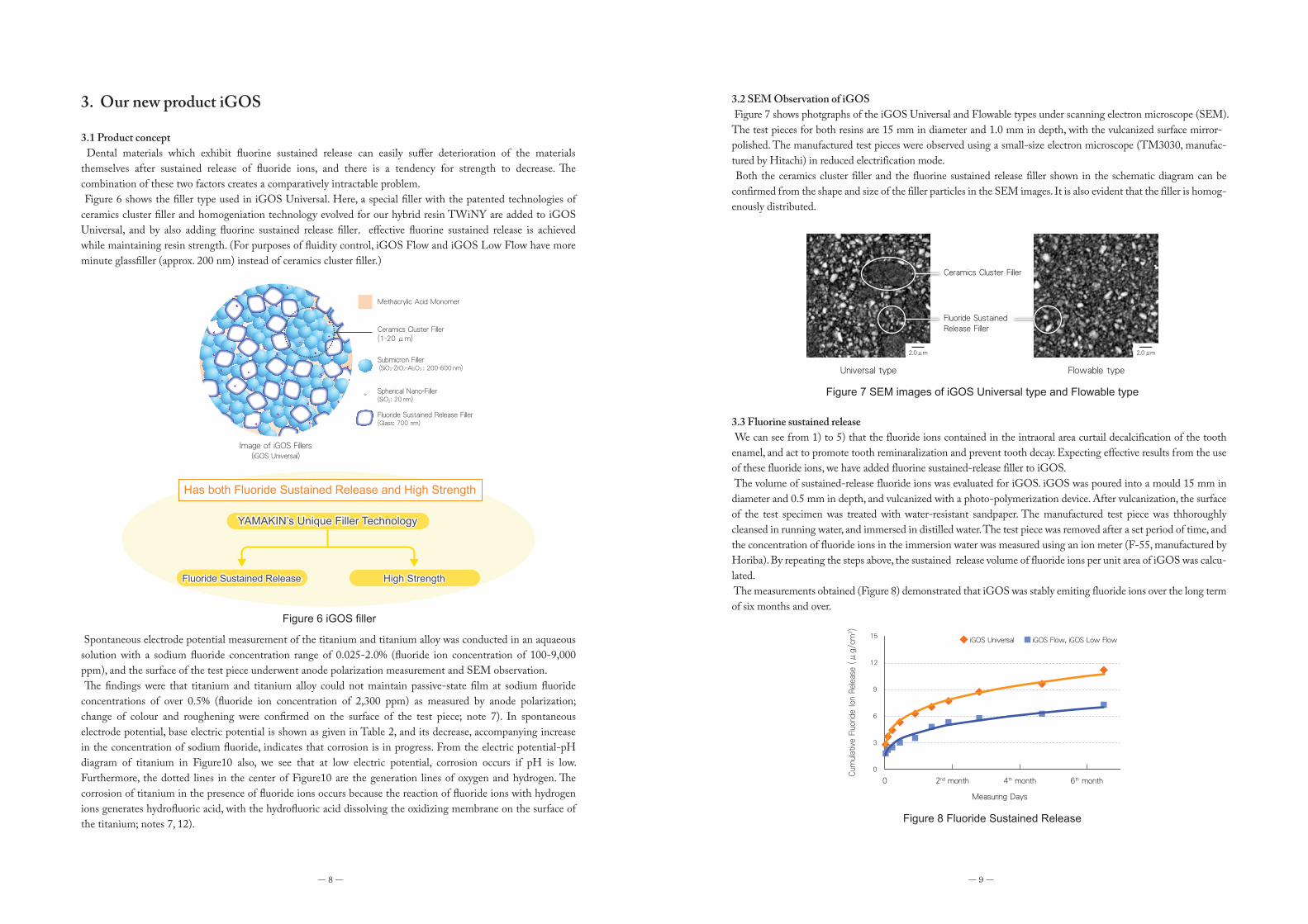
3.14 Clinical cases �e clinical cases below cover only a part of the processes involved in composite resin restoration; check the ttached literature for details of the usage and working methods actually used.
■ Restoration of a Class I cavity (Photo provided by: Dr Kenya Maeda, Yamakita Dental Clinic)
■ Restoration of a Class II cavity (Photo provided by: Dr Masahiro Uka, Uka Department of Cardiovascular Medicine and Dentistry
■ Restoration of a Class II cavity (2)
■ Restoration of a Class IV cavity
① cavity formation ② application of iGOS-Bond ③ filling in with iGOS Flow ④ after tretament
① before treatment ② cavity formation ③ application of iGOS-Bond ④ filling in with iGOS Flow
⑤ exposure to light ⑥ after photo-vulcanization ⑦ after treatment
① before treatment ② before restoration ③ cavity formation ④ etching
⑤ application of iGOS-Bond ⑥ exposure to light ⑦ filling in with iGOS Flow ⑧ after photo-vulcanization
⑨ filling in with iGOS Universal ⑩ after photo-vulcanization ⑪ after treatment
① before treatment ② cavity formation ③ application of iGOS-Bond ④ exposure to light
⑤ filling in with iGOS Flow ⑥ after photo-vulcanization ⑦ after treatment
Pulp is protected using lining material between Step① and ②.
■ Restoration of a Class III cavity (Photo provided by: Dr Kenya Maeda, Yamakita Dental Clinic)
■ Restoration of Class V cavity (Photo provided by: Dr Kenya Maeda, Yamakita Dental Clinic)
3.15 Biological safety �e first priority for dental materials is that they do not exert harmful impacts on patients – in other words, that they are biologically safe. In order to evaluate the biological safety of iGOS and iGOS-Bond, human monocytic THP.1 cells, derived from an acutemonocytic leukemia patient (obtained from the Department of Oral and Maxillofacial Surgery, Kochi Medical School, Kochi University, Japan), were used to test for cytotoxicity.
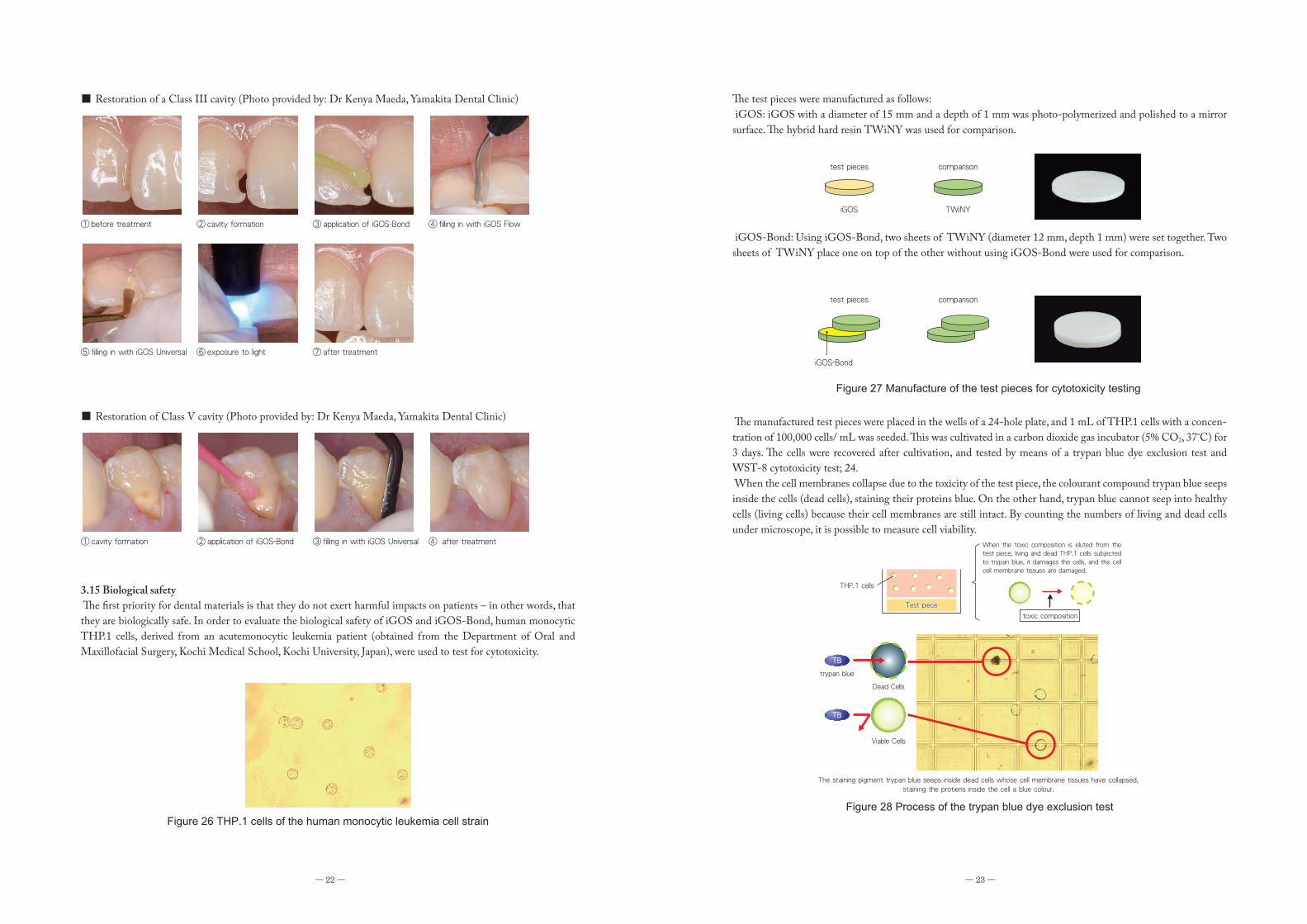
�e test pieces were manufactured as follows: iGOS: iGOS with a diameter of 15 mm and a depth of 1 mm was photo-polymerized and polished to a mirror surface. �e hybrid hard resin TWiNY was used for comparison.
iGOS-Bond: Using iGOS-Bond, two sheets of TWiNY (diameter 12 mm, depth 1 mm) were set together. Two sheets of TWiNY place one on top of the other without using iGOS-Bond were used for comparison.
�e manufactured test pieces were placed in the wells of a 24-hole plate, and 1 mL of THP.1 cells with a concen-tration of 100,000 cells/ mL was seeded. �is was cultivated in a carbon dioxide gas incubator (5% CO2, 37˚C) for 3 days. �e cells were recovered after cultivation, and tested by means of a trypan blue dye exclusion test and WST-8 cytotoxicity test; 24. When the cell membranes collapse due to the toxicity of the test piece, the colourant compound trypan blue seeps inside the cells (dead cells), staining their proteins blue. On the other hand, trypan blue cannot seep into healthy cells (living cells) because their cell membranes are still intact. By counting the numbers of living and dead cells under microscope, it is possible to measure cell viability.
Figure 26 THP.1 cells of the human monocytic leukemia cell strain
Figure 27 Manufacture of the test pieces for cytotoxicity testing
test pieces
iGOS
comparison
TWiNY
test pieces
iGOS-Bond
comparison
Figure 28 Process of the trypan blue dye exclusion test
The staining pigment trypan blue seeps inside dead cells whose cell membrane tissues have collapsed, staining the protiens inside the cell a blue colour.
TB
trypan blue
Dead Cells
TB
Viable Cells
THP.1 cells
Test piece
toxic composition
When the toxic composition is eluted from the test piece, living and dead THP.1 cells subjected to trypan blue, it damages the cells, and the cell cell membrane tissues are damaged.
― 22 ― ― 23 ―
① before treatment ② cavity formation ③ application of iGOS-Bond ④ filling in with iGOS Flow
⑤ filling in with iGOS Universal ⑥ exposure to light ⑦ after treatment
① cavity formation ② application of iGOS-Bond ③ filling in with iGOS Universal ④ after treatment

― 24 ― ― 25 ―
After recovery, cultivated cells had trypan blue mixed in on the test piece, and living and dead cells were counted individually using a haemocytometer. Cell viability was determined by calculating the proportion of living cells within the total cell count (totalling both dead and living cells).
We have already reported (note 25, 26) that TWiNY, which was used as a comparison, displayed a high level of safety in this experiment. Confirmation of the THP.1 cells grown on all of the test pieces for iGOS (Universal, Flow and Low Flow types) and iGOS-Bond + TWiNY demonstrated a cell viability of equivalent height to the compara-tive material (TWiNY).
WST-8 cytotoxicity test; note 27, 28) �is experiment exploited the fact that the indicator WST-8 is reduced to orange-coloured WST-8 formazan by the dehydrogenase (NAD+, NAD (P) + dehydrogenase) in living cells. By measuring the depth of the orange colour as the optical density, the impact of the test pieces on the metabolic activity of the cells could be analysed. Where the orange colour was dense (absorbance was high) cytotoxicity was low, and where it was paler (the optical density was low) cytotoxicity was judged to be high.
�e cells cultivated on the test piece were transferred to the wells of a 96-hole culture plate, and WST-8 reagent was added. After they had been left standing for 2 hours at 37˚C, the optical density (450 nm) of the generated formazan (orange) . was measured
All of the test pieces for iGOS (Universal, Flow and Low Flow types) and iGOS-Bond + TWiNY demonstrated an optical density equivalent to the comparative material, and no impact of the test pieces on the metabolic activity of the cells could be confirmed.
4. Afterword
Conventional composite resins face the technical problem that [material] strength declines when fluorine sustained release is increased, meaning that the sustained release volume of fluirine must be curbed in order to increase strength. By evolving the technology building on the development experience of hard resins for dental crowns in the past, it has been possible to overcome this problem with iGOS. Both fluorine sustained release and [material] strength are secured. In the Univesal type, this is achieved through a combimation of Yamamoto Precious Metal’s in-house ceramics cluster filler technology with fluorine sustained release filler; in the Flowable type, it is achieved through a combination of fluorine sustained release filler and glass filler (200 nm). It is thought that the combination of fluorine sustained release filler does not only allow for the sustained release of fluoride ions, but that it also makes iGOS rechargable. Furthermore, while cytotoxicity testing of iGOS produced findings broadly comparable to other hard resins etc., this material displays a number of deeply interesting qualities, such as suppression of S. mutans adhesion.
S. mutans adhesion suppression testing and cytotoxicity testing are carried out as part of a joint research product with the Department of Oral and Maxillofacial Surgery, Kochi Medical School, Kochi University, Japan.
Figure 29 Survival rates for THP.1 cells
Figure 30 Process of WST-8 cytotoxicity testing
WST-8 is changed to orange WST-8 formazan by the metabolic activity (dehydrogenase) of the cells
NN
NN
H
MeO
NO2SO3-
SO3-
NO2
N
N+
N
N
MeO
NO2SO3-
SO3-
NO2
WST-8 WST-8 formazan
Dehydrogenase
NN
NN
H
MeO
NO2SO3-
SO3-
NO2
NN
NN
H
MeO
NO2SO3-
SO3-
NO2
High toxicity: cell metabolic activity declines, along with the quantities of WST-8 formazan generated.
Low toxicity: cell metabolic activity is maintained, and large quantities of WST-8 formazan are generated.
Orange: pale
Orange: deep
Figure 31 Metabolic activity of THP.1 cells
0.000
0.500
1.000
1.500
2.000
2.500
450 n
m O
ptical d
ensity
TWiNY iGOSUniversal
iGOSFlow
iGOSLow Flow
0.000
0.500
1.000
1.500
2.000
2.500
450 n
m O
ptical d
ensity
TWiNY iGOS Bond+
TWiNY
0
20
40
60
80
100
120
Cell v
iability
(%)
TWiNY iGOS Bond+
TWiNY
0
20
40
60
80
100
120
Cell v
iability
(%)
TWiNY iGOSUniversal
iGOSFlow
iGOSLow Flow

References1) Hicks J, Garcia G, Milano M, Flaitz C: Compomer materials and secondary caries formation.
Am. J. Dent., 13(5), 231-234, 2000.
2) Han L, Edward C, Okamoto A, Iwaku M: A comparative study of fluoride-releasing adhesive resin materials. Dent. Mater. J., 21(1), 9-19, 2002.
3) Itota Toshiyuki,Iwai Yoichiro,Okamoto Mimiko,Tashiro Yoko,Nakabo Satoshi,Nishimura Yoshihiro,Nagamine Michihiro,Torii Yasuhiro,Yoshiyama Masahiro: Remineralization of decalcified dentine by means of fluorine sustained release adhesion system. Jpn J Conserve Dent, 44, 175-181, 2001. [in Japanese]
4) Okuyama K, T. Nakata, P. N. R. Pereira, C. Kawamoto, H. Komatsu, and H. Sano: Prevention of artificial caries: effect of bonding agent, resin composite and topical fluoride Application Oper, Dent., 31(1), 135-142, 2006.
5) Kijimura Daiki, Komatsu Hisanori, Matsuda Yasuhiro,Okuyama Katsufumi,Sano Hidehiko: Evaluation through pH cycle of the inhibitory effect on tooth decay of fluorine sustained release resin. Jpn J Conserve Dent, 52, 39-50, 2009. [In Japanese]
6) ISO/TS 14569-2: 2001, Dental materials-Guidance on testing of wear resistance-Part 1: Wear by tooth brushing.
7) Oda Yutaka, Kawada Eiji, Yoshinari Masao, Hasegawa Koji,Okabe Toru: The influence of the concentration of fluorine ions on the corrosion of titanium and titanium alloy. 15(4): 317-322, 1996. [In Japanese]
8) Oda Yutaka: Does the bio-material“titanium”undergo corrosion and colour change? J. J. Dent. soc., 55(12): 1167-1176, 2003.[In Japanese]
9) Nakagawa Masaharu: Issues regarding titanium in the environment of the oral cavity. J. J Dent. soc., 58(6): 531-541, 2005. [In Japanese]
10) Nakagawa M., Matsuya S., Udoh K.: Effects of fluoride and dissolved oxygen concentrations on the corrosion behavior of pure titanium and titanium alloys. Dent. Mater. J., 21(2): 83-92, 2002.
11) Tsuruta Shozo, Ozeki Junko, Koyama Kenichi, Hasegawa Jiro: The influence of the concentration of fluorine in prototype dentifrices on the abrasion of titanium. Aichi Gakuin J. Dent. Sci., 39(2): 175-180, 2001. [In Japanese]
12) Irie Naomichi, Aoki Harumi, Yoshida Ryuichi: Change of colour and quantity in titanium and titanium alloy: comparison of immersion testing and brushing testing of dentifrices containing fluorine. J. Jpn. Soc. Dent. Prod., 21(1): 14-25, 2007. [In Japanese]
13) Yamanaka Kanae: The concentration of fluoride ions in commercially available tea drinks products marketed in PET bottles. ir.tdc.ac.jp/irucaa/bitstream/10130/1721/1/56_43.pdf (Accessed on October 20, 2014)
14) Ministry of Health, Labour and Welfare: Water Quality Standard Items and Reference Values (51 Items).http://www.mhlw.go.jp/stf/seisakunitsuite/bunya/topics/bukyoku/kenkou/suido/kijun/kijunchi.html. (Accessed on October 20, 2014) [in Japanese]
15) Anraku Teruo, Yamazoe Masatoshi, Matsuura Ritaro: Safety testing report no. 19 on dental precious metal alloys Vol.9. Yamamoto Precious Metal, Co., Ltd., 15, 2011. [In Japanese]
16) Hamada Shigeyuki, Oshima Takashi: New Science of Caries, First ed., 23, 2006, Ishiyaku Shuppan, Tokyo. [In Japanese]
17) Shibata Akiko, Minami Jiro, Nakamura Shigeru, Terano Motohiro, Suenaga Hidenori, Fujii Hiroyuki: Analysis of the concentration of organic acids in sputum of patients suffering from allergies to metals. J. Jpn. Prosthodont. Soc., 46: 17120 02. [In Japanese]
― 26 ― ― 27 ―
The original performance could not be exhibited depending on the cases.
Related Products
Lineup
Resin-based Dental Restorative Material
Has both Fluoride Sustained Release and High Strength.
iGOS Universal 4g (2 ml)Single Package Single Package Single Package
Single Package
iGOS Flow 2.6g (1.5 ml)Accessory: Needle Tip 10 pcs.
iGOS Low Flow 2.6g (1.5 ml)Accessory: Needle Tip 10 pcs.
iGOSUniversal
iGOSFlow
iGOS-BOND (5ml): 1 bottle·Disposable Applicator Brush: 50 pcs.·Disposable Plate: 25 pcs.
Set Package
Set Package iGOS-BOND (5ml): 2 bottlesDisposable Applicator Brush: 50 pcs.Disposable Plate: 50 pcs.
Accessory
Needle Tip: 20 pcs.Accessory
iGOS-BOND (5ml)
Multi Primer LIQUID (7ml)For Metal and Ceramics
Dental adhesive which achieves high adhesion inside the mouth under wet condition.
iGOS-Bond and Multi Primer Liquid are flammable.
Bonding Composite Resin Material to metal, ceramic and cured resin for direct repairing.
Dental Adhesive for Enamel and Dentine
Bonding Material for Dental Metal Bonding Material for Dental Ceramics Bonding Material for Dental Resin
iGOSLow Flow
(Low Flowable Type)(High Flowable Type)
·Starter Pack (Universal) .......... A2, A3, OA2, OA3, E, iGOS-BOND·Starter Pack (Flow) ................ A2, A3, OA2, OA3, E, iGOS-BOND·Starter Pack (Low Flow) ......... A2, A3, OA2, OA3, E, iGOS-BOND·iGOS Universal Dentine 3pcs Pack (3pcs. of the Same Shade) ................ A2, A3, A3.5·iGOS Flow Dentine 3pcs Pack (3pcs. of the Same Shade) ................... A2, A3, A3.5·iGOS Low Flow Dentine 3pcs Pack (3pcs. of the Same Shade)............. A2, A3, A3.5·Repair Pack ............................ iGOS-BOND, Multi Primer Liquid
Set Package
Shade LineupProduct Name
•Bleaching White•Enamel
•Bleaching White•Enamel
•Bleaching White•Enamel
Type Others
4g (2 ml)
2.6g (1.5 ml)
2.6g (1.5 ml)
16
13
13
Number ofShade
Contentg (ml)
iGOS Universal
iGOS Flow
iGOS Low Flow
DentineOpaque
DentineOpaque
DentineOpaque
A2
●●●●●●
A3
●●●●●●
A1
●
●
●
A3.5
●
●
●
A4
●
●
●
A5
●
●● ● ●
●● ● ●
B1
●
B2
●
B3
●
C2
●
C3
●
D2
●
Dental Adhesive
Applicable for
Precious Metals
○ ○ ○ ○ ○ ○ ○ ○ ×
Au Alloy Au-Ag-PdAlloy
TiTi Alloy
Ni-CrAlloy
Co-CrAlloy
PorcelainResin Resin
Zirconia(ZrO2)
Non-precious Metals Ceramics Composite Resin
Containsinorganic
fillers
Withoutinorganic
fillers)( )(
The actual color of the product, model and package may differ from the photographs due to printing ink and shooting conditions.

Editor Teruo AnrakuPublisher Shigenari Yamamoto Date of publication Jul. 1 2017
18) The Japan Institute of Metals and Materials: Compendium of Metals, sixth ed., 819-820, 2000, Maruzen, Tokyo. [in Japanese]
19) JIS T 6514: 2013, Composite resins for use in dental restoration and prosthodontic tooth construction. [in Japanese]
20) JIS T 6517: 2011, Hard resins for dental crowns. [in Japanese]
21) Japan Society of Education of Dental Technology (ed.): New Textbook for the Dental Technician: Dental Science and Engineering, Ishiyaku Shuppan, 16-17, 2006. [in Japanese]
22) Hara Mai, Koyama Hiroshi, Satoh Toru, Takuma Yuusuke, Yoshinari Masao, The abrasion properties of transluscent zirconia and bovine tooth enamel. Shika Gakuho, 112 (4), 538, 2012. [in Japanese]
23) Tsukatani T, Suenaga H, Higuchi T, Akao T, Ishiyama M, Ezoe K, Matsumoto K, Colourimetric cell proliferation assay for microorganisms in microtiter plate using water-soluble tetrazolium salts. J Microbiol. Methods., 75(1), 109-116, 2008.
24) Correa GT, Veranio GA, Silva LE, Hirata Junior R, Coil JM, Scelza MF: Cytotoxicity evaluation of two root canal sealers and a commercial calcium hydroxide paste on THP1 cell line by Trypan Blue assay. J. Appl. Oral Sci., 17(5), 457-461, 2009.
25) Matsuura Ritaro, Mikagi Eriko, Horiguchi Koji, Anraku Teruo, Yamamoto Tetsuya: Involvement of residual monomers in cytotoxicity of hard resins for dental crowns. J. Jpn. Soc. Dent. Mat. Dev. 29(5), 464, 2010. [In Japanese]
26) Matsuura Ritaro, Mikagi Eriko, Anraku Teruo, Yamamoto Tetsuya: Biological investigation of cytotoxicity of addition agents in hard resins for dental crowns. 28(1), 1-7, 2009. [In Japanese]
27) Ishiyama M, Miyazono Y, Sasamoto K, Ohkura Y, Ueno K: A highly water-soluble disulfonated tetrazolium salt as a chromogenic indicator for NADH as well as cell viability. Talanta., 44(7), 1299-1305, 1997.
28) Tominaga H, Ishiyama M, Ohseto F, Sasamoto K, Hamamoto T, Suzuki K, Watanabe M: A water-soluble tetrazolium salt useful for colorimetric cell viability assay. Anal. Commu., 36(2), 47-50, 1999.
《Previously Published Product Reports》Basic information and product report on zirconia (Feb. 2014)Basic information and product report on titanium ( Jun. 2016)Basic information and product report on hybrid resin for CAD/CAM use (Sep. 2014)Basic Information and Product Report on Dental Bonding Material ( Jan. 2016)
《Previously Published Technical Reports》Technical report on ZEO Ce Light (Aug. 2002)Technical report on Luna-Wing (May 2007)Technical report on TWiNY ( July 2010)
《Previously Published Safety Test Reports》Vol. 1 Pursuing international standards in quality and safety (Dec. 2004)Vol. 2 ZEO METAL series elution test and in vitro cytotoxicity test ( Jun. 2005)Vol. 3 Elution test and in vitro cytotoxicity test of precious-metal alloys and gold alloys for metal ceramic restoration use (Dec. 2005)Vol. 4 Biological evaluation of Luna-Wing ( Jun. 2006)Vol. 5 Report on physical properties and safety of high-carat gold alloys (Oct. 2007)Vol. 6 Examination of the biological impact of the physical properties of dental-material alloys and gold alloys for hard resin and metal ceramic restoration use (May. 2008)Vol. 7 Report on the physical properties and safety of the gold alloy Nexo-Cast (Oct. 2008)Vol. 8 Biological evaluation of the hybrid composite resin TWiNY ( Jun. 2010)Vol. 9 Chemical and biological characteristics of precious-metal alloys: elution characteristics produced through mixture with titanium (Feb. 2011)Vol. 10 Physical properties and safety of the precious-metal alloy for metal ceramic restoration use 「Brightis」 (Oct. 2011)Vol. 11 Physical properties and safety of the dental adhesive Multi Primer (Mar. 2014)
Vol. 1 The polymerization of dental materials: the basis of radical polymerization (1) (Oct. 2009)Vol. 2 The polymerization of dental materials: the basis of radical polymerization (2) (Feb. 2010)Vol. 3 The polymerization of dental materials: restoration-material monomers (1) (Mar. 2010)Vol. 4 The polymerization of dental materials: restoration-material monomers (2) ( Jul. 2010)Vol. 5 The polymerization of dental materials: the influence of oxygen (Aug. 2011)Vol. 6 The polymerization of dental materials: primers and developers (Oct. 2012)Vol. 7 Polymerization silane coupling agent: methacrylic resin (acrylic resin) ( Jun. 2013)Vol. 8 Shrinkage of Dental Composite in Polymerization (Nov. 2014)Vol. 9 Application of Iodonium Salt as Initiator Component in Dental Material (Mar. 2017)
《Previously Published Macromolecule Technology Reports》
Vol. 1 Dental surgery and bisphosphonate formulation (Aug. 2010)Vol. 2 Reactive oxygen: its generation, elimination and effects (Nov. 2011)Vol. 3 �e hypoxic world ( Jul. 2012)Vol. 4 Recent progress in the regeneration of tooth material (Feb. 2014)Vol. 5 Application of Fluoride and its Effect (Oct. 2016)
《All Previously Published Science Reports》