basic fibroblast growth factor induces the expression of ... · among this family,...
TRANSCRIPT
Basic Fibroblast Growth Factor Induces The Expression of MatrixMetalloproteinase-3 in Human Periodontal Ligament Cells Through theMEK2 Mitogen-Activated Protein Kinase Pathway
Department of Preventive Dentistry, Faculty of Dentistry,Hiroshima University, Hiroshima, Japan
Atsushi Shimazu, Masayuki Morishita
Basic fibroblast growth factor (bFGF, FGF-2) is one of the potentmitogens for periodontal ligament (PDL) cells. However, the role of bFGFon the matrix metalloproteinase (MMP) -3 expression in PDL cells isunknown. In this study, the effect of bFGF on MMP-3 expression in PDLcells and the mechanism of this process were examined. Human PDL cellswere exposed to bFGF at various concentrations (0.01 - 10 ng/ml) inmonolayer cultures. bFGF increased [3H]-thymidine incorporation andsuppressed proteoglycan synthesis concentration-dependently. However,similar concentration ranges of bFGF increased the release of thecell-associated proteoglycans into the medium. Furthermore, bFGFincreased MMP-3 mRNAlevels concentration-dependently as examinedby reverse transcription-polymerase chain reaction. Induction of MMP-3after the stimulation with bFGF was observed as early as 12 h withmaximal at 24 h. Thereafter, the MMP-3 mRNAlevel gradually decreaseduntil 72 h. Cycloheximide blocked the induction of MMP-3 by bFGF,indicating the requirement of de novo protein synthesis for thisstimulation. Furthermore, MMP-3 expression induced by bFGF wasabrogated by U0126, a specific inhibitor of MEK1/2 and ERK1/2 inmitogen-activated protein (MAP) kinase pathway, not by PD98059, aspecific inhibitor of MEK1. In addition, bFGF up-regulated thephosphorylated ERK1/2 in 5 min with the maximal at 20 min as examinedby Western blotting, and U0126 inhibited the ERK1/2 phosphorylationinduced by bFGF. These findings suggest that bFGF induces MMP-3expression in PDL cells through the activation of the MEK2 in MAPkinase pathway. bFGF stimulation on MMP-3 synthesis may be involvedin the control of the cell-associated proteoglycans in PDL cells duringperiodontal regeneration and degradation.
Keywords; periodontal ligament (PDL), basic fibroblast growth factor (bFGF,FGF-2), matrix metalloproteinase (MMP) -3, mitogen-activated protein (MAP)kinase.
38

IntroductionDuring the periodontal regeneration process, periodontal ligament (PDL)
cells play an important role in forming new attachments between these tissues aswell as cementogenesis and osteogenesis. PDL cells initially extend and migrateon the root surface, and then the cells proliferate and secret various extracellularmatrix (ECM) components (1, 2). In this process, various hormones and growthfactors modulate the proliferation and differentiation of PDL cells. Among them,basic fibroblast growth factor (bFGF, FGF-2) is a member of theheparin-binding growth factor family (3) and the most potent mitogen for PDLcells (4-6). bFGF increases the cell number and inhibits alkaline phosphataseactivity in these cells. When bFGF is applied to alveolar bone defects, bFGFinduces new PDL and new bone formation without ankylosis or root resorption(7). In addition, PDL cells express both bFGF and FGF receptor mRNAs andproteins. bFGF increases the number of PDL cells without affecting theirpotency for calcification (7). bFGF may function as both autocrme and paracrinefactors in the early wound-healing process of PDL. bFGF may be one of the keyfactors in periodontal regeneration.
Recently, we reported that PDL cells express FGF receptor mRNAas wellas syndecans, a family of heparan sulfate proteoglycans (HSPGs) core proteins,and that the expression levels of these molecules decrease during thedifferentiation process of PDL cells (6). Differentiated PDL cells show a lowerresponse to bFGF. PDL cells control their growth and differentiation bychanging the levels of FGF receptors and syndecans. Furthermore, removingHSPGs from PDL cells results in a decrease in the stimulatory effect of bFGF(6). HSPGs form complexes with bFGF on the FGF receptors to activate cells(8). HSPGs function as a low affinity receptor for bFGF and store bFGF in theECM for protection against proteolytic degradation (9, 10).
Matrix metalloproteinases (MMPs) are a family of proteolytic zincenzymes responsible for the degradation of various ECM components (1 1, 12).Among this family, MMP-3 (stromelysin-1) targets and digests proteoglycan,types IV and IX collagen, laminin, fibronectin, and gelatin (ll, 12). MMP-3plays a role in connective tissue breakdown associated with severalinflammatory diseases (12). Interleukin-16 (IL-1J3), which is an inflammatorycytokine produced by monocytes and macrophages, induces the release ofMMP-3in PDL cells in vitro (13, 14). IL-16 and MMP-3 are found in inflamedperiodontal tissues and the gingival crevicular fluid of periodontitis ( 13).
Previous studies suggested that bFGF and HSPGs may play a role in thecontrol of growth and differentiation of PDL cells. However, it is unknownwhether and how bFGF regulates HSPGs turnover in periodontal regeneration.Therefore, this study was designed to investigate the effect of bFGF on theMMP-3expression and the mechanism of this process in human PDL cells.
39
Material and methodsPDL cell cultures and treatment with bFGFHumanPDL cells were isolated from healthy periodontal ligaments of the firstpremolars of individuals undergoing tooth extraction for orthodontic treatment.Informed consent was obtained from all patients before surgical procedures.Healthy periodontal tissue was removed from the middle third of the rootsurface, and then transferred to 100 mm plastic culture dishes. The explantswere cultured in Dulbecco's modified Eagle's medium (Sigma, St. Louis, MO)supplemented with 10% fetal calf serum (FCS; GIBCO, Grand Island, NY), 100units/ml of penicillin and 100 pig/ml of streptomycin in a humidified atmosphereof 95% air and 5% CO2 at 37°C. When the cells growing from the explantsbecame confluent, they were harvested with 0.125% trypsin in phosphatebuffered saline and transferred to plastic culture dishes at a 1:3 split ratio. Forexperiments, the cells were trypsinized and cultured in 16 mm wells of a 24-wellplate or 100 mmculture dishes at a density of 5 x 10^ cells/ml in alpha modifiedminimum essential medium (D-MEM; Sigma) supplemented with 10% FCS andantibiotics until confluence. After confluence, the cells were washed twice withserum-free D-MEMand further cultured with serum free D-MEMfor another 24h. Then the cells were exposed to various concentrations (0.01 - 10 ng/ml) ofbFGF (PeproTech EC, London, UK). Experiments were carried out with cellsfrom the fourth to eighth passaged cultures and all cell lines provided similarresults in each experiment.
Effect of bFGF on DNA synthesisPDL cells were labeled with [3H]-thymidine (Japan Atomic Energy Institute,Tokyo, Japan; final concentration, 10 //Ci/ml) for the last 4 h of the 24 htreatment as described previously (6). At the end of labeling, the cell layers werewashed three times with PBS, twice with 10% trichloroacetic acid and twicewith ethanol/diethyl Wher (3: 1, vol/vol) on ice. The residues in the wells weresolubilized with 0.1 N NaOH, the solution was neutralized with 6 N HC1, andradioactivity was measured in a liquid scintillation spectrometer (Aloka, Tokyo,Japan).
Effect of bFGF on proteoglycan synthesisPDL cells were labeled with [35S]-sulfate (Japan Atomic Energy Institute; finalconcentration, 20 ^Ci/ml) for 24 h as described previously (15). Proteoglycansynthesis was determined by measurement of [35s]-sulfate incorporation intomaterial precipitated with cetylpridinium chloride after pronase E treatment.
Effect of bFGF on the release of proteoglycans from the cell matrix layerinto the mediumPDL cells were labeled with [35S]-sulfate (Japan Atomic Energy Institute; final
40

concentration, 20 //Ci/ml) for 48 h. The cell matrix layers were washed 5 timeswith medium as described previously (15), and the cells were then incubated foranother 48 h in the presence or absence of various concentrations of bFGF. Atthe end of treatment, the medium containing the released proteoglycans wascollected and the amount of the released proteoglycans was determined bymeasurement of [35s]-surfate incorporation into material precipitated withcetylpridinium chloride.
RNA isolation and MMP-3 detection by reverse transcription-polymerasechain reaction (RT-PCR)PDL cells treated with bFGF were incubated for various lengths of time prior toisolating total RNAs. Total RNAs were isolated from these PDL cells by theguanidinium isothiocyanate method (16) and treated with DNAase I (Promega,Madison, WI) to avoid contamination of genomic DNA. The primers used forRT-PCR were designed based on the published sequence data for correspondinghuman MMP-3 (sense primer; 5'-TAA AGA CAG GCA CTT TTG GCG C,antisense primer; 5'-CTG CAT CGA TTT TCC TCA CGG T, expected size;245 bp) (12), glyceraldehyde-3-phosphate dehydrogenase (GAPDH) (senseprimer; 5'-CCA TGG AGA AGG CTG GGG, antisense primer; 5'-CAA AGTTGT CAT GGA TGA CC, expected size; 210 bp) (17). Aliquots of 1 pig of totalRNA were reverse transcribed using avian myeloblastosis virus reversetranscriptase (Life Sciences, St. Petersburg, FL) with these 3' -specific primers in25 mMTris-HCl (pH 8.3), 50 mM KC1, 2 mM DTT, 5 mMMgCl2 and 1 mM ofeach dGTP, dTTP, dCTP and dATP at 64°C for 30 min, 99°C for 5 min and 4°Cfor 5 min. Amplification was performed for 1 cycle at 95°C for 12 min, 28cycles at 94°C for 30 sec and 64°C for 1 min, 1 cycle at 64°C for 10 min and4°C for 5 min using Ampri7ag Gold polymerase (Perkin-Elmer, Foster City,CA) with the 3' and 5'- specific primers in 15 mM Tris-HCl (pH 8.3), 50 mMKC1, 0.4 mMDTT, 2.5 mMMgCl2, and 200 piM of each dATP, dCTP, dGTPand dTTP. The PCR products were separated on 2% agarose gels containingethidium bromide, and then observed on an ultraviolet transilluminator. PCRproducts in the gels were scanned and the images were quantified using imageprocessing and analysis software (NIH Image version 1.62, National Institutesof Health, Bethesda, MD).
Sequencing of PCR productsAll PCR products were separated in 2% low melting-point agarose gels, purifiedwith a DNA fragment extraction kit (QIAGEN, Hilden, Germany) and clonedinto the PGEM-5Zf(+) vector (Promega). Sequencing was carried out by thesingle primer extension method with an Applied Biosystems 377 Sequencer(Perkin-Elmer). The identities of the cloned DNAs were confirmed bycomparison with sequences in the GenBank/EMBL/DDBJ database.
41
Immunoblotting analysis for phosphorylation of ERK1/2PDL cells were treated with 10 ng/ml of bFGF for the times indicated. The cellswere washed with ice-cold 150 mM NaCl and lysed with 150 mM NaCl, 1 mMphenylmethylsulfonyl fluoride, 100 mM Na3VO4, 20 /fg/ml aprotinin, 25 pig/mlleupeptin, and 1 % Triton-X 100. The proteins were concentrated by centrifugalfiltration (10 kDa cut-off, Millipore, Bedford, MA), and the proteinconcentration was measured using a BCA protein assay kit (Pierce, Rockford,IL). Then 40 ]A.g samples were subjected to 10% SDS-PAGE using theprocedure of Laemmli (18) under nonreducing conditions, and proteinsseparated in the gel were subsequently electrotransferred onto PVDFmembranes (Immobilon, Millipore). The membranes were incubated withpolyclonal rabbit antibody against phosphorylated ERK1/2 (pTEpY, 1 : 4,000,Promega) in 1% casein and TBS (0.15 M NaCl, 20 mMTris-HCl, pH 8.0) for 1h at room temperature. After washing with TBS, the membranes were incubatedwith biotinylated goat anti-rabbit IgG (1 : 2,000, Vector Laboratories,Burlingame, CA) for 30 min, followed by washing with TBS, and then coupledto a peroxidase biotin-avidin complex (Vector). The membranes were incubatedin the color reagent (50 mM Tris-HCl, pH 8.0, 0.1% 3,3-diaminobenzidinetetrahydrochloride, and H^Oj), until brown reaction products were obtained.Reactions were terminated by flushing with water and drying.
Statistical analysisValues were assessed by analysis of variance (ANOVA) to evaluate thedifferences and were considered significant at * p<0.05 and ** p<0.01. Valueswere expressed as means ± SEM (standard error of the mean).
42

ResultsHuman PDL cells were exposed to various concentrations (0.01 - 10
ng/ml) of bFGF for 24 h. bFGF increased [^HJ-thymidine incorporation intoDNA in a concentration-dependent manner (Fig. 1A). This stimulatory effectwas detected at 0.01 ng/ml (p<0.05) and maximal at 10 ng/ml (p<0.01). On theother hand, bFGF decreased proteoglycan synthesis in PDL cells in aconcentration-dependent manner (Fig. IB). The maximum inhibitory effect wasdetected at 1 ng/ml (p<0.01).
To determine whether bFGF influences the degradation of thecell-associated proteoglycans in PDL cells, PDL cells were labeled with[35s]-sulfate for 48 h. Then various concentrations of bFGF were added to the
medium, and the released proteoglycans into the medium were determined.bFGF increased the release of proteoglycans into the medium in aconcentration-dependent manner (Fig. 2). This effect was detected at a 0. 1 ng/ml(p<0.01) and maximal at 10 ng/ml (p<0.01).
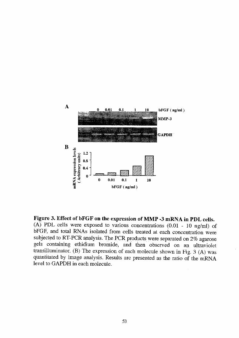
Next, we examined whether MMP-3 was induced by bFGF in PDL cells,because MMP-3 is known to degrade proteoglycans. The effect on theexpression of MMP-3 was assessed by RT-PCR. MMP-3 mRNAwas barelydetected in control PDL cells (Fig. 3A, B). However, the expression of MMP-3was observed as an expected 245 bp band in bFGF-exposed cells and themaximal expression was observed at 10 ng/ml (Fig. 3A, B). The DNA sequenceof the cloned PCR product was identical to the human MMP-3 sequence (datanot shown). In addition, the induction of MMP-3 after the stimulation withbFGF in PDL cells was observed as early as 12 h and became maximal at 24 h(Fig. 4A, B). Thereafter, the expression gradually decreased until 72 h.
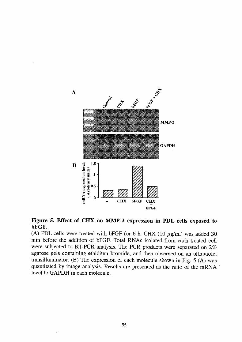
To elucidate whether the bFGF stimulation in MMP-3 expression requiresnewly synthesized protein, the induction of MMP-3 by bFGF was assessed inthe presence or absence of a protein synthesis inhibitor, cycloheximide (CHX,10 //g/ml). Induction of MMP-3 mRNAby bFGF was completely blocked byCHX (Fig. 5A, B). These results suggest that de novo protein synthesis isrequired for MMP-3 expression.
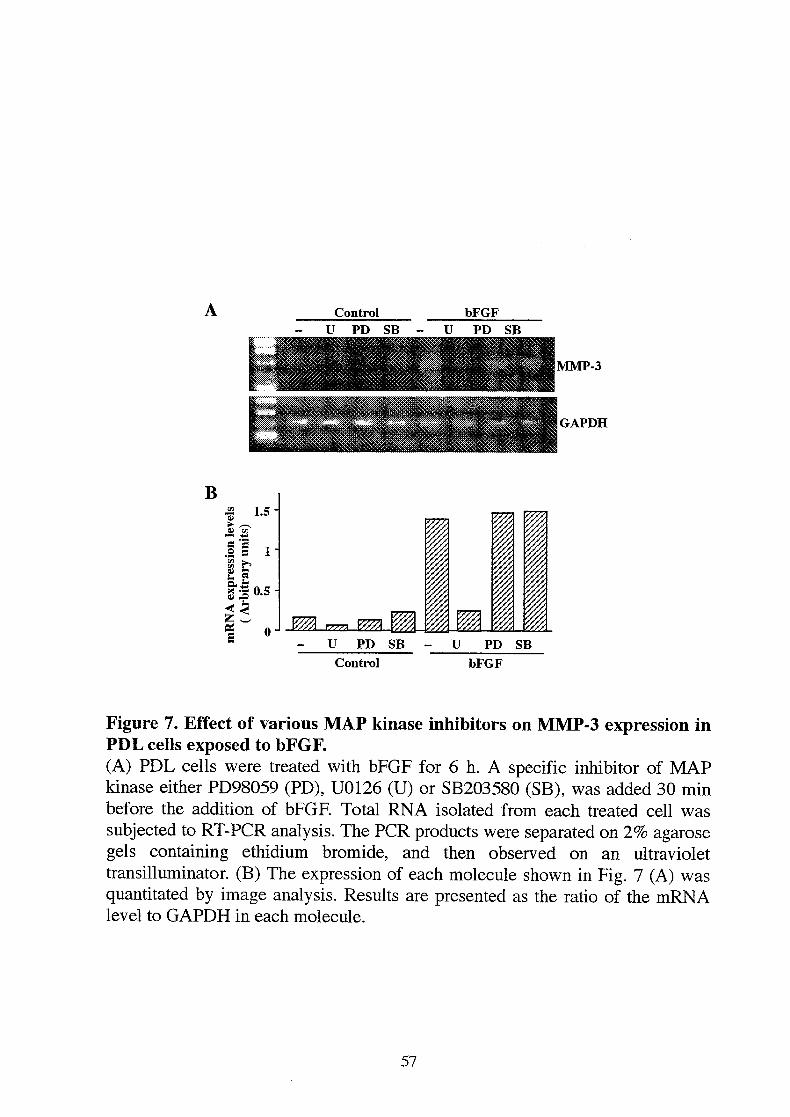
Recent studies showed that activation of mitogen-activated protein (MAP)kinase pathways is required for bFGF-dependent MMP-9 expression in mouseosteoclasts (19). To clarify whether and which MAP kinase pathway mediatesMMP-3expression in PDL cells exposed to bFGF, we used several MAP kinaseinhibitors (Fig. 6). PD98059 is a specific inhibitor of MEK1, and partiallyinhibits MEK2, which inhibits ERK1 activation. U0126 is a specific inhibitor ofMEK1/2 which inhibits ERK1/2 activation. SB203580 is a specific inhibitor ofp38 MAP kinase which acts as a competitive inhibitor. Expression of MMP-3 bybFGF was completely blocked by U0126, however PD98059 and SB203580showed no significant effect on the MMP-3 expression (Fig. 7A, B). Thesefindings suggest that activation of MEK2 in the ERK/MAP kinase pathway is
43

required for bFGF-dependent MMP-3 expression.To confirm whether bFGF activates ERK1/2 in PDL cells and whether
U0126 inhibits the phosphorylated ERK1/2 induced by bFGF, thephosphorylation of ERK1/2 was monitored by Western blotting. bFGFup-regulated the phosphorylated ERK1/2 in 5 min with the maximal at 20 min,and the phosphorylated ERK1/2 were observed as 44 and 42 kDa bands (Fig.8A). Furthermore, U0126 completely blocked the phosphorylation of ERK1/2induced by bFGF (Fig. 8B). However PD98059 revealed a little inhibition in thephosphorylated ERK1/2. These findings suggest that bFGF activates the MEK2in human PDL cells.
44
DiscussionVarious hormones and growth factors are involved in the control of ECM
synthesis and ECM degradation in PDL (2). bFGF has been shown to modulateECM synthesis including type I collagen, laminin and tropoelastin (5, 20, 21).However, the role of bFGF on the cell-associated proteoglycans in PDL isunknown. In the present study, we found that bFGF was involved in the controlof the cell-associated proteoglycans as well as proliferation of PDL cells.Incubation of PDL cells with bFGF increased the release of the cell-associatedproteoglycans concentration-dependently. The increase in the release ofproteoglycans by bFGF from the cell-matrix layer was not due to increasedsynthesis of proteoglycans. Interestingly, bFGF increased MMP-3 mRNAexpression in PDL cells within 12 h, bFGF at 10 ng/ml also increased theproduction of MMP-3 protein by PDL cells within 24 h when analyzed bycasein-zymography (data not shown). Furthermore, bFGF increased the releaseof proteoglycans and MMP-3 expression at similar concentrations in PDL cells.Thus, bFGF may stimulate the release of proteoglycans via the increase inMMP-3production. Moreover, this rapid response of MMP-3 expression afterbFGF treatment suggests that bFGF signaling may directly regulate MMP-3expression in PDL cells. However, this action by bFGF required newlysynthesized proteins. It remains unknown what newly synthesized protein(s) areinvolved in the bFGF action on MMP-3expression.
FGF receptors activate multiple intracellular signaling, including theMAP kinase pathway in several cells (19, 22, 23). Three groups of MAP kinasepathways have been identified; the ERK/MAP kinase pathway, the JNK/SAPKpathway, and the p38 pathway (24, 25). All three groups of the MAP kinasepathways are activated by several growth factors and inflammatory cytokines,and medicate various cellular events including growth, differentiation, and stressresponses. However, the activation of each type of MAP kinase depends on thetypes of the cells and the stimulus. Recently, it was demonstrated that mouseosteoclasts require activation of the p38 MAP kinase pathway forbFGF-dependent MMP-9 expression (19), whereas human primary fibroblastsrequire the activation of both the ERK/MAP and p38 MAP kinase pathways forbFGF-dependent MMP-1 expression (22). Furthermore, mouse osteoblastic cellsrequire the activation of ERK/MAP kinase pathway, but not the p38 MAPkinase pathway for bFGF-dependent release of vascular endothelial growthfactor (23). Several MAP kinase inhibitors are used for analyzing thesesignaling pathways, such as U0126, PD98059, and SB203580 (25-27). In thepresent study, U0126 abrogated bFGF-dependent MMP-3 expression inRT-PCR, and the phosphorylation of ERK1/2 by Western blotting in PDL cells.However, PD98059 showed little effect in both experiments, although U0126and PD98059 are the inhibitors in ERK/MAP kinase pathway. PD98059 is amore selective inhibitor and mainly binds to nonactivated MEK1 and inhibitsthe MEK phosphorylation by Raf (25). U0126 is a significantly higher affinity
45
inhibitor in the ERK/MAP kinase pathway. U0126 inhibits both thephosphorylation of MEK1/2 by inhibiting Raf binding, and the phosphorylationof ERK1/2 by inhibiting the phosphorylation of MEK1/2 (27, 28). Therefore,the findings by RT-PCR and Western blotting using U0126 and PD98059demonstrated that the bFGF signaling was mediated through thephosphorylation of MEK2, and the phosphorylation of MEK2 appears to beessential for the MMP-3 expression in PDL cells.
Previous studies have reported that bFGF increases MMP-1 (collagenase)synthesis in rat bone cells (20) and human smooth muscle cells (29), andMMP-9 (gelatinase B) synthesis in rabbit chondrocytes (30) and mouseosteoclasts (19). bFGF may enhance MMP-3expression in PDL cells, but not inother cell types. PDL cells may have distinct characteristics compared with theother connective tissue cells, including gingival and lung tissues (31, 32). PDLcells show very rapid turnover. The rapid turnover of the ECM in PDL mayoffer advantages for regulating tooth eruption and the physiological movementof teeth in the jaw.
Although bFGF induced the release of proteoglycans and the expressionof MMP-3 in human PDL cells, it is still uncertain wherther MMP-3 is the majorenzyme degrading proteoglycans in PDL cells. There are several candidates inMMP family for digesting HSPGs. Stromelysin family is composed ofstromelysin-1 (MMP-3), stromelysin-2 (MMP-10), and stromelysin-3(MMP-ll), and has broader specificity and can degrade proteoglycans,basement membrane collagens, and matrix glycoproteins (33). This family hasbeen observed in carcinomas and inflamed synovial tissues in rheumatoidarthritis (34, 35). They play a role in connective tissue destruction associatedwith cancers and inflammatory diseases. Nevertheless, no information isavailable on oral tissues in vitro or in vivo, except for MMP-3. MMP-3distributes in inflamed periodontal tissues and is detected in the gingivalcrevicular fluid of periodontitis (13). Matrilysin (MMP-7) degrades variousnon-collagenous ECMs and basement membrane components, and can activateseveral other latent collagenolytic MMPs (36). Matrilysin increases thelocalization in inflamed gingival tissue in adult periodontitis, and the gingivalcrevicular fluid in adult periodontitis contains high amounts of matrilysin (36).However, it remains unclear whether bFGF induces these MMPs in PDL cells.
PDL cells decrease the expression levels of FGF receptors and syndecansduring the differentiation process (6). Differentiated PDL cells show a lowerresponse to bFGF. The reductions in these molecules in PDL cells may beimportant events leading to the differentiated state. Furthermore, digestingHSPGs in PDL cells inhibited the mitogenic effect of bFGF (6). These findingssuggested that the changes in HSPGs modulate the action of bFGF on PDL cells,and HSPGs exist as a functional regulatory molecule, not just as ECMcomponents in PDL cells. It is unknown wherther MMP-3 degrades syndecansand other HSPGs in PDL cells. If MMP-3, induced by bFGF, could degrade
46
syndecans and other HSPGs, MMP-3 may play a role in negative feedbackregulation on bFGF-FGFR interactions. Recently, Whitelock et al. reported thatMMPs degrade HSPGs and induce the release of bFGF from HSPGs (37). Thus,MMP-3, induced by bFGF in PDL cells, may be part of a mechanism thatreleases active bFGF from HSPGs-bFGF complex.
In conclusion, we found that bFGF markedly enhanced the expression ofMMP-3and the release of the cell-associated proteoglycans in human PDL cellcultures. bFGF may be involved in the control of the cell-associatedproteoglycans as well as proliferation in PDL cells, and may have a crucial rolein periodontal regeneration. The information obtained in the present study willbe useful for understanding the role of FGF in PDL.
Acknowle dg ementsThis study was supported in part by a grant-in-aid from the Ministry of
Education, Culture, Sports, Science and Technology of Japan. We thank theResearch Center for Molecular Medicine, Hiroshima University School ofMedicine for the use of their facilities.
47
References1. Egelberg J. Regeneration and repair of periodontal tissues. J Periodont Res
1987;22: 233-242.
2. Narayanan AS, Bartold PM. Biochemistry of periodontal connective tissuesand their regeneration: a current perspective. Connect Tissue Res1996;34: 191-201.
3. Rifkin DB, Moscatelli D. Recent developments in the cell biology of basicfibroblast growth factor. / Cell Biol 1989;109: l-6.
4. Okamoto T, Yatsuzuka N, Tanaka Y, Kan M, Yamanaka T, Sakamoto A,Takata T, Akagawa Y, Sato GH, Sato JD, Takada K. Growth anddifferentiation of periodontal ligament-derived cells in serum-free definedculture. In Vitro Cell Develop Biol Animal 1997;33:302-309.
5. Takayama S, Murakami S, Miki Y, Ikezawa K, Tasaka S, Terashima A,Asano T, Okada H. Effects of basic fibroblast growth factor on humanperiodontal ligament cells. J Periodont Res 1997;8:667-675.
6. Shimazu A, Bachchu M, Morishita M, Noshiro M, Kato Y, Iwamoto Y.Expression of syndecan-2, -4, and fibroblast growth factor receptor type 1 inhuman periodontal ligament fibroblasts and down-regulation of theremembrane proteins during maturation in culture. / Dent Res1999;78: 1791-1799.
7. Murakami S, Takayama S, Ikezawa K, Shimabukuro Y, Kitamura M, NozakiT, Terashima A, Asano T, Okada H. Regeneration of periodontal tissues bybasic fibroblast growth factor. J Periodont Res 1 999;34:425-430.
8. Yayon A, Klagsbrun M, Esko JD, Leder P, Ornitz DM. Cell surface,heparin-like molecules are required for binding of basic fibroblast growthfactor to its high affinity receptor. Cell 1991 ;64:841-848.
9. Bashkin P, Doctrow S, Klagsbrun M, Svahn CM, Folkman J, Vlodavsky I.Basic fibroblast growth factor binds to subendothelial extracellular matrixand is released by heparitinase and heparin-like molecules. Biochemistry1989;28: 1737-1743.
lO.Turnbull JE, Fernig DG, Ke Y, Wilkinson MC, Gallagher JT. Identificationof the basic fibroblast growth factor binding sequence in fibroblast heparansulfate. J Biol Chem 1992;267: 10337- 10341.
ll.Okada Y, Nagase H, Harris ED, Jr. Matrix metalloproteinases 1, 2, and 3from rheumatoid synovial cells are sufficient to destroy joints. / Rheumatol1 987;41 -42.
12.Saus J, Quinones S, Otani Y, Nagase H, Harris ED, Jr., Kurkinen M. Thecomplete primary structure of human matrix metalloproteinase-3. Identitywith stromelysin. / Biol Chem 1988;263:6742-6745.
13.Stashenko P, Fujiyoshi P, Obernesser MS, Prostak L, Haffajee AD,Socransky SS. Levels of interleukin 1 beta in tissue from sites of activeperiodontal disease. J Clin Periodontol 199 1 ; 18:548-554.
14.Nakaya H, Oates TW, Hoang AM, Kamoi K, Cochran DL. Effects of
48
interleukin- 1 beta on matrix metalloproteinase-3 levels in human periodontalligament cells. J Periodontol 1997;68:517-523.
15.Shimazu A, Jikko A, Iwamoto M, Koike T, Yan W, Okada Y, Shinmei M,Nakamura S, Kato Y. Effects of hyaluronic acid on the release ofproteoglycan from the cell matrix in rabbit chondrocyte cultures in thepresence and absence of cytokines. Arthritis Rheum 1 993 ;36:247-253.
16.Chomczynski P, Sacchi N. Single-step method of RNA isolation by acidguanidinium thiocyanate-phenol-chloroform extraction. Anal Biochem1987;162: 156-159.
17.Tso JY, Sun XH, Kao TH, Reece KS, Wu R. Isolation and characterizationof rat and human glyceraldehyde-3-phosphate dehydrogenase cDNAs:genomic complexity and molecular evolution of the gene. Nucleic Acids Res1 985; 13:2485-2502.
18.Laemmli UK. Cleavage of structural proteins during the assembly of thehead of bacteriophage T4. Nature 1970;227: 680-685.
19.Chikazu D, Hakeda Y, Ogata N, Nemoto K, Itabashi A, Takato T,Kumegawa M, Nakamura K, Kawaguchi H. Fibroblast growth factor(FGF)-2 directly stimulates mature osteoclast function through activation ofFGF receptor 1 and p42/p44 MAP kinase. / Biol Chem2000;275:3 1444-3 1450.
2O.Varghese S, Ramsby ML, Jeffrey JJ, Canalis E. Basic fibroblast growthfactor stimulates expression of interstitial collagenase and inhibitors ofmetalloproteinases in rat bone cells. Endocrinology 1995; 136:2 156-21 62.
21.Palmon A, Roos H, Reichenberg E, Grosskop A, Bar Kana I, Pitaru S,Redlich M. Basic fibroblast growth factor suppresses tropoelastin geneexpression in cultured human periodontal fibroblasts. / Periodont Res2001 ;36: 65-70.
22.Brauchle M, Gluck D, Di Padova F, Han J, Gram H. Independent role of p38and ERK1/2 mitogen-activated kinases in the upregulation of matrixmetalloproteinase-1. Exp Cell Res 2000;258: 135- 144.
23.Tokuda H, Kozawa O, Uematsu T. Basic fibroblast growth factor stimulatesvascular endothelial growth factor release in osteoblasts: divergent regulationby p42/p44 mitogen-activated protein kinase and p38 mitogen-activatedprotein kinase. / Bone Miner Res 2000;15:2371-2379.
24.Hill CS, Treisman R. Transcriptional regulation by extracellular signals:mechanisms and specificity. Cell 1995;80: 199-21 1.
25.Alessi DR, Cuenda A, Cohen P, Dudley DT, Saltiel AR. PD 098059 is aspecific inhibitor of the activation of mitogen- activated protein kinase kinasein vitro and in vivo. J Biol Chem 1995;270:27489-27494.
26.Dudley DT, Pang L, Decker SJ, Bridges AJ, Saltiel AR. A synthetic inhibitorof the mitogen-activated protein kinase cascade. Proc Natl Acad Sci U S A1 995 ;92: 7686-7689.
27.DeSilva DR, Jones EA, Favata MF, Jaffee BD, Magolda RL, Trzaskos JM,
49

Scherle PA. Inhibition of mitogen-activated protein kinase kinase blocks Tcell proliferation but does not induce or prevent anergy. J Immunol1998; 160:4175-4181.
28.Favata MF, Horiuchi KY, Manos EJ, Daulerio AJ, Stradley DA, Feeser WS,Van Dyk DE, Pitts WJ, Earl RA, Hobbs F, Copeland RA, Magolda RL,Scherle PA, Trzaskos JM. Identification of a novel inhibitor ofmitogen-activated protein kinase kinase. J Biol Chem1998;273: 1 8623- 1 8632.
29.Kennedy SH, Qin H, Lin L, Tan EM. Basic fibroblast growth factor regulatestype I collagen and collagenase gene expression in human smooth musclecells. Am J Pathol 1995;146:764-771.
30.Sasaki K, Hattori T, Fujisawa T, Takahashi K, Inoue H, Takigawa M. Nitricoxide mediates interleukin-1-induced gene expression of matrixmetalloproteinases and basic fibroblast growth factor in cultured rabbitarticular chondrocytes. J Biochem Tokyo 1998; 123:43 1 -439.
3 1.Laurent GJ. Dynamic state of collagen: pathways of collagen degradation invivo and their possible role in regulation of collagen mass. Am J Physiol1987;252:Cl-9.
32.Sodek J, Ferrier JM. Collagen remodelling in rat periodontal tissues:compensation for precursor reutilization confirms rapid turnover of collagen.Coll Relat Res 1988;8: 1 1-21.
33.Reynolds JJ, Meikle MC. The functional balance of metalloproteinases andinhibitors in tissue degradation: relevance to oral pathologies. / R Coll SurgEdinb 1997;42: 154-160.
34.Birkedal-Hansen B, Pavelic ZP, Gluckman JL, Stambrook P, Li YQ,Stetler-Stevenson WG. MMP and TIMP gene expression in head and necksquamous cell carcinomas and adjacent tissues. Oral Dis 2000;6:376-382.
35.Konttinen YT, Ainola M, Valleala H, Ma J, Ida H, Mandelin J, Kinne RW,Santavirta S, Sorsa T, Lopez-Otin C, Takagi M. Analysis of 16 differentmatrix metalloproteinases (MMP-1 to MMP-20) in the synovial membrane:different profiles in trauma and rheumatoid arthritis. Ann Rheum Dis1999;58:691 -697.
36.Tervahartiala T, Pirila E, Ceponis A, Maisi P, Salo T, Tuter G, Kallio P,Tornwall J, Srinivas R, Konttinen YT, Sorsa T. The in vivo expression of thecollagenolytic matrix metalloproteinases (MMP-2, -8, -13, and -14) andmatrilysin (MMP-7) in adult and localized juvenile periodontitis. J Dent Res2000;79: 1969- 1977.
37.Whitelock JM, Murdoch AD, Iozzo RV, Underwood PA. The degradation ofhuman endothelial cell-derived perlecan and release of bound basic fibroblastgrowth factor by stromelysin, collagenase, plasmin, and heparanases. J BiolChem 1996;271: 10079-10086.
50

A**# T..
**X
ftftt fcJ 1 m
a.
mHi
T
n
<yw &i in
Figure 1. Effects of bFGF on DNA and proteoglycan syntheses in PDL cells.(A) PDL cells were cultured in 16 mm wells of a 24-well plate in D-MEMsupplemented with 10% FCS until confluence. After confluence, the cells werefurther cultured with serum free D-MEMfor 24 h. Then the cells were exposedto various concentrations (0.01 - 10 ng/ml) of bFGF for 24 h and labeled with[^Hj-thymidine for the final 4 h, and the incorporated radioactivity was
determined. (B) PDL cells were labeled with [35s]-sulfate for 24 h, andproteoglycan synthesis was determined by measurement of [35s]-sulfateincorporation into material precipitated with cetylpridinium chloride afterpronase E treatment. Points and bars are means ± SEM for four cultures. Similarresults were obtained in three independent experiments. * p<0.05, ** p<0.01,si gnificantly different from control cultures.
51

sau
6HI
32
#I20J
3
£
0.01 0.1 1
bFGF (ng/ml)
10
Figure 2. Effect of bFGF on the release of proteoglycans from the cellmatrix layer in PDL cells.PDL cells were labeled with p^S]-sulfate for 48 h, washed, and then incubatedfor 48 h with various concentrations of bFGF. After 48 h incubation, releasedproteoglycans into the medium were determined by measurement of[35S]-sulfate incorporation into material precipitated with cetylpridiniumchloride. Points and bars are means ± SEM for four cultures. Similar resultswere obtained in three independent experiments. ** p<0.01, significantlydifferent from control cultures.
52
B
0 0.01 0.1 10 bFGF ( ng/ml )1
MMP-3
GAPDH
0 ) 1.2
i§£^
0.8s i i- 0.4
oJ
R.
5* 3a£
V///A0.01 0.1 1
bFGF ( ng/ml )
10
Figure 3. Effect of bFGF on the expression of MMP -3 mRNAin PDL cells.(A) PDL cells were exposed to various concentrations (0.01 - 10 ng/ml) ofbFGF, and total RNAs isolated from cells treated at each concentration weresubjected to RT-PCR analysis. The PCR products were separated on 2% agarosegels containing ethidium bromide, and then observed on an ultraviolettransilluminator. (B) The expression of each molecule shown in Fig. 3 (A) wasquantitated by image analysis. Results are presented as the ratio of the mRNAlevel to GAPDH in each molecule.
53

B it0.6 "
0.5 "
0.4 "is ^ 0.31
3«<
0.2 -j
72 Time (hours)
MMP-3
GAPDH
12 24 36
Time (hours)
48 72
Figure 4. Expression of MMP-3 mRNA in PDL cells after stimulating withbFGF.(A) PDL cells were treated with 10 ng/ml of bFGF for 0, 12, 24, 36, 48 or 72 h.Total RNAs were isolated at the indicated time points, and subjected to RT-PCRanalysis. The PCR products were separated on 2% agarose gels containingethidium bromide, and then observed on an ultraviolet transilluminator. (B) Theexpression of each molecule shown in Fig. 4 (A) was quantitated by imageanalysis. Results are presented as the ratio of the mRNA level to GAPDH ineach molecule.
54
B
#"V
MMP-3
GAFDH
Figure 5. Effect of CHX on MMP-3 expression in PDL cells exposed tobFGF.(A) PDL cells were treated with bFGF for 6 h. CHX (10 /*g/ml) was added 30min before the addition of bFGF. Total RNAs isolated from each treated cellwere subjected to RT-PCR analysis. The PCR products were separated on 2%agarose gels containing ethidium bromide, and then observed on an ultraviolettransilluminator. (B) The expression of each molecule shown in Fig. 5 (A) wasquantitated by image analysis. Results are presented as the ratio of the mRNAlevel to GAPDH in each molecule.
55

Control bFGFU PD SB - U PD SB
mm
MMP-3
I GAPDH
Bå 3 LH
!ii\£I?*10.5-J fcl^;
0J tjfjtJA ,
/y/5i IU PD SB - U PD SB
Control bFGF
Figure 7. Effect of various MAP kinase inhibitors on MMP-3 expression inPDL cells exposed to bFGF.(A) PDL cells were treated with bFGF for 6 h. A specific inhibitor of MAPkinase either PD98059 (PD), U0126 (U) or SB203580 (SB), was added 30 minbefore the addition of bFGF. Total RNA isolated from each treated cell wassubjected to RT-PCR analysis. The PCR products were separated on 2% agarosegels containing ethidium bromide, and then observed on an ultraviolettransilluminator. (B) The expression of each molecule shown in Fig. 7 (A) wasquantitated by image analysis. Results are presented as the ratio of the mRNAlevel to GAPDH in each molecule.
57
Control bF GFU PD SB - U PD SB
MMP-3
GAPDH
Bå S 1-5
cj in
!§ H8&
% 0J .... iwy,̂
IU PD SB - U PD SB
Control bFGF
Figure 7. Effect of various MAP kinase inhibitors on MMP-3 expression inPDL cells exposed to bFGF.(A) PDL cells were treated with bFGF for 6 h. A specific inhibitor of MAPkinase either PD98059 (PD), U0126 (U) or SB203580 (SB), was added 30 minbefore the addition of bFGF. Total RNA isolated from each treated cell wassubjected to RT-PCR analysis. The PCR products were separated on 2% agarosegels containing ethidium bromide, and then observed on an ultraviolettransilluminator. (B) The expression of each molecule shown in Fig. 7 (A) wasquantitated by image analysis. Results are presented as the ratio of the mRNAlevel to GAPDH in each molecule.
57

0 5 10 20 60 180 (min)
B
I I 1 I 1 1 I 1 I
- ^ - E R K l^ - ^ :r k 2
^ - E R K l
I m I !m H HI m tasim SSBMm wm llm i i I i i I I I IH HM BgaggBEBBEB3M B jflU IJ5agfa-sa |B |1 m w .H H 蝣n H m
Im 蝣H sIm H 蝣 蝣 蝣
蝣 H m H H a n H
C o n tr o l b F G F
DU P D U
**サ *サ :. x"5 - ォ ー E R K 2
Figure 8. Effect of bFGF on ERK1/2 phosphorylation in PDL cells.(A) PDL cells were treated with bFGF for the indicated times. Then 40 pigsamples from cell layers were subjected to 10% SDS-PAGE, and the gels weresubsequently electrotransferred onto PVDF membranes. The membranes wereincubated with polyclonal rabbit antibody against phosphorylated ERK1/2 andphosphorylation of ERK1/2 was detected. (B) PDL cells were treated with bFGFfor 20 min. A specific inhibitor of MAP kinase either U0126 (U) or PD98059(PD) was added 60 min before the addition of bFGF.
58