bailey & love · essential clinical anatomy · bailey & love ... · uap uap ua lap ua lap...
TRANSCRIPT
Bailey & Love · Essential Clinical Anatomy · Bailey & Love · Essential Clinical Anatomy Essential Clinical Anatomy · Bailey & Love · Essential Clinical Anatomy · Bailey & Love Bailey & Love · Essential Clinical Anatomy · Bailey & Love · Essential Clinical AnatomyChapter4
• The pleura ............................................................................63 • The lungs .............................................................................64 • Lymphatic drainage of the thorax ..............................70 • Autonomic nervous system ...........................................71
• MCQs .....................................................................................75 • USMLE MCQs ....................................................................77 • EMQs ......................................................................................77 • Applied questions .............................................................78
�e pleura and lungs
THE PLEURAThe pleura is a �broelastic serous membrane lined by squa-mous epithelium forming a sac on each side of the chest. Each pleural sac is a closed cavity invaginated by a lung. Parietal pleura lines the chest wall, and visceral (pulmonary) pleura covers the lungs. These two pleural layers are continuous around the root of the lung and are separated by a thin �lm of serous �uid, permitting them to glide easily on each other. The layers are prevented from separating by the �uid’s sur-face tension and the negative pressure in the thoracic cavity. Thus, when the thoracic cage expands, the lung also must expand, and air is inhaled.
Parietal pleura lines the ribs, costal cartilages, intercostal spaces, lateral surface of the mediastinum and upper surface of the diaphragm. Superiorly, it extends above the thoracic inlet into the neck as the cervical dome of pleura; inferiorly, around the margin of the diaphragm, it forms a narrow gut-ter, the costodiaphragmatic recess; anteriorly, the left costal and mediastinal surfaces are in contact, extending in front of the heart to form the costomediastinal recess. Mediasti-nal pleura invests the main bronchi and pulmonary vessels and passes on to the surface of the lung to become visceral pleura, which covers the lung and extends into its interlobar �ssures.
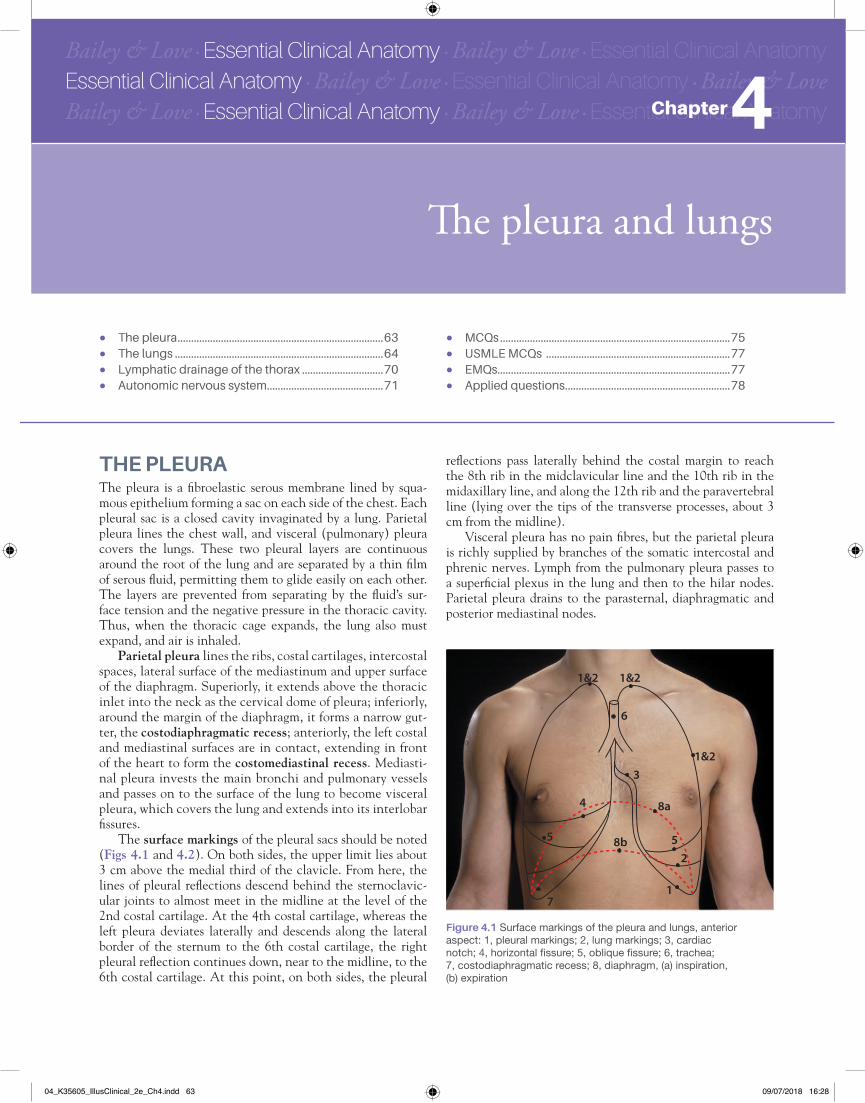
The surface markings of the pleural sacs should be noted (Figs 4.1 and 4.2). On both sides, the upper limit lies about 3 cm above the medial third of the clavicle. From here, the lines of pleural re�ections descend behind the sternoclavic-ular joints to almost meet in the midline at the level of the 2nd costal cartilage. At the 4th costal cartilage, whereas the left pleura deviates laterally and descends along the lateral border of the sternum to the 6th costal cartilage, the right pleural re�ection continues down, near to the midline, to the 6th costal cartilage. At this point, on both sides, the pleural
re�ections pass laterally behind the costal margin to reach the 8th rib in the midclavicular line and the 10th rib in the midaxillary line, and along the 12th rib and the paravertebral line (lying over the tips of the transverse processes, about 3 cm from the midline).
Visceral pleura has no pain �bres, but the parietal pleura is richly supplied by branches of the somatic intercostal and phrenic nerves. Lymph from the pulmonary pleura passes to a super�cial plexus in the lung and then to the hilar nodes. Parietal pleura drains to the parasternal, diaphragmatic and posterior mediastinal nodes.
1&2 1&2
1&2
1
2
3
4
5 58b
8a
6
7
Figure 4.1 Surface markings of the pleura and lungs, anterior aspect: 1, pleural markings; 2, lung markings; 3, cardiac notch; 4, horizontal �ssure; 5, oblique �ssure; 6, trachea; 7, costodiaphragmatic recess; 8, diaphragm, (a) inspiration, (b) expiration
04_K35605_IllusClinical_2e_Ch4.indd 63 09/07/2018 16:28

CHAPTER 4 The pleura and lungs
PART 1 | THE THORAX
64
(a)
(b)
The pleural sac is a potential space that can in patho-logical conditions �ll with �uid or air; with blood after intrathoracic haemorrhage (haemothorax); with in�am-matory exudate (pleural effusion) or pus (pyothorax) (Fig. 4.3); or with air (pneumothorax) (Fig. 1.6, p. 00) after chest wall trauma that has torn the lung or after the rupture of a lung bulla that has burst the visceral pleura. A distended pleural cavity may interfere with lung expansion. Fluid may be drained from the pleural cavity by insertion of a needle or tube, attached to an underwater seal, into the 7th intercos-tal space in the midaxillary line (Figs 1.7a and 1.9, p. 00). Insertion below this level runs the risk of the needle pene-trating the diaphragm and the underlying liver or spleen. To avoid danger to the neurovascular bundle, it is best to insert the needle along the top of the rib, which avoids the vessels lying in the subcostal groove. Emergency aspiration of air is most safely achieved by inserting a needle, attached to an underwater seal or �utter valve, into the 2nd or 3rd intercostal space in the midclavicular line.
Punch biopsy needles inserted through the intercostal space allow specimens of pleura to be obtained for histological examination. Pleurisy – in�ammation of the pleura – causes pain that is magni�ed by respiratory movements. The pain is referred by sensory �bres within the parietal pleura to the cutaneous distribution of the nerve supplying it. Thus costal in�ammation is referred to the chest wall or, in the case of lower nerves, to the upper abdominal wall, and in�ammation of the diaphragmatic pleura is referred to the area supplied by the nerve root (C4) from which originates the phrenic nerve, i.e. to the tip of the shoulder.
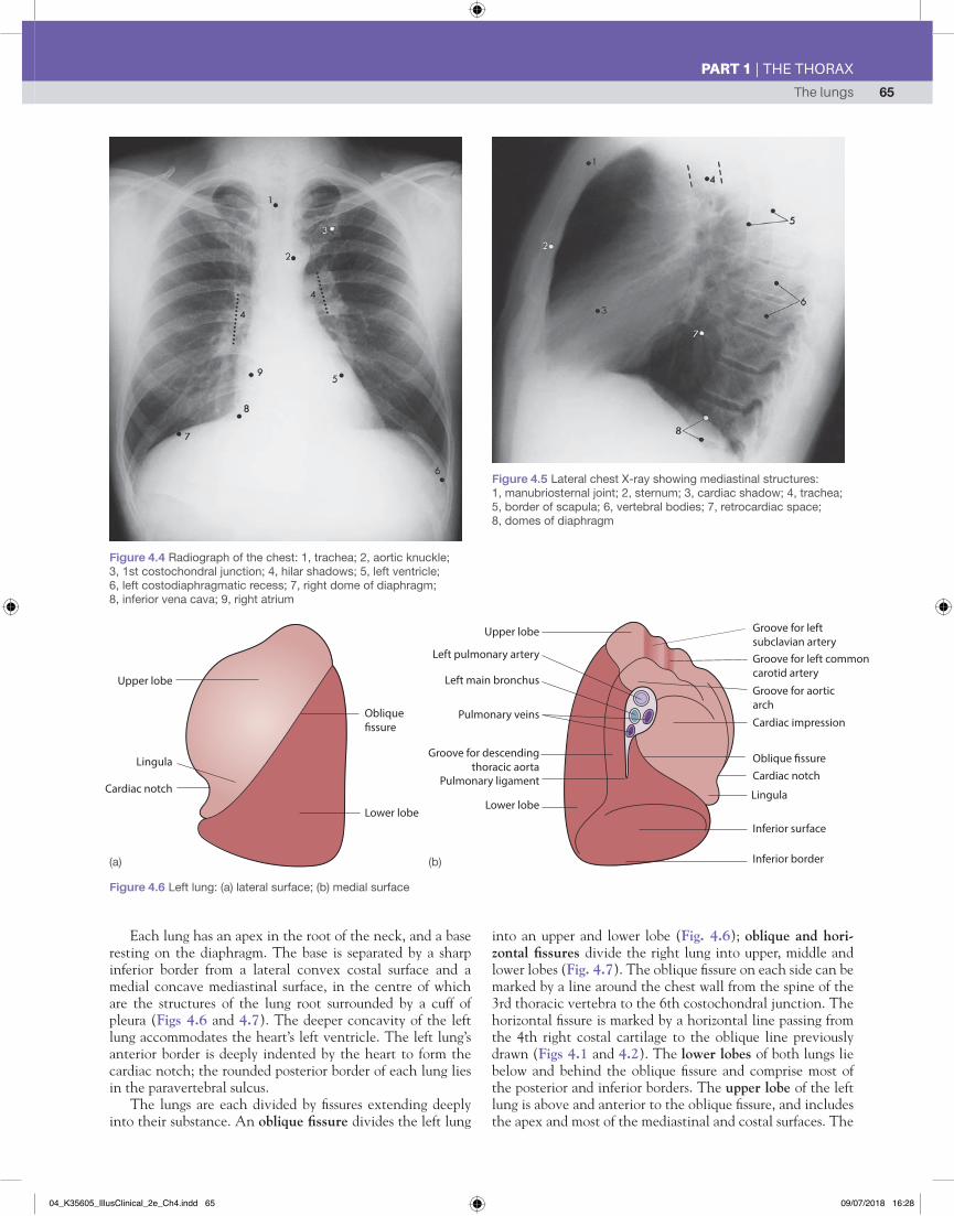
THE LUNGSThese paired organs lie in separate pleural sacs attached to the mediastinum at the hila (Figs 4.1, and Figs 4.6 and 4.7 below). Spongy and elastic in composition, they conform to the contours of the thoracic cavity. The right lung weighs about 620 g, and the left about 560 g. They have a charac-teristic mottled appearance on radiographs – lung tissue is clear; denser shadows at the hilus are caused by hilar lymph nodes, and radiating shadows by blood vessels. A posteroan-terior (PA) chest X-ray will also reveal the normal aortic arch (knuckle), the inferior vena cava and the outline of the heart. A lateral view reveals details of the mediastinal structures (Figs 4.4 and 4.5).
1
1&2
1&21&2
2
4a
4b
3
Figure 4.2 Surface markings of the pleura and lungs, posterior aspect: 1, pleural markings; 2, lung markings; 3, oblique �ssure; 4, diaphragm, (a) inspiration, (b) expiration. The black dotted line shows the incision for opening the chest through 5th intercostal space
Figure 4.3 (a) Left pleural effusion obliterating much of the lower lung markings; (b) localized interlobar �uid collection (arrow)
04_K35605_IllusClinical_2e_Ch4.indd 64 09/07/2018 16:28
The lungs 65
PART 1 | THE THORAX
(a) (b)
Each lung has an apex in the root of the neck, and a base resting on the diaphragm. The base is separated by a sharp inferior border from a lateral convex costal surface and a medial concave mediastinal surface, in the centre of which are the structures of the lung root surrounded by a cuff of pleura (Figs 4.6 and 4.7). The deeper concavity of the left lung accommodates the heart’s left ventricle. The left lung’s anterior border is deeply indented by the heart to form the cardiac notch; the rounded posterior border of each lung lies in the paravertebral sulcus.
The lungs are each divided by �ssures extending deeply into their substance. An oblique �ssure divides the left lung
into an upper and lower lobe (Fig. 4.6); oblique and hori-zontal �ssures divide the right lung into upper, middle and lower lobes (Fig. 4.7). The oblique �ssure on each side can be marked by a line around the chest wall from the spine of the 3rd thoracic vertebra to the 6th costochondral junction. The horizontal �ssure is marked by a horizontal line passing from the 4th right costal cartilage to the oblique line previously drawn (Figs 4.1 and 4.2). The lower lobes of both lungs lie below and behind the oblique �ssure and comprise most of the posterior and inferior borders. The upper lobe of the left lung is above and anterior to the oblique �ssure, and includes the apex and most of the mediastinal and costal surfaces. The
Figure 4.4 Radiograph of the chest: 1, trachea; 2, aortic knuckle; 3, 1st costochondral junction; 4, hilar shadows; 5, left ventricle; 6, left costodiaphragmatic recess; 7, right dome of diaphragm; 8, inferior vena cava; 9, right atrium
Figure 4.5 Lateral chest X-ray showing mediastinal structures: 1, manubriosternal joint; 2, sternum; 3, cardiac shadow; 4, trachea; 5, border of scapula; 6, vertebral bodies; 7, retrocardiac space; 8, domes of diaphragm
Lower lobe
Upper lobe
Lingula
Cardiac notch
Oblique�ssure
Upper lobe Groove for left subclavian arteryGroove for left commoncarotid artery
Groove for aorticarch
Cardiac impression
Oblique �ssureCardiac notch
Lingula
Inferior surface
Inferior border
Lower lobe
Groove for descendingthoracic aorta
Pulmonary ligament
Pulmonary veins
Left main bronchus
Left pulmonary artery
Figure 4.6 Left lung: (a) lateral surface; (b) medial surface
04_K35605_IllusClinical_2e_Ch4.indd 65 09/07/2018 16:28

CHAPTER 4 The pleura and lungs
PART 1 | THE THORAX
66
Upper lobe
Horizontal�ssure
Middle lobe Lower lobe
Oblique�ssure
Apex
Groove for rightsubclavian artery
Impression ofazygos vein
Oblique �ssure
Groove foroesophagus
Pulmonaryligament
Pulmonary veins
Groove for inferiorvena cava
Base of lung
Horizontal�ssure
Anteriorborder of
heart
Division of rightmain bronchus
Groove forthe superior
vena cava
Division ofright pulmonary
artery
Figure 4.7 Right lung: (a) lateral surface; (b) medial surface
Medial (MM)Middle lobe Lateral (LM)
Posterior basal(LP)
Medial basal(LM)
Apical basal(LAP)
Lateral basal (LL)
Anterior basal (LA)
Anterior (UA)
Posterior (UP)
Apical (UAP)
Right Left
Apico - posterior (UAP)
Anterior (UA)
Superior (SL)
Inferior (IL)Lingular
Anterior basal (LA)
Lateral basal (LL)
UAPUAP
UA
UALAPLAP
LP
LP
UP
LL LL
LA LA
LMIL
IL
SL
SL
Left lung L medial L lateral
R Medial Right lung
R Lateral
UAPUAP
UP
UP
LAP
LAP
LP
LM
MM ML MM
UAUA
LALL
Figure 4.8 (a) Bronchial tree. (b) and (c) Bronchopulmonary segments; (b) right lung – mediastinal and costovertebral surfaces; (c) left lung – medial and lateral surfaces
(a)
(a)
(b) (c)
(b)
04_K35605_IllusClinical_2e_Ch4.indd 66 09/07/2018 16:28
The lungs 67
PART 1 | THE THORAX
equivalent part of the right lung is divided by the horizontal �ssure into a large upper lobe and a smaller, anterior, wedge-shaped middle lobe, which lies deep to the right breast. A thin anteroinferior part of the left upper lobe, adjacent to the cardiac notch, is known as the lingula and is the left-sided equivalent of the middle lobe. Fissures may be incomplete or absent, and additional lobes are occasionally present.
The hilum or root of each lung contains a main bronchus, a pulmonary artery, two pulmonary veins, the pulmonary nerve plexus and lymph nodes, all enveloped by the pleural cuff; an inferior narrow extension of the cuff is known as the pulmonary ligament (Figs 4.6 and 4.7). The bronchus lies behind the pulmonary artery, and the two veins are below and anterior.
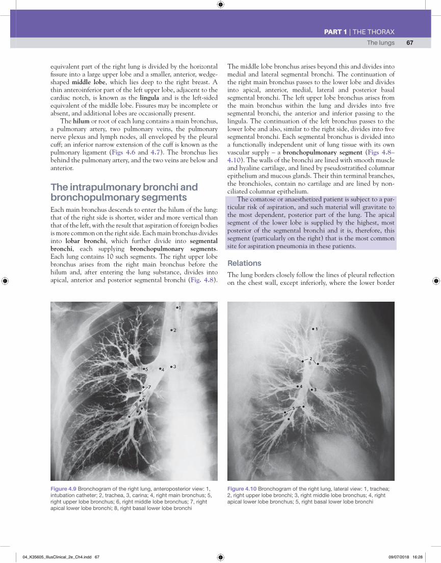
The intrapulmonary bronchi and bronchopulmonary segmentsEach main bronchus descends to enter the hilum of the lung: that of the right side is shorter, wider and more vertical than that of the left, with the result that aspiration of foreign bodies is more common on the right side. Each main bronchus divides into lobar bronchi, which further divide into segmental bronchi, each supplying bronchopulmonary segments. Each lung contains 10 such segments. The right upper lobe bronchus arises from the right main bronchus before the hilum and, after entering the lung substance, divides into apical, anterior and posterior segmental bronchi (Fig. 4.8).
The middle lobe bronchus arises beyond this and divides into medial and lateral segmental bronchi. The continuation of the right main bronchus passes to the lower lobe and divides into apical, anterior, medial, lateral and posterior basal segmental bronchi. The left upper lobe bronchus arises from the main bronchus within the lung and divides into �ve segmental bronchi, the anterior and inferior passing to the lingula. The continuation of the left bronchus passes to the lower lobe and also, similar to the right side, divides into �ve segmental bronchi. Each segmental bronchus is divided into a functionally independent unit of lung tissue with its own vascular supply – a bronchopulmonary segment (Figs 4.8–4.10). The walls of the bronchi are lined with smooth muscle and hyaline cartilage, and lined by pseudostrati�ed columnar epithelium and mucous glands. Their thin terminal branches, the bronchioles, contain no cartilage and are lined by non-ciliated columnar epithelium.
The comatose or anaesthetized patient is subject to a par-ticular risk of aspiration, and such material will gravitate to the most dependent, posterior part of the lung. The apical segment of the lower lobe is supplied by the highest, most posterior of the segmental bronchi and it is, therefore, this segment (particularly on the right) that is the most common site for aspiration pneumonia in these patients.
Relations
The lung borders closely follow the lines of pleural re�ection on the chest wall, except inferiorly, where the lower border
● 1
●●
● 3● 4
2
5 ●
●
●
Figure 4.9 Bronchogram of the right lung, anteroposterior view: 1, intubation catheter; 2, trachea, 3, carina; 4, right main bronchus; 5, right upper lobe bronchus; 6, right middle lobe bronchus; 7, right apical lower lobe bronchi; 8, right basal lower lobe bronchi
Figure 4.10 Bronchogram of the right lung, lateral view: 1, trachea; 2, right upper lobe bronchi; 3, right middle lobe bronchus; 4, right apical lower lobe bronchus; 5, right basal lower lobe bronchi
04_K35605_IllusClinical_2e_Ch4.indd 67 09/07/2018 16:28

CHAPTER 4 The pleura and lungs
PART 1 | THE THORAX
68
of the lung lies about two intercostal spaces above the pleural re�ection (costodiaphragmatic recess), and in front, where, near to the cardiac notch, the anterior border of the left lung lies some 3 cm lateral to the pleural re�ection (costomediasti-nal recess) (Figs 4.1 and 4.2). The costal surfaces are related to the thoracic wall; the base is separated by the diaphragm from the right lobe of the liver on the right, and the liver, stomach and spleen on the left. The apex, covered by the dome of the pleura, lies under the suprapleural membrane, a �brous sheet extending from the transverse process of the 7th cervical vertebra to the inner border of the 1st rib. The subclavian vessels arch over the membrane. Posteriorly lie the anterior primary ramus of the �rst thoracic nerve, passing to the brachial plexus, and the sympathetic trunk, both lying on the neck of the 1st rib.
The medial relations differ on each side. On the left (Figs 4.6b and 4.11), a large concavity for the left ventricle con-tinues superiorly with a groove for the aortic arch, which passes in front of the hilum. Above the arch, the lung is in contact, from before backwards, with the left brachiocephalic vein, left common carotid artery, left subclavian artery and oesophagus. On the right (Figs 4.7b and 4.12), a shallow concavity in front of the hilum for the right atrium is continu-ous above with a groove for the superior vena cava, and below with a shorter groove for the inferior vena cava. The azygos vein grooves the lung as it arches forwards above the hilum. The oesophagus is in contact throughout its length near to
the posterior border, except where the azygos vein separates it from the lung. The oesophagus lies between the superior vena cava and the trachea.
Blood supply
Lung tissue is supplied by the bronchial arteries – branches of the descending aorta – and some of this blood returns to the heart via the pulmonary veins. Other bronchial veins drain to the azygos or hemiazygos veins. The pulmonary artery con-veys poorly oxygenated blood to the alveoli by branches that accompany the bronchial tree. From the alveolar capillary, network arise veins that accompany the bronchi to form the upper and lower pulmonary veins.
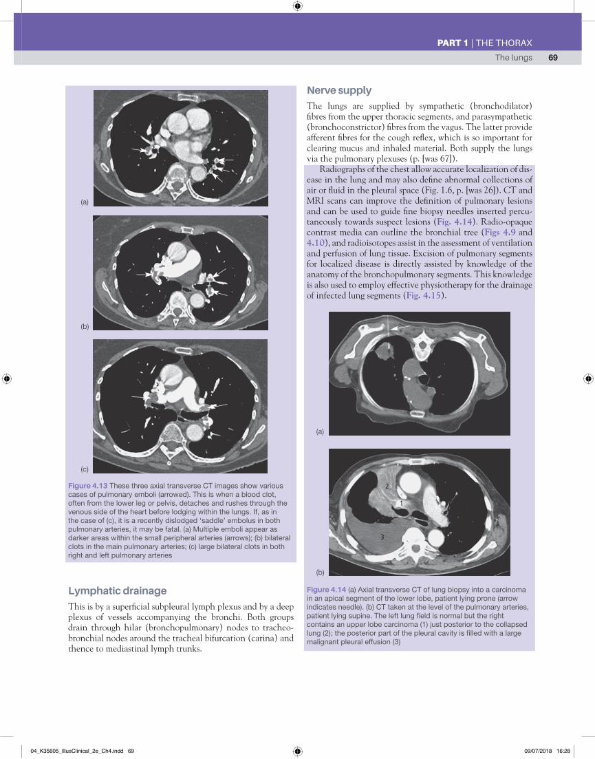
Pulmonary embolism may be a fatal condition. It is caused by large blood clots (thrombi) that originate in the deep veins of the legs or pelvis, becoming dislodged and then being conveyed through the right side of the heart to rest in and occlude large or segmental pulmonary arteries (Fig. 3.5, p. [was 51], and Fig. 4.13), or worse, the pulmonary trunk. The result is partial or complete obstruction of the arterial supply to a segment or lobe of the lung, which, although ventilated, is no longer perfused with blood. If the embolus blocks the main pulmonary artery, acute respiratory distress and cyanosis results and death often follows within a few minutes. Segmen-tal emboli cause death of lung tissue, a pulmonary infarct that, if small, resolves over a few weeks.
Brachiocephalicartery
Left commoncarotid artery
Left subclavianartery
Descendingthoracic aorta
Left mainbronchus
Pulmonaryligament
Left side of heart
Pulmonaryveins
Pulmonaryartery
Leftbrachiocephalic
vein
Azygos vein
Rightbrachiocephalicvein
Superiorvena cava
Division of rightpulmonary artery
Right pulmonaryveins
Right side ofheart
Pulmonaryligament
Rightupper andlower lobe
bronchi
Figure 4.11 Medial relations of the left lung Figure 4.12 Medial relations of the right lung
04_K35605_IllusClinical_2e_Ch4.indd 68 09/07/2018 16:28
The lungs 69
PART 1 | THE THORAX
Lymphatic drainage
This is by a super�cial subpleural lymph plexus and by a deep plexus of vessels accompanying the bronchi. Both groups drain through hilar (bronchopulmonary) nodes to tracheo-bronchial nodes around the tracheal bifurcation (carina) and thence to mediastinal lymph trunks.
Nerve supply
The lungs are supplied by sympathetic (bronchodilator) �bres from the upper thoracic segments, and parasympathetic (bronchoconstrictor) �bres from the vagus. The latter provide afferent �bres for the cough re�ex, which is so important for clearing mucus and inhaled material. Both supply the lungs via the pulmonary plexuses (p. [was 67]).
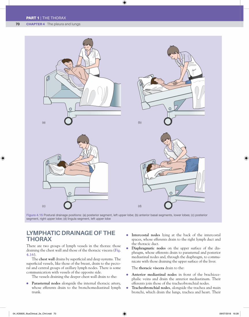
Radiographs of the chest allow accurate localization of dis-ease in the lung and may also de�ne abnormal collections of air or �uid in the pleural space (Fig. 1.6, p. [was 26]). CT and MRI scans can improve the de�nition of pulmonary lesions and can be used to guide �ne biopsy needles inserted percu-taneously towards suspect lesions (Fig. 4.14). Radio-opaque contrast media can outline the bronchial tree (Figs 4.9 and 4.10), and radioisotopes assist in the assessment of ventilation and perfusion of lung tissue. Excision of pulmonary segments for localized disease is directly assisted by knowledge of the anatomy of the bronchopulmonary segments. This knowledge is also used to employ effective physiotherapy for the drainage of infected lung segments (Fig. 4.15).
Figure 4.13 These three axial transverse CT images show various cases of pulmonary emboli (arrowed). This is when a blood clot, often from the lower leg or pelvis, detaches and rushes through the venous side of the heart before lodging within the lungs. If, as in the case of (c), it is a recently dislodged ‘saddle’ embolus in both pulmonary arteries, it may be fatal. (a) Multiple emboli appear as darker areas within the small peripheral arteries (arrows); (b) bilateral clots in the main pulmonary arteries; (c) large bilateral clots in both right and left pulmonary arteries
2
1
3
Figure 4.14 (a) Axial transverse CT of lung biopsy into a carcinoma in an apical segment of the lower lobe, patient lying prone (arrow indicates needle). (b) CT taken at the level of the pulmonary arteries, patient lying supine. The left lung �eld is normal but the right contains an upper lobe carcinoma (1) just posterior to the collapsed lung (2); the posterior part of the pleural cavity is �lled with a large malignant pleural effusion (3)
(a)
(b)
(c)
(a)
(b)
04_K35605_IllusClinical_2e_Ch4.indd 69 09/07/2018 16:28
CHAPTER 4 The pleura and lungs
PART 1 | THE THORAX
70
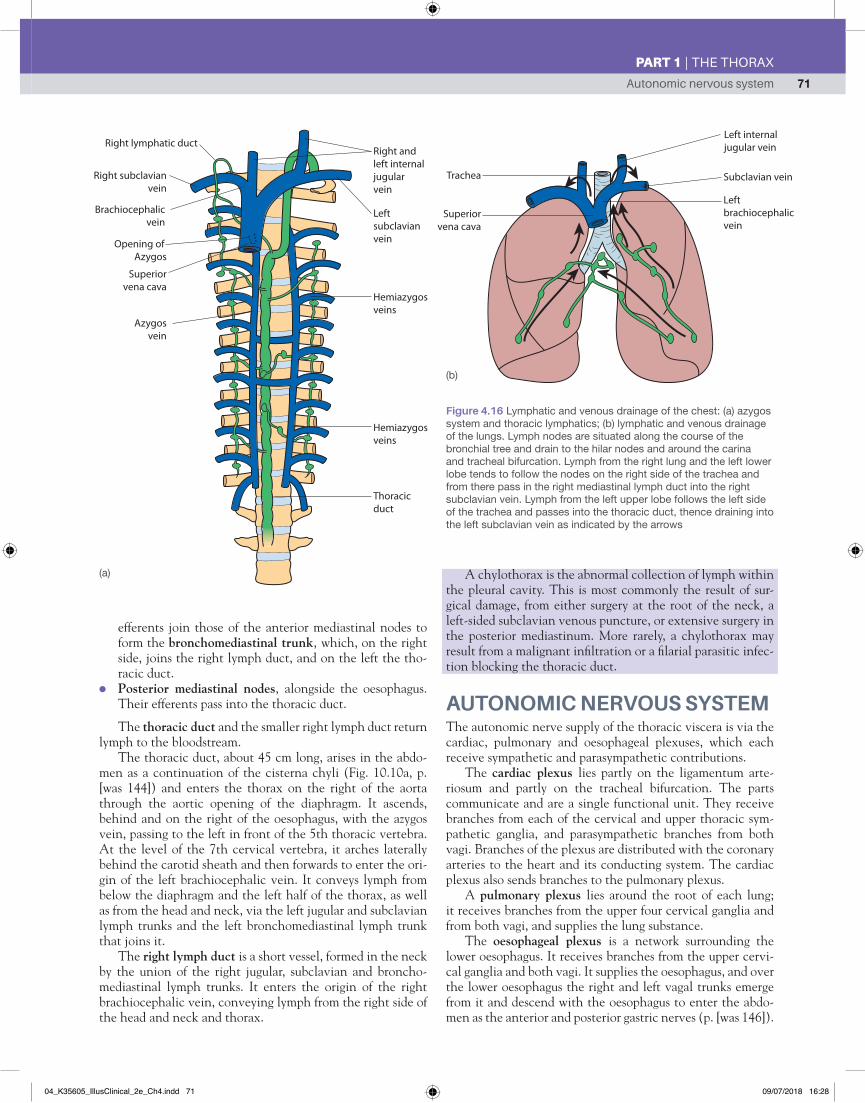
LYMPHATIC DRAINAGE OF THE THORAXThere are two groups of lymph vessels in the thorax: those draining the chest wall and those of the thoracic viscera (Fig. 4.16).
The chest wall drains by super�cial and deep systems. The super�cial vessels, like those of the breast, drain to the pecto-ral and central groups of axillary lymph nodes. There is some communication with vessels of the opposite side.
The vessels draining the deeper chest wall drain to the:
● Parasternal nodes alongside the internal thoracic artery, whose efferents drain to the bronchomediastinal lymph trunk.
● Intercostal nodes lying at the back of the intercostal spaces, whose efferents drain to the right lymph duct and the thoracic duct.
● Diaphragmatic nodes on the upper surface of the dia-phragm, whose efferents drain to parasternal and posterior mediastinal nodes and, through the diaphragm, to commu-nicate with those draining the upper surface of the liver.
The thoracic viscera drain to the:
● Anterior mediastinal nodes in front of the brachioce-phalic veins and drain the anterior mediastinum. Their efferents join those of the tracheobronchial nodes.
● Tracheobronchial nodes, alongside the trachea and main bronchi, which drain the lungs, trachea and heart. Their
Figure 4.15 Postural drainage positions: (a) posterior segment, left upper lobe; (b) anterior basal segments, lower lobes; (c) posterior segment, right upper lobe; (d) lingula segment, left upper lobe
(a) (b)
(c) (d)
04_K35605_IllusClinical_2e_Ch4.indd 70 09/07/2018 16:28
Autonomic nervous system 71
PART 1 | THE THORAX
efferents join those of the anterior mediastinal nodes to form the bronchomediastinal trunk, which, on the right side, joins the right lymph duct, and on the left the tho-racic duct.
● Posterior mediastinal nodes, alongside the oesophagus. Their efferents pass into the thoracic duct.
The thoracic duct and the smaller right lymph duct return lymph to the bloodstream.
The thoracic duct, about 45 cm long, arises in the abdo-men as a continuation of the cisterna chyli (Fig. 10.10a, p. [was 144]) and enters the thorax on the right of the aorta through the aortic opening of the diaphragm. It ascends, behind and on the right of the oesophagus, with the azygos vein, passing to the left in front of the 5th thoracic vertebra. At the level of the 7th cervical vertebra, it arches laterally behind the carotid sheath and then forwards to enter the ori-gin of the left brachiocephalic vein. It conveys lymph from below the diaphragm and the left half of the thorax, as well as from the head and neck, via the left jugular and subclavian lymph trunks and the left bronchomediastinal lymph trunk that joins it.
The right lymph duct is a short vessel, formed in the neck by the union of the right jugular, subclavian and broncho-mediastinal lymph trunks. It enters the origin of the right brachiocephalic vein, conveying lymph from the right side of the head and neck and thorax.
A chylothorax is the abnormal collection of lymph within the pleural cavity. This is most commonly the result of sur-gical damage, from either surgery at the root of the neck, a left-sided subclavian venous puncture, or extensive surgery in the posterior mediastinum. More rarely, a chylothorax may result from a malignant in�ltration or a �larial parasitic infec-tion blocking the thoracic duct.
AUTONOMIC NERVOUS SYSTEMThe autonomic nerve supply of the thoracic viscera is via the cardiac, pulmonary and oesophageal plexuses, which each receive sympathetic and parasympathetic contributions.
The cardiac plexus lies partly on the ligamentum arte-riosum and partly on the tracheal bifurcation. The parts communicate and are a single functional unit. They receive branches from each of the cervical and upper thoracic sym-pathetic ganglia, and parasympathetic branches from both vagi. Branches of the plexus are distributed with the coronary arteries to the heart and its conducting system. The cardiac plexus also sends branches to the pulmonary plexus.
A pulmonary plexus lies around the root of each lung; it receives branches from the upper four cervical ganglia and from both vagi, and supplies the lung substance.
The oesophageal plexus is a network surrounding the lower oesophagus. It receives branches from the upper cervi-cal ganglia and both vagi. It supplies the oesophagus, and over the lower oesophagus the right and left vagal trunks emerge from it and descend with the oesophagus to enter the abdo-men as the anterior and posterior gastric nerves (p. [was 146]).
Right and left internal jugularvein
Left subclavianvein
Right subclavianvein
Right lymphatic duct
Brachiocephalicvein
Thoracic duct
Hemiazygosveins
Hemiazygosveins
Superiorvena cava
Opening of Azygos
Azygosvein
Left internaljugular vein
Subclavian vein
Leftbrachiocephalicvein
Trachea
Superiorvena cava
Figure 4.16 Lymphatic and venous drainage of the chest: (a) azygos system and thoracic lymphatics; (b) lymphatic and venous drainage of the lungs. Lymph nodes are situated along the course of the bronchial tree and drain to the hilar nodes and around the carina and tracheal bifurcation. Lymph from the right lung and the left lower lobe tends to follow the nodes on the right side of the trachea and from there pass in the right mediastinal lymph duct into the right subclavian vein. Lymph from the left upper lobe follows the left side of the trachea and passes into the thoracic duct, thence draining into the left subclavian vein as indicated by the arrows
(a)
(b)
04_K35605_IllusClinical_2e_Ch4.indd 71 09/07/2018 16:28

CHAPTER 4 The pleura and lungs
PART 1 | THE THORAX
72
Each thoracic sympathetic trunk (Fig. 4.17a,b) lies alongside the vertebral column behind the parietal pleura. It is continuous above with the cervical trunk and below with the lumbar sympathetic trunk. It usually possesses 12 ganglia, each contributed by a thoracic nerve, but half of the � rst tho-racic ganglion is fused to the 7th cervical to form a larger stellate ganglion on the neck of the 1st rib. Each ganglion receives preganglionic � bres in a white ramus communicans from its corresponding spinal nerve, and sends postganglionic � bres back to that nerve as a grey ramus communicans.
Sympathetic denervation (sympathectomy) of the upper limb is employed to dilate cutaneous blood vessels or inhibit sweating. It is achieved by surgically removing the 2nd and 3rd thoracic ganglia. The 1st ganglion is left intact to preserve sympathetic innervation to the head and neck and prevent the development of Horner’s syndrome (Fig. 4.18). The fea-tures of the syndrome are unilateral ptosis, � ushed and dry skin on the same side of the face, enophthalmos and a small pupil (meiosis). The syndrome is due to damage to the upper cervical trunk. It is often caused by cancerous in� ltration from the apex of the lung (Fig. 4.19), but may also be caused by surgical removal of the stellate ganglion or a stellate gan-glion block.
Figure 4.17 The sympathetic nervous system in the chest. (a) Sympathetic nerves in the thorax and root of neck. (b) Typical thoracic sympathetic ganglion. (c) Posterior thoracic wall dissection after removal of organs to show vertebral bodies and sympathetic chains: 1, vertebral bodies; 2, rib; 3, internal intercostal muscles; 4, disarticulated heads of ribs to show demi-facets; 5, sympathetic chain; 6, sympathetic ganglia; 7, intercostal, segmental neurovascular bundles; 8, posterior intercostal arteries; 9, intervertebral discs; 10, psoas major muscle
Cervicalsympathetic chain
Right commoncarotid artrey Cervical
oesophagus Right recurrentlaryngeal nerve
Rightsubclavian artery
Right vagus
Trachea
Left commoncarotid artery
Left recurrentlaryngeal nerve
Left subclavianartery
Transected archof the aorta
Greatersplanchnic nerves
Left recurrentlaryngeal nervepassing aroundthe aorta
Left vagus
Descendingthoracic aorta
Thoracicsympathetic chain
Splanchnic nervespiercing diaphragm
Thoracicoesophagus
Thoracic sympatheticganglion
Grey ramuscommunicantes(postganglionic)
White ramuscommunicantes(preganglionic)
Sympathetic chainThoracic spinal nerve
1
87
5
5
4 23
7
6
6
8
8
5
5
9
5
10
(a)
(b) (c)
Figure 4.18 Right Horner’s syndrome
04_K35605_IllusClinical_2e_Ch4.indd 72 09/07/2018 16:28

Autonomic nervous system 73
PART 1 | THE THORAX
The sympathetic trunk supplies:
● rami communicantes to each of its spinal nerves; ● branches to the cardiac, pulmonary and oesophageal plex-
uses from the upper four ganglia;
Development of the trachea, bronchi and lungs
(b)
Figure 4.19 Apical cancer of the lung (Pancoast’s syndrome): (a) chest X-ray, right-sided lesion; (b) CT scan (arrow), left-sided lesion
(a)
● branches to form the greater, lesser and least splanchnic nerves (Fig. 4.17 and Fig. 10.13, p. [146]). These descend medial to the sympathetic chain and enter the abdomen by piercing the diaphragmatic crura. They contribute branches to the coeliac and other preaortic ganglia.
The lungs and tracheobronchial tree develop from the respiratory diverticulum, an outgrowth of tissue that buds off the ventral aspect of the proximal fore-gut in the region of the 6th pharyngeal arch during week 4 of development (Fig. 4.20). The lining of the
larynx and tracheobronchial tree is therefore derived from endoderm. The respiratory diverticulum grows caudally and branches initially into the left and right lung buds, each containing the bronchial buds that form the primary bronchi (Figs. 4.21). The bronchial buds continue to subdivide, first into secondary bron-chi (three on the right, and two on the left) followed by segmental bronchi (10 on the right, and eight on the left), and continuing to sequentially branch both in utero and postpartum to create the bronchial tree. The number of secondary and tertiary bronchi that develop reflects the number of lobes and bronchopul-monary segments present in the fully developed lung.
Aorticarches
Head
Cranial Caudal
Hearttube
Pericardium
Arterial�ow
Venousin�ow
Figure 4.20 [caption to follow - leave 3 lines] Figure 4.21 [caption to follow - leave 3 lines]
04_K35605_IllusClinical_2e_Ch4.indd 73 09/07/2018 16:28
CHAPTER 4 The pleura and lungs
PART 1 | THE THORAX
74
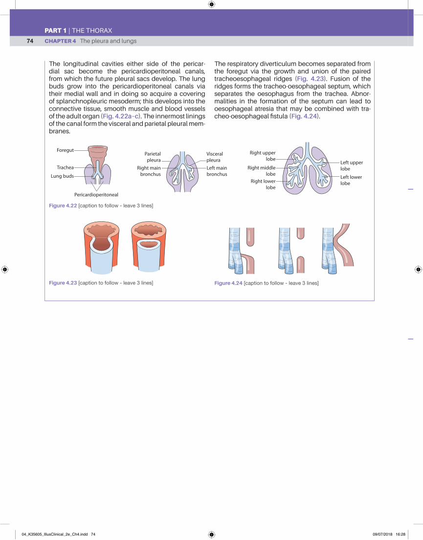
The longitudinal cavities either side of the pericar-dial sac become the pericardioperitoneal canals, from which the future pleural sacs develop. The lung buds grow into the pericardioperitoneal canals via their medial wall and in doing so acquire a covering of splanchnopleuric mesoderm; this develops into the connective tissue, smooth muscle and blood vessels of the adult organ (Fig. 4.22a–c). The innermost linings of the canal form the visceral and parietal pleural mem-branes.
The respiratory diverticulum becomes separated from the foregut via the growth and union of the paired tracheoesophageal ridges (Fig. 4.23). Fusion of the ridges forms the tracheo-oesophageal septum, which separates the oesophagus from the trachea. Abnor-malities in the formation of the septum can lead to oesophageal atresia that may be combined with tra-cheo-oesophageal fi stula (Fig. 4.24).
ForegutParietal
pleuraVisceralpleura Left upper
lobe
Right upperlobe
Right middlelobe
Right lowerlobe
Left lowerlobe
Right mainbronchus
Left mainbronchus
Pericardioperitoneal
Trachea
Lung buds
Figure 4.22 [caption to follow - leave 3 lines]
Figure 4.23 [caption to follow - leave 3 lines] Figure 4.24 [caption to follow - leave 3 lines]
04_K35605_IllusClinical_2e_Ch4.indd 74 09/07/2018 16:28

Mcqs 75
PART 1 | THE THORAX
MCQS1. The surface markings of the pleural sacs: T/F
a do not extend above the clavicle (____)b meet anteriorly in the midline (____)c are similar on both sides (____)d extend to the 8th rib in the midaxillary line (____)e extend to the 12th rib in the paravertebral
line(____)
Answers1.a F – On both sides, the upper limit of the pleural sacs lies
3 cm above the mid-clavicle.b T – The pleura meet in the midline at the level of the 2nd
to 4th costal cartilages.c F – At the 4th costal cartilage, the right pleura descends
close to the midline, whereas the left pleura deviates laterally to accommodate the heart to descend to the 6th costal cartilage along the lateral border of the sternum.
d F – Both deviate laterally to reach the 8th rib in the midclavicular line and the 10th rib in the midaxillary line.
e T – The lower limit of the pleural re�ection often descends to just below the 12th rib in the paravertebral line.
2. The right lung: T/Fa is smaller than the left (____)b is divided into upper and lower lobes and
the lingula(____)
c possesses 10 bronchopulmonary seg-ments
(____)
d is related to the oesophagus only in the lower part of its medial surface
(____)
e is related to the liver (____)
Answers2.a F – The right weighs about 620 g and the left about 560
g.b F – Oblique and horizontal �ssures divide it into upper,
middle and lower lobes. The lingula exists only in the left lung and is the equivalent of the middle lobe on the right.
c T – Bronchopulmonary segments are functionally independent units of lung. In the right lung, there are three in the upper lobe, two in the middle and �ve in the lower lobe. In the left lung, there are �ve in the upper and four in the lower lobe, the medial segment being replaced by the left ventricle of the heart.
d F – The whole length of the thoracic oesophagus lies close to the medial surface of the right lung, separated only by the azygos vein crossing over the hilum of the lung.
e T – It is separated from the liver only by the diaphragm and the pleural and peritoneal coverings.
3. Lung tissue: T/Fa receives oxygenated arterial blood from
branches of the aorta(____)
b drains venous blood into the azygos veins (____)c has no lymph drainage (____)d is lined throughout by ciliated epithelium (____)e is supplied by the vagus nerve (____)
Answers3.a T – It receives blood from small bronchial arteries arising
from the descending thoracic aorta.b T – Drainage is via bronchial veins draining into the
azygos and hemiazygos veins.c F – There is an extensive subpleural lymph plexus and
a deep lymph plexus that accompanies the bronchi. These drain into hilar and tracheobronchial nodes and then into the mediastinal lymph trunks.
d F – Ciliated epithelium lines only the extra- and intrapulmonary bronchi. The distal bronchioles are lined by non-ciliated columnar epithelium and the alveoli by squamous epithelium.
e T – Innervation is via branches that pass through the pulmonary plexuses.
04_K35605_IllusClinical_2e_Ch4.indd 75 09/07/2018 16:28

CHAPTER 4 The pleura and lungs
PART 1 | THE THORAX
76
4. The sympathetic chain: T/Fa is formed by contributions from the ventral
roots of T1–L2(____)
b carries parasympathetic �bres below L2 (____)c lies anterior to the common iliac vessels (____)d lies anterior to the lumbar arteries and veins (____)e lies posterior to the psoas fascia (____)
Answers4.a T – The sympathetic chain extends from the base of the
skull to the coccyx.b F – It carries no parasympathetic �bres.c F – It lies behind the iliac vessels.d T – It lies anterior to segmental vessels such as the
lumbar vessels.e F – Inferiorly, it lies on the psoas fascia.
5. The thoracic duct: T/Fa arises in the thorax (____)b lies anterior to the vertebral column (____)c drains into the left brachiocephalic vein (____)d drains only thoracic structures (____)e is joined by the right lymph duct (____)
Answers5.a F – It arises in the abdomen from the cisterna chyli,
entering the thorax through the aortic opening of the diaphragm ...
b T – ... alongside the azygos vein, behind the oesophagus.c T – It arches forwards from behind the carotid sheath
over the subclavian artery to enter the vein.d F – It drains all structures below the diaphragm and
the left half of the body and head and neck above it through the left superior mediastinal, jugular and subclavian lymph trunks.
e F – The right lymph duct is often absent but, when present, enters the right brachiocephalic vein separately, draining lymph from the right side of the head, neck and thorax.
04_K35605_IllusClinical_2e_Ch4.indd 76 09/07/2018 16:28
Emqs 77
PART 1 | THE THORAX
USMLE MCQS 1. A comatose or anaesthetized patient is particularly at
risk of aspiration, and this is frequently followed by the development of right lower lobe pneumonia. Why is this?a the right main bronchus is longer than the the left
main bronchus b the right main bronchus is narrower than the left
main bronchus c the right main bronchus is more vertical than the
left main bronchusd the right lung has a higher pulmonary vascular
resistance than the left lunge venous drainage is poorer from the right lower
lobe than from other lobes
Answerc The right bronchus is more vertical, shorter and wider than
the left,so aspirated material is more likely to pass into it and thence to the most dependent part of the right lobe. There are no differences in pulmonary vascular resistance or venous drainage between the right lower lobe and any other pulmonary lobe, and these play no part.
2. A patient is admitted with a cough, fever and painful breathing. A pleural rub is heard on auscultation. Lo-bar pneumonia with pleurisy is diagnosed. In addition to respiratory support and antibiotics, the doctor has decided to give pain relief by the injection of local anaesthetic into which nerve(s)?a phrenic nerveb intercostal nervesc recurrent laryngeal nervesd cardiopulmonary nervese vagus nerve
Answerb Pleuritic pain is the consequence of in�ammation of
the parietal pleural, which is supplied by the intercostal nerves. The phrenic nerve supplies only the diaphragmatic part of the visceral pleura and is not amenable to nerve blocks. None of the other nerves innervates the pleura.
EMQSEach question has an anatomical theme linked to the chapter, and a list of 10 related items (A–J) placed in alphabetical order: these are followed by �ve statements (1–5). Match one or more of the items A–J to each of the �ve statements.
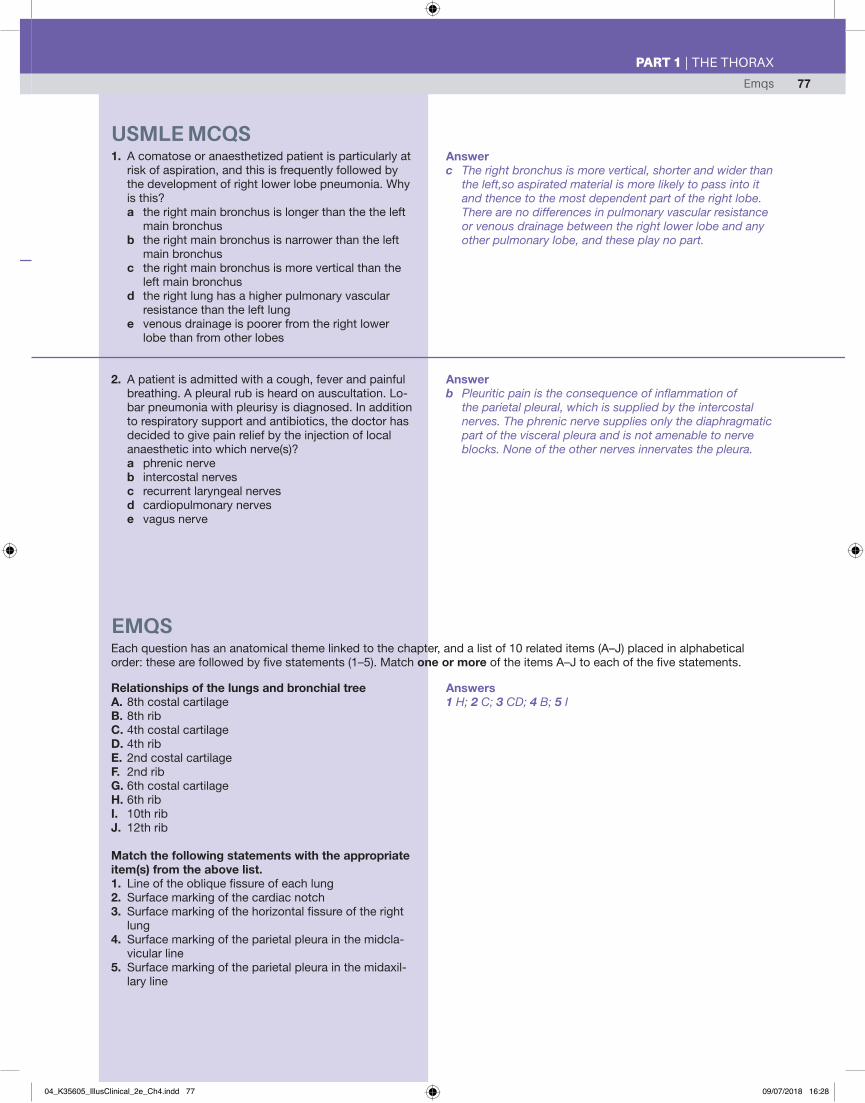
Relationships of the lungs and bronchial treeA. 8th costal cartilageB. 8th ribC. 4th costal cartilageD. 4th ribE. 2nd costal cartilageF. 2nd ribG. 6th costal cartilageH. 6th ribI. 10th ribJ. 12th rib
Match the following statements with the appropriate item(s) from the above list.1. Line of the oblique �ssure of each lung2. Surface marking of the cardiac notch3. Surface marking of the horizontal �ssure of the right
lung4. Surface marking of the parietal pleura in the midcla-
vicular line5. Surface marking of the parietal pleura in the midaxil-
lary line
Answers1 H; 2 C; 3 CD; 4 B; 5 I
04_K35605_IllusClinical_2e_Ch4.indd 77 09/07/2018 16:28
CHAPTER 4 The pleura and lungs
PART 1 | THE THORAX
78
APPLIED QUESTIONS1. You are informed that a patient has a pleural effusion.
Where is the �uid situated?1. A pleural effusion is an abnormal collection of �uid in the
pleural cavity, which normally contains only a thin �lm of �uid suf�cient for lubrication of the opposing visceral and parietal layers of pleura. The exact position of any abnormal collection is in�uenced by gravity and the patient’s posture. In bedridden patients, therefore, it tends to collect at the base of the pleural cavity posteriorly, where it will be detected by percussion and auscultation.
2. How would you drain the pleural cavity of air, and through which structures would your needle or cathe-ter pass?
2. A pneumothorax is drained by a needle or, more commonly, a tube attached to an underwater seal apparatus inserted at one of two sites, either the 2nd intercostal space in the midclavicular line or the 7th intercostal space in the midaxillary line. With the upper approach, the needle or trocar and cannula pass through skin, super�cial fascia and pectoralis major and minor, before the intercostal muscle �bres and, �nally, the parietal pleura. The lower approach avoids penetration of the pectoral muscles and produces less discomfort for the patient.
3. Your patient has unwanted bronchial secretions in her posterior basal segments. Into which position should she be placed to maximize postural drainage?
3. Effective postural drainage is achieved by positioning the patient so that the lung secretions drain from the diseased segment with the aid of gravity. Basal segments therefore require the patient to be tipped head downwards and, for the posterior basal segment, into a prone position. A physiotherapist helps the drainage by chest wall percussion over the diseased segment.
4. Why may a misplaced central venous catheter cause a chylothorax?
4. It is prudent to avoid performing a left subclavian or brachiocephalic puncture because of the presence of the thoracic duct terminating in the angle between the internal jugular and subclavian veins. If the pleura is punctured at the same time as the duct is damaged, a chylothorax may result from leakage of lymph into the pleural cavity.
5. Why may an apical carcinoma of the lung cause pain in the little �nger and a drooping upper eyelid on the same side?
5. An apical carcinoma often affects structures lying in contact with the suprapleural membrane (Sibson’s fascia), namely the sympathetic trunk, the stellate ganglion and the 1st thoracic nerve root. Damage to the sympathetic nerves at this level may produce a Horner’s syndrome – a constricted pupil (meiosis), �ushed and dry skin on the side of the face, drooping of the upper eyelid (ptosis) and retraction of the eyeball (enophthalmos). The ptosis is caused by partial paralysis of levator palpebrae superioris, whose smooth muscle is innervated by sympathetic �bres. The �nger pain may be caused by direct involvement of T1 nerve, whose dermatome lies along the medial border of the forearm and hand. This combination is known as Pancoast’s syndrome.
3. A young boy throws a peanut in the air and, in attempting to catch it in his mouth, inhales it. Into which bronchus is it likely to pass?
3. It is most likely to be inhaled into the right main bronchus, which is shorter, wider and nearly in the same line as the trachea. Once inhaled, material such as peanuts, pins or even gastric contents tends to pass into the right middle or lower lobe. However, in the unconscious patient lying on the right side, inhaled material frequently collects in the posterior segment of the right upper lobe.
04_K35605_IllusClinical_2e_Ch4.indd 78 09/07/2018 16:28